alternatives to the use of fetal bovine serum: human ... · altex 28, 4/11 305 alternatives to the...
TRANSCRIPT

Altex 28, 4/11 305
Alternatives to the Use of Fetal Bovine Serum: Human Platelet Lysates as a Serum Substitute in Cell Culture MediaCaroline Rauch 1, Elisabeth Feifel 1, Eva-Maria Amann 2, Hans Peter Spötl 2, Harald Schennach 2, Walter Pfaller 1, and Gerhard Gstraunthaler 1 1Division of Physiology, Innsbruck Medical University, Innsbruck, Austria; 2Central Institute of Blood transfusion and Immunology, University Hospital, Innsbruck, Austria
1 Introduction
Propagation of human and animal cells in vitro requires ade-quate culture conditions and culture media. the culture medium has to supply all essential nutrients for cell metabolism, growth and proliferation. Basal media are frequently supplemented with animal serum, mostly fetal bovine serum (FBS), which promotes cell growth and proliferation (Gstraunthaler, 2003; lindl und Gstraunthaler, 2008). Animal serum is an extremely complex mixture of a large number of constituents, including low and high molecular weight biomolecules with different physiologically balanced growth-promoting and growth-inhib-iting activities. the major functions of serum in culture media are to provide hormonal factors stimulating cell growth and pro-liferation, to promote differentiated functions, and to provide transport proteins, minerals, trace elements, lipids, attachment and spreading factors, and stabilizing and detoxifying factors needed for maintaining pH or to inhibit proteases either directly, such as α-antitrypsin, or indirectly, by acting as an unspecific sink for proteases and other (toxic) molecules. FBS is superior
to serum from adult animals because of its low gamma-globulin content, as a high content of antibodies may inhibit growth and proliferation.
Recently, major concerns were raised about the welfare of donor fetuses during harvesting of fetal blood (Brunner et al., 2010; van der Valk et al., 2004, 2010). Bovine fetuses from which blood is drawn for FBS production are obtained from pregnant cows sent to slaughter. When a pregnant cow is dis-covered at the slaughterline, the fetus is separated at the abattoir, and fetal blood is collected under aseptic conditions by punctur-ing the beating heart of the unanesthetized fetus using large di-ameter needles. Thus, fetal blood collection involves significant manipulation of the fetus (Jochems et al., 2002).
the use of in vitro human cell culture for tissue engineer-ing, (adult) stem cell technology, and cell-based therapy has gained in importance during the last decade (leist et al., 2008; Park and Eve, 2009; Taupin, 2007). Because of the undefined composition of FBS, the risk of contaminations, animal wel-fare concerns regarding its harvest and production, along with the problems of limited availability and the cost factor, the
SummaryThe search for alternatives to the use of fetal bovine serum (FBS) in cell and tissue culture media has become a major goal in terms of the 3R principles in order to reduce or to avoid harvesting of FBS from bovine fetuses, and, in terms of Good Manufacturing Practice (GMP), to ensure safe and animal product-free conditions for biomedical tissue engineering and (adult) stem cell therapy, respectively. In the present study, we investigated the feasibility of using platelet lysates (PL) as a substitute for FBS, based on the fact that most of the potent mitogenic factors present in serum are derived from activated thrombocytes. Platelet lysates were obtained from outdated human donor platelet concentrates. Methods were established to activate human donor platelets in order to achieve a maximum yield of platelet α-granule growth factors. Platelet lysates were successfully introduced to grow and maintain anchorage-dependent and -independent human and animal cell lines. For cell culture experiments, cells were either grown in culture media supplemented with 10% FBS, 5% PL, or under serum-free conditions. Growth experiments, viability assays, and platelet lysate-induced activation of ERK1/2 mitogen-activated protein kinase revealed platelet lysates as a valuable alternative to FBS in cell culture media.
Keywords: cell culture media, fetal bovine serum, serum-free cell culture, platelet lysates, non-animal alternatives
Received July 6, 2011; accepted in revised form October 18, 2011.
r-altex_2011_4_305_316_Rauch.indd 305 24.11.2011 19:54:40 Uhr

Rauch et al.
Altex 28, 4/11306
chet, 2000; Kamath et al., 2001; Reed et al., 2000; Rendu and Brohard-Bohn, 2001). Of the methods tested (coagulation with CaCl2, activation with ADP, fibrillar collagen type I, thrombin, thrombin receptor activating peptide, or zeolite), freeze-thaw-ing of platelet suspension turned out to be the most effective procedure (Barano and Hammond, 1985). the method induces platelet disruption by hypo-osmotic shock and three repeated freezing/thawing cycles at -20°C.
Counting of platelets Determination of platelet counts was performed by using the CASY® technology Model tt (innovatis AG, Bielefeld, Ger-many). Platelets were diluted in 0.9% NaCl for measurement with a 60 µm diameter measuring capillary.
Cell culture For tissue culture experiments mainly renal epithelial cells, particularly immortal llC-PK1, llC-PK1-FBPase+, MDCK, OK, and HK-2 cells were used (Gstraunthaler, 1988). Further-more, the human leukemia cell lines Raji and tHP-1 were used as models for suspension cultures. Cultures were incubated at 37°C with 5% CO2. Routinely, cultures were fed three times a week. Confluent monolayers were subcultured with 0.25% trypsin and 0.02% eDtA in Ca2+- and Mg2+-free buffered sa-line. experimental cultures were grown in 10 cm plastic tissue culture dishes until confluent monolayers were achieved using 7 ml culture medium.
Cell culture media were prepared from DMeM (Dulbecco’s Modified Eagle’s Medium) base (Cat. No. D-5030, Sigma-Aldrich, Vienna, Austria) supplemented with 5.5 mM D-glu-cose, 2 mM l-glutamine, 26.2 mM NaHCO3 and 17.8 mM NaCl to correct for medium osmolarity (Gstraunthaler et al., 2000), 10% FBS (Biochrom, Berlin, Germany), 1 mM sodium pyruvate, 0.011 g/l phenol red and 100 U/ml penicillin and 100 µg/ml streptomycin. Suspension cultures were grown in RPMI-1640 medium supplemented with 10% FBS and antibiot-ics. For culturing cells with platelet lysates, FBS was replaced by 5% Pl. Serum-free cultures were without addition of either FBS or Pl. All media additives were of tissue culture grade and were obtained from Sigma-Aldrich. tissue culture plastic ware and culture dishes were from Greiner (Greiner Bio-One, Krems-münster, Austria).
Cell counting Cell densities of epithelial cultures were determined as cell counts per area as described elsewhere (Pfaller et al., 1990; lindl und Gstraunthaler, 2008).
Cell extraction For Western blotting, cells were harvested in freshly prepared ice-cold RIPA-buffer (Feifel et al., 2002). the lysate was cen-trifuged at 10,000 g and 4°C for 5 min. the supernatant was transferred into new tubes and stored at -20°C. the phosphatase inhibitors sodium orthovanadate and β-glycerophosphate were added to maintain the proteins in the phosphorylated state. the protease-inhibitor cocktail (Cat. No. P8340, Sigma-Aldrich) was added to prevent proteolysis.
switch to serum-free alternatives is promoted by regulatory authorities, industry, and the research community in general (Falkner et al., 2006; Brunner et al., 2010; van der Valk et al., 2010). Examples for the limited usage of FBS are the field of multipotent mesenchymal stromal cells (MSCs) (lange et al., 2007) and human embryonic stem cells (heSC). Concerns were raised that the incorporation of the nonhuman N-gly-colylneuraminic acid (Neu5Gc), present in animal serum, ex-cludes FBS from application in human eS for cellular therapy (Martin et al., 2005; taupin, 2006).
Other laboratories have already suggested that platelet lysates are a valuable, non-xenogenic alternative to FBS in cell culture (Bernardo et al., 2007; Doucet et al., 2005; Johansson et al., 2003). Physiologically, activated platelets are known to deliver a broad spectrum of growth factors and other active molecules by exocytosis at the site of injury. the growth factors released from α-granules include platelet-derived growth factor (PDGF), epithelial growth factor (eGF), vascular endothelial growth factor (VEGF), basic fibroblast growth factor (bFGF), hepato-cyte growth factor (HGF), and transforming growth factor β1 (TGF-β1) (King and Reed, 2002; Maguire and Fitzgerald, 2003; Reed et al., 2000; Rendu and Brohard-Bohn, 2001).
However, almost all lysates used so far contained high vol-umes of donor serum, i.e. potential allograft material (Abrams, 2005; Bieback et al., 2009; Spreafico et al., 2009). In the present study it was investigated whether separation of platelets from donor serum by washing in saline before their in vitro process-ing would provide lysates that promote cell growth and can be used as an alternative to FBS. As cell culture models continuous human and animal cell lines, grown as monolayers or in suspen-sion, were used (Gstraunthaler, 1988).
2 Materials and methods
Preparation of human platelet lysates Outdated human donor platelets obtained by different apher-esis methods (Amicus™, Fenwal, lake Zurich, Il, USA; tri-ma™, CaridianBCt, lakewood, CO, USA) were provided by the blood bank of the Innsbruck Medical University Hospital. Platelet donor bags (one bag per donor) were each processed separately. Platelet concentrates, containing at least 2 x 10¹¹ do-nor platelets, were transferred under sterile conditions into 250 ml centrifugation cups and centrifuged at 6,000 x g for 20 min in order to remove platelet additive solution (PAS) (Shanwell et al., 2003). the supernatant was aspirated and the platelets were washed with 0.9% NaCl. Platelets were resuspended in 15 ml 0.9% NaCl with a final cell count of ~1.5 x 10¹⁰ platelets/ml. the suspension was stored at -20°C before lysate preparation by three freeze/thawing cycles. Aliquots of the platelet lysate were again stored at -20°C for use within 4 weeks. Before addition to serum-free culture media, aliquots were thawed and spun at 8,000 x g for 10 min and supernatants were taken.
To identify the most efficient platelet activation method in order to achieve a maximum yield of growth factors, differ-ent physiological/biochemical or mechanical methods of in vitro platelet activation were tested (Burnouf et al., 2008; Ga-
r-altex_2011_4_305_316_Rauch.indd 306 24.11.2011 19:54:40 Uhr

Rauch et al.
Altex 28, 4 /11 307
Viability Assay the WSt-8 solution (Cat.No. 96992, Sigma-Aldrich) was add-ed to cells grown in 6-well culture plates to a final concentration of 10% and incubated for 2 h at 37°C. WSt-8 is reduced by dehydrogenases in cells to give a yellow-colored product (for-mazan), correlating to the number of metabolically active cells in the culture. Formazan formation was measured at 450 nm.
Scanning Electron Microscopy Control donor platelets and platelets after one cycle of freeze/thawing were fixed in glutaraldehyde before filtration and subsequent preparation for scanning electron microscopy. In brief, control and activated platelet suspensions were pipet-ted into 0.1 M sodium cacodylate/2.5% glutaraldehyde, pH 7.4 in a 15 ml test tube and incubated for 30 min on ice. Af-terwards, platelets were sucked onto Millipore filters (pore size: 0.2 and 2.0 µm) that were mounted on a glass screen in a filtration unit. Dried filters were transferred to 6-well cul-ture plates and covered with cacodylate/glutaraldehyde for 30 min. Thereafter, filters were washed with PBS, post-fixed with OsO4 for 30 min and washed again with PBS. Filters were then dehydrated with 30%, 50%, 75%, and 95% metha-nol, each for 10 min, and 3 times with 100% methanol at Rt. After critical point drying, filters were sputtered with gold-palladium for scanning electron microscopy. SeM was carried out on a Jeol SM25 at 15 kV.
3 Results
3.1 Effects of treatment on plateletsMorphological changes of platelets the morphological changes of human thrombocytes by freeze/thawing were visualized by scanning electron microscopy. Rep-resentative photographs are displayed in Figure 1. Resting plate-
Protein Quantification Assay the BCA™ Protein Assay (Pierce, Rockford, Il, USA) is a detergent-compatible formulation based on bicinchoninic acid (BCA) for the colorimetric detection and quantitation of total protein.
SDS-PAGE SDS-PAGe was performed under standard denaturing con-ditions using the Mini-PROteAN 3 electrophoresis System (Bio-Rad laboratories, Inc.). equal amounts of protein were loaded into each lane of 8% polyacrylamide gels for P-selectin and 9% gels for eRK, respectively. Rainbow Marker (RPN 756, Amersham life Science, Ge Healthcare europe, Freiburg, Germany) was used as a molecular weight standard.
Western Blotting Gels were blotted immediately after electrophoresis onto polyvinylidene fluoride (PVDF) microporous membrane (Immobilon-P, Millipore). For immunodetection, membranes were blocked for 1 h with 5% (w/v) non-fat dry milk in tris-buffered saline (pH 7.6) at room temperature and processed according to the instructions of the antibody manufacturers. Anti-P-selectin antibody was from R&D Systems, Abingdon, UK, and pan-eRK and anti-phospho-eRK1/2 antibodies were from New england Biolabs, Frankfurt, Germany. Visualiza-tion of blots was carried out with the Phototope®-HRP West-ern Blot Detection System (lumiGlO® Cell Signaling, New england Biolabs).
ELISA Growth factors bFGF, EGF, HGF, IGF-1, PDGF-AB, TGF-β1 and VeGF were determined by commercially available immu-noassays from R&D Systems (Quantikine Assay kits for growth factors and Parameter Assay kits for hormones, R&D Systems, Abingdon, UK).
Fig. 1: Representative scanning electron micrographs of human thrombocytes on Millipore filters Platelets in resting state (left), and after one cycle of freeze/thawing (right). Magnification 10,000 x (pore size 2.0 µm for controls and 0.2 µm for activated platelets, respectively).
r-altex_2011_4_305_316_Rauch.indd 307 24.11.2011 19:54:40 Uhr
Rauch et al.
Altex 28, 4/11308
steps do not activate the platelets, and that freeze/thawing is sufficient for maximal release of α-granule growth factors and P-selectin, respectively.
3.2 Growth factor content of platelet lysatesConcentration of different growth factors in platelet lysates Concentrations of α-granule growth factors bFGF, EGF, HGF, IGF-1, PDGF-AB, TGF-β1 and VEGF were quantified via enzyme-linked immunosorbent assay (elISA) in 11 batches of platelet lysates (Kamath et al., 2001; Reed et al., 2000). Growth
lets show a spheroid-discoid shape, while platelets after one cycle of freeze/thawing form long pseudopodia-like structures.
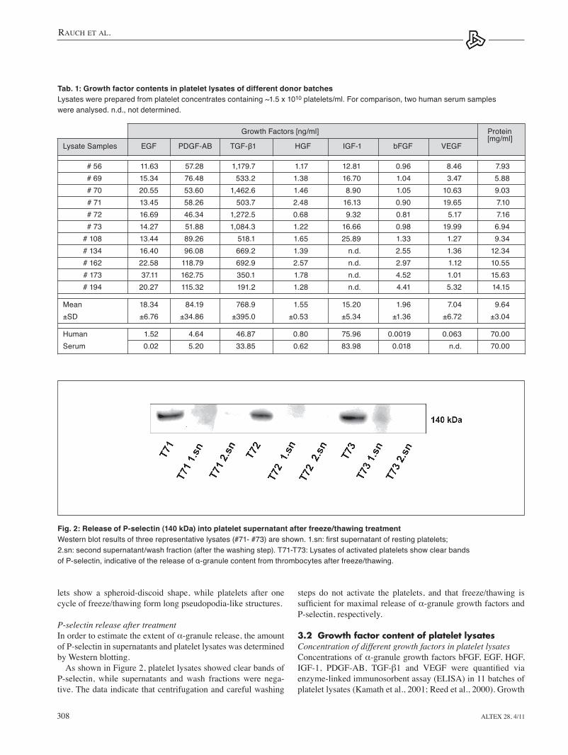
P-selectin release after treatment In order to estimate the extent of α-granule release, the amount of P-selectin in supernatants and platelet lysates was determined by Western blotting.
As shown in Figure 2, platelet lysates showed clear bands of P-selectin, while supernatants and wash fractions were nega-tive. the data indicate that centrifugation and careful washing
Fig. 2: Release of P-selectin (140 kDa) into platelet supernatant after freeze/thawing treatmentWestern blot results of three representative lysates (#71- #73) are shown. 1.sn: first supernatant of resting platelets; 2.sn: second supernatant/wash fraction (after the washing step). T71-T73: Lysates of activated platelets show clear bands of P-selectin, indicative of the release of α-granule content from thrombocytes after freeze/thawing.
Tab. 1: Growth factor contents in platelet lysates of different donor batches Lysates were prepared from platelet concentrates containing ~1.5 x 10¹⁰ platelets/ml. For comparison, two human serum samples were analysed. n.d., not determined.
Growth Factors [ng/ml] Protein
Lysate Samples EGF PDGF-AB TGF-β1 HGF IGF-1 bFGF VEGF [mg/ml]
# 56 11.63 57.28 1,179.7 1.17 12.81 0.96 8.46 7.93 # 69 15.34 76.48 533.2 1.38 16.70 1.04 3.47 5.88 # 70 20.55 53.60 1,462.6 1.46 8.90 1.05 10.63 9.03 # 71 13.45 58.26 503.7 2.48 16.13 0.90 19.65 7.10 # 72 16.69 46.34 1,272.5 0.68 9.32 0.81 5.17 7.16 # 73 14.27 51.88 1,084.3 1.22 16.66 0.98 19.99 6.94 # 108 13.44 89.26 518.1 1.65 25.89 1.33 1.27 9.34 # 134 16.40 96.08 669.2 1.39 n.d. 2.55 1.36 12.34 # 162 22.58 118.79 692.9 2.57 n.d. 2.97 1.12 10.55 # 173 37.11 162.75 350.1 1.78 n.d. 4.52 1.01 15.63 # 194 20.27 115.32 191.2 1.28 n.d. 4.41 5.32 14.15
Mean 18.34 84.19 768.9 1.55 15.20 1.96 7.04 9.64±SD ±6.76 ±34.86 ±395.0 ±0.53 ±5.34 ±1.36 ±6.72 ±3.04
Human 1.52 4.64 46.87 0.80 75.96 0.0019 0.063 70.00Serum 0.02 5.20 33.85 0.62 83.98 0.018 n.d. 70.00
r-altex_2011_4_305_316_Rauch.indd 308 24.11.2011 19:54:41 Uhr

Rauch et al.
Altex 28, 4 /11 309
factor concentrations in lysate preparations are listed in tab. 1. For comparison the levels of these factors determined in two unrelated human serum samples are given.
Platelet-derived growth factor (PDGF), a strong mitogen, is a major constituent of α-granules. Concentrations found in plate-let lysates are in the range of 50-100 ng/ml (tab. 1), indicating a 10- to 20-fold enrichment compared to normal serum levels. epidermal growth factor (eGF), which is present in a number of serum-free culture media formulations in recommended con-centrations of 1-100 ng/ml, is also a strong mitogen (Barnes and Sato, 1980; Gstraunthaler, 2003). Mean eGF contents in platelet lysates are in the range of ~20 ng/ml compared to ~1 ng/ml in human serum.
the different batches show concentration differences. these differences are likely attributable to individual donor factors, such as age and sex. Overall, the platelet lysates contained higher levels of α-granule factors PDGF-AB, EGF, bFGF, HGF, TGF-β1 and VEGF than the serum samples, whereas the level of the liver-derived serum marker IGF-1 was high-er in the serum sample. Platelet lysates also showed a lower protein content than human serum, which is consistent with the removal of immunoglobulins and albumin in the washing procedure.
Based on these results, the following quality criteria for plate-let lysates could be defined: high contents of PDGF-AB, VEGF, EGF, bFGF, and TGF-β1, and low IGF-1 content and low pro-tein content [~10 mg/ml].
Growth factor stability during storage of platelet lysates to test the stability of platelet lysates during long-term storage, lysates were stored at -20°C for five months. Every month, the content of eGF as a representative lysate growth factor mark-er was determined. Results show a strong stability of eGF in
platelet lysates over the test period under this storage condition (Fig. 3).
Growth-promoting effects of platelet lysates on cell lines For investigation of the growth-promoting effect of platelet lysates, cell culture experiments with a variety of human and animal cell lines were performed. Different concentrations of standardized platelet lysates (1%, 5% and 10% v/v) were added as serum replacement to serum-free basal culture media. Results from pilot experiments revealed that addition of 5% was suf-ficient to promote cell proliferation (data not shown). The cells were cultured for up to 28 days, phase contrast micrographs were taken at day 3, 7, 14, and 28.
the results after 14 days with llC-PK1 and llC-PK1-FBPase+ porcine kidney cells, MDCK canine kidney cells, HK-2 human kidney cells, and OK opossum kidney cells (Gstraunthaler, 1988) are depicted in Figure 4. As can be seen, 5% platelet lysate supplementation fully supported growth and proliferation of the renal epithelial cell lines comparable to cultures with 10% FBS.
Raji cells (human lymphoblastoid cell line) and tHP-1 cells (human acute monocytic leukemia cell line) were used as models for suspension-grown cultures (Fig. 5). 5% Platelet lysate also fully supported the growth of the human suspen-sion cell lines.
Furthermore, primary cultures of human chondrocytes, iso-lated from cartilage tissue in a tissue engineering laboratory, proliferated in media with 10% platelet lysates in human col-lagen-coated culture dishes. Human cornea epithelia were also successfully maintained in platelet lysate-supplemented media (data not shown).
For investigation whether cell attachment and growth can be enhanced by collagen-coating, we used collagen-coated cell dish-
Fig. 3: Growth factor stability of EGF in platelet lysates stored at -20°C for five monthsValues are expressed as means ±SD of four lysate samples.
r-altex_2011_4_305_316_Rauch.indd 309 24.11.2011 19:54:41 Uhr

Rauch et al.
Altex 28, 4/11310
Fig. 4: Cell growth experiments using animal and human renal epithelial cell lines (LLC-PK1 pig kidney cell line, LLC-PK1-F+ strain, MDCK Madin-Darby canine kidney cell line, HK-2 human kidney cell line, OK opossum kidney cells)Cells were grown on DMEM with either 10% FBS, without any supplementation (serum-free, SF) or with the addition of 5% platelet lysate (PL). Phase contrast micrographs were taken after 14 days in culture. Magnification 20 x.
r-altex_2011_4_305_316_Rauch.indd 310 24.11.2011 19:54:42 Uhr

Rauch et al.
Altex 28, 4 /11 311
3.3 Cell growth promotion by platelet lysatesGrowth curves of LLC-PK1 and MDCK cells to quantify the cell growth experiments, representative growth curves for llC-PK1 and MDCK cultures were determined by in situ-cell counting for llC-PK1 and WSt-8 proliferation as-say for MDCK cultures, respectively (lindl und Gstraunthaler, 2008). the results in Figure 6 clearly revealed that in both meth-ods the growth-promoting effect of 5% platelet lysate is almost identical to that of 10% FBS.
es in comparison to tissue culture-treated polystyrol dishes. Coat-ed dishes with proteins of the extracellular matrix are frequently used in serum-free or serum-reduced cell cultivation for improve-ment of cell attachment and growth, because serum-free media partially contain lower attachment proteins than FBS containing media (Gstraunthaler, 2003; lindl und Gstraunthaler, 2008). For MDCK cells collagen-coating did not improve cell proliferation, whereas for llC-PK1 cells collagen-coating could enhance the growth rate in platelet lysate containing media (data not shown).
Fig. 5: Cell growth experiment using human cell lines grown in suspension culture (Raji, a lymphoblastoid cell line derived from a Burkitt lymphoma and THP-1, a human acute monocytic leukemia cell line)Cells were grown in RPMI-1640 medium with either 10% FBS, without any supplementation (serum-free, SF) or with the addition of 5% platelet lysate (PL). Phase contrast micrographs were taken after 14 days in culture. Magnification 20 x.
Fig. 6: Growth curves of LLC-PK1 (left) and MDCK cells (right) under FBS or PL supplementation Cultures were grown on DMEM with either 10% FBS (black), without any supplementation (serum-free, SF, dashed line) or with the addition of 5% platelet lysate (PL, grey). Left panel: Cell counts (cells/mm2) of LLC-PK1 cultures; right panel: WST-8 assay of MDCK cells. Data are expressed as means ±SD of three independent experiments.
r-altex_2011_4_305_316_Rauch.indd 311 24.11.2011 19:54:43 Uhr

Rauch et al.
Altex 28, 4/11312
h. Platelet lysate-treated cells show a slightly higher stimulation after 5 min compared to FBS. Addition of serum-free medium to serum-deprived cultures did not stimulate the MAP kinases.
4 Discussion
the 3Rs are guiding principles for the use of animals in research and testing, first described by Russell and Burch more than 50 years ago (Balls, 2010; Balls et al., 1995). their intention was to reduce pain and fear of experimental animals defined by the 3Rs concept (Refine, Reduce, and Replace). Refinement means any decrease in the incidence or severity of inhumane procedures. Reduction means the reduction in the numbers of animals used to obtain information of a given amount and precision. Replace-ment means the substitution of any experimental animals by in vitro and/or in silico methods (Fletcher et al., 2011; Mahadevan et al., 2011).
the search for alternatives to replace fetal bovine serum (FBS) has become a major goal in the field of cell and tissue
Stimulation of ERK1/2 MAP kinases In order to visualize the mitogenic effect of growth factors in platelet lysates, phospho-specific ERK Western blots using llC-PK1 cells were performed. For this blotting technique, Phospho-specific antibodies against ERK1 and ERK2 were used. eRK1 and eRK2 are members of the MAPK family, which belong to a specific transcriptional activation cascade involved in many cell regulating processes. eRK1/2 are acti-vated by specific phosphorylation that initiates the transcrip-tion of genes leading to cell growth and proliferation (Feifel et al., 2002; Schramek, 2002).
the Western blots presented in Figure 7 show the induction of eRK1/2 phosphorylation in FBS- or platelet lysate-stimulated llC-PK1 cells. llC-PK1 cultures were serum-deprived for 24 h and then stimulated by the addition of 10% FBS, 5% plate-let lysate or serum-free medium without any supplementation with growth factors or hormones. the bands show distinct phos-phorylation of eRK1 and eRK2. Phosphorylation, and thus eRK activation starts about two minutes after stimulation, with a peak after ten minutes and a decrease to basal values after 3
Fig. 7: Western blot analysis of specific ERK1/2 phosphorylation, and thus activation, in whole cell extracts of LLC-PK1 cells upon addition of FBS and platelet lysate, respectively Subconfluent LLC-PK1 cultures were serum-depleted for 24 h and then stimulated by the addition of 10% FBS or 5% PL (upper panels). At the time points indicated, cells were harvested and subjected to SDS-PAGE and Western blotting. Addition of serum-free medium served as a control. Equal protein loading was assessed by staining with a pan ERK2 antibody (lower panels).
r-altex_2011_4_305_316_Rauch.indd 312 24.11.2011 19:54:43 Uhr

Rauch et al.
Altex 28, 4 /11 313
of biochemical activators. the morphological status of platelets was visualized by scan-
ning electron microscopy (SeM), and platelet degranulation was determined by specific P-selectin release (Fig. 1, 2). P-selectin, also known as CD62P or PADGeM is a transmembrane protein of the α-granule that is released together with the granule con-tent into the supernatant (Mcever, 1995; Blann and lip, 1997).
the lysates obtained by freeze/thawing showed high concen-trations of the α-granule derived growth factors EGF, bFGF, HGF, PDGF-AB, TGF-β1, and VEGF. As expected, the content of liver-derived IGF-1 was lower in the lysates than in human serum samples. the low protein concentration of about 10 mg/ml suggests a low concentration of extraneous material, thus reducing the risk of causing immunological reactions.
Platelet-derived growth factor concentrations have also been investigated by several other laboratories (Christgau et al., 2006; Cognasse et al., 2006; Doucet et al., 2005; Frechette et al., 2005; Gruber et al., 2002; Kaps et al., 2002; lacoste et al., 2003; Ogino et al., 2006; Schallmoser et al., 2007; Shen et al., 2006; Weibrich et al., 2002; Zimmermann et al., 2003). the results of these analyses are not comparable with our results, because of different preparation methods: All except el Backly et al. and Gruber et al. (el Backly et al., 2011; Gruber et al., 2002) stored platelets in serum, which also contains growth factors, and/or in platelet additive solution.
We further assessed the stability of the growth factors in the platelet supernatant during long-term storage. eGF was chosen as a marker as it is commonly used in cell culture media. Results showed that eGF is stable at -20°C for at least five months (Fig. 3).
culture. the efforts to develop a substitute for FBS are pri-marily based on two intentions. On the one hand, harvest and production of FBS is ethically problematic, and thus a replace-ment is needed (Brunner et al., 2010; Jochems et al., 2002; van der Valk et al., 2004). On the other hand, the rapidly expanding fields of in vitro cell and tissue culture, biomedical tissue en-gineering, and (adult) stem-cell therapy require defined, safe, and animal component-free culture conditions (Atala, 2007; van der Valk et al., 2010). In case of the scientific aspect the substitution of sera would lead to a better quality and reproduc-ibility of experimental data as suggested by Good Cell Culture Practice (Balls et al., 2006; Coecke et al., 2005; Gstraunthaler, 2006, 2010; Hartung et al., 2002). Serum or FBS is known to be a chemically ill-defined supplement, bearing qualitative and quantitative variations that exhibit a possible source of micro-bial contamination with fungi, bacteria, viruses, mycoplasma, and prions (Brunner et al., 2010; Gstraunthaler, 2003; van der Valk et al., 2010).
More than 30 years ago (Hayashi and Sato, 1976) researchers started efforts to find alternatives to animal serum (Brunner et al., 2010; Gstraunthaler, 2003; lindl und Gstraunthaler, 2008). Since then a whole range of substitutes have become com-mercially available or are still under investigation, including chemically-defined media substitutes, tissue extracts (e.g., pi-tuitary extracts), ocular fluid (Filipic et al., 2002), bovine milk fractions or bovine colostrum (Belford et al., 1995), plant ex-tracts (vegetal serum) (Pazos et al., 2004), and platelet lysates (Johansson et al., 2003).
Serum, rather than plasma, promotes the growth and prolifera-tion of cultured cells (Gospodarowicz and Ill, 1980). In the past, much effort was spent to identify the factors in serum that stimu-late cell growth in vitro. An important step forward in the search for serum growth factors has been the finding that the most po-tent mitogenic factors present in serum are derived from activated thrombocytes (Balk et al., 1981; Gospodarowicz and Ill, 1980).
Platelets are known to be a rich source of growth factors (Marx et al., 1998; Weibrich et al., 2002; Anitua et al., 2004), thus suggesting platelet lysates as a valuable animal serum substitute. Platelet lysates were tested on mesenchymal stem cells (MSCs), concluding that the lysates were more efficient in terms of costs and proliferation rate than using exogenous recombinant growth factors and retained their immunosuppres-sive (Doucet et al., 2005) as well as their differentiation capabil-ity (lange et al., 2007).
Donor platelets have a shelf life of five days for therapeutic applications, but they retain all growth factors for at least three weeks (Chan et al., 2005). Outdated donor platelets obtained by apheresis were separated from the plasma or platelet addi-tive solution (PAS) (Shanwell et al., 2003) by centrifugation and gentle resuspension in physiological saline. the counting of platelets by the CASY® Cell Counter revealed a marginal loss of platelets during the preparation procedure. the platelet suspen-sion yielded an average platelet count of 1.5 x 10¹⁰/ml.
Pilot experiments established freeze/thawing as the method of choice for enriching α-granule derived components. Besides being the most economic method, a further advantage of this physical method is that the platelet lysates do not contain traces
Fig. 8: MAP kinase signaling cascade leading to cell proliferation Mitogens (e.g., EGF, whole serum) activate Raf, which phosporylates MEK (MAPK/ERK kinase), that leads to a specific phosphorylation of ERK1/2. As a result, the terminal kinases activate downstream transcription factors that initiate specific gene expression programs for cell division and proliferation, respectively.
r-altex_2011_4_305_316_Rauch.indd 313 24.11.2011 19:54:44 Uhr

Rauch et al.
Altex 28, 4/11314
terms of the 3Rs and as a promising substitute for FBS in all in vitro methodologies, like cell and tissue culture, in vitro toxic-ity studies, prevalidation and validation, tissue engineering, and cell therapy (leist et al., 2008).
References Abrams, C. S. (2005). Intracellular signaling in platelets. Curr.
Opin. Hematol. 12, 401-405. Anitua, e., Andia, I., Ardanza, B., et al. (2004). Autologous
platelets as a source of proteins for healing and tissue regen-eration. Thromb. Haemost. 91, 4-15.
Atala, A. (2007). engineering tissues, organs and cells. J. Tissue Eng. Regen. Med. 1, 83-96.
Balk, S. D., levine, S. P., Young, l. l., et al. (1981). Mitogenic factors present in serum but not in plasma. Proc. Natl. Acad. Sci. USA 78, 5656-5660.
Balls, M., Goldberg, A. M., Fentem, J. H., et al. (1995). the three Rs: the way forward. the report and recommendations of eCVAM Workshop 11. ATLA 23, 838-866.
Balls, M., Coecke, S., Bowe, G., et al. (2006): the importance of Good Cell Culture Practice (GCCP). ALTEX 23, Spec. Is-sue, 270-273.
Balls, M. (2010). the principles of humane experimental tech-nique: timeless insights and unheeded warnings. ALTEX 27, Spec. Issue, 19-23.
Barano, J. l. S. and Hammond, J. H. (1985). Serum-free me-dium enhances growth and differentiation of cultured pig granulosa cells. Endocrinol. 116, 51-58.
Barnes, D. and Sato, G. (1980). Methods for growth of cultured cells in serum-free medium. Anal. Biochem. 102, 255-270.
Bartholomew, A., Sturgeon, C., Siatskas, M., et al. (2002). Mes-enchymal stem cells suppress lymphocyte proliferation in vitro and prolong skin graft survival in vivo. Exp. Hematol. 30, 42-48.
Belford, D. A., Rogers, M.-l., Regester, G. O., et al. (1995). Milk-derived growth factors as serum supplements for the growth of fibroblasts and epithelial cells. In Vitro Cell. Dev. Biol. 31, 752-760.
Bernardo, M. e., Avanzini, M. A., Perotti, C., et al. (2007). Op-timization of in vitro expansion of human multipotent mes-enchymal stromal cells for cell-therapy approaches: further insights in the search for fetal calf serum substitute. J. Cell. Physiol. 211, 121-130.
Bieback, K., Hecker, A., Kocaömer, A., et al. (2009). Human alternatives to fetal bovine serum for the expansion of mes-enchymal stromal cells from bone marrow. Stem Cells 27, 2331-2341.
Blann, A. D. and lip, G. Y. H. (1997). Hypothesis: Is soluble P-selectin a new marker of platelet activation? Atherosclerosis 128, 135-138.
Brunner, D., Frank, J., Appl, H., et al. (2010). Serum-free cell culture: the serum-free media interactive online database. ALTEX 27, 53-62.
Burnouf, t., tseng, Y. H., Kuo, Y. P., and Su, C. Y. (2008). Sol-vent/detergent treatment of platelet concentrates enhances the release of growth factors. Transfusion 48, 1090-1098.
In order to biochemically determine the proliferative po-tential of platelet lysates, the stimulation of extracellular sig-nal-regulated MAP kinase (eRK1/2) by platelet lysates was determined. eRK1/2, activated by extracellular stimuli such as growth factors or FBS, are part of the MAPK (mitogen-activated protein kinase) signaling network that regulates growth, proliferation, differentiation, and survival of almost all cells (treisman, 1996; Chang and Karin, 2001; Pearson et al., 2001) (Fig. 8). Addition of 5% platelet lysates to serum-starved, quiescent llC-PK1 cells induced phosphorylation, and thus activation, of eRK1/2 within minutes. the activation potential of platelet lysates was comparable with that of FBS (Figure 7). In recent functional studies, llC-PK1 and MDCK epithelia cultured in the presence of platelet lysates generated a transepithelial electrical resistance (teeR) comparable to FBS (manuscript in preparation). teeR is a sensitive indicator for epithelial differentiation, assembly of cell-cell junctional com-plexes, and vectorial transepithelial solute transport across cul-tured epithelial layers (Gstraunthaler, 1988).
Results from our experiments indicate a high potential for platelet lysates as a growth-promoting culture media substitute for a multitude of cell lines. Moreover, the application of plate-let lysate would minimize the risk of microbial contaminations (Kuznetsov et al., 2000), reduce immunological reactions (Bar-tholomew et al., 2002), and be more economical than FBS or recombinant growth factors (Doucet et al., 2005).
Aside from use as a universal serum replacement, platelet lysate could also serve as a substitute for autologous human serum in tissue engineering and biomedical cell therapy (Kli-manskaya et al., 2008; Minuth et al., 1998; Stock and Vacanti, 2001). tissue engineering requires authorized GMP (Good Manufacturing Practice) protocols and autologous culture con-ditions for the cultivation and expansion of human donor cells (Halme and Kessler, 2006; McDevitt and Palecek, 2008). In or-der to avoid additional stress for patients by extracting a 400 ml unit of blood for autologous serum production, this could be overcome with the use of platelet apheresis and subsequent platelet lysate production.
In case of platelet transfusion therapy, allogenic platelet con-centrates from voluntary donors with blood groups and Rhesus factors identical with the recipients are applied. thus, also for cultivation and expansion of donor cells, it could be feasible to use allogenic platelet lysate with matching blood group and Rhesus factor (Kaufman, 2009; Shehata et al., 2009; Stroncek and Rebulla, 2007). In this context, the collaboration with a tissue engineering laboratory showed that our platelet lysate could support the growth of human chondrocytes. Also, hu-man corneal epithelia were successfully maintained in platelet lysate-supplemented media (data not shown).
ESAC, the ECVAM Scientific Advisory Committee, recent-ly endorsed a statement that strongly recommends – whenever possible – the use of non-animal alternatives to FBS in new in vitro test methods: “For methods forwarded to ECVAM for validation/prevalidation where this is not fulfilled, a justifica-tion for future use must be provided, including measures taken to seek non-animal alternatives to FBS” (eSAC, 2008).
thus, platelet lysates can serve as a full “Replacement” in
r-altex_2011_4_305_316_Rauch.indd 314 24.11.2011 19:54:44 Uhr

Rauch et al.
Altex 28, 4 /11 315
mRNAs in gluconeogenic llC-PK1-FBPase+ cells. Am. J. Physiol. Renal Physiol. 278, F227-237.
Gstraunthaler, G. (2003). Alternatives to the use of fetal bovine serum: serum-free cell culture. ALTEX 20, 275-281.
Gstraunthaler, G. (2006). Standardization in cell and tissue cul-ture – the need for specific GLP guidelines in the cell culture laboratory (Good Cell Culture Practice – GCCP). ALTEX 23, Spec. Issue, 274-277.
Gstraunthaler, G. (2010). the Bologna Statement on Good Cell Culture Practice (GCCP) – 10 years later. ALTEX 27, Spec. Issue, 141-146.
Hartung, t., Balls, M., Bardouille, C., et al. (2002). Good Cell Culture Practice. eCVAM Good cell culture practice task force report 1. ATLA 30, 407-414.
Hayashi, I. and Sato, G.-H. (1976). Replacement of serum by hormones permits growth of cells in a defined medium. Na-ture 259, 132-134.
Jochems, C. E. A., van der Valk, J. B. F., Stafleu, F. R., and Baumans, V. (2002). the use of fetal bovine serum: ethical or scientific problem? ATLA 30, 219-227.
Johansson, l., Klinth, J., Holmqvist, O., and Ohlson, S. (2003). Platelet lysate: a replacement for fetal bovine serum in animal cell culture? Cytotechnol. 42, 67-74.
Kamath, S., Blann, A. D., and lip, G. Y. H. (2001). Platelet activation: assessment and quantification. Eur. Heart J. 22, 1561-1571.
Kaps, C., loch, A., Haisch, A., et al. (2002). Human platelet su-pernatant promotes proliferation but not differentiation of ar-ticular chondrocytes. Med. Biol. Eng. Comput. 40, 485-490.
Kaufman, R. M. (2009). Platelet ABO matters. Transfusion 49, 5-7.
King, S. M. and Reed, G. l. (2002). Development of platelet secretory granules. Sem. Cell Dev. Biol. 13, 293-302.
Klimanskaya, I., Chung, Y., Meisner, l., et al. (2005). Human embryonic stem cells derived without feeder cells. Lancet 365, 1636-1641.
Kuznetsov, S. A., Mankani, M. H., and Robey, P. G. (2000). ef-fect of serum on human bone marrow stromal cells: ex vivo expansion and in vivo bone formation. Transplantation 70, 1780-1787.
lacoste, e., Martineau, I., and Gagnon, G. (2003). Platelet con-centrates: effects of calcium and thrombin on endothelial cell proliferation and growth factor release. J. Periodontol. 74, 1498-1507.
lange, C., Cakiroglu, F., Spiess, A.-N., et al. (2007). Acceler-ated and safe expansion of human mesenchymal stromal cells in animal serum-free medium for transplantation and regen-erative medicine. J. Cell. Physiol. 213, 18-26.
leist, M., Bremer, S., Brundin, P., et al. (2008). the biological and ethical basis of the use of human embryonic stem cells for in vitro test systems or cell therapy. ALTEX 25, 163-190.
lindl, t. and Gstraunthaler, G. (2008). Zell- und Gewebekultur. Von den Grundlagen zur Laborbank. Heidelberg: Spektrum Akademischer Verlag.
Maguire, P. B. and Fitzgerald, D. J. (2003). Platelet proteomics. J. Thromb. Haemost. 1, 1593-1601.
Mahadevan, B., Snyder, R. D., Waters, M. D., et al. (2011). Ge-
Chan, R. K., liu P., lew, D. H., et al. (2005). expired liquid pre-served platelet releasates retain proliferative activity. J. Surg. Res. 126, 55-58.
Chang, l. and Karin, M. (2001). Mammalian MAP kinase sig-nalling cascades. Nature 410, 37-40.
Christgau, M., Moder D., Hiller, K. A., et al. (2006). Growth factors and cytokines in autologous platelet concentrate and their correlation to periodontal regeneration outcomes. J. Clin. Periodontol. 33, 837-845.
Coecke, S., Balls, M., Bowe, G., et al. (2005). Guidance on Good Cell Culture Practice. A report of the second eCVAM task force on Good Cell Culture Practice. ATLA 33, 261-287.
Cognasse, F., Boussoulade F., Chavarin, P., et al. (2006). Re-lease of potential immunomodulatory factors during platelet storage. Transfusion 46, 1184-1189.
Doucet, C., ernou, I., Zhang, Y., et al. (2005). Platelet lysates promote mesenchymal stem cell expansion: a safety substi-tute for animal serum in cell-based therapy applications. J. Cell. Physiol. 205, 228-236.
el Backly, R., Ulivi, V., tonachini, l., et al. (2011). Platelet lysate induces in vitro wound healing of human keratinocytes associated with a strong proinflammatory response. Tissue Eng. A 17, 1787-1800.
eSAC (2008). Statement on the use of FCS and other animal-derived supplements. http://ecvam.jrc.it/index.cfm?voce=s&idvoce=27&idmm=4&idsm=27
Falkner, e., Appl, H., eder, C., et al. (2006). Serum free cell culture: the free access online database. toxicol. In Vitro 20, 395-400.
Feifel e., Obexer P., Andratsch M., et al. (2002). p38 MAPK mediates acid-induced transcription of PePCK in llC-PK1-FBPase+ cells. Am. J. Physiol. Renal Physiol. 283, F678-F688.
Filipic, B., Shehata, M., toth, S., et al. (2002). Novel serum re-placement based on bovine ocular fluid: a useful tool for culti-vation of different animal cells in vitro. ALTEX 19, 15-20.
Fletcher, K., Shah, R. R., thomas, A., et al. (2011). Novel ap-proaches to assessing cardiac safety – proceedings of a work-shop: regulators, industry and academia discuss the future of in silico cardiac modelling to predict the proarrhythmic safety of drugs. Drug Saf. 34, 439-443.
Frechette, J. P., Martineau, I., and Gagnon, G. (2005). Platelet-rich plasmas: growth factor content and roles in wound heal-ing. J. Dent. Res. 84, 434-439.
Gachet, C. (2000). Platelet activation by ADP: the role of ADP antagonists. Ann. Med. 32, Suppl. 1, 15-20.
Gospodarowicz, D. and Ill, C. R. (1980). Do plasma and serum have different abilities to promote cell growth? Proc. Natl. Acad. Sci. USA 77, 2726-2730.
Gruber, R., Varga, F., Fischer, M. B., and Watzek, G. (2002). Platelets stimulate proliferation of bone cells: involvement of platelet-derived growth factor, microparticles and mem-branes. Clin. Oral Implants Res. 13, 529-535.
Gstraunthaler, G. J. (1988). epithelial cells in tissue culture (Re-view). Renal Physiol. Biochem. 11, 1-42.
Gstraunthaler, G., Holcomb, t., Feifel, e., et al. (2000). Dif-ferential expression and acid-base regulation of glutaminase
r-altex_2011_4_305_316_Rauch.indd 315 24.11.2011 19:54:44 Uhr

Rauch et al.
Altex 28, 4/11316
Shen, e. C., Chou t. C., Gau, C. H., et al. (2006). Releasing growth factors from activated human platelets after chitosan stimulation: a possible bio-material for platelet-rich plasma preparation. Clin. Oral Implants Res. 17, 572-578.
Spreafico, A., Chellini, F., Frediani, B., et al. (2009). Biochemical investigation of the effects of human platelet releasates on hu-man articular chondrocytes. J. Cell. Biochem. 108, 1153-1165.
Stock, U. A. and Vacanti, J. P. (2001). tissue engineering: cur-rent state and prospects. Annu. Rev. Med. 52, 443-451.
Stroncek, D. F. and Rebulla, P. (2007). Platelet transfusions. Lancet 370, 427-438.
taupin, P. (2006). Derivation of embryonic stem cells for cel-lular therapy: challenges and new strategies. Med. Sci. Monit. 12, RA75-78.
taupin, P. (2007). Stem cells engineering for cell-based therapy. J. Neural. Eng. 4, R59-63.
treisman, R. (1996). Regulation of transcription by MAP kinase cascades. Curr. Opin. Cell Biol. 8, 205-215.
van der Valk, J., Mellor, D., Brands, R., et al. (2004). the humane collection of fetal bovine serum and possibilities for serum-free cell and tissue culture. Toxicol. In Vitro 18, 1-12.
van der Valk, J., Brunner, D., De Smet, K., et al. (2010). Opti-mization of chemically defined cell culture media – replacing fetal bovine serum in mammalian in vitro methods. Toxicol. In Vitro 24, 1053-1063.
Weibrich, G., Kleis, W. K., Hafner, G., and Hitzler, W. e. (2002). Growth factor levels in platelet-rich plasma and correlations with donor age, sex, and platelet count. J. Craniomaxillofac. Surg. 30, 97-102.
Zimmermann, R., Arnold, D., Strasser, e., et al. (2003). Sample preparation technique and white cell content influence the de-tectable levels of growth factors in platelet concentrates. Vox Sang. 85, 283-289.
Acknowledgements the authors thank Mrs. edna Nemati for the superb scanning electron microscopy work. this study was generously support-ed by the Pollux Private Foundation.
Correspondence toGerhard Gstraunthaler, PhDInnsbruck Medical University Division of Physiology Fritz-Pregl-Strasse 3 6020 Innsbruck Austria http://physiologie.i-med.ac.at e-mail: [email protected]
netic toxicology in the 21st century: Reflections and future directions. Environ. Mol. Mutagen. 52, 339-354.
Martin, M. J., Muotri, A., Gage, F., and Varki, A. (2005). Hu-man embryonic stem cells express an immunogenic nonhu-man sialic acid. Nat. Med. 11, 228-232.
Marx, R. e., Carlson, e. R., eichstaedt, R. M., et al. (1998). Platelet-rich plasma: Growth factor enhancement for bone grafts. Oral Surg. Oral Med. Oral Pathol. Oral Radiol. En-dod. 85, 638-646.
McDevitt, t. C. and Palecek, S. P. (2008). Innovation in the culture and derivation of pluripotent human stem cells. Curr. Opin. Biotechnol. 19, 527-533.
Mcever, R. P. (1995). Regulation of function and expression of P-selectin. Agents Actions, Suppl. 47, 117-119.
Minuth, W. W., Sittinger, M., and Kloth, S. (1998). tissue engi-neering: generation of differentiated artificial tissues for bio-medical applications. Cell Tissue Res. 291, 1-11.
Ogino, Y., Ayukawa, Y., Kukita, t., and Koyano, K. (2006). the contribution of platelet-derived growth factor, transforming growth factor-beta1, and insulin-like growth factor-I in plate-let-rich plasma to the proliferation of osteoblast-like cells. Oral Surg. Oral Med. Oral Pathol. Oral Radiol. Endod. 101, 724-729.
Park, D. H. and eve, D. J. (2009). Regenerative medicine: ad-vances in new methods and technologies. Med. Sci. Monit. 15, RA233-251.
Pazos, P., Boveri, M., Gennari, A., et al. (2004). Culturing cells without serum: lessons learnt using molecules of plant ori-gin. ALTEX 21, 67-72.
Pearson, G., Robinson, F., Beers Gibson, t., et al. (2001). Mi-togen-activated protein (MAP) kinase pathways: regulation and physiological functions. Endocr. Rev. 22, 153-183.
Pfaller, W., Gstraunthaler, G., and loidl, P. (1990). Morphology of the differentiation and maturation of llC-PK1 epithelia. J. Cell. Physiol. 142, 247-254.
Reed, G. l., Fitzgerald, M. l., and Polgar, J. (2000). Molecular mechanisms of platelet exocytosis: insights into the “secrete” life of thrombocytes. Blood 96, 3334-3342.
Rendu, F. and Brohard-Bohn, B. (2001). the platelet release re-action: granules’ constituents, secretion and functions. Plate-lets 12, 261-273.
Schallmoser, K., Bartmann, C., Rohde, e., et al. (2007). Human platelet lysate can replace fetal bovine serum for clinical-scale expansion of functional mesenchymal stromal cells. Transfu-sion 47, 1436-1446.
Schramek, H. (2002). MAP kinases: from intracellular signals to physiology and disease. News Physiol. Sci. 17, 62-67.
Shanwell, A., Falker, C., and Gullikkson, H. (2003). Storage of platelets in additive solutions: the effects of magnesium and potassium on the release of RANteS, beta-thromboglobulin, platelet factor 4 and interleukin-7, during storage. Vox Sang. 85, 206-212.
Shehata, N., tinmouth, A., Naglie, G. et al. (2009). ABO-iden-tical versus nonidentical platelet transfusion: a systematic re-view. Transfusion 49, 2442-2453.
r-altex_2011_4_305_316_Rauch.indd 316 24.11.2011 19:54:44 Uhr