alternative cied (re)implantation techniques · indication) 6) use fewest leads consistent with...
TRANSCRIPT
Alternative CIED
(Re)Implantation Techniques
WELCOME!
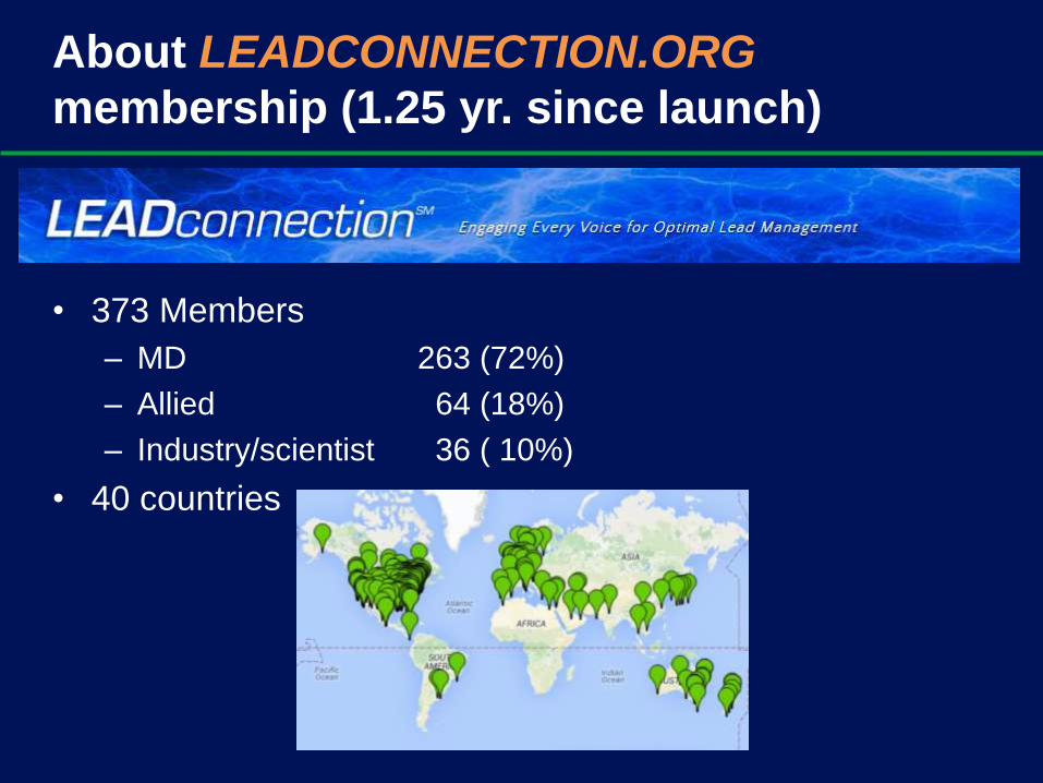
About LEADCONNECTION.ORG
membership (1.25 yr. since launch)
• 373 Members
– MD 263 (72%)
– Allied 64 (18%)
– Industry/scientist 36 ( 10%)
• 40 countries

Today’s Faculty
Bruce L. Wilkoff, MD, ChairDirector of Cardiac Pacing & Tachyarrhythmia Devices
Department of Cardiovascular Medicine
Cleveland Clinic
Professor of Medicine
Cleveland Clinic Lerner College of Medicine of Case Western Reserve University
Cleveland, OH
Charles J. Love, MDProfessor of Medicine
Director of Cardiac Rhythm Device Services
Division of Cardiology
New York University Langone Medical Center, New York, NY
President, International Board of Heart Rhythm Examiners
Jag Singh, MD, DPhilAssociate Chief, Cardiology Division
Massachusetts General Hospital
Professor of Medicine
Harvard Medical School
Boston, MA

Webinar Information
• Sponsorship and Support
– Boston Scientific, Medtronic, EBR Systems, and Spectranetics
provided the funding for this CME activity
– Postgraduate Institute for Medicine has accredited this activity
– Medtelligence organized this activity
• Webinar tips
– Using headphones (eg, earbuds) will improve your listening
experience
– Send your questions anytime during the webinar by entering them
into the chat box located at the lower left-hand side of the screen
• Note you can use the translator button
– Exit the webinar by clicking on the Exit button at the top right
corner

Claiming Credit
• A link to obtain CME credit for this webinar will be emailed
to the address provided when you registered and posted
on LEADCONNECTION.ORG
• If you don’t receive the link by August 9, please send an
email to: [email protected]
• Complete the brief evaluation and claim your credit
– This activity is 1 credit
– An enduring recording of this webinar is 1 credit, and will be
available on LEADCONNECTION.ORG

Indications for Alternative
Implantation Techniques
Bruce L. Wilkoff M.D.Cleveland Clinic
Professor of Medicine
Cleveland Clinic Lerner College of Medicine of CWRU
Director of Cardiac Pacing and Tachyarrhythmia Devices
Cleveland Clinic


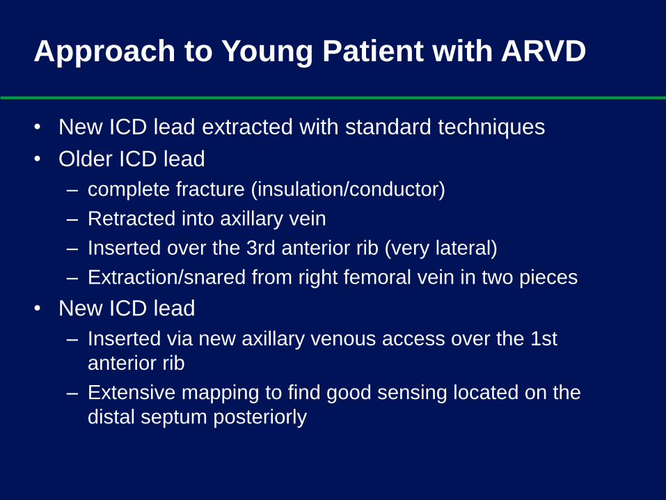
Approach to Young Patient with ARVD
• New ICD lead extracted with standard techniques
• Older ICD lead
– complete fracture (insulation/conductor)
– Retracted into axillary vein
– Inserted over the 3rd anterior rib (very lateral)
– Extraction/snared from right femoral vein in two pieces
• New ICD lead
– Inserted via new axillary venous access over the 1st
anterior rib
– Extensive mapping to find good sensing located on the
distal septum posteriorly

Assumptions & Limitations
Produces Creativity
1) Strength of CIED Indicated Therapy
2) Risk with CIED < Risk without CIED
3) Low Impact on Comorbid Conditions
4) Sustainable Implementation
5) Reversibility/Flexibility

Strength of CIED Indicated Therapy
1) Goals:
1) Survival
2) Quality of Life
3) Predictable Outcomes
4) Facilitates Therapy of Co-morbid Conditions

Risk with CIED < Risk without CIED
1) Risks
1) Arrhythmia
2) Surgery
3) Bleeding
4) Infection
5) Clotting

Low Impact on Comorbid Conditions
1) Venous stenosis/occlusion
2) Anticoagulation
3) Valvular regurgitation
4) Ventricular function/Cardiac Synchronization
5) Infection
6) Chronic Kidney Disease and/or Dialysis
7) Indicated or previous cardiothoracic surgery

Sustainable Implementation
1) Current leads are reliable
2) Veins will sustain lead and vascular access needs
3) Lowest risk of infection
4) Supplies all required therapy
5) Ready for next device change

Reversibility/Flexibility
1) Program around issues
2) Extraction feasible

Indications for Lead Extraction
1. Infection 2. Lead Dysfunction 3. Vascular Access

One Year Mortality with InfectionTarakji KG et al. Europace. 2014;16:1490-5.
33%
7%
24%

Scenario #1a
Pacemaker Infection – Young Patient
1) Symptomatic bradycardia (Atrial or AV Block)
2) Pacemaker Infection
1) Extraction!!!
2) Reimplant other side, iliac, epicardial if still needed
3) Use fewest leads consistent with good therapy
1) Leadless, single chamber, dual chamber, CRT

Scenario #1b
Pacemaker Infection – Older Patient
1) Symptomatic bradycardia (Atrial or AV Block)
2) Pacemaker Infection
1) Consider comorbidities (If Prognosis >2 yrs, consider young)
2) Extraction!!!
3) Evaluate for continued requirement for therapy
4) Extraction & Reimplant other side, iliac, epicardial if still
needed!
5) Comorbidities (Dialysis, renal dysfunction, other OHS
indication)
6) Use fewest leads consistent with good therapy
1) Leadless, single chamber, dual chamber, CRT

Scenario #1c
ICD Infection – Young Patient
1) Primary or Secondary Prevention ICD
2) ICD Infection
1) Evaluate for alternative approach therapy (Ablation, Rx)
2) Extraction!!!
3) Reimplant other side, iliac, epicardial if still needed vs
SICD
4) Use fewest leads consistent with good therapy
1) SubQ, single chamber, dual chamber, CRT
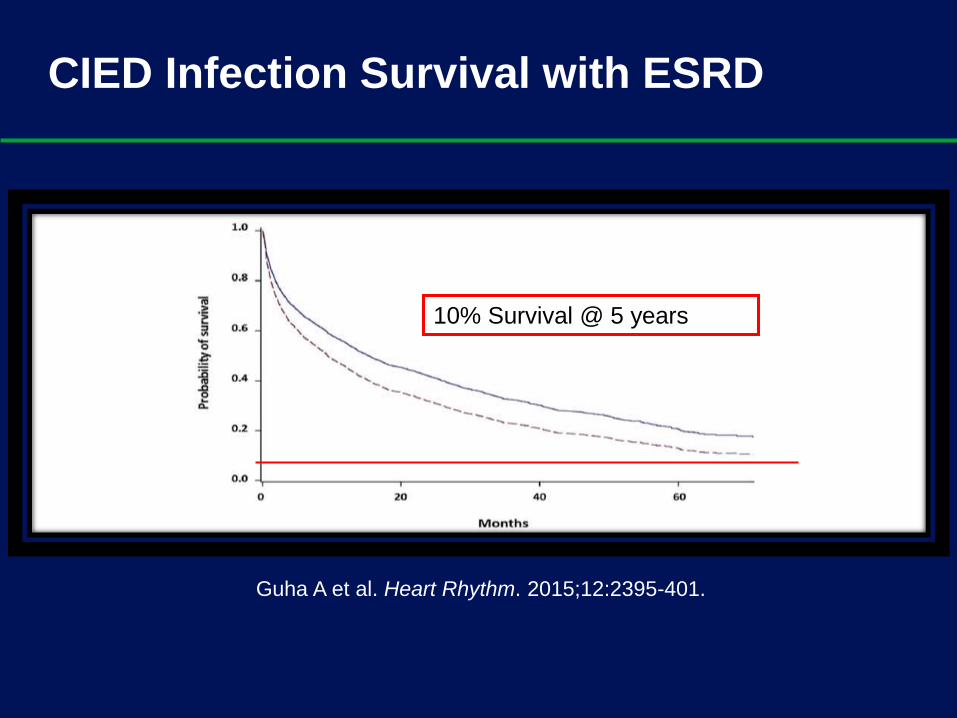
CIED Infection Survival with ESRD
Guha A et al. Heart Rhythm. 2015;12:2395-401.
10% Survival @ 5 years

Scenario #1d
CIED Infection – Older Patient
1) Primary or Secondary Prevention ICD
2) ICD Infection
1) Evaluate for alternative approach therapy (Ablation,
Rx)
2) Extraction! Consider no reimplantation
3) Reimplant other side, iliac, epicardial if still needed
vs SICD
4) Use fewest leads consistent with good therapy
1) SubQ, single chamber, dual chamber, CRT

Leads vs Pulse Generators
• Leads
– Saturated w/ Body Fluids
– Mechanically Stressed
– Intrathoracic
– Implant Technique Sensitive
– Patient Activity Sensitive
– High Frequency of Recalls
– HIGH RISK
• Pulse Generators
– Hermetic Sealed
– Mechanically protected
– Extrathoracic
– Implant Technique
Insensitive
– Patient Activity Insensitive
– Lower Frequency of Recalls
– LOW RISK

Only 72% survive after a 10-year period
How Long do Pacemaker Leads Last?
Fortescue EB et al. Heart Rhythm. 2004;1:150-9.
Failure 28% at 10 years

ICD leads: How long do they last?
Kleeman T et al. Circulation. 2007:115:2474-80.
Failure 15% at 5 years, 40% at 8 years

Scenario #2a
Lead is Unreliable – Young Patient
1) Symptomatic bradycardia (Atrial or AV Block)
2) Pacemaker Lead failure
1) Evaluate for alternative approach therapy
2) Extraction!
3) Reimplant same side if still needed
4) Use fewest leads consistent with good therapy
1) Leadless, single chamber, dual chamber, CRT

Scenario #2b
Lead is Unreliable – Older Patient
1) Symptomatic bradycardia (Atrial or AV Block)
2) Pacemaker Lead failure
1) Consider comorbidities (If Prognosis >10 yrs, consider
young)
2) Evaluate for alternative approach therapy
3) Extraction & Reimplant same side if still needed!
4) Implant on other side if prognosis very poor
5) Use fewest leads consistent with good therapy
1) Leadless, single chamber, dual chamber, CRT
Scenario #2c
Lead is Unreliable – Young Patient
1) Primary or Secondary Prevention ICD
2) ICD Lead failure
1) Evaluate for alternative approach therapy (Ablation, Rx)
2) Extraction!!!
3) Reimplant same side if still needed
4) Use fewest leads consistent with good therapy
1) SubQ, single chamber, dual chamber, CRT

Scenario #2d
Lead is Unreliable – Older Patient
1) Primary or Secondary Prevention ICD
2) ICD Lead failure
1) Evaluate for alternative approach therapy (Ablation, Rx)
2) Extraction unless prognosis poor (<3 years)
3) Reimplant same side if still needed
4) Use fewest leads consistent with good therapy
1) SubQ, single chamber, dual chamber, CRT

PoorJudgment

Scenario #3a
Veins are Occluded – Young Patient
1) Symptomatic bradycardia (Atrial or AV Block)
2) Needs another Pacemaker lead to deliver therapy
1) Evaluate for alternative approach therapy
2) Extraction! vs Venoplasty
1) Leads unuseful vs useful
3) Reimplant same side!!!
4) Use fewest leads consistent with good therapy
1) Leadless, single chamber, dual chamber, CRT
Scenario #3b
Veins are Occluded – Older Patient
1) Symptomatic bradycardia (Atrial or AV Block)
2) Needs another Pacemaker lead to deliver therapy
1) Consider comorbidities
(If Prognosis >10 yrs, consider young)
2) Extraction! vs Venoplasty
1) Leads unuseful vs useful
3) Extraction & Reimplant same side!!
4) Implant on other side if Prognosis <3 yrs
5) Use fewest leads consistent with good therapy
1) Leadless, single chamber, dual chamber, CRT

Scenario #3c
Veins are Occluded – Young Patient
1) Primary or Secondary Prevention ICD
2) Needs another ICD lead to deliver therapy
1) Evaluate for alternative approach therapy (Ablation, Rx)
2) Extraction!!!
3) Reimplant same side!!!
4) Use fewest leads consistent with good therapy
1) SubQ, single chamber, dual chamber, CRT

Scenario #3d
Veins are Occluded – Older Patient
1) Primary or Secondary Prevention ICD
2) Needs another ICD lead to deliver therapy
1) Evaluate for alternative approach therapy (Ablation, Rx)
2) Extraction unless prognosis poor (<3 years)
3) Reimplant same side!!!
4) Use fewest leads consistent with good therapy
1) SubQ, single chamber, dual chamber, CRT

All Cause Mortality 30 Days – Post Extraction
Category Odds Ratio P value
Body mass index <25 kg/m2 1.8 0.043
End Stage Renal Disease 4.8 <0.001
NYHA FC 0.006
II 1.3
III 2.0
IV 8.5
Hemoglobin 3.3 <0.001
INR 1.3 0.01
Infection 1.3 0.002
Dual Coil ICD Lead 2.7 <0.001
Brunner MP, Wilkoff BL, et al. Heart Rhythm. 2014;11:419-25.
Assumptions & Limitations
Produces Creativity and Reality Check
1) Strength of CIED Indicated Therapy
2) Risk with CIED < Risk without CIED
3) Low Impact on Comorbid Conditions
4) Sustainable Implementation
5) Reversibility/Flexibility

Indications for Alternative
Implantation Techniques
Bruce L. Wilkoff M.D.Cleveland Clinic
Professor of Medicine
Cleveland Clinic Lerner College of Medicine of CWRU
Director of Cardiac Pacing and Tachyarrhythmia Devices
The Cleveland Clinic Foundation

Charles J. Love, MD FACC FAHA FHRS CCDSProfessor of Clinical MedicineDirector, Cardiac Rhythm Device ServicesNew York University Langone Medical CenterNew York, NY USAPresident, International Board of Heart Rhythm Examiners
ICD & Pacemaker Implantation
after Extraction

Disclosures
• Medtronic: Consultant
• St. Jude Medical: Consultant
• Spectranetics: Consultant

Site Considerations
• Pectoral
• Sub-Mammary
• Lateral Mammary
• Axillary
• Abdomen
• Epicardial
• Limited Atriotomy
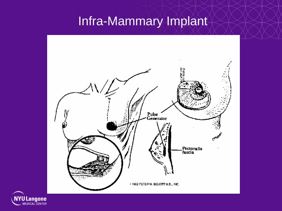
Infra-Mammary Implant

Infra-Mammary
• Very cosmetic
• Be wary of bra cup irritation
• Lateral approach is an effective
alternative (similar to axillary site)
– Device placed in the pre or sub-
pectoral position
• Fixation of lead body may be difficult

Axillary Approach

Abdomen Placement
• May be used for epicardial leads• Useful when femoral vein approach is
used

Femoral Implant
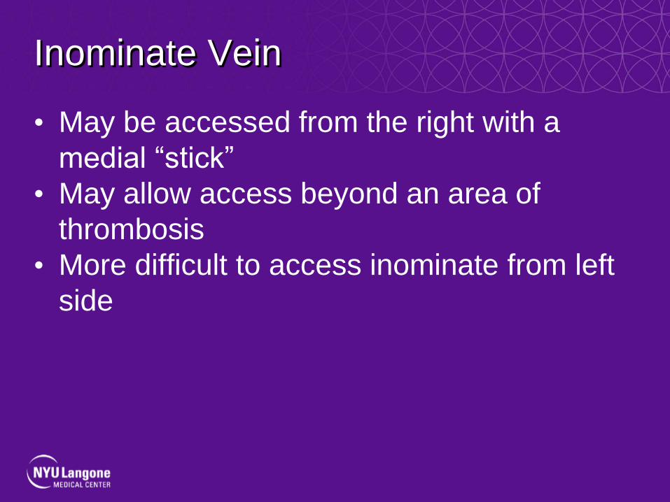
Inominate Vein
• May be accessed from the right with a
medial “stick”
• May allow access beyond an area of
thrombosis
• More difficult to access inominate from left
side

Internal Jugular Implant

Transiliac / Femoral approach
• Useful when all superior vein occluded, or
when congenital anomaly prevents access
via superior routes
– vein is accessed via cutdown and/or
introducer technique
– active fixation leads placed
– pacemaker inserted in lower abdominal
wall
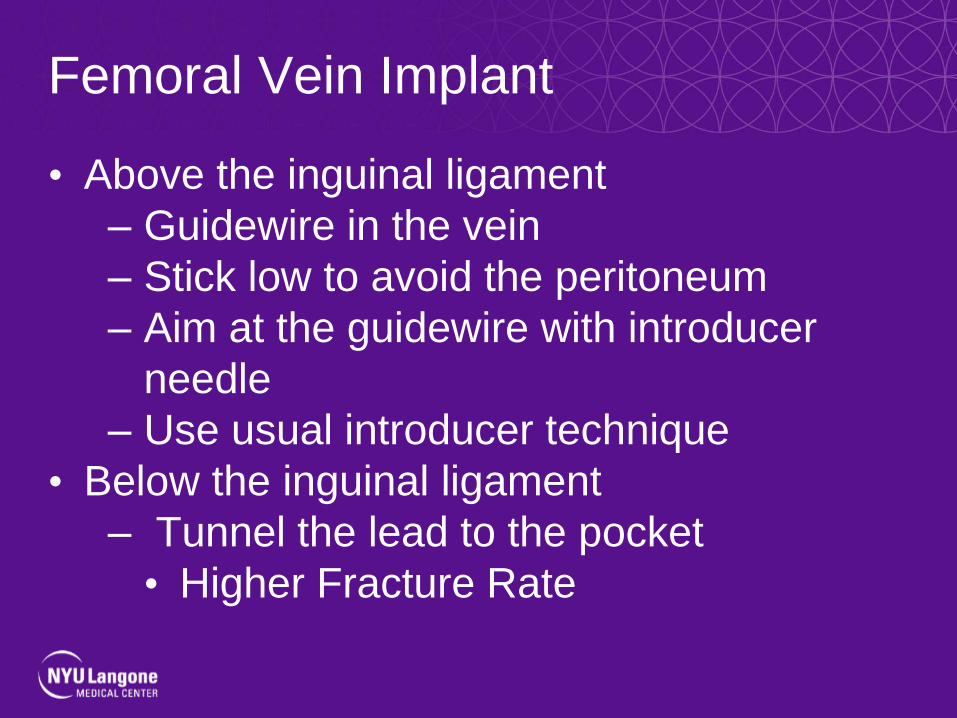
Femoral Vein Implant
• Above the inguinal ligament
– Guidewire in the vein
– Stick low to avoid the peritoneum
– Aim at the guidewire with introducer
needle
– Use usual introducer technique
• Below the inguinal ligament
– Tunnel the lead to the pocket
• Higher Fracture Rate

Femoral Vein Implant

Abdomen Implant
Caution

Inferior Vena Cava
• Useful when all superior vein occluded, or
when congenital anomaly prevents access
via superior routes
– “mini-laparotomy”; right flank incision
– retro-peritoneal identification of IVC
– active fixation leads placed through
purse-string sutures
– ?less risk of fracture vs femoral
approach

IVC Implant

Transatrial Approach
• Endocardial leads may be utilized• Dual chamber pacing may be performed• Chronic leads may be removed
– General anesthesia– Incision over 3rd/4th costochondral
cartilage – Purse-string suture in atrium /
appendage– Introducer / sheath placed, then
lead(s)– Pacer pocket made via incision
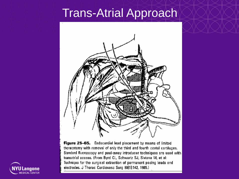
Trans-Atrial Approach
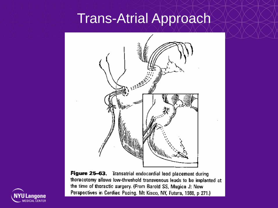
Trans-Atrial Approach

Minimally invasive epicardial lead
placement
• 2 or 3 one cm incisions are made in the intercostal space
• Thoracoscope inserted into one• Lead advanced through the other• Selective intubation of right and left
mainstem bronchi required• Alternative to subxiphoid, thoracotomy and
sternotomy approaches

Subcutaneous-ICD System
• Designed to sense,
detect and treat
malignant ventricular
tachyarrhythmias
– Primary Prevention
– Secondary Prevention
• S-ICD System
is entirely
subcutaneous
Burke, S-ICD, HRS 2012, Boston, MA

SC-ICD
• No Fluoroscopy needed
• No venous stick
• No leads in the venous system
• No leads in the heart
• No leads across the heart valve

Leadless VVIR Pacemaker
• Intended for patients that have a Class I or II indication for a single-chamber ventricular pacemaker
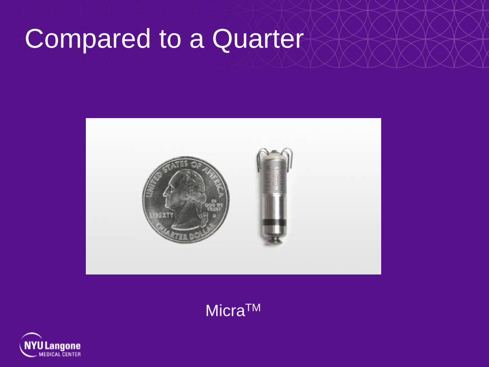
Compared to a Quarter
MicraTM

Or a Euro
NanostimTM

Typical Delivery System + Introducer
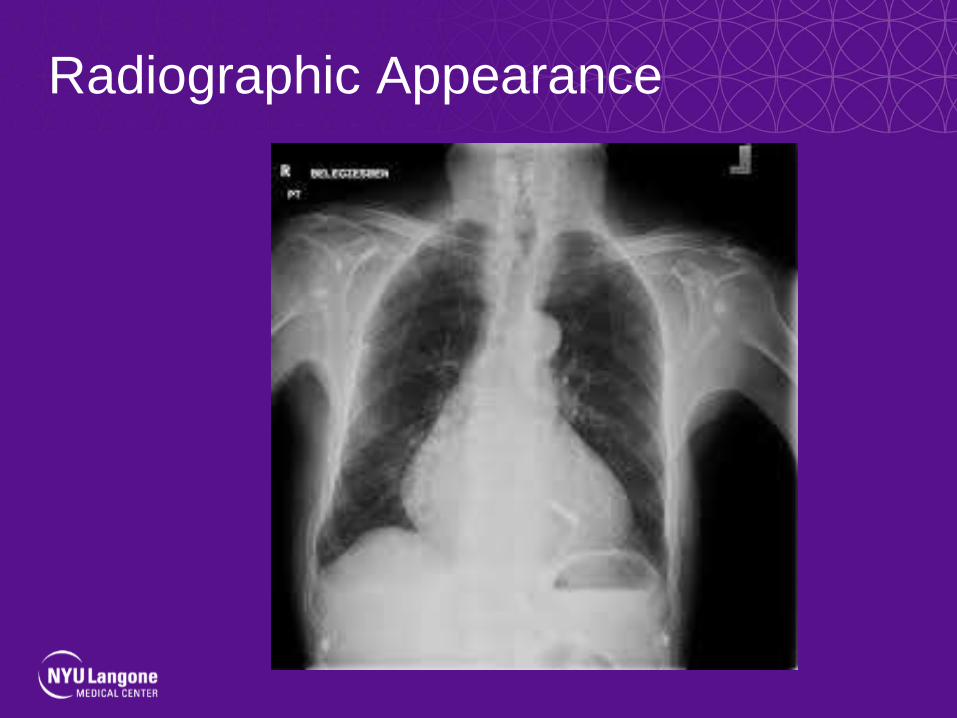
Radiographic Appearance

Post Extraction CRT ImplantNovel techniques & Technologies
Jag Singh MD DPhil FHRS
Associate Chief, Cardiology DivisionProfessor of Medicine, Harvard Medical School
Massachusetts General Hospital, Boston
Deputy Editor, Journal Am Coll Cardiol: Clinical EP
Disclosures:
Consultant: Biotronik, Boston Scientific, Impulse Dynamics, Liva Nova, Medtronic, Respicardia Inc, St. Jude
Medical, theHeart.org, Research Grants: St. Jude Medical, Boston Scientific

Overview
• Post-extraction challenges, need an individualized
implantation strategy
• Imaging:
• Intra-procedural coronary venography important for assessing
options
• Sometimes pre-procedural imaging, in a staged procedure, may
be useful
• Coronary venous interventional strategies
• Venoplasty & Stenting
• Evolving LV endocardial pacing strategies

Re-implanting the LV leadPotential Challenges
Tackling an unwilling anatomy
• Venous stenosis
• Distal Thrombotic Occlusion of Vein
Path of no return
• Thrombotic occlusion of main branch
• No alternative branches
• Coronary Sinus Occlusion
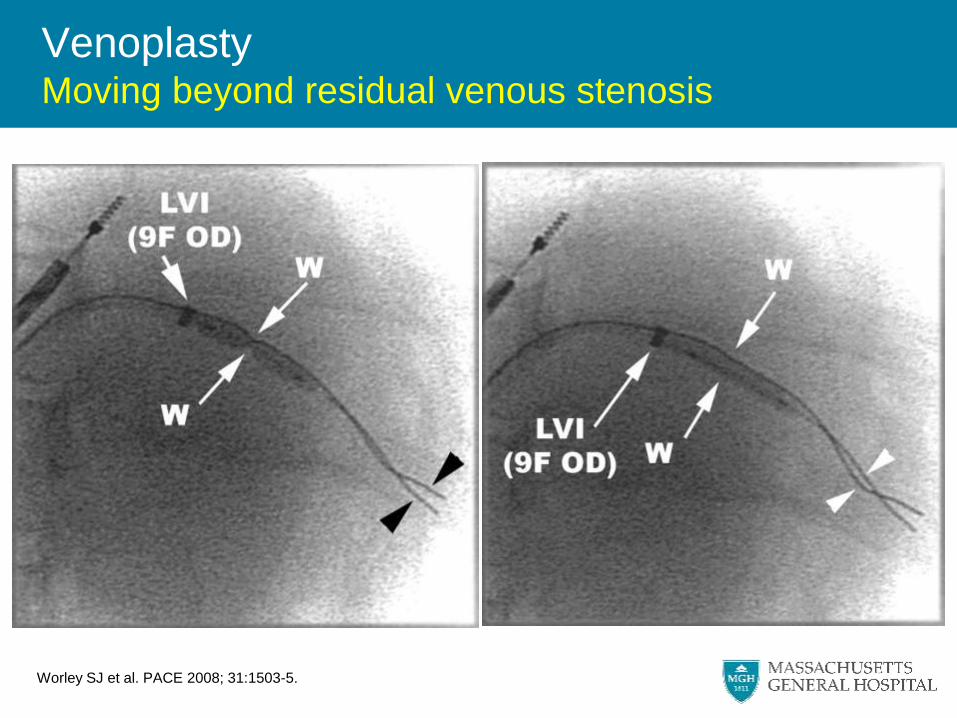
VenoplastyMoving beyond residual venous stenosis
Worley SJ et al. PACE 2008; 31:1503-5.

Altered Anatomy: Securing the Lead Proximally Stenting as an option
László Gellér et al. Heart Rhythm 2011; 8:845-50.

Intra-luminal InteractionStent, Vascular tissue & Lead
• Explanted Heart
examples
• Intact intimal tissue
layer seen
• No sign of occluding
proliferative tissue
• No lead injury
Future Implications for
extraction?
• Manually extracted
with gentle traction
Balazs T et al. J Cardiovasc Electrophysiol 2013; 24:468-70

Value of pre-procedural ImagingDemonstrates options
Truong QA / Singh JP: Critical Pathways in Cardiology 2008; 7:185-90. •23
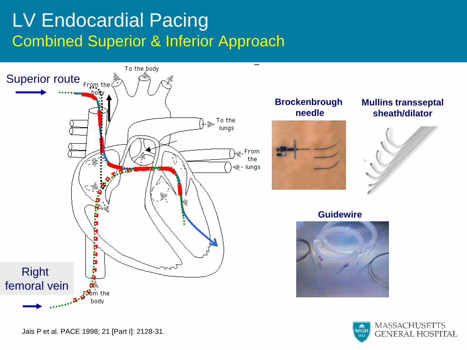
Mullins transseptal
sheath/dilator
Brockenbrough
needle
Guidewire
Right
femoral vein
Superior route
LV Endocardial Pacing Combined Superior & Inferior Approach
Jais P et al. PACE 1998; 21 [Part I]: 2128-31.

Van Gelder BM et al. Heart Rhythm 2007;4:454-60.
Endocardial LV lead Implantation:Combined approach

LV Endocardial PacingShoulder approach
• Complete CRT Implant via Shoulder approach with
LV lead implantation individualized to area of most
delayed electrical activation (Animal Study)
• ALSYNC Study provides more insight
•Singh JP and Gras D; EHJ 2011
•Barrett CD, Singh JP, et al; HRS 2011
•Exner D, Auricchio A and Singh JP. Heart Rhythm 2012
• Endocardial approach
provides the needed
individualization
• Best site is variable
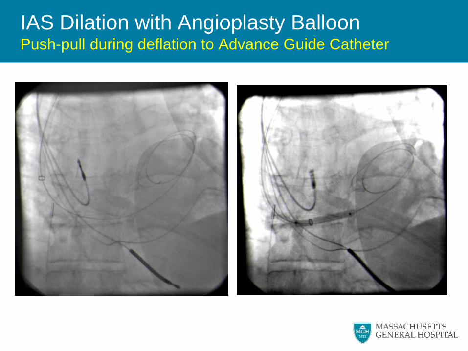
IAS Dilation with Angioplasty BalloonPush-pull during deflation to Advance Guide Catheter

LV Electro-anatomical Map & CRT Leads
- Shoulder Access to LV via Transseptal route
• Animal Study
• Complete CRT Implant via Shoulder approach with
LV lead implantation individualized to area of most
delayed electrical activation
• Endocardial approach
provides the needed
individualization
• Best site is variable
•Singh JP and Gras D; EHJ 2011
•Barrett CD, Singh JP, et al; HRS 2011
•Exner D, Auricchio A and Singh JP. Heart Rhythm 2012

ALSYNC: LV Endocardial approachTechnique
Morgan JM et al. European Heart Journal 2016; 37:2118-27.
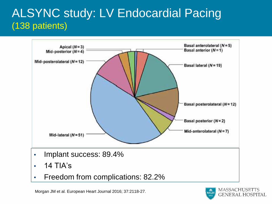
ALSYNC study: LV Endocardial Pacing(138 patients)
• Implant success: 89.4%
• 14 TIA’s
• Freedom from complications: 82.2%
Morgan JM et al. European Heart Journal 2016; 37:2118-27.

Kassai I et al. Alternative Method for Cardiac Resynchronization: Transapical Lead Implantation,
Ann Thorac Surg 2009; 87:650-2.)
Trans-apical Endocardial LV lead placementAlternative strategies: ? role
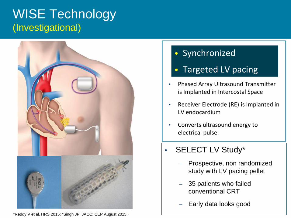
• Synchronized
• Targeted LV pacing
• Phased Array Ultrasound Transmitter is Implanted in Intercostal Space
• Receiver Electrode (RE) is Implanted in LV endocardium
• Converts ultrasound energy to electrical pulse.
WISE Technology(Investigational)
• SELECT LV Study*
– Prospective, non randomized
study with LV pacing pellet
– 35 patients who failed
conventional CRT
– Early data looks good
*Reddy V et al. HRS 2015; *Singh JP. JACC: CEP August 2015.

In Summary
• Post-extraction challenges, need an individualized
implantation strategy
• Imaging:
• Intra-procedural coronary venography important for assessing
options
• Sometimes pre-procedural imaging, in a staged procedure, may
be useful
• Coronary venous interventional strategies
• Venoplasty & Stenting
• Evolving LV endocardial pacing strategies

Claiming Credit
• A link to obtain CME credit for this webinar will be emailed
to the address provided when you registered and posted
on LEADCONNECTION.ORG
• If you don’t receive the link by August 9, please send an
email to: [email protected]
• Complete the brief evaluation and claim your credit
– This activity is 1 credit
– An enduring recording of this webinar is 1 credit, and will be
available on LEADCONNECTION.ORG