alternative approach to extraction of maxillary fourth
TRANSCRIPT
Journal of Veterinary Research Advances Short Communication
ISSN: 2582-774X Open access
Visit at: http://jvra.org.in Vol 03 No 02, p 01-06/1
Alternative approach to extraction of maxillary fourth premolar in dogs
Anthony Caiafa and Kayoko Kuroda 1James Cook University, School of Veterinary and Biomedical Sciences, Queensland, Australia
2Northside Veterinary Care, Strathpine, Queensland, Australia
Corresponding author: [email protected]
Received on: 11/04/2021 Accepted on: 28/06/2021 Published on: 13/07/2021
ABSTRACT
Aim: The study was aimed to identify an alternative approach to the extraction of the maxillary 4th premolar tooth in dogs, that offers a more predictable and reproducible approach to extracting this tooth, whilst reducing intra-operative and post-operative complications, such as root fracture or flap dehiscence. Method and materials: Twelve client-owned dogs (July to November 2020) were presented for extraction of the maxillary 4th premolar tooth, due to traumatic injury or significant pathology associated with the tooth. Results: The alternative approach to the extraction of the maxillary 4th premolar was performed without complication in the 12 client-owned dogs, and with dental equipment readily available in the majority of companion animal practices. Conclusion: The alternative approach to the surgical extraction of the maxillary 4th premolar offers a reproducible and easily mastered procedure, whilst limiting complications associated with extracting multirooted teeth.
Keywords: Crown amputation, dog, extraction, furcation, maxillary fourth premolar.
Cite This Article as: Caiafa A and Kuroda K (2021). Alternative approach to extraction of maxillary fourth premolar in dogs. J. Vet. Res. Adv. 03(02): 01-06.
Introduction
Exodontia (tooth extraction) is one of the most common oral surgical procedures performed by small animal veterinary surgeons (Reiter, 2018). Extraction of the maxillary fourth premolar tooth (PM4) may be indicated due to severe periodontal or endodontic disease, as well as dentoalveolar trauma, tooth and/or root anomalies and malocclusions resulting in trauma to oral soft tissues. The extraction of maxillary PM4 should be performed when other more advanced options of saving the tooth are not appropriate or or where financial constraints arise (Carmichael, 2002 and Smith, 1998).
There are very few reports that describe the approach to the removal of the maxillary 4th premolar in the dog and cat (Carmichael, 2002, Charlier, 2019, Gorrel, 2004, Lommer, 2012, Reilter, 2018 and Smith, 1998). This study describes an alternative approach to the surgical extraction of the maxillary 4th premolar tooth in the dog. This alternative approach was shown to be repeatable, predictable and effective. Copyright: Caiafa and Kuroda. Open Access. This article is distributed under the terms of the Creative Commons Attribution 4.0 International License (http://creativecommons.org/licenses/by/4.0/), which permits unrestricted use, distribution, and reproduction in any medium, provided you give appropriate credit to the original author(s) and the source, provide a link to the Creative Commons license, and indicate if changes were made.
The technique should also minimize intra-operative complications as well as post-operative morbidity, when compared to other techniques previously described (Charlier, 2019 and Gorrel, 2004). The main objective of present research was to provide the armamentarium for exodontia and to offer an alternative and reproducible approach to the surgical extraction of this tooth.
Case Presentations Clint owned dogs with toothaches, fractures and other dental problems were subjected for procedures. Before planning and undertaking any tooth extraction, owner’s consent was taken to perform exodontia. Then perform a comprehensive oral health assessment, including intraoral radiographs and periodontal probing, to examine and assess the tooth and any associated pathology, as well as inspecting the surrounding anatomy.
Treatment and discussion Multimodal pain management, including provision of regional anaesthesia, is highly recommended for pre-emptive analgesia and for patient comfort, as well as to reduce the amount of inhalation anaesthesia used during the procedure. All 12 patients underwent the same anaesthetic protocol,

Journal of Veterinary Research Advances Open access
Visit at: http://jvra.org.in Vol 03 No 02, p 01-06/2
including pre-anaesthetic blood tests and premedication with Acepromazine at a dosage of 0.025 mg/kg (ACP 2; Ceva) and Methadone at a dosage of 0.3mg/kg (Methadone; Troy) given subcutaneously, 30 minutes prior to the induction. The intravenous induction agent (Alfaxan; Jurox) for general anaesthesia was administered and titrated to effect, followed by placement and securing of a cuffed endotracheal tube of appropriate size for the patient. A pulse oximeter and respiratory monitoring device was used on all patients to monitor oxygen saturation status, pulse and respiratory rate during the procedure. Patients were maintained on between 1% to 2% of isoflurane (Isothesia; Piramal) and 1 to 3L/min of oxygen throughout the procedure according to size of the animal. Retrievable packing materials (e.g. using several layers of bunched up swabs with cotton tape tie attached) were placed into the oropharynx, to prevent entry of debris, blood, and irrigant from the high-speed handpiece. Prior to the extraction procedure, the oral cavity was rinsed with 0.14% chlorhexidine solution (Hexarinse; Virbac) to reduce surgical site bacterial contamination. Gross calculus and large dental deposits attached to the crown, as well as exposed root surfaces, were removed using a combination of calculus removing forceps and an ultrasonic scaler. This aids in the visualisation of the dental hard tissue structures, as well as, preventing dislodgment of infected material into the alveolus or soft tissues during the extraction procedure. Creating a triangular flap: Intrasulcular incision and vertical releasing incision A caudal maxillary nerve block was performed for all patients, by using either a 25G (0.5 x16mm) or 27G (0.4 x 13mm) hypodermic needle with 1mL syringe attached. The preferred local anesthetic agent was Lignocaine hydrochloride without adrenaline (Lignocaine20; Troy) at a total dosage of 4mg/Kg. divided into numbers of local anaesthesia required. A circumferential intrasulcular releasing incision was made around the maxillary PM4. The intrasulcular incision was extended to the mid-buccal region of the maxillary 1st molar tooth (Fig 1).
A diverging vertical releasing incision was made close to the mesial border of mesiobuccal (MB) root of PM4. It was created a triangular mucoperiosteal flap which better preserves blood supply to the flap due to there being only one mesial vertical releasing incision. Care was not
taken to inadvertently incise over the infraorbital foramen. A gloved finger was placed over the foramen to protect the underlying neurovascular bundle exiting the foramen, from being incised by the scalpel blade (Fig 2). The scalpel blade incised the mucoperiosteum, in an apical to coronal direction and towards the free gingival margin. The mucoperiosteum and attached gingiva were then gently elevated off the bone using a sharp periosteal elevator. Particular attention was paid to preserve the mesial corner of the gingival flap, as this area can be very fragile and easily torn or damaged by inappropriate tissue handling. Removal of the buccal bone plate over the distal and mesiobuccal roots The buccal bone covering the mesiobuccal and distal roots was removed using either a size 2 or 4 round friction grip (FG) bur in a high speed handpiece, with a copious amount of coolant, to avoid iatrogenic thermal damage to the underlying bone. The bone removal aided in the visualisation of the furcation, separating the mesial and distal roots. It was preferred to preserve the alveolar bone at the furcation site, as much as possible, in order to aid bone regeneration at the extraction site. Approximately 50% or more in some cases, of the buccal bony plate over the mesial and distal roots was removed. Extraction of the distal root (PM4) It was strongly recommended to extract the largest of the three roots (distal root) first, before sectioning and extracting the two mesial roots. Sectioning and separating the distal root from the mesial roots should start from the buccal furcation and head coronally towards the cusp of the PM4. Sectioning can be achieved by using a 22mm length 701 (L) or 25mm length 701/702 tapered fissure bur, in the high-speed handpiece, with copious water irrigation.
The distal root was sectioned in such way to allow its’ delivery in a straight line. When insufficient interdental space between the crowns of the maxillary PM4 and 1st molar was encountered, preventing the introduction of a dental luxator/elevator, space was created by using a fine tapered fissure bur (699 tapered fissure bur) to remove the last 2 mm of the distal surface, with the bur running parallel to the distal root of PM4 (Fig 3a and 3b).
Small though deep gutters (or grooves) were placed beside the mesial and distal surfaces of the distal root being careful not to damage the roots of

Journal of Veterinary Research Advances Open access
Visit at: http://jvra.org.in Vol 03 No 02, p 01-06/3
the 1st molar tooth. Again, a fine bur, like a 699 tapered fissure bur (Fig 4), was used to create these gutters, thus allowing the introduction of a dental elevator/luxator. This allows the instrument to follow the path made by the bur.
Following alveolectomy and sectioning of the tooth, as described, dental luxators/elevators were used to incise and tear the attached periodontal ligament, as well as applying a leverage force (elevator only) close to the apex of the root. Once the distal root became very mobile, extraction forceps were placed, as far apically as possible, to remove the tooth root from the alveolus. Mesial crown amputation technique After the distal root of PM4 was extracted, the remaining mesial crown of the tooth was decoronated (amputated) which allowed easier visualisation of the furcation between the MB and MP roots. The mesial crown amputation can be performed by using a 22mm length, 701 (L) or 701/702 25mm surgical length bur. The crown decoronation is usually performed down to the level of the cementoenamel junction (CEJ) of the mesial part of the crown, but can be extended further apically, if necessary. This crown amputation technique will allow clear visualisation of the furcation area between MB and MP roots, thus aiding in separating these two mesial roots. Extreme care should be taken when performing the mesial amputation so that the neighbouring tooth (PM3) and the surrounding soft tissues are not damaged by the bur.
In some breeds, as well as individual patients, the MP cusp may be too small or indistinct to visualise, or the tooth may be rotated, which can make the identification and orientation of the furcation challenging (Fig 5a, 5b and Fig 6). This crown amputation technique, described above, should significantly improve the visualisation and identification of the furcation, regardless of the breed of dog and/or anatomical variation present. Therefore, the step can aid in the prevention of one of the more common complications of exodontias, namely iatrogenic root fracture (Lommer, 2012) (Fig 6 to Fig 10).
In the past, a more “conventional” method for the extraction of the mesial roots in maxillary fourth premolar was performed by sectioning through the furcation of the mesiobuccal and mesiopalatal roots without performing mesial crown amputation (Carmichael, 2002, Charlier, 2019, Reiter, 2018 and Smith, 1998). In this
“conventional” method, confirmation of root separation was done by assessing independent movement of the MB and MP roots (Fig 11a and 11b). The “conventional” method could lead to incorrect sectioning of the MB and MP roots, leading to the common complication of root fracture, seen during the extraction of the maxillary PM4 (or any multirooted tooth). Separation of mesiobuccal (MB) and mesiopalatal (MP) root through the furcation Using the crown amputation technique, the MB and MP roots were separated through the furcation, with a tapered fissure bur, in the high-speed handpiece. The authors strongly recommend using a 699 tapered fissure bur for this method, as this FG bur is narrow in diameter, and should not remove a large amount of bone or tooth structure. The sectioning through the furcation should be performed with the bur perpendicular to the furcation and to the occlusal plane (Fig 12). Mesiobuccal (MB) root extraction The MB root was the next root to be extracted (Fig 13). Alveolar bone overlying the MB root was removed. Bony gutters were then carefully placed on the mesial and distal aspect of the root. Removal of 50% or more of the buccal bone plate using a 2 or 4 round FG bur assists in the extraction procedure. It is also recommended to carefully create small gutters, again with a 699 tapered fissure bur, along the mesial and distal borders of the MB root prior to extraction of this root, as previously described for the distal root. Mesiopalatal (MP) root extraction Once the MB root has been extracted, the remaining MP root is visualised and extracted. A small amount of the alveolar bone covering the buccal aspect of the MP root was removed, by again, using a 699 tapered fissure bur or size 2 round bur (Fig 14). Gutters can be created beside the MP root to assist with the introduction of a luxator/elevator. The MP root can be extracted in a similar manner to the MB root, although, for the MP root, some luxation is required on the palatal aspect of this root. It is important that a finger stop be utilised at all times when using the dental luxator, to avoid iatrogenic damage to the surrounding tissues, if slipping of the instrument occurs (Gorrel, 2004). The MP root may also have some curvature or dilaceration, especially towards the apex, which can increase the risk of root fracture.
Journal of Veterinary Research Advances Open access
Visit at: http://jvra.org.in Vol 03 No 02, p 01-06/4
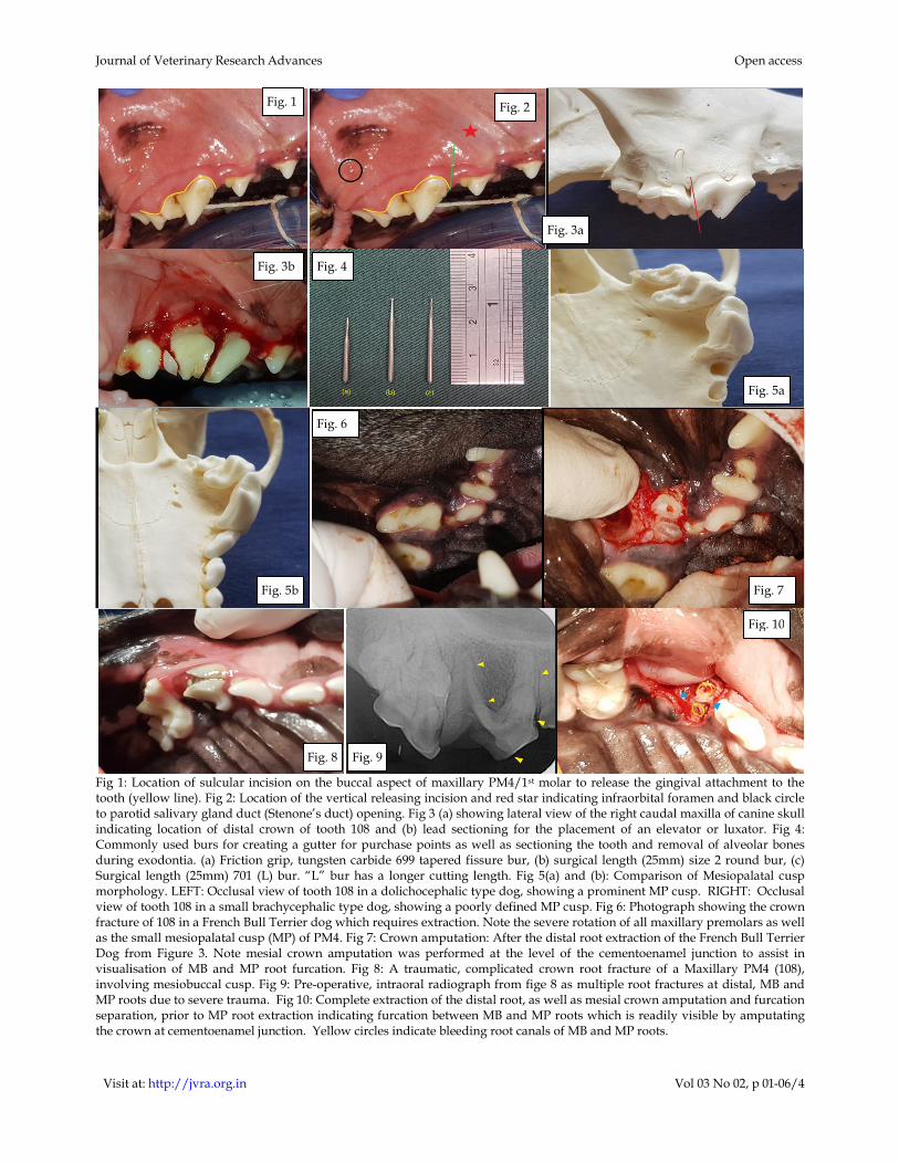
Fig 1: Location of sulcular incision on the buccal aspect of maxillary PM4/1st molar to release the gingival attachment to the tooth (yellow line). Fig 2: Location of the vertical releasing incision and red star indicating infraorbital foramen and black circle to parotid salivary gland duct (Stenone’s duct) opening. Fig 3 (a) showing lateral view of the right caudal maxilla of canine skull indicating location of distal crown of tooth 108 and (b) lead sectioning for the placement of an elevator or luxator. Fig 4: Commonly used burs for creating a gutter for purchase points as well as sectioning the tooth and removal of alveolar bones during exodontia. (a) Friction grip, tungsten carbide 699 tapered fissure bur, (b) surgical length (25mm) size 2 round bur, (c) Surgical length (25mm) 701 (L) bur. “L” bur has a longer cutting length. Fig 5(a) and (b): Comparison of Mesiopalatal cusp morphology. LEFT: Occlusal view of tooth 108 in a dolichocephalic type dog, showing a prominent MP cusp. RIGHT: Occlusal view of tooth 108 in a small brachycephalic type dog, showing a poorly defined MP cusp. Fig 6: Photograph showing the crown fracture of 108 in a French Bull Terrier dog which requires extraction. Note the severe rotation of all maxillary premolars as well as the small mesiopalatal cusp (MP) of PM4. Fig 7: Crown amputation: After the distal root extraction of the French Bull Terrier Dog from Figure 3. Note mesial crown amputation was performed at the level of the cementoenamel junction to assist in visualisation of MB and MP root furcation. Fig 8: A traumatic, complicated crown root fracture of a Maxillary PM4 (108), involving mesiobuccal cusp. Fig 9: Pre-operative, intraoral radiograph from fige 8 as multiple root fractures at distal, MB and MP roots due to severe trauma. Fig 10: Complete extraction of the distal root, as well as mesial crown amputation and furcation separation, prior to MP root extraction indicating furcation between MB and MP roots which is readily visible by amputating the crown at cementoenamel junction. Yellow circles indicate bleeding root canals of MB and MP roots.
Fig. 1 Fig. 2
Fig. 4
Fig. 3a
Fig. 3b
Fig. 5a
Fig. 5b
Fig. 6
Fig. 7
Fig. 8 Fig. 9
Fig. 10

Journal of Veterinary Research Advances Open access
Visit at: http://jvra.org.in Vol 03 No 02, p 01-06/5
Fig. 11(a) and (b): Skull model showing the sectioning through the furcation of mesiopalatal and mesiobuccal root of 108 in a conventional method. Fig. 12: Photograph showing after the sectioning through the furcation between MB and MP root of left maxillary 4th premolar tooth (208) by using 699 tapered fissure bur. Fig 13: Photograph showing post MB root extraction for the left maxillary 4th premolar (208). MP root with all four bony walls still intact at this stage. Fig 14: Photograph showing removal of the buccal bone plate (yellow arrow) present between MB and MP root for the preparation of MP root extraction during the extraction of right maxillary 4th premolar (108).
Alveolus and flap closure techniques Once all three roots of maxillary PM4 were extracted, post-operative intraoral radiographs were taken to confirm complete extraction, as well as forming a part of the medical record. Any sharp alveolar bone edges or projections were smoothed over by using a medium or coarse grit diamond football FG bur or a size 2 or 4 round FG bur prior to closure. All three alveoli were debrided using a dental curette to debride the periodontal ligament, granulation tissues and any bony debris present, followed by flushing of the alveoli with sterile 0.9% saline solution. Prior to closure, a periosteal release at the base of the mucoperiosteal flap was performed, to prevent any excessive tension in the flap. The mucoperiosteal flap was then closed with a fast-absorbing, synthetic suture material, in a simple interrupted suture pattern. Simple interrupted sutures were placed about 2 to 3 mm
apart. If excess bleeding is observed, a haemostatic agent, such as a silver impregnated gelatin sponge, can be placed prior to closure of alveoli. Post-operative instruction and follow-up examination For all 12 patients, a non-steroidal anti-inflammatory drug (NSAID) was given subcutaneously at the end of the procedure and prescribed for two to three days post-operatively given orally, unless it was contraindicated for that patient. The clients were instructed to feed their pet soft food, avoid playing with chew toys, and to administer a chlorhexidine rinse orally once or twice daily during the initial wound healing phase. For all patients, a re-examination of the surgical site was performed between 10 to 14 days post-operatively, to assess for surgical complications, such as pain, wound dehiscence and/or infection.
Charlier (2019) and Gorrel (2004) corroborated with approaches to oral surgery and extractions while Reiter (2018) also advocated closed and open
Fig. 11a
Fig. 12
Fig. 14 Fig. 13
Fig. 11b

Journal of Veterinary Research Advances Open access
Visit at: http://jvra.org.in Vol 03 No 02, p 01-06/6
tooth extraction. In this present study, complications from the tooth extraction procedures were not observed and all of the dogs recovered uneventfully. Lommer (2012), however, reported complications after tooth extractions can occur in dogs, often due to poor extraction planning.
Conclusion While performing extraction of the maxillary fourth premolar tooth in the dog, there were numerous anatomical and pathological variations that needed to be considered. This study has focused on the techniques and the sequence of steps involved in the extraction of the maxillary 4th premolar tooth, including an alternative crown amputation technique that has proven to be repeatable, predictable and an effective method of tooth extraction. This alternative method can also be applied to the extraction of other multirooted teeth in the dog and cat. .
Reference Carmichael DT (2002). Surgical Extraction of the
Maxillary Fourth Premolar Tooth in the Dog. J. Vet. Dent. 19(4): 231-233.
Charlier C (2019). Oral surgery- Extractions. In: Wigg’s Veterinary Dentistry: Principles and Practice 2nd edn. Lobprise HB and Dodd, JR (Eds.). John Wiley & Sons, Inc., PP: 229-245.
Gorrel C (2004). Veterinary Dentistry for the General Practitioner. London: Saunders Elsevier 157-174.
Lommer MJ (2012). Complications of extractions. In: Oral and Maxillofacial Surgery in Dogs and Cats Verstraete FJM and Lommer MJ (Eds.). 2012. Saunders Elsevier Philadelphia, PA, PP: 153-159.
Reiter AM (2018). Closed and open tooth extraction. In: Reiter, AM andGracis M (Eds.). 2018. BSAVA Manual of Canine and Feline Dentistry and Oral Surgery 4th edn. BSAVA, Quedgeley, Gloucester, PP: 304-337.
Smith MM (1998). Exodontics. Vet. Clin. N. Am. Small. 28(5): 1297- 319.
******