alterations of overused supraspinatus tendon: a possible role of glycosaminoglycans and...
TRANSCRIPT
Alterations of Overused Supraspinatus Tendon: A Possible Role ofGlycosaminoglycans and HARP/Pleiotrophin in Early Tendon Pathology
Mohamed Attia,1,2 Alexander Scott,3 Arlette Duchesnay,1 Gilles Carpentier,1 Louis J. Soslowsky,4 Minh Bao Huynh,1
Toin H. Van Kuppevelt,5 Camille Gossard,6 Jose Courty,1 Marie-Claude Tassoni,2 Isabelle Martelly1
1Laboratoire CRRET CNRS EAC 7149, Universite Paris-Est Creteil, 61 Av du General de Gaulle, 94010 Creteil Cedex, France, 2Cogitobio,Departement de mecanobiologie, Cachan, France, 3Department of Physical Therapy, Centre for Hip Health and Mobility, University of BritishColumbia, Vancouver, BC, Canada, 4McKay Orthopaedic Research Laboratory, University of Pennsylvania, Philadelphia, Pennsylvania, 5TheDepartment of Biochemistry, Radboud University Nijmegen Medical Centre, Nijmegen Centre for Molecular Life Sciences, Nijmegen, TheNetherlands, 6Osteobio, Ecole Superieure d’Osteopathie et de Biomecanique Appliquee, Cachan, France
Received 4 February 2011; accepted 23 May 2011
Published online 17 June 2011 in Wiley Online Library (wileyonlinelibrary.com). DOI 10.1002/jor.21479
ABSTRACT: Supraspinatus tendon overuse injuries lead to significant pain and disability in athletes and workers. Despite the preva-lence and high social cost of these injuries, the early pathological events are not well known. We analyzed the potential relationbetween glycosaminoglycan (GAG) composition and phenotypic cellular alteration using a rat model of rotator cuff overuse. Totalsulfated GAGs increased after 4 weeks of overuse and remained elevated up to 16 weeks. GAG accumulation was preceded by up-regulation of decorin, versican, and aggrecan proteoglycans (PGs) mRNAs and proteins and biglycan PG mRNA after 2 weeks. At 2weeks, collagen 1 transcript decreased whereas mRNAs for collagen 2, collagen 3, collagen 6, and the transcription factor Sox9 wereincreased. Protein levels of heparin affine regulatory peptide (HARP)/pleiotrophin, a cytokine known to regulate developmental chon-drocyte formation, were enhanced especially at 4 weeks, without up-regulation of HARP/pleiotrophin mRNA. Further results suggestthat the increased GAGs present in early lesions may sequester HARP/pleiotrophin, which could contribute to a loss of tenocyte’sphenotype. All these modifications are characteristic of a shift towards the chondrocyte phenotype. Identification of these early changesin the extra-cellular matrix may help to prevent the progression of the pathology to more disabling, degenerative alterations. � 2011Orthopaedic Research Society. Published by Wiley Periodicals, Inc. J Orthop Res 30:61–71, 2012.
Keywords: supraspinatus tendon; proteoglycans; glycosaminoglycans; chondrogenesis; HARP/pleiotrophin
Tendon is composed primarily of type 1 collagen mole-cules organized into fibrils that constitute the tension-bearing structure in association with biglycan anddecorin. Decorin, a small dermatan sulfate (DS) richproteoglycan (PG) associated with collagen fibrils,regulates their diameter and longitudinal organizationinto fibers.1,2 The collagen fibrils are embedded in thecolloid extra-cellular matrix, containing most of the70–80% tendinous water bound to the compression-bearing sulfated glycosaminoglycans (GAGs) of PGs.Tendon cells are surrounded by versican, a large chon-droitin sulfate (CS) rich PG, which buffers load trans-mitted from the collagen matrix.3 Overuse humanchronic degenerative tendinopathy due to repetitiveloading is characterized by structural and biochemicalalterations including collagen fibrils disarray, separa-tion, and disorganization, fibrocartilaginous cellularmetaplasia, GAGs accumulation4,5 and PGs variationsat mRNA levels and protein content.6,7 However tendi-nopathic biopsies are mostly available at late stages ofpathology; knowledge of early pathological events isstill incomplete.
A rat model of tendon overuse has been developedthat generates changes in histology and mechanicalproperties reproducing key characteristics of human
supraspinatus tendinopathy.8,9 This early experimen-tal tendinosis is associated with collagen fragmenta-tion, GAG accumulation, proliferating tenocytes, andhigher expression of cartilage matrix markersmRNA.10,11 These events are not primarily mediatedby the presence of inflammatory cells, althoughmRNAs of some inflammatory mediators weredetected.12 Because of their long polysaccharidicanionic chains, GAGs retain water molecules thatinfluence biomechanical tissue environment13 and alsointeract with proteins, such as cytokines, both mecha-nisms finally regulating tenocyte metabolism. It couldthus be hypothesized that changes in GAG composi-tion may be involved in early stages of tendon overusepathology. These changes may temporarily interferewith the tendon’s tensile load-bearing capacity, thusserving as a prelude to subsequent recurring collagenmicrotears and cell-mediated pathoetiological events.
The rat model appears suitable for studying thetime course of early changes in tendon matrix composi-tion and to establish the relationship between chemi-cal and mechanical modulators of cellular phenotype.Our objective was to analyze when, in overused supra-spinatus tendon, changes in GAGs composition occurand whether these changes are associated with previ-ously suggested tenocyte phenotypic alterations.
MATERIALS AND METHODSRat Running Protocol and Sample CollectionMale Sprague–Dawley rats (500 � 20 g) (Janvier, Le GenestSaint-Isle, France) were used. Twelve control animals were
Marie-Claude Tassoni and Isabelle Martelly contributed equallyto this work.Correspondence to: Isabelle Martelly (T: þ33-1-45171457; F: þ33-1-45171816 E-mail: [email protected])
� 2011 Orthopaedic Research Society. Published by Wiley Periodicals, Inc.
JOURNAL OF ORTHOPAEDIC RESEARCH JANUARY 2012 61

allowed normal cage activity. Twelve other rats weresubjected to daily treadmill downhill running.9 After 2 and 4weeks, control (n ¼ 6) and running (n ¼ 6) rats were eutha-nized, and the supraspinatus tendons were dissected freefrom their muscular and bony attachments, weighed, frozenin liquid nitrogen, and stored at �808C until use for biochem-ical studies. For immunohistochemistry on fixed rat supra-spinatus tendon, paraffin blocks from a previous cohort ofrats (n ¼ 32) trained according to the same protocol wereused.11 At each time (4, 8, 12, and 16 weeks), five runningand three control rats were studied. We used these tissueblocks to reduce the required number of animals, followingprinciples of the Canadian Council on Animal Care (192/2002).
GAG Extraction and QuantificationGAG extraction and quantification was performed accordingto.14 Aliquots of total GAG extracts were treated with nitrousacid or 0.5 U/ml chondroitinase B (Sigma, St-Quentin-Fallavier, France), and the remaining GAGs was quantifiedwith the same DMMB method. Using these chemical andenzymatic treatments, the heparan sulfate (HS), DS, and CScontents were calculated:
HS ¼ Total GAGs� DS
CS
� �; DS ¼ Total GAGs� CS
HS
� �and
CS ¼ Total GAGs�DS�HS:
Histological Analysis of Total Sulfated GAG by DMMBComplexationAt each time point, paraffin sections (7 mm) were deparaffi-nized, and DMMB solution was used labeling of total sulfatedGAGs on tissue sections after incubation overnight in thedark at room temperature. The labeling was validated usingchondroitinase ABC. A CoolSNAP camera (Princeton Instru-ments; Acton, MA) was used for images acquisition. Imageanalysis was performed using a color segmentation procedureprogrammed with ImageJ.15
Immunolabeling of GAGs and PGsAt each time point, deparaffinized sections were blocked withPBS–BSA 3% (w/v) and incubated overnight with phage dis-play single chain anti-HS, anti-DS, and anti-CS (produced byT. Van Kuppevelt). Bound antibodies were detected withmouse anti-VSV tag IgG antibody P5D4. PGs were detectedusing anti-decorin, anti-versican, and anti-aggrecan antibod-ies, followed by Alexa 488-conjugated goat anti-mouse IgG(Fluoprobes Interchim, Montlucon, France). Fluorescenceimages were obtained using a CCD monochrome camera(CFW-1310M; Scion; Frederick, MD) fitted to a BH-2epifluorescence microscope (Olympus; Rungis, France).
Protein Extraction and Western Blot AnalysisThe frozen supraspinatus tendons were homogenized inRIPA buffer containing 60 mM Tris–HCl pH 7.5, 150 mMNaCl, 10 mM EDTA, 0.1% SDS, 0.5% sodium deoxycholatesupplemented with 1/100 protease inhibitor cocktail (Sigma).Tissue was lysed overnight at 48C then centrifuged at12,000g for 10 min. Twenty micrograms proteins were ana-lyzed by Laemmli SDS–PAGE (Biorad, Marne la Coquette,France) and then transferred to Immobilon-P PVDF mem-brane (Millipore, Guyancourt, France) for detection of:
decorin (pAb LF-113, 1/5,000, kind gift from Larry Fisher,NIH, Bethesda, MD), versican (pAb, 1/200, Santa Cruz Bio-technology, Le Perray en Yvelines, France), aggrecan (pAb,1/100, Santa Cruz Biotechnology), heparin affine regulatorypeptide (HARP) (pAb, 1/1,000, R&D system, Inc, Oxon, UK),GAPDH (mAb, 1/10,000, Applied Biosystems, Courtaboeuf,France), and a-tubulin (pAb, 1/5,000, Sigma). Appropriatehorseradish-peroxidase-conjugated secondary antibodieswere visualized with BM chemiluminescence substrate(Roche Diagnostics, Meylan, France).
mRNA Isolation and Reverse Transcriptase Polymerase ChainReaction (RT-PCR)Total RNA was extracted from supraspinatus tendons usingTrizol reagent (Invitrogen, Cergy-Pontoise, France) accordingto manufacturer’s instructions. Reverse transcription wasperformed on 1 mg mRNA extracts using the SuperScript IIRNase H (Invitrogen). The resulting cDNA were used forPCR amplification. Primers were as shown in Table I. Themethod was validated by preliminary experiments thatestablished the optimal conditions. RT-PCR products weresubjected to electrophoresis on 2% (w/v) agarose gels contain-ing 0.5 mg/ml ethidium bromide. Gels were photographedusing a Chimigenius system (Syngene, Cambridge, UK) andband intensities were quantified using ImageJ.15
Heparin Versus GAGs Competition Assay Towards GrowthFactorsA competition ELISA using HARP, also called pleiotrophinor fibroblast growth factor-2 (FGF-2) was performed. Thisassay allows evaluation of the ability of GAGs from tendons,taken as competitor, to prevent the binding of HARP or FGF-2 to heparin.16 This is a qualitative assay for GAGs, whichcompetitive power depends on their composition. A heparin–BSA complex was coated overnight at 48C on 96-well plates.Wells were blocked with PBS–BSA 3% (w/v). HARP, or FGF-2 (50 ng/ml) in PBS–BSA 1% (w/v) was added to wells simul-taneously with GAGs (4 mg/ml) from each time point. Plateswere incubated overnight at 48C for competition then washedand further incubated for 2 h at room temperature with pAbagainst HARP or FGF-2 (R&D system, Inc), then incubatedfor 2 h with the appropriate peroxidase-labeled secondaryantibody. Peroxidase activity was determined according tomanufacturer’s protocol (Thermo Scientific, Brebieres,France). Binding of HARP or FGF-2 to heparin in theabsence of GAG competitor was defined as 100% binding.
Statistical AnalysisData are expressed as mean � SD. Means were comparedwith Student’s t-test using GraphPad software (San Diego,CA). Significant differences were determined at p < 0.001(���), p < 0.01 (��), and p < 0.05 (�).
RESULTS
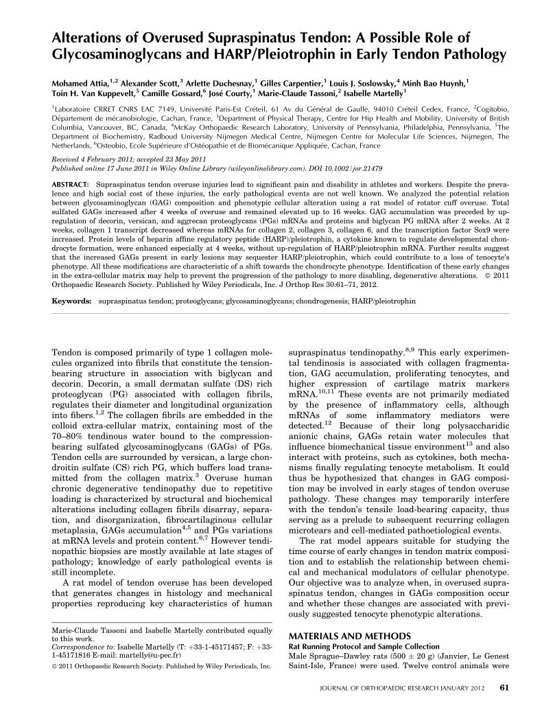
Changes in Extra-Cellular Matrix Components in OverusedSupraspinatus TendonCompared to controls, Col 1a1 mRNA expressionslightly decreased at 2 weeks, then more substantiallyat 4 weeks (by �4 fold). Col 2a1 transcripts increasedprogressively from 2 to 4 weeks, whereas Col 3a1 andCol 6a1 mRNA expressions significantly increased at2 weeks but returned to the control level at 4 weeks(Fig. 1). Levels of decorin and biglycan transcripts
62 ATTIA ET AL.
JOURNAL OF ORTHOPAEDIC RESEARCH JANUARY 2012

increased at both 2 and 4 weeks of overuse comparedto controls (Fig. 2a). The levels of mRNA of the largePGs versican and aggrecan also increased at bothtimes (Fig. 2a). The corresponding protein levelsincreased concomitantly (biglycan could not be tested)(Fig. 3a). These increases persisted for at least 8 weeks(Fig. 3b). The mRNA of transcription factor Sox9almost absent in controls was highly expressed at2 weeks and remained high at 4 weeks (Fig. 2b).Table II shows results for all the molecules examined.
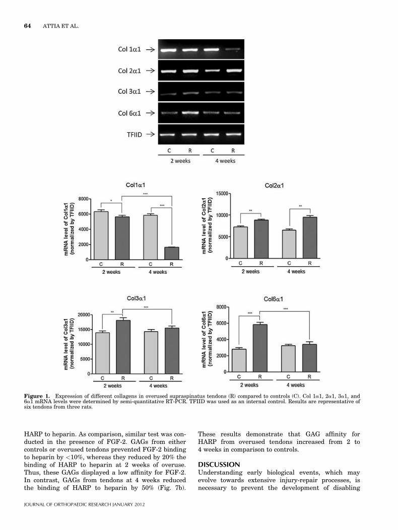
Changes in Sulfated GAGs in Overused TendonSince overuse enhanced the expression of several PGs,we examined the amount and composition of GAGs inoverused tendons and control animals (Fig. 4 andTable II). Whereas after 2 weeks of running theamount of total sulfated GAGs measured biochemicallywas not significantly altered compared to controls, itincreased by almost three times in 4 weeks-overusedtendons (Fig. 4a). This increase was also observed byhistology. The high level of sulfated GAGs observed at4 weeks then decreased progressively but stillremained significantly higher than in controls at16 weeks (Fig. 5). In controls, the main GAG specieswas DS (�80%), whereas HS or CS each represented�10% of total GAG (Fig. 4a). In overused tendons, theproportion of CS slightly increased whereas that of DSdecreased at 2 weeks, and more at 4 weeks (Fig. 4a).However, these GAG species increased by �2.5-fold at
4 weeks and thus accounted for the observed GAGaccumulation. This was corroborated by the increasein the 4S sulfotransferase (CH4ST) mRNA as early as2 weeks (Fig. 4b); CHST14 is specifically involved inDS synthesis.
The use of phage display antibodies against HS, CS,and DS showed a labeling of parallel long stripeslocated between collagen fibers in the environment oftenocytes (Fig. 6). The enhanced CS and DS labelingcompared to controls at 4 weeks was still visible at8 weeks. Altogether, GAGs accumulation appeared tocorrespond to the observed increase in transcripts andcore protein of PGs.
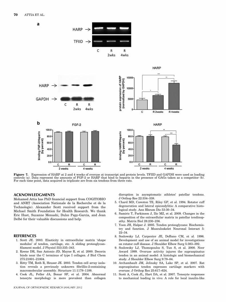
HARP Protein is Enhanced and Sequestered in OverusedSupraspinatus TendonHARP is well known as a chondrogenesis promotingfactor during development. Although the level ofHARP transcript was not altered during the first4 weeks of training, the protein level was increased upto threefold in tendon extracts at 2 and 4 weeks(Fig. 7a). Since HARP has affinity to HS/DS GAG spe-cies,17 we investigated the possibility that GAG compo-sition and affinity to HARP was altered in overusedtendons.
One possibility to detect such qualitative GAGchanges towards HARP is to perform an ELISAcompetition assay based on the ability of GAGs fromtendons, taken as competitor, to prevent the binding of
Table I. Primer Sequences and Products Size
Genes PrimersAnnealing
temperature (8C) CyclesProductsize (bp)
Collagen 1a1 50-CTGCTGGTCCTAAGGGAGAG-30 (forward) 62 35 37250-CAATACCAGGAGCACCATTG-30 (reverse)
Collagen2a1 50-GGAAGAGCGGAGACFACFGG-30 (forward) 64 40 39050-CCCTCATCTCCACATCATTG-30 (reverse)
Collagen 3a1 50-GATTCCCTGGATCTCCTGGTG-30 (forward) 62 35 30950-TCCTGGCTCTCCCTTTGCTC-30 (reverse)
Collagen 6a1 50-ATACCGGCGCAATTTCACGGC-30 (forward) 64 40 31150-AACCCTTCTCTCCACGGCTTCC-30 (reverse)
Decorin 50-GGAAFGAAGGGTCTCGGAFA-30 (forward) 62 35 39450-GACFCACGGCAGFGTAGGAA-30 (reverse)
Biglycan 50-TGFCCTTTTGGCTGCCACTGC-30 (forward) 64 40 35050-AACACGCCCFTGGGCACTTTG-30 (reverse)
Versican 50-AGACATGA 1 GGGGAAGGAAG-30 (forward) 64 40 35150-AGAGGGAAGCATGTCTGGTT-30 (reverse)
Aggrecan 50-ClGGCATTACGTTTGTGGAC-30 (forward) 64 40 37450-AGCAGTAGGAGCCAGGGTTA-30 (reverse)
CHST14 50-CGCAGTGACTTGGTGTTTCT-30 (forward) 34 34 36350-ATCAGCTTCCAGCCTCTCAT-30 (reverse)
SOX9 50-ATGACCGACGAGCAGGAGAAGG-30 (forward) 64 40 3750-CGITGIGCAGATGCGGGIACTG-30 (reverse)
HARP 50-TCGTCCCAGCAATACCAGCAGC-30 (forward) 62 35 39050-ACAGTCGGCATTGTGCAGAGC-30 (reverse)
TFIID 50-CGTCTAGTGGCCCAGATCTGT-30 (forward) 60 43 34750-CGGTACIGCAFCFTGAFTGFCA-30 (reverse)
GAGS IN OVERUSED SUPRASPINATUS TENDON 63
JOURNAL OF ORTHOPAEDIC RESEARCH JANUARY 2012
HARP to heparin. As comparison, similar test was con-ducted in the presence of FGF-2. GAGs from eithercontrols or overused tendons prevented FGF-2 bindingto heparin by <10%, whereas they reduced by 20% thebinding of HARP to heparin at 2 weeks of overuse.Thus, these GAGs displayed a low affinity for FGF-2.In contrast, GAGs from tendons at 4 weeks reducedthe binding of HARP to heparin by 50% (Fig. 7b).
These results demonstrate that GAG affinity forHARP from overused tendons increased from 2 to4 weeks in comparison to controls.
DISCUSSIONUnderstanding early biological events, which mayevolve towards extensive injury-repair processes, isnecessary to prevent the development of disabling
Figure 1. Expression of different collagens in overused supraspinatus tendons (R) compared to controls (C). Col 1a1, 2a1, 3a1, and6a1 mRNA levels were determined by semi-quantitative RT-PCR. TFIID was used as an internal control. Results are representative ofsix tendons from three rats.
64 ATTIA ET AL.
JOURNAL OF ORTHOPAEDIC RESEARCH JANUARY 2012

tendinopathy pathology. We used a rat overuse rotatorcuff model to analyze early changes in GAGs, PGs,and collagen composition related to phenotypic cellularalteration. Total sulfated GAG significantly increasedafter 4 weeks in overused tendons but not at an earliertime point. This increase lasted up to 16 weeks
as shown by histology. The increase followed up-regulations at 2 weeks of the PGs transcripts ofdecorin, biglycan, versican, and aggrecan, which char-acterizes cartilage tissue. At this time point, collagen2, collagen 3, and especially collagen 6 mRNAs werealso up-regulated, whereas collagen 1 transcript
Figure 2. Expression of PGs in overused tendons compared to control. mRNA levels of decorin, biglycan, versican, aggrecan (a), andSox9 (b) were determined by semi-quantitative RT-PCR. TFIID was used as an internal control. Results are representative of sixtendons from three rats.
GAGS IN OVERUSED SUPRASPINATUS TENDON 65
JOURNAL OF ORTHOPAEDIC RESEARCH JANUARY 2012

decreased from 2 to 4 weeks. Interestingly, the tran-scription factor Sox9, known to regulate chondrogene-sis, was also highly up-regulated as early as 2 weeks.These molecular alterations were characteristic of aphenotypic shift of tendon cells toward chondrogenesisas observed previously study.10
Biochemical data show that in tendons, the mainGAG species is DS. At 4 weeks, overused tendons, DS,and CS GAGs species both accounted for GAGs accu-mulation, and their respective proportions slightlychanged. These results differ from those in humanpost mortem supraspinatus tendon in which CS wasfound as the most abundant GAG species.18 This dis-crepancy might arise from different biomechanicalenvironments of rat and human tendons or fromrecent improvements in the technology used to dis-criminate GAG species. The increase in total sulfatedGAGs was preceded in time by up-regulations of bothsmall (decorin and biglycan) and large PGs (versicanand aggrecan), these biochemical and molecular databeing obtained from the whole tendon. Changes inPGs and GAGs expressions in overused tendon may bepart of an adaptive response to local modifications ofcompression-elongation exerted on tenocytes. Onemight argue that up-regulation of decorin and versican
would occur in the tensile region, whereas the up-regulation of biglycan and aggrecan would occur in theosteotendinous fibrocartilaginous region. Alternative-ly, a shift of tendon to fibrocartilage may occur withoveruse. Histology at 8 weeks of overuse showed thatGAGs increased at the middle part of the tendon alongwith pericellular labeling of CS, versican, and aggre-can. These observations are in favor of chondrocytemetaplasia in the middle part of the tendon, known tobe the most altered region with overuse.11
Alterations of collagens with overuse, particularlyof collagen 2, confirm the fibrocartilaginous alterationin the pericellular matrix. Collagen 6 accumulation isalso a feature of chondrogenesis,19 forming a hexago-nal network organized by biglycan20 and connectingwith collagen 2 fibers, aggrecan, decorin, and biglycanserving as adaptors.21 The chondrogenic transcriptionfactor Sox9, which increased 2 weeks of overuse, is up-regulated in tendon cells in association with mechani-cal stress22 or cyclic hydrostatic pressure.23 It can bealso induced in tendon cells by TGF-b.24
We further examined which cytokines mightpromote chondrogenesis in overused tendon. Severalcytokines, including TGF-b or FGF family members,involved in developmental chondrogenesis,25 are al-tered in overused supraspinatus tendon.26 Insulin-likegrowth factor 1 (IGF-1) is another cytokine that morespecifically increases in longer term mechanically load-ed tendons (>4 weeks) and may favor tenocytes prolif-eration and survival.11 Our results suggest thatanother growth factor, HARP, may also be involved inthe observed change in tendon phenotype. The amountof HARP protein was increased in overused tendonsespecially at 4 weeks. Changes in HARP protein wasprobably a local response of cells, presumably teno-cytes, to overuse, since the amount of HARP did notincrease in blood plasma (data not shown). WhereasHARP transcript was not increased, the increasedamount of HARP protein, in keeping with Westernblot data, suggested an enhanced sequestration of thisgrowth factor in extra cellular matrix with overuse.The overall proportions of GAG species changed slight-ly with overuse, but subtle changes in the structure ofGAG molecules may have also occurred. Using compet-itive ELISA technique, we showed that indeed GAGsfrom overused tendons at 4 weeks present an in-creased affinity for HARP protein compared to con-trols. Interestingly, GAG affinity for FGF-2 was notaltered with overuse. An increased expression of theN-galactosamine 4-O-sulfotransferase transcript(CHST14) was also observed at 2 weeks. This increase,which probably leads to higher enzymatic activity,might contribute to an over-sulfation of CS/DS GAGswith a resulting higher affinity to HARP.27 This mightcontribute to an enhanced sequestration of HARP inthe matrix.
Since it could not be excluded that the up-regulation of HARP could be secondary to overuse, itcan be hypothesized that HARP is, at least partially,
Table II. Overview Changes of all Analyzed Molecules
MoleculesRunners 2 weeksversus controls
Runners 4 weeksversus controls
Collagens1a1 mRNA - - -2a1 mRNA þ þ3a1 mRNA þ ¼6a1 mRNA þ þ ¼
DecorinmRNA þ þ þProtein ¼ þ þ
BiglycanmRNA þ þ þ þ
VersicanmRNA þ þ þ þProtein ¼ þ þ
AggrecanmRNA þ þ þ þProtein þ þ þ þ
SOX9mRNA þ þ þ
GlycosaminoglycansTotal ¼ þ þDS ¼ þ þCS ¼ þHS ¼ ¼CHST14 þ þ ¼
HARPmRNA ¼ ¼Protein þ þ þ
Levels of expression; - or - -: less, ¼: equivalent, þ or þ þ: morethan controls.
66 ATTIA ET AL.
JOURNAL OF ORTHOPAEDIC RESEARCH JANUARY 2012

the cause of increased GAGs, collagen type 2, and PGssuch as biglycan as observed in bovine cartilage28 or inhuman fibroblasts.29 Indeed this cytokine is known tobe involved in chondrogenesis during development.30
Our results are the first to indicate the role of HARPin adult tendon. Further studies are ongoing to clarify
its role in a tenocyte phenotypic shift to chondrocytephenotypes.
In conclusion, we present new information on therole of GAGs and HARP in early molecular eventsassociated with supraspinatus tendon overuse. In thecontext of high frequency repeated mechanical loading,
Figure 3. Western blots analysis in control and overused tendons at 2 and 4 weeks (a) and immunolabeling at 4 and 8 weeks (b) ofdecorin, versican, and aggrecan. GAPDH or a-tubulin were used as loading controls. Results are representative of six tendons fromthree rats. Scale bar 50 mm.
GAGS IN OVERUSED SUPRASPINATUS TENDON 67
JOURNAL OF ORTHOPAEDIC RESEARCH JANUARY 2012

the increase in amount and sulfation of GAGs mayenhance the water retention and thus the viscoelasti-city of the tendon.1 These responses may cause animbalance in the synthesis and degradation of matrix
components, eventually leading to structural altera-tions and degeneration of tendon. Anticipated detec-tion of these events, some of which may be reversible,could help to develop injury prevention strategies.
Figure 4. Sulfated GAGs quantification in controls and overused tendons. Total GAGs, DS, CS, and HS were quantified as detailedin the text. GAGs species shown as amount and percent of total GAGs (a). mRNA levels of dermatan 4-O-sulfotransferase 1 (CHST14)(b). TFIID was used as an internal control for RT-PCR. Results are representative of six tendons from three rats.
68 ATTIA ET AL.
JOURNAL OF ORTHOPAEDIC RESEARCH JANUARY 2012
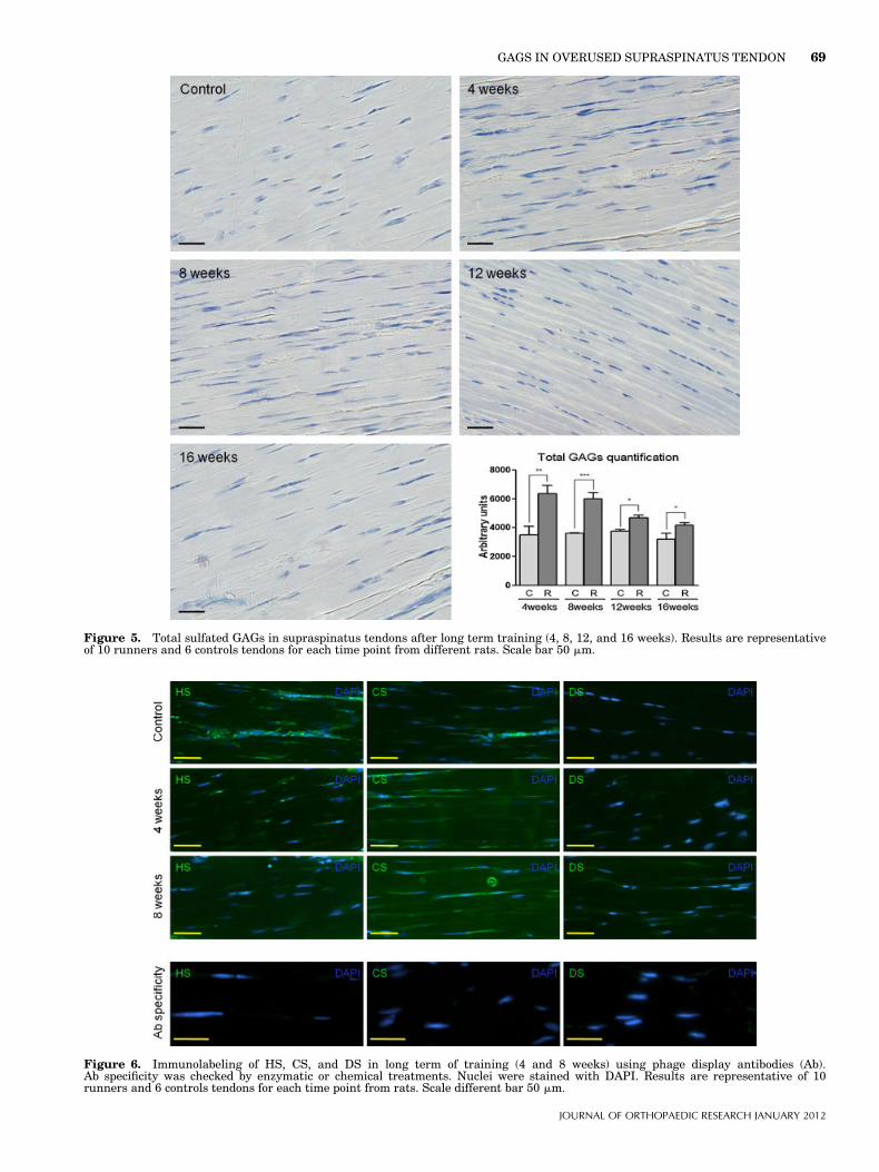
Figure 5. Total sulfated GAGs in supraspinatus tendons after long term training (4, 8, 12, and 16 weeks). Results are representativeof 10 runners and 6 controls tendons for each time point from different rats. Scale bar 50 mm.
Figure 6. Immunolabeling of HS, CS, and DS in long term of training (4 and 8 weeks) using phage display antibodies (Ab).Ab specificity was checked by enzymatic or chemical treatments. Nuclei were stained with DAPI. Results are representative of 10runners and 6 controls tendons for each time point from rats. Scale different bar 50 mm.
GAGS IN OVERUSED SUPRASPINATUS TENDON 69
JOURNAL OF ORTHOPAEDIC RESEARCH JANUARY 2012
ACKNOWLEDGMENTSMohamed Attia has PhD financial support from COGITOBIOand ANRT (Association Nationale de la Recherche et de laTechnologie). Alexander Scott received support from theMichael Smith Foundation for Health Research. We thankEric Huet, Suzanne Menashi, Dulce Papy-Garcia, and JeanDelbe for their valuable discussions and help.
REFERENCES1. Scott JE. 2003. Elasticity in extracellular matrix ‘shape
modules’ of tendon, cartilage, etc. A sliding proteoglycan-filament model. J Physiol 553:335–343.
2. Keene DR, San Antonio JD, Mayne R, et al. 2000. Decorinbinds near the C terminus of type I collagen. J Biol Chem275:21801–21804.
3. Ritty TM, Roth R, Heuser JE. 2003. Tendon cell array isola-tion reveals a previously unknown fibrillin-2-containingmacromolecular assembly. Structure 11:1179–1188.
4. Cook JL, Feller JA, Bonar SF, et al. 2004. Abnormaltenocyte morphology is more prevalent than collagen
disruption in asymptomatic athletes’ patellar tendons.J Orthop Res 22:334–338.
5. Chard MD, Cawston TE, Riley GP, et al. 1994. Rotator cuffdegeneration and lateral epicondylitis: A comparative histo-logical study. Ann Rheum Dis 53:30–34.
6. Samiric T, Parkinson J, Ilic MZ, et al. 2009. Changes in thecomposition of the extracellular matrix in patellar tendinop-athy. Matrix Biol 28:230–236.
7. Yoon JH, Halper J. 2005. Tendon proteoglycans: Biochemis-try and function. J Musculoskelet Neuronal Interact 5:22–34.
8. Soslowsky LJ, Carpenter JE, DeBano CM, et al. 1996.Development and use of an animal model for investigationson rotator cuff disease. J Shoulder Elbow Surg 5:383–392.
9. Soslowsky LJ, Thomopoulos S, Tun S, et al. 2000. NeerAward 1999. Overuse activity injures the supraspinatustendon in an animal model: A histologic and biomechanicalstudy. J Shoulder Elbow Surg 9:79–84.
10. Archambault JM, Jelinsky SA, Lake SP, et al. 2007. Ratsupraspinatus tendon expresses cartilage markers withoveruse. J Orthop Res 25:617–624.
11. Scott A, Cook JL, Hart DA, et al. 2007. Tenocyte responsesto mechanical loading in vivo: A role for local insulin-like
Figure 7. Expression of HARP at 2 and 4 weeks of overuse at transcript and protein levels. TFIID and GAPDH were used as loadingcontrols (a). Data represent the amounts of FGF-2 or HARP that bind to heparin in the presence of GAGs taken as a competitor (b).For each time point, data acquired in triplicate are from six tendons from three rats.
70 ATTIA ET AL.
JOURNAL OF ORTHOPAEDIC RESEARCH JANUARY 2012
growth factor 1 signaling in early tendinosis in rats.Arthritis Rheum 56:871–881.
12. Perry SM, McIlhenny SE, Hoffman MC, et al. 2005. Inflam-matory and angiogenic mRNA levels are altered in a supra-spinatus tendon overuse animal model. J Shoulder ElbowSurg 14:79S–83S.
13. Vogel KG. 2003. Tendon structure and response to changingmechanical load. J Musculoskelet Neuronal Interact 3:323–325, discussion 333–324.
14. Barbosa I, Garcia S, Barbier-Chassefiere V, et al. 2003.Improved and simple micro assay for sulfated glyco-saminoglycans quantification in biological extracts and itsuse in skin and muscle tissue studies. Glycobiology 13:647–653.
15. Rasband W. 1997–2009. Image J. U.S. National Institutes ofHealth, Bethesda, Maryland, USA, http://rsb.info.nih.gov/ij/.
16. Najjam S, Gibbs RV, Gordon MY, et al. 1997. Characteriza-tion of human recombinant interleukin 2 binding to heparinand heparan sulfate using an ELISA approach. Cytokine9:1013–1022.
17. Vacherot F, Delbe J, Heroult M, et al. 1999. Glycosaminogly-cans differentially bind HARP and modulate its biologicalactivity. J Biol Chem 274:7741–7747.
18. Riley GP, Harrall RL, Constant CR, et al. 1994. Glycosami-noglycans of human rotator cuff tendons: Changes with ageand in chronic rotator cuff tendinitis. Ann Rheum Dis 53:367–376.
19. Vigfusdottir AT, Pasrija C, Thakore PI, et al. 2010. Role ofpericellular matrix in mesenchymal stem cell deformationduring chondrogenic differentiation. Cell Mol Bioeng 3:387–397.
20. Wiberg C, Heinegard D, Wenglen C, et al. 2002. Biglycanorganizes collagen VI into hexagonal-like networks resem-bling tissue structures. J Biol Chem 277:49120–49126.
21. Wiberg C, Klatt AR, Wagener R, et al. 2003. Complexes ofmatrilin-1 and biglycan or decorin connect collagen VI
microfibrils to both collagen II and aggrecan. J Biol Chem278:37698–37704.
22. Thomopoulos S, Das R, Birman V, et al. 2010. Fibrocartilagetissue engineering: The role of the stress environment oncell morphology and matrix expression. Tissue Eng Part A17:1039–1053.
23. Shim JW, Elder SH. 2006. Influence of cyclic hydrostaticpressure on fibrocartilaginous metaplasia of achilles tendonfibroblasts. Biomech Model Mechanobiol 5:247–252.
24. Lorda-Diez CI, Montero JA, Martinez-Cue C, et al. 2009.Transforming growth factors beta coordinate cartilage andtendon differentiation in the developing limb mesenchyme.J Biol Chem 284:29988–29996.
25. Quintana L, zur Nieden NI, Semino CE. 2009. Morphogenet-ic and regulatory mechanisms during developmental chon-drogenesis: New paradigms for cartilage tissue engineering.Tissue Eng Part B Rev 15:29–41.
26. Molloy TJ, Kemp MW, Wang Y, et al. 2006. Microarrayanalysis of the tendinopathic rat supraspinatus tendon:Glutamate signaling and its potential role in tendon degen-eration. J Appl Physiol 101:1702–1709.
27. Maeda N, Fukazawa N, Hata T. 2006. The binding ofchondroitin sulfate to pleiotrophin/heparin-binding growth-associated molecule is regulated by chain length and over-sulfated structures. J Biol Chem 281:4894–4902.
28. Tapp H, Hernandez DJ, Neame PJ, et al. 1999. Pleiotrophininhibits chondrocyte proliferation and stimulates proteogly-can synthesis in mature bovine cartilage. Matrix Biol 18:543–556.
29. Yamada H, Inazumi T, Tajima S, et al. 1997. Stimulation ofcollagen expression and glycosaminoglycan synthesis bymidkine in human skin fibroblasts. Arch Dermatol Res289:429–433.
30. Dreyfus J, Brunet-de Carvalho N, Duprez D, et al. 1998.HB-GAM/pleiotrophin: Localization of mRNA and protein inthe chicken developing leg. Int J Dev Biol 42:189–198.
GAGS IN OVERUSED SUPRASPINATUS TENDON 71
JOURNAL OF ORTHOPAEDIC RESEARCH JANUARY 2012