alterations in fluids, electrolytes, and acid-base balance
TRANSCRIPT

84
CHAPTER
6 Alterations in Fluids,Electrolytes, and
Acid-Base Balance
Composition and Compartmental Distribution ofBody Fluids
Introductory ConceptsDiffusion and Osmosis
Compartmental Distribution of Body FluidsIntracellular Fluid VolumeExtracellular Fluid Volume
Capillary/Interstitial Fluid ExchangeEdemaThird-Space Accumulation
Sodium and Water BalanceRegulation of Sodium and Water Balance
Regulation of Sodium BalanceRegulation of Water Balance
Alterations in Isotonic Fluid VolumeIsotonic Fluid Volume DeficitIsotonic Fluid Volume Excess
Alterations in Sodium ConcentrationHyponatremiaHypernatremia
Potassium BalanceRegulation of Potassium Balance
HypokalemiaHyperkalemia
Calcium and Magnesium BalanceCalcium Balance
Regulation of Serum CalciumHypocalcemiaHypercalcemia
Magnesium BalanceRegulation of MagnesiumHypomagnesemiaHypermagnesemia
Acid-Base BalanceIntroductory Concepts
Acid-Base ChemistryMetabolic Acid and Bicarbonate ProductionRegulation of pHLaboratory Tests
Alterations in Acid-Base BalanceMetabolic Versus Respiratory Acid-Base DisordersPrimary Versus Compensatory MechanismsMetabolic AcidosisMetabolic AlkalosisRespiratory AcidosisRespiratory Alkalosis
F luids and electrolytes are present in body cells, in the tis-sue spaces between the cells, and in the blood that fillsthe vascular compartment. Body fluids serve to transport
gases, nutrients, and wastes; help to generate the electricalactivity needed to power body functions; take part in thetransforming of food into energy; and otherwise maintain theoverall function of the body. Although fluid volume andcomposition remain relatively constant in the presence of awide range of changes in intake and output, conditions suchas environmental stresses and disease can increase fluid loss,impair its intake, and otherwise interfere with mechanismsthat regulate fluid volume, composition, and distribution.
COMPOSITION AND COMPARTMENTALDISTRIBUTION OF BODY FLUIDS
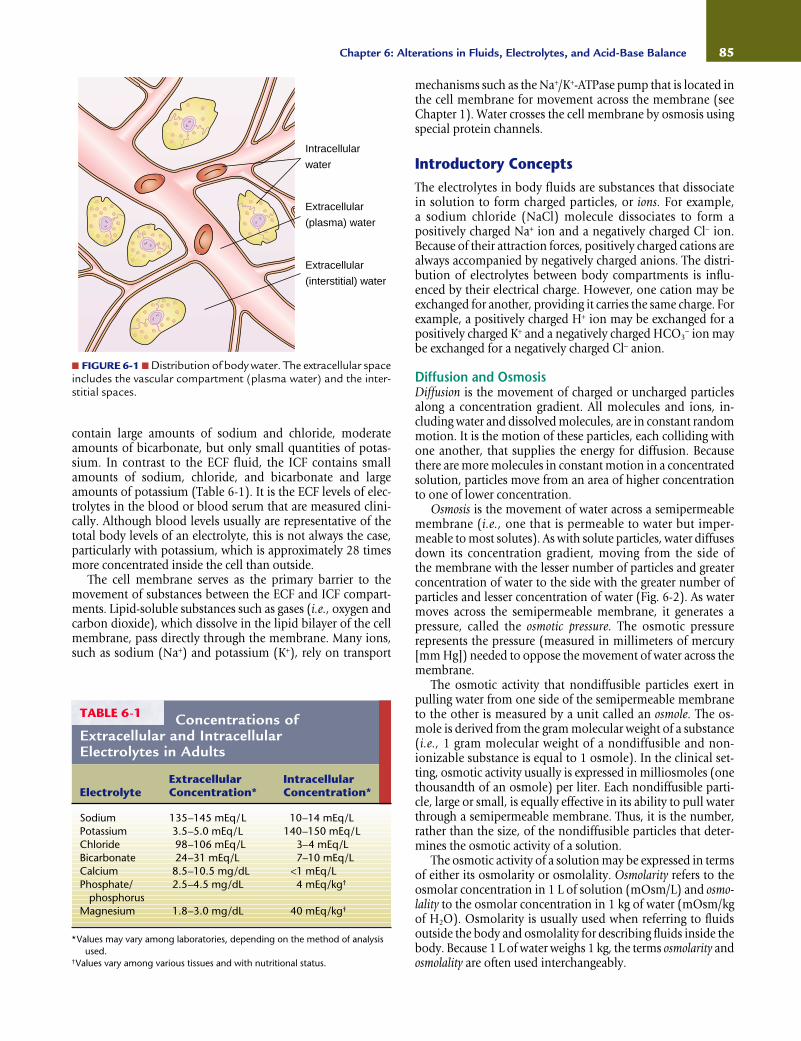
Body fluids are distributed between the intracellular fluid (ICF)and extracellular fluid (ECF) compartments. The ICF compart-ment consists of fluid contained within all of the billions ofcells in the body. It is the larger of the two compartments, con-taining approximately two thirds of the body water in healthyadults. The remaining one third of body water is in the ECFcompartment, which contains all the fluids outside the cells, in-cluding that in the interstitial or tissue spaces and blood vessels(Fig. 6-1). The ECF, including the plasma and interstitial fluids,
85Chapter 6: Alterations in Fluids, Electrolytes, and Acid-Base Balance
contain large amounts of sodium and chloride, moderateamounts of bicarbonate, but only small quantities of potas-sium. In contrast to the ECF fluid, the ICF contains smallamounts of sodium, chloride, and bicarbonate and largeamounts of potassium (Table 6-1). It is the ECF levels of elec-trolytes in the blood or blood serum that are measured clini-cally. Although blood levels usually are representative of thetotal body levels of an electrolyte, this is not always the case,particularly with potassium, which is approximately 28 timesmore concentrated inside the cell than outside.
The cell membrane serves as the primary barrier to themovement of substances between the ECF and ICF compart-ments. Lipid-soluble substances such as gases (i.e., oxygen andcarbon dioxide), which dissolve in the lipid bilayer of the cellmembrane, pass directly through the membrane. Many ions,such as sodium (Na+) and potassium (K+), rely on transport
mechanisms such as the Na+/K+-ATPase pump that is located inthe cell membrane for movement across the membrane (seeChapter 1). Water crosses the cell membrane by osmosis usingspecial protein channels.
Introductory ConceptsThe electrolytes in body fluids are substances that dissociatein solution to form charged particles, or ions. For example, a sodium chloride (NaCl) molecule dissociates to form a positively charged Na+ ion and a negatively charged Cl− ion.Because of their attraction forces, positively charged cations arealways accompanied by negatively charged anions. The distri-bution of electrolytes between body compartments is influ-enced by their electrical charge. However, one cation may beexchanged for another, providing it carries the same charge. Forexample, a positively charged H+ ion may be exchanged for apositively charged K+ and a negatively charged HCO3
− ion maybe exchanged for a negatively charged Cl− anion.
Diffusion and OsmosisDiffusion is the movement of charged or uncharged particlesalong a concentration gradient. All molecules and ions, in-cluding water and dissolved molecules, are in constant randommotion. It is the motion of these particles, each colliding withone another, that supplies the energy for diffusion. Becausethere are more molecules in constant motion in a concentratedsolution, particles move from an area of higher concentrationto one of lower concentration.
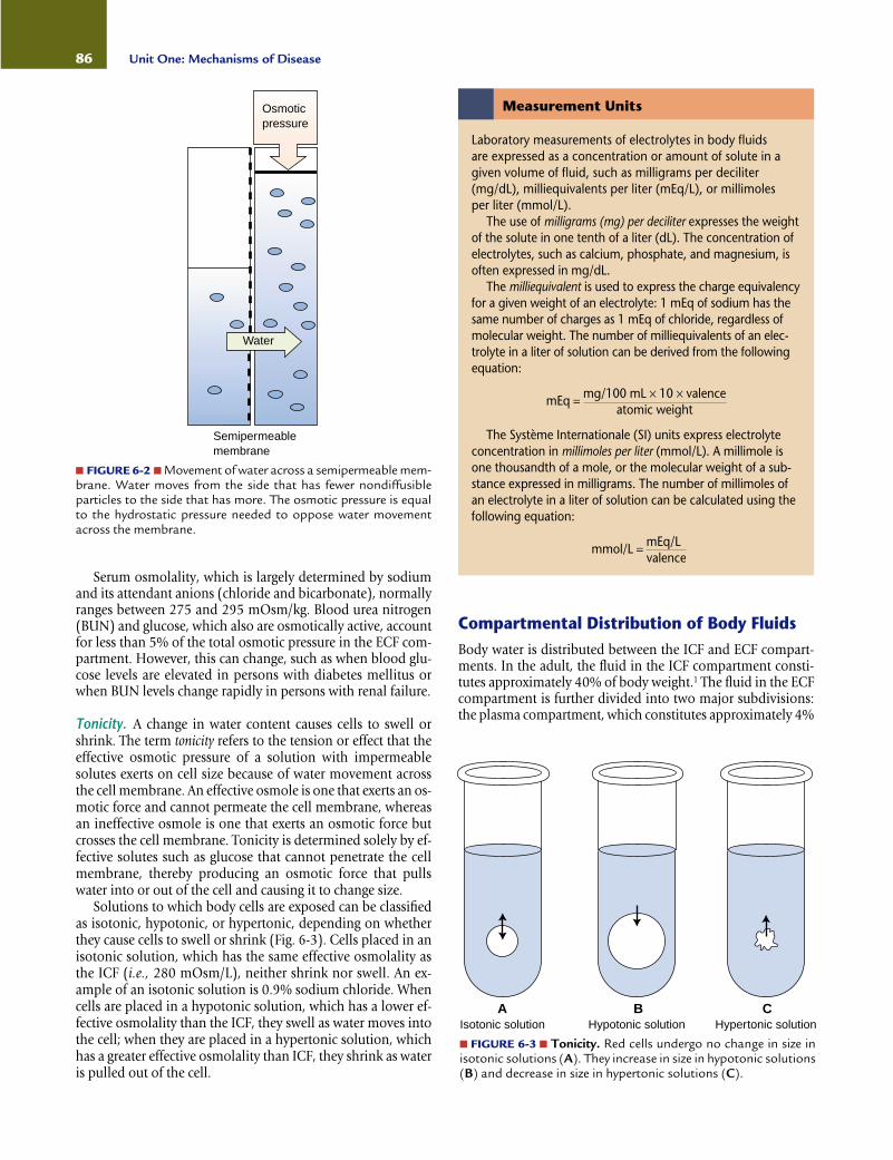
Osmosis is the movement of water across a semipermeablemembrane (i.e., one that is permeable to water but imper-meable to most solutes). As with solute particles, water diffusesdown its concentration gradient, moving from the side of the membrane with the lesser number of particles and greaterconcentration of water to the side with the greater number ofparticles and lesser concentration of water (Fig. 6-2). As watermoves across the semipermeable membrane, it generates apressure, called the osmotic pressure. The osmotic pressure represents the pressure (measured in millimeters of mercury[mm Hg]) needed to oppose the movement of water across themembrane.
The osmotic activity that nondiffusible particles exert inpulling water from one side of the semipermeable membraneto the other is measured by a unit called an osmole. The os-mole is derived from the gram molecular weight of a substance(i.e., 1 gram molecular weight of a nondiffusible and non-ionizable substance is equal to 1 osmole). In the clinical set-ting, osmotic activity usually is expressed in milliosmoles (onethousandth of an osmole) per liter. Each nondiffusible parti-cle, large or small, is equally effective in its ability to pull waterthrough a semipermeable membrane. Thus, it is the number,rather than the size, of the nondiffusible particles that deter-mines the osmotic activity of a solution.
The osmotic activity of a solution may be expressed in termsof either its osmolarity or osmolality. Osmolarity refers to theosmolar concentration in 1 L of solution (mOsm/L) and osmo-lality to the osmolar concentration in 1 kg of water (mOsm/kgof H2O). Osmolarity is usually used when referring to fluidsoutside the body and osmolality for describing fluids inside thebody. Because 1 L of water weighs 1 kg, the terms osmolarity andosmolality are often used interchangeably.
■ FIGURE 6-1 ■ Distribution of body water. The extracellular spaceincludes the vascular compartment (plasma water) and the inter-stitial spaces.
Intracellular
water
Extracellular
(plasma) water
Extracellular
(interstitial) water
Concentrations ofExtracellular and IntracellularElectrolytes in Adults
TABLE 6-1
Sodium 135–145 mEq/L 10–14 mEq/LPotassium 3.5–5.0 mEq/L 140–150 mEq/LChloride 98–106 mEq/L 3–4 mEq/LBicarbonate 24–31 mEq/L 7–10 mEq/LCalcium 8.5–10.5 mg/dL <1 mEq/LPhosphate/ 2.5–4.5 mg/dL 4 mEq/kg†
phosphorusMagnesium 1.8–3.0 mg/dL 40 mEq/kg†
*Values may vary among laboratories, depending on the method of analysisused.
†Values vary among various tissues and with nutritional status.
Extracellular IntracellularElectrolyte Concentration* Concentration*
Compartmental Distribution of Body FluidsBody water is distributed between the ICF and ECF compart-ments. In the adult, the fluid in the ICF compartment consti-tutes approximately 40% of body weight.1 The fluid in the ECFcompartment is further divided into two major subdivisions:the plasma compartment, which constitutes approximately 4%
86 Unit One: Mechanisms of Disease
■ FIGURE 6-2 ■ Movement of water across a semipermeable mem-brane. Water moves from the side that has fewer nondiffusibleparticles to the side that has more. The osmotic pressure is equalto the hydrostatic pressure needed to oppose water movementacross the membrane.
Water
Osmoticpressure
Semipermeablemembrane
Water
Serum osmolality, which is largely determined by sodiumand its attendant anions (chloride and bicarbonate), normallyranges between 275 and 295 mOsm/kg. Blood urea nitrogen(BUN) and glucose, which also are osmotically active, accountfor less than 5% of the total osmotic pressure in the ECF com-partment. However, this can change, such as when blood glu-cose levels are elevated in persons with diabetes mellitus orwhen BUN levels change rapidly in persons with renal failure.
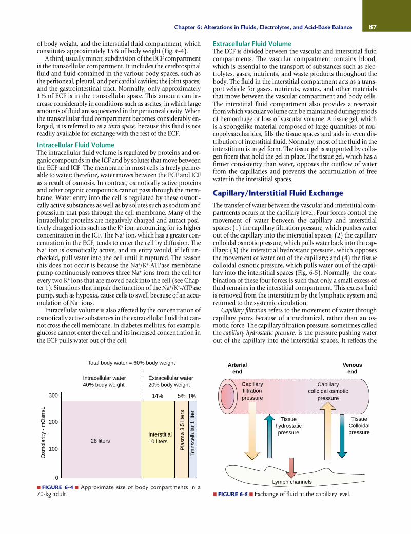
Tonicity. A change in water content causes cells to swell orshrink. The term tonicity refers to the tension or effect that theeffective osmotic pressure of a solution with impermeablesolutes exerts on cell size because of water movement across the cell membrane. An effective osmole is one that exerts an os-motic force and cannot permeate the cell membrane, whereasan ineffective osmole is one that exerts an osmotic force butcrosses the cell membrane. Tonicity is determined solely by ef-fective solutes such as glucose that cannot penetrate the cellmembrane, thereby producing an osmotic force that pullswater into or out of the cell and causing it to change size.
Solutions to which body cells are exposed can be classifiedas isotonic, hypotonic, or hypertonic, depending on whetherthey cause cells to swell or shrink (Fig. 6-3). Cells placed in anisotonic solution, which has the same effective osmolality asthe ICF (i.e., 280 mOsm/L), neither shrink nor swell. An ex-ample of an isotonic solution is 0.9% sodium chloride. Whencells are placed in a hypotonic solution, which has a lower ef-fective osmolality than the ICF, they swell as water moves intothe cell; when they are placed in a hypertonic solution, whichhas a greater effective osmolality than ICF, they shrink as wateris pulled out of the cell.
Measurement Units
Laboratory measurements of electrolytes in body fluids are expressed as a concentration or amount of solute in agiven volume of fluid, such as milligrams per deciliter(mg/dL), milliequivalents per liter (mEq/L), or millimoles per liter (mmol/L).
The use of milligrams (mg) per deciliter expresses the weightof the solute in one tenth of a liter (dL). The concentration ofelectrolytes, such as calcium, phosphate, and magnesium, isoften expressed in mg/dL.
The milliequivalent is used to express the charge equivalencyfor a given weight of an electrolyte: 1 mEq of sodium has thesame number of charges as 1 mEq of chloride, regardless ofmolecular weight. The number of milliequivalents of an elec-trolyte in a liter of solution can be derived from the followingequation:
mEq = mg/100 mL × 10 × valenceatomic weight
The Système Internationale (SI) units express electrolyteconcentration in millimoles per liter (mmol/L). A millimole isone thousandth of a mole, or the molecular weight of a sub-stance expressed in milligrams. The number of millimoles ofan electrolyte in a liter of solution can be calculated using thefollowing equation:
mmol/L = mEq/Lvalence
Isotonic solution Hypotonic solution Hypertonic solution
■ FIGURE 6-3 ■ Tonicity. Red cells undergo no change in size inisotonic solutions (A). They increase in size in hypotonic solutions(B) and decrease in size in hypertonic solutions (C).
A B C
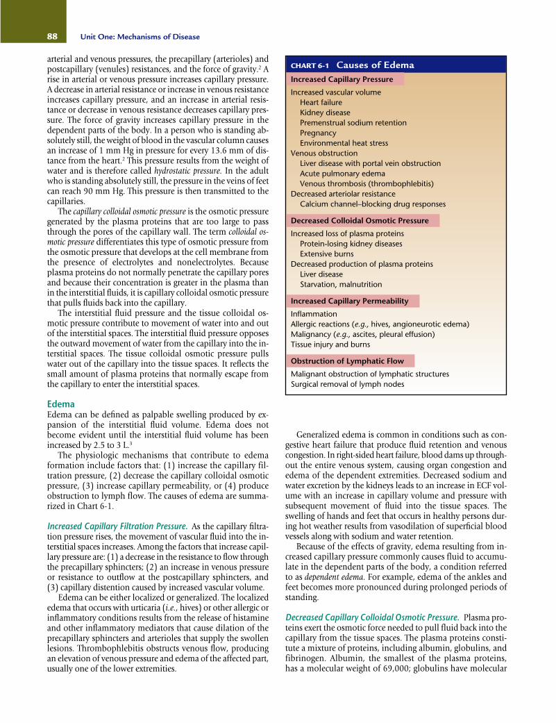
of body weight, and the interstitial fluid compartment, whichconstitutes approximately 15% of body weight (Fig. 6-4).
A third, usually minor, subdivision of the ECF compartmentis the transcellular compartment. It includes the cerebrospinalfluid and fluid contained in the various body spaces, such asthe peritoneal, pleural, and pericardial cavities; the joint spaces;and the gastrointestinal tract. Normally, only approximately1% of ECF is in the transcellular space. This amount can in-crease considerably in conditions such as ascites, in which largeamounts of fluid are sequestered in the peritoneal cavity. Whenthe transcellular fluid compartment becomes considerably en-larged, it is referred to as a third space, because this fluid is notreadily available for exchange with the rest of the ECF.
Intracellular Fluid VolumeThe intracellular fluid volume is regulated by proteins and or-ganic compounds in the ICF and by solutes that move betweenthe ECF and ICF. The membrane in most cells is freely perme-able to water; therefore, water moves between the ECF and ICFas a result of osmosis. In contrast, osmotically active proteinsand other organic compounds cannot pass through the mem-brane. Water entry into the cell is regulated by these osmoti-cally active substances as well as by solutes such as sodium andpotassium that pass through the cell membrane. Many of theintracellular proteins are negatively charged and attract posi-tively charged ions such as the K+ ion, accounting for its higherconcentration in the ICF. The Na+ ion, which has a greater con-centration in the ECF, tends to enter the cell by diffusion. TheNa+ ion is osmotically active, and its entry would, if left un-checked, pull water into the cell until it ruptured. The reasonthis does not occur is because the Na+/K+-ATPase membranepump continuously removes three Na+ ions from the cell forevery two K+ ions that are moved back into the cell (see Chap-ter 1). Situations that impair the function of the Na+/K+-ATPasepump, such as hypoxia, cause cells to swell because of an accu-mulation of Na+ ions.
Intracellular volume is also affected by the concentration ofosmotically active substances in the extracellular fluid that can-not cross the cell membrane. In diabetes mellitus, for example,glucose cannot enter the cell and its increased concentration inthe ECF pulls water out of the cell.
Extracellular Fluid VolumeThe ECF is divided between the vascular and interstitial fluidcompartments. The vascular compartment contains blood,which is essential to the transport of substances such as elec-trolytes, gases, nutrients, and waste products throughout thebody. The fluid in the interstitial compartment acts as a trans-port vehicle for gases, nutrients, wastes, and other materialsthat move between the vascular compartment and body cells.The interstitial fluid compartment also provides a reservoirfrom which vascular volume can be maintained during periodsof hemorrhage or loss of vascular volume. A tissue gel, whichis a spongelike material composed of large quantities of mu-copolysaccharides, fills the tissue spaces and aids in even dis-tribution of interstitial fluid. Normally, most of the fluid in theinterstitium is in gel form. The tissue gel is supported by colla-gen fibers that hold the gel in place. The tissue gel, which has afirmer consistency than water, opposes the outflow of waterfrom the capillaries and prevents the accumulation of freewater in the interstitial spaces.
Capillary/Interstitial Fluid ExchangeThe transfer of water between the vascular and interstitial com-partments occurs at the capillary level. Four forces control themovement of water between the capillary and interstitialspaces: (1) the capillary filtration pressure, which pushes waterout of the capillary into the interstitial spaces; (2) the capillarycolloidal osmotic pressure, which pulls water back into the cap-illary; (3) the interstitial hydrostatic pressure, which opposesthe movement of water out of the capillary; and (4) the tissuecolloidal osmotic pressure, which pulls water out of the capil-lary into the interstitial spaces (Fig. 6-5). Normally, the com-bination of these four forces is such that only a small excess offluid remains in the interstitial compartment. This excess fluidis removed from the interstitium by the lymphatic system andreturned to the systemic circulation.
Capillary filtration refers to the movement of water throughcapillary pores because of a mechanical, rather than an os-motic, force. The capillary filtration pressure, sometimes calledthe capillary hydrostatic pressure, is the pressure pushing waterout of the capillary into the interstitial spaces. It reflects the
87Chapter 6: Alterations in Fluids, Electrolytes, and Acid-Base Balance
300
200
100
0
Osm
olar
ity -
mO
sm/L
Total body water = 60% body weight
Intracellular water40% body weight
Extracellular water20% body weight
14% 5% 1%
28 litersInterstitial10 liters
Pla
sma
3.5
liter
s
Tran
scel
lula
r 1
liter
■ FIGURE 6-4 ■ Approximate size of body compartments in a 70-kg adult. ■ FIGURE 6-5 ■ Exchange of fluid at the capillary level.
Capillaryfiltrationpressure
Arterialend
Tissuehydrostaticpressure
TissueColloidalpressure
Venousend
Capillarycolloidal osmotic
pressure
Lymph channels
arterial and venous pressures, the precapillary (arterioles) andpostcapillary (venules) resistances, and the force of gravity.2 Arise in arterial or venous pressure increases capillary pressure.A decrease in arterial resistance or increase in venous resistanceincreases capillary pressure, and an increase in arterial resis-tance or decrease in venous resistance decreases capillary pres-sure. The force of gravity increases capillary pressure in the dependent parts of the body. In a person who is standing ab-solutely still, the weight of blood in the vascular column causesan increase of 1 mm Hg in pressure for every 13.6 mm of dis-tance from the heart.2 This pressure results from the weight ofwater and is therefore called hydrostatic pressure. In the adultwho is standing absolutely still, the pressure in the veins of feetcan reach 90 mm Hg. This pressure is then transmitted to thecapillaries.
The capillary colloidal osmotic pressure is the osmotic pressuregenerated by the plasma proteins that are too large to passthrough the pores of the capillary wall. The term colloidal os-motic pressure differentiates this type of osmotic pressure fromthe osmotic pressure that develops at the cell membrane fromthe presence of electrolytes and nonelectrolytes. Becauseplasma proteins do not normally penetrate the capillary poresand because their concentration is greater in the plasma thanin the interstitial fluids, it is capillary colloidal osmotic pressurethat pulls fluids back into the capillary.
The interstitial fluid pressure and the tissue colloidal os-motic pressure contribute to movement of water into and outof the interstitial spaces. The interstitial fluid pressure opposesthe outward movement of water from the capillary into the in-terstitial spaces. The tissue colloidal osmotic pressure pullswater out of the capillary into the tissue spaces. It reflects thesmall amount of plasma proteins that normally escape fromthe capillary to enter the interstitial spaces.
EdemaEdema can be defined as palpable swelling produced by ex-pansion of the interstitial fluid volume. Edema does not become evident until the interstitial fluid volume has been increased by 2.5 to 3 L.3
The physiologic mechanisms that contribute to edemaformation include factors that: (1) increase the capillary fil-tration pressure, (2) decrease the capillary colloidal osmoticpressure, (3) increase capillary permeability, or (4) produceobstruction to lymph flow. The causes of edema are summa-rized in Chart 6-1.
Increased Capillary Filtration Pressure. As the capillary filtra-tion pressure rises, the movement of vascular fluid into the in-terstitial spaces increases. Among the factors that increase capil-lary pressure are: (1) a decrease in the resistance to flow throughthe precapillary sphincters; (2) an increase in venous pressureor resistance to outflow at the postcapillary sphincters, and (3) capillary distention caused by increased vascular volume.
Edema can be either localized or generalized. The localizededema that occurs with urticaria (i.e., hives) or other allergic orinflammatory conditions results from the release of histamineand other inflammatory mediators that cause dilation of theprecapillary sphincters and arterioles that supply the swollenlesions. Thrombophlebitis obstructs venous flow, producingan elevation of venous pressure and edema of the affected part,usually one of the lower extremities.
Generalized edema is common in conditions such as con-gestive heart failure that produce fluid retention and venouscongestion. In right-sided heart failure, blood dams up through-out the entire venous system, causing organ congestion andedema of the dependent extremities. Decreased sodium andwater excretion by the kidneys leads to an increase in ECF vol-ume with an increase in capillary volume and pressure withsubsequent movement of fluid into the tissue spaces. Theswelling of hands and feet that occurs in healthy persons dur-ing hot weather results from vasodilation of superficial bloodvessels along with sodium and water retention.
Because of the effects of gravity, edema resulting from in-creased capillary pressure commonly causes fluid to accumu-late in the dependent parts of the body, a condition referredto as dependent edema. For example, edema of the ankles andfeet becomes more pronounced during prolonged periods ofstanding.
Decreased Capillary Colloidal Osmotic Pressure. Plasma pro-teins exert the osmotic force needed to pull fluid back into thecapillary from the tissue spaces. The plasma proteins consti-tute a mixture of proteins, including albumin, globulins, andfibrinogen. Albumin, the smallest of the plasma proteins, has a molecular weight of 69,000; globulins have molecular
88 Unit One: Mechanisms of Disease
CHART 6-1 Causes of EdemaIncreased Capillary Pressure
Increased vascular volumeHeart failureKidney diseasePremenstrual sodium retentionPregnancyEnvironmental heat stress
Venous obstructionLiver disease with portal vein obstructionAcute pulmonary edemaVenous thrombosis (thrombophlebitis)
Decreased arteriolar resistanceCalcium channel–blocking drug responses
Decreased Colloidal Osmotic Pressure
Increased loss of plasma proteinsProtein-losing kidney diseasesExtensive burns
Decreased production of plasma proteinsLiver diseaseStarvation, malnutrition
Increased Capillary Permeability
InflammationAllergic reactions (e.g., hives, angioneurotic edema)Malignancy (e.g., ascites, pleural effusion)Tissue injury and burns
Obstruction of Lymphatic Flow
Malignant obstruction of lymphatic structuresSurgical removal of lymph nodes
weights of approximately 140,000; and fibrinogen has a molec-ular weight of 400,000.2 Because of its lower molecular weight,1 g of albumin has approximately twice as many osmoticallyactive molecules as 1 g of globulin and almost six times asmany osmotically active molecules as 1 g of fibrinogen. Inaddition, the concentration of albumin (approximately 4.5g/dL) is greater than that of the globulins (2.5 g/dL) and fi-brinogen (0.3 mg/dL).
Edema caused by decreased capillary colloidal osmoticpressure usually is the result of inadequate production or ab-normal loss of plasma proteins, mainly albumin. The plasmaproteins are synthesized in the liver. In persons with severe liverfailure, impaired synthesis of albumin results in a decrease incolloidal osmotic pressure. In starvation and malnutrition,edema develops because there is a lack of the amino acidsneeded in plasma protein synthesis.
The most common site of plasma protein loss is the kidney.In kidney diseases such as nephrosis, the glomerular capillariesbecome permeable to the plasma proteins, particularly albu-min, which is the smallest of the proteins. When this happens,large amounts of albumin are filtered out of the blood and lostin the urine. An excessive loss of plasma proteins also occurswhen large areas of skin are injured or destroyed. Edema is acommon problem during the early stages of a burn, resultingfrom capillary injury and loss of plasma proteins.
Because the plasma proteins are evenly distributed through-out the body and are not affected by the force of gravity, edemacaused by decreased capillary colloidal osmotic pressure tendsto affect tissues in nondependent as well as dependent partsof the body. There is swelling of the face as well as the legs andfeet.
Increased Capillary Permeability. When the capillary pores be-come enlarged or the integrity of the capillary wall is damaged,capillary permeability is increased. When this happens, plasmaproteins and other osmotically active particles leak into the in-terstitial spaces, increasing the tissue colloidal osmotic pressureand thereby contributing to the accumulation of interstitialfluid. Among the conditions that increase capillary permeabil-ity are burn injury, capillary congestion, inflammation, andimmune responses.
Obstruction of Lymph Flow. Osmotically active plasma pro-teins and other large particles that cannot be reabsorbedthrough the pores in the capillary membrane rely on the lym-phatic system for movement back into the circulatory system.Edema caused by impaired lymph flow is commonly referredto as lymphedema. Malignant involvement of lymph structuresand removal of lymph nodes at the time of cancer surgery arecommon causes of lymphedema. Another cause of lymph-edema is infection involving the lymphatic channels andlymph nodes.
Manifestations. The effects of edema are determined largely byits location. Edema of the brain, larynx, or lungs is an acute,life-threatening condition. Although not life threatening,edema may interfere with movement by limiting joint motion.Swelling of the ankles and feet often is insidious in onset andmay or may not be associated with disease. At the tissue level,edema increases the distance for diffusion of oxygen, nutrients,and wastes. Edematous tissues usually are more susceptible to
injury and the development of ischemic tissue damage, in-cluding pressure ulcers. Edema can also compress blood ves-sels. For example, the skin of a severely swollen finger can actas a tourniquet, shutting off the blood flow to the finger. Edemacan also be disfiguring, causing psychological effects and dis-turbances in self-concept.
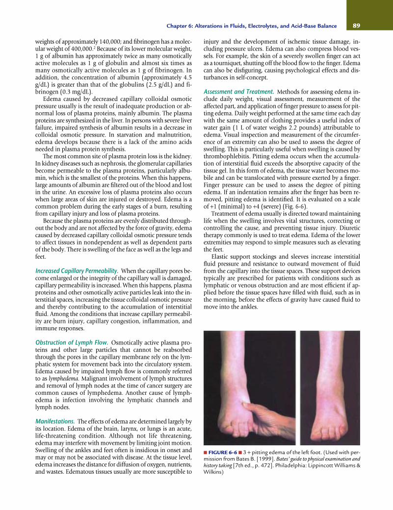
Assessment and Treatment. Methods for assessing edema in-clude daily weight, visual assessment, measurement of the affected part, and application of finger pressure to assess for pit-ting edema. Daily weight performed at the same time each daywith the same amount of clothing provides a useful index ofwater gain (1 L of water weighs 2.2 pounds) attributable toedema. Visual inspection and measurement of the circumfer-ence of an extremity can also be used to assess the degree ofswelling. This is particularly useful when swelling is caused bythrombophlebitis. Pitting edema occurs when the accumula-tion of interstitial fluid exceeds the absorptive capacity of thetissue gel. In this form of edema, the tissue water becomes mo-bile and can be translocated with pressure exerted by a finger.Finger pressure can be used to assess the degree of pittingedema. If an indentation remains after the finger has been re-moved, pitting edema is identified. It is evaluated on a scale of +1 (minimal) to +4 (severe) (Fig. 6-6).
Treatment of edema usually is directed toward maintaininglife when the swelling involves vital structures, correcting orcontrolling the cause, and preventing tissue injury. Diuretictherapy commonly is used to treat edema. Edema of the lowerextremities may respond to simple measures such as elevatingthe feet.
Elastic support stockings and sleeves increase interstitialfluid pressure and resistance to outward movement of fluidfrom the capillary into the tissue spaces. These support devicestypically are prescribed for patients with conditions such aslymphatic or venous obstruction and are most efficient if ap-plied before the tissue spaces have filled with fluid, such as inthe morning, before the effects of gravity have caused fluid tomove into the ankles.
89Chapter 6: Alterations in Fluids, Electrolytes, and Acid-Base Balance
■ FIGURE 6-6 ■ 3 + pitting edema of the left foot. (Used with per-mission from Bates B. [1999]. Bates’ guide to physical examination andhistory taking [7th ed., p. 472]. Philadelphia: Lippincott Williams &Wilkins)
In summary, body fluids are distributed between the ICFand ECF compartments of the body. Two thirds of body fluidsare contained in the body cells of the ICF compartment, andone third is contained in the vascular compartment, intersti-tial spaces, and third-space areas of the ECF compartment.Electrolytes and nonelectrolytes move by diffusion across cellmembranes that separate the ICF and ECF compartments.Water moves by osmosis across semipermeable membranes,moving from the side of the membrane that has the lessernumber of particles and greater concentration of water to theside that has the greater number of particles and lesser con-centration of water. The osmotic tension or effect that a solu-tion exerts on cell volume in terms of causing the cell to swellor shrink is called tonicity.
Intracellular volume is regulated by the large numbers ofproteins and other inorganic solutes that cannot cross thecell’s membrane and solutes such as sodium, potassium, andglucose that selectively move between the ICF and ECF de-pendent upon concentration gradients and transport mecha-nisms. ECF volume, which is distributed between the vascularand interstitial compartments, is regulated by the eliminationof sodium and water by the kidney.
Edema represents an increase in interstitial fluid volume.The physiologic mechanisms that predispose to edema for-mation are increased capillary filtration pressure, decreasedcapillary colloidal osmotic pressure, increased capillary per-meability, and obstruction of lymphatic flow. The effect thatedema exerts on body function is determined by its location;cerebral edema can be a life-threatening situation, butswollen feet can be a normal discomfort that accompanieshot weather. Fluid can also accumulate in the transcellularcompartment—the joint spaces, pericardial sac, the peri-toneal cavity, and the pleural cavity. Because this fluid is noteasily exchanged with the rest of the ECF, it is often referredto as third-space fluid.
Third-Space AccumulationThird spacing represents the loss or trapping of ECF in the trans-cellular space. The serous cavities are part of the transcellularcompartment (i.e., third space) located in strategic body areaswhere there is continual movement of body structures—thepericardial sac, the peritoneal cavity, and the pleural cavity. Theexchange of ECF among the capillaries, the interstitial spaces,and the transcellular space of the serous cavity uses the samemechanisms as capillaries elsewhere in the body. The serouscavities are closely linked with lymphatic drainage systems. Themilking action of the moving structures, such as the lungs, con-tinually forces fluid and plasma proteins back into the circula-tion, keeping these cavities empty. Any obstruction to lymphflow causes fluid accumulation in the serous cavities. As withedema fluid, third-space fluids represent an accumulation ortrapping of body fluids that contribute to body weight but notto fluid reserve or function.
The prefix hydro- may be used to indicate the presence of ex-cessive fluid, as in hydrothorax, which means excessive fluid inthe pleural cavity. The accumulation of fluid in the peritonealcavity is called ascites. The transudation of fluid into the serouscavities is also referred to as effusion. Effusion can containblood, plasma proteins, inflammatory cells (i.e., pus), and ECF.
SODIUM AND WATER BALANCE
The movement of body fluids between the ICF and ECF com-partments occurs at the cell membrane and depends on regu-lation of ECF water and sodium. Water provides approxi-mately 90% to 93% of the volume of body fluids and sodiumsalts approximately 90% to 95% of the ECF solutes. Normally,equivalent changes in sodium and water are such that thevolume and osmolality of the ECF is maintained within a nor-mal range. Because it is the concentration of sodium (in milli-grams per liter) that controls ECF osmolality, changes insodium are usually accompanied by proportionate changes inwater volume.
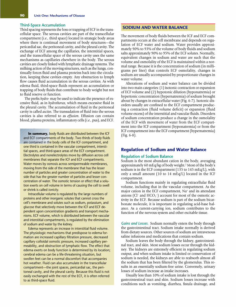
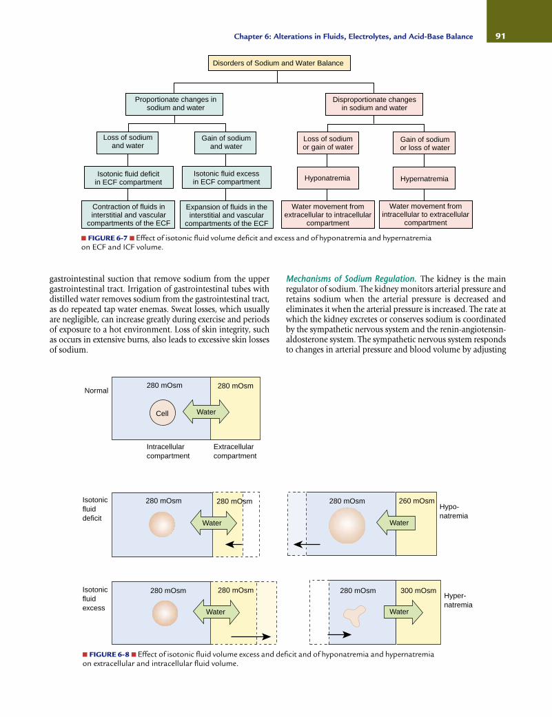
Alterations of sodium and water balance can be dividedinto two main categories: (1) isotonic contraction or expansionof ECF volume and (2) hypotonic dilution (hyponatremia) orhypertonic concentration (hypernatremia) of sodium broughtabout by changes in extracellular water (Fig. 6-7). Isotonic dis-orders usually are confined to the ECF compartment produc-ing a contraction (fluid volume deficit) or expansion (fluidvolume excess) of the interstitial and vascular fluids. Disordersof sodium concentration produce a change in the osmolalityof the ECF with movement of water from the ECF compart-ment into the ICF compartment (hyponatremia) or from theICF compartment into the ECF compartment (hypernatremia)(Fig. 6-8).
Regulation of Sodium and Water BalanceRegulation of Sodium BalanceSodium is the most abundant cation in the body, averagingapproximately 60 mEq/kg of body weight.2 Most of the body’ssodium is in the ECF compartment (135 to 145 mEq/L), withonly a small amount (10 to 14 mEq/L) located in the ICFcompartment.
Sodium functions mainly in regulating extracellular fluidvolume, including that in the vascular compartment. As themajor cation in the ECF compartment, Na+ and its attendantanions (Cl− and HCO3
−) account for most of the osmotic ac-tivity in the ECF. Because sodium is part of the sodium bicar-bonate molecule, it is important in regulating acid-base bal-ance. As a current-carrying ion, sodium contributes to thefunction of the nervous system and other excitable tissue.
Gains and Losses. Sodium normally enters the body throughthe gastrointestinal tract. Sodium intake normally is derivedfrom dietary sources. Other sources of sodium are intravenoussaline infusions and medications that contain sodium.
Sodium leaves the body through the kidney, gastrointesti-nal tract, and skin. Most sodium losses occur through the kid-ney. The kidneys are extremely efficient in regulating sodiumoutput, and when sodium intake is limited or conservation ofsodium is needed, the kidneys are able to reabsorb almost allthe sodium that has been filtered by the glomerulus. This re-sults in an essentially sodium-free urine. Conversely, urinarylosses of sodium increase as intake increases.
Usually less than 10% of sodium intake is lost through thegastrointestinal tract and skin. Sodium losses increase withconditions such as vomiting, diarrhea, fistula drainage, and
90 Unit One: Mechanisms of Disease
gastrointestinal suction that remove sodium from the uppergastrointestinal tract. Irrigation of gastrointestinal tubes withdistilled water removes sodium from the gastrointestinal tract,as do repeated tap water enemas. Sweat losses, which usuallyare negligible, can increase greatly during exercise and periodsof exposure to a hot environment. Loss of skin integrity, suchas occurs in extensive burns, also leads to excessive skin lossesof sodium.
Mechanisms of Sodium Regulation. The kidney is the mainregulator of sodium. The kidney monitors arterial pressure andretains sodium when the arterial pressure is decreased andeliminates it when the arterial pressure is increased. The rate atwhich the kidney excretes or conserves sodium is coordinatedby the sympathetic nervous system and the renin-angiotensin-aldosterone system. The sympathetic nervous system respondsto changes in arterial pressure and blood volume by adjusting
91Chapter 6: Alterations in Fluids, Electrolytes, and Acid-Base Balance
■ FIGURE 6-7 ■ Effect of isotonic fluid volume deficit and excess and of hyponatremia and hypernatremiaon ECF and ICF volume.
Disorders of Sodium and Water Balance
Proportionate changes insodium and water
Loss of sodiumand water
Isotonic fluid deficitin ECF compartment
Contraction of fluids ininterstitial and vascular
compartments of the ECF
Gain of sodiumand water
Isotonic fluid excessin ECF compartment
Expansion of fluids in theinterstitial and vascular
compartments of the ECF
Disproportionate changesin sodium and water
Loss of sodiumor gain of water
Hyponatremia
Water movement fromextracellular to intracellular
compartment
Gain of sodiumor loss of water
Hypernatremia
Water movement fromintracellular to extracellular
compartment
Normal
Isotonicfluiddeficit
Isotonicfluidexcess
280 mOsm 280 mOsm
Cell Water
Intracellularcompartment
Extracellularcompartment
280 mOsm 280 mOsm
Water
280 mOsm
260 mOsm
Water
Hypo-natremia
280 mOsm
280 mOsm
Water
280 mOsm 300 mOsm
Water
Hyper-natremia
■ FIGURE 6-8 ■ Effect of isotonic fluid volume excess and deficit and of hyponatremia and hypernatremiaon extracellular and intracellular fluid volume.
the glomerular filtration rate and the rate at which sodium isfiltered from the blood. Sympathetic activity also regulatestubular reabsorption of sodium and renin release. The renin-angiotensin-aldosterone system exerts its action through angio-tensin II and aldosterone (see Chapter 16). Angiotensin II actsdirectly on the renal tubules to increase sodium reabsorption. Italso acts to constrict renal blood vessels, thereby decreasing theglomerular filtration rate and slowing renal blood flow so thatless sodium is filtered and more is reabsorbed. Angiotensin IIis also a powerful regulator of aldosterone, a hormone secretedby the adrenal cortex. Aldosterone acts at the level of the cor-tical collecting tubules of the kidneys to increase sodium re-absorption while increasing potassium elimination.
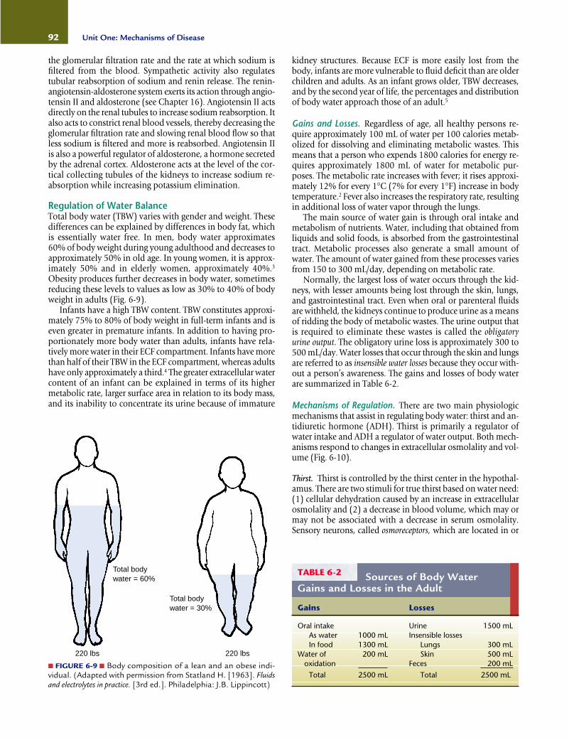
Regulation of Water BalanceTotal body water (TBW) varies with gender and weight. Thesedifferences can be explained by differences in body fat, whichis essentially water free. In men, body water approximates60% of body weight during young adulthood and decreases toapproximately 50% in old age. In young women, it is approx-imately 50% and in elderly women, approximately 40%.3
Obesity produces further decreases in body water, sometimesreducing these levels to values as low as 30% to 40% of bodyweight in adults (Fig. 6-9).
Infants have a high TBW content. TBW constitutes approxi-mately 75% to 80% of body weight in full-term infants and iseven greater in premature infants. In addition to having pro-portionately more body water than adults, infants have rela-tively more water in their ECF compartment. Infants have morethan half of their TBW in the ECF compartment, whereas adultshave only approximately a third.4 The greater extracellular watercontent of an infant can be explained in terms of its highermetabolic rate, larger surface area in relation to its body mass,and its inability to concentrate its urine because of immature
kidney structures. Because ECF is more easily lost from thebody, infants are more vulnerable to fluid deficit than are olderchildren and adults. As an infant grows older, TBW decreases,and by the second year of life, the percentages and distributionof body water approach those of an adult.5
Gains and Losses. Regardless of age, all healthy persons re-quire approximately 100 mL of water per 100 calories metab-olized for dissolving and eliminating metabolic wastes. Thismeans that a person who expends 1800 calories for energy re-quires approximately 1800 mL of water for metabolic pur-poses. The metabolic rate increases with fever; it rises approxi-mately 12% for every 1°C (7% for every 1°F) increase in bodytemperature.2 Fever also increases the respiratory rate, resultingin additional loss of water vapor through the lungs.
The main source of water gain is through oral intake andmetabolism of nutrients. Water, including that obtained fromliquids and solid foods, is absorbed from the gastrointestinaltract. Metabolic processes also generate a small amount ofwater. The amount of water gained from these processes variesfrom 150 to 300 mL/day, depending on metabolic rate.
Normally, the largest loss of water occurs through the kid-neys, with lesser amounts being lost through the skin, lungs,and gastrointestinal tract. Even when oral or parenteral fluidsare withheld, the kidneys continue to produce urine as a meansof ridding the body of metabolic wastes. The urine output thatis required to eliminate these wastes is called the obligatoryurine output. The obligatory urine loss is approximately 300 to500 mL/day. Water losses that occur through the skin and lungsare referred to as insensible water losses because they occur with-out a person’s awareness. The gains and losses of body waterare summarized in Table 6-2.
Mechanisms of Regulation. There are two main physiologicmechanisms that assist in regulating body water: thirst and an-tidiuretic hormone (ADH). Thirst is primarily a regulator ofwater intake and ADH a regulator of water output. Both mech-anisms respond to changes in extracellular osmolality and vol-ume (Fig. 6-10).
Thirst. Thirst is controlled by the thirst center in the hypothal-amus. There are two stimuli for true thirst based on water need:(1) cellular dehydration caused by an increase in extracellularosmolality and (2) a decrease in blood volume, which may ormay not be associated with a decrease in serum osmolality.Sensory neurons, called osmoreceptors, which are located in or
92 Unit One: Mechanisms of Disease
Total bodywater = 60%
Total bodywater = 30%
220 lbs 220 lbs
■ FIGURE 6-9 ■ Body composition of a lean and an obese indi-vidual. (Adapted with permission from Statland H. [1963]. Fluidsand electrolytes in practice. [3rd ed.]. Philadelphia: J.B. Lippincott)
Sources of Body WaterGains and Losses in the Adult
TABLE 6-2
Oral intake Urine 1500 mLAs water 1000 mL Insensible lossesIn food 1300 mL Lungs 300 mL
Water of 200 mL Skin 500 mLoxidation Feces 200 mL
Total 2500 mL Total 2500 mL
Gains Losses
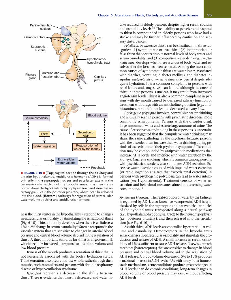
near the thirst center in the hypothalamus, respond to changesin extracellular osmolality by stimulating the sensation of thirst(Fig. 6-10). Thirst normally develops when there is as little as a1% to 2% change in serum osmolality.6 Stretch receptors in thevascular system that are sensitive to changes in arterial bloodpressure and central blood volume also aid in the regulation ofthirst. A third important stimulus for thirst is angiotensin II,which becomes increased in response to low blood volume andlow blood pressure.
Dryness of the mouth produces a sensation of thirst that isnot necessarily associated with the body’s hydration status.Thirst sensation also occurs in those who breathe through theirmouths, such as smokers and persons with chronic respiratorydisease or hyperventilation syndrome.
Hypodipsia represents a decrease in the ability to sensethirst. There is evidence that thirst is decreased and water in-
take reduced in elderly persons, despite higher serum sodiumand osmolality levels.7,8 The inability to perceive and respondto thirst is compounded in elderly persons who have had astroke and may be further influenced by confusion and sen-sory disturbances.
Polydipsia, or excessive thirst, can be classified into three cat-egories: (1) symptomatic or true thirst, (2) inappropriate orfalse thirst that occurs despite normal levels of body water andserum osmolality, and (3) compulsive water drinking. Sympto-matic thirst develops when there is a loss of body water and re-solves after the loss has been replaced. Among the most com-mon causes of symptomatic thirst are water losses associatedwith diarrhea, vomiting, diabetes mellitus, and diabetes in-sipidus. Inappropriate or excessive thirst may persist despite ade-quate hydration. It is a common complaint in persons withrenal failure and congestive heart failure. Although the cause ofthirst in these persons is unclear, it may result from increasedangiotensin levels. Thirst is also a common complaint in per-sons with dry mouth caused by decreased salivary function ortreatment with drugs with an anticholinergic action (e.g., anti-histamines, atropine) that lead to decreased salivary flow.
Psychogenic polydipsia involves compulsive water drinkingand is usually seen in persons with psychiatric disorders, mostcommonly schizophrenia. Persons with the disorder drinklarge amounts of water and excrete large amounts of urine. Thecause of excessive water drinking in these persons is uncertain.It has been suggested that the compulsive water drinking mayshare the same pathology as the psychosis because personswith the disorder often increase their water drinking during pe-riods of exacerbation of their psychotic symptoms.9 The condi-tion may be compounded by antipsychotic medications thatincrease ADH levels and interfere with water excretion by thekidneys. Cigarette smoking, which is common among personswith psychiatric disorders, also stimulates ADH secretion. Ex-cessive water ingestion coupled with impaired water excretion(or rapid ingestion at a rate that exceeds renal excretion) inpersons with psychogenic polydipsia can lead to water intoxi-cation (see Hyponatremia). Treatment consists of water re-striction and behavioral measures aimed at decreasing waterconsumption.10
Antidiuretic Hormone. The reabsorption of water by the kidneysis regulated by ADH, also known as vasopressin. ADH is syn-thesized by cells in the supraoptic and paraventricular nucleiof the hypothalamus; transported along a neural pathway(i.e., hypothalamohypophysial tract) to the neurohypophysis(i.e., posterior pituitary); and then released into the circula-tion (see Fig. 6-10).11
As with thirst, ADH levels are controlled by extracellular vol-ume and osmolality. Osmoreceptors in the hypothalamussense changes in extracellular osmolality and stimulate the pro-duction and release of ADH. A small increase in serum osmo-lality of 1% is sufficient to cause ADH release. Likewise, stretchreceptors (baroreceptors) that are sensitive to changes in bloodpressure and central blood volume aid in the regulation ofADH release. A blood volume decrease of 5% to 10% producesa maximal increase in ADH levels.12 As with many other homeo-static mechanisms, acute conditions produce greater changes inADH levels than do chronic conditions; long-term changes inblood volume or blood pressure may exist without affectingADH levels.
93Chapter 6: Alterations in Fluids, Electrolytes, and Acid-Base Balance
■ FIGURE 6-10 ■ (Top) sagittal section through the pituitary andanterior hypothalamus. Antidiuretic hormone (ADH) is formedprimarily in the supraoptic nucleus and to a lesser extent in theparaventricular nucleus of the hypothalamus. It is then trans-ported down the hypothalamohypophysial tract and stored in se-cretory granules in the posterior pituitary, where it can be releasedinto the blood. (Bottom) pathways for regulation of extracellularwater volume by thirst and antidiuretic hormone.
Hypothalamo-hypophysial tract
Anterior lobePosterior lobe
Pituitarygland
Supraopticnucleus
Osmoreceptors
Paraventricularnucleus
Capillaryplexus
Blood volume
Secretion ofADH
Reabsorption ofwater by the kidney
Feedback
Extracellularwater volume
Thirst
Water ingestion
Serum osmolality
An abnormal increase in ADH synthesis and release occursin a number of stress situations. Severe pain, nausea, trauma,surgery, certain anesthetic agents, and some narcotics (e.g.,morphine and meperidine) increase ADH levels. Nausea is apotent stimulus of ADH secretion; it can increase ADH levels10 to 1000 times those required for maximal diuresis.13 Amongthe drugs that affect ADH are nicotine, which stimulates its re-lease, and alcohol, which inhibits it. Two important conditionsalter ADH levels: diabetes insipidus and inappropriate secre-tion of ADH.
Diabetes insipidus (DI) is caused by a deficiency of or a de-creased response to ADH. Persons with DI are unable to con-centrate their urine during periods of water restriction; they ex-crete large volumes of urine, usually 3 to 20 L/day, dependingon the degree of ADH deficiency or renal insensitivity to ADH.This large urine output is accompanied by excessive thirst. Aslong as the thirst mechanism is normal and fluid is readilyavailable, there is little or no alteration in the fluid levels of per-sons with DI. The danger arises when the condition developsin someone who is unable to communicate the need for wateror is unable to secure the needed water. In such cases, inade-quate fluid intake rapidly leads to hypertonic dehydration andincreased serum osmolality.
There are two types of DI: central or neurogenic and ne-phrogenic DI. Neurogenic DI occurs because of a defect in thesynthesis or release of ADH and nephrogenic DI occurs be-cause the kidneys do not respond to ADH.14 In neurogenic DI,loss of 75% to 80% of ADH-secretory neurons is necessary be-fore polyuria becomes evident. Most persons with neurogenicDI have an incomplete form of the disorder and retain someability to concentrate their urine. Temporary neurogenic DImay follow head injury or surgery near the hypophysial tract.Nephrogenic DI is characterized by impairment of the urine-concentrating ability of the kidney and free-water conservation.It may occur as a genetic trait that affects the ADH receptors inthe kidney, as a side effect of drugs such as lithium,15 or as theresult of electrolyte disorders such as potassium depletion orchronic hypercalcemia.
The syndrome of inappropriate ADH (SIADH) results from afailure of the negative feedback system that regulates the releaseand inhibition of ADH.16 In persons with this syndrome, ADHsecretion continues even when serum osmolality is decreased,causing marked water retention and dilutional hyponatremia.
SIADH may occur as a transient condition, such as in a stresssituation, or as a chronic condition, resulting from disorderssuch as a lung tumor. Stimuli, such as surgery, pain, stress, andtemperature changes, are capable of stimulating ADH releasethrough the central nervous system (CNS). Drugs induce SIADHin different ways; some drugs are thought to increase hypo-thalamic production and release of ADH, and others are be-lieved to act directly on the renal tubules to enhance the actionof ADH. More chronic forms of SIADH may be the result oflung tumors, chest lesions, and CNS disorders. Tumors, partic-ularly bronchogenic carcinoma and cancers of the lymphoidtissues, prostate, and pancreas, are known to produce and re-lease ADH independent of normal hypothalamic control mech-anisms. Other intrathoracic conditions, such as advancedtuberculosis, severe pneumonia, and positive-pressure breath-ing, can also cause SIADH. The suggested mechanism forSIADH in positive-pressure ventilation is activation of baro-receptors (e.g., aortic baroreceptors, cardiopulmonary recep-
tors) that respond to marked changes in intrathoracic pressure.Human immunodeficiency virus (HIV) infection is emerging asa new cause of SIADH. It has been reported that as many as35% of persons with acquired immunodeficiency syndrome(AIDS) who are admitted to the acute care setting will haveSIADH related to Pneumocystis carinii pneumonia, central ner-vous system infections, or malignancies.17
The manifestations of SIADH are those of dilutional hypo-natremia. Urine output decreases despite adequate or increasedfluid intake. Urine osmolality is high, and serum osmolality islow. Hematocrit and the serum sodium and BUN levels are alldecreased because of the expansion of the ECF volume. Theseverity of symptoms usually is proportional to the extent ofsodium depletion and water intoxication.
Alterations in Isotonic Fluid VolumeIsotonic fluid volume disorders represent an expansion or con-traction of the ECF brought about by proportionate changes inboth sodium and water.
Isotonic Fluid Volume DeficitFluid volume deficit is characterized by a decrease in ECF, in-cluding circulating blood volume. The term isotonic fluid volumedeficit is used to differentiate fluid deficit in which there are pro-portionate losses in sodium and water from water deficit andthe hyperosmolar state associated with hypernatremia. Unlessother fluid and electrolyte imbalances are present, the concen-tration of serum electrolytes remains essentially unchanged.When the effective circulating blood volume is compromised,the condition is often referred to as hypovolemia.
Causes. Isotonic fluid volume deficit results when water andelectrolytes are lost in isotonic proportions. It is almost alwayscaused by a loss of body fluids and is often accompanied by a
94 Unit One: Mechanisms of Disease
KEY CONCEPTS
SODIUM AND WATER BALANCE
■ It is the amount of water and its effect on sodiumconcentration in the ECF that serves to regulate thedistribution of fluid between the ICF and the ECFcompartments.
■ Isotonic changes in body fluids that result from pro-portionate gains or losses of sodium and water arelargely confined to the ECF compartment. Many ofthe manifestations of isotonic fluid deficit or excessreflect changes in vascular and interstitial fluidvolume.
■ Hyponatremia or hypernatremia brought about bydisproportionate losses or gains in sodium or waterexert their effects on the ICF compartment, causingwater to move in or out of body cells. Many of themanifestations of changes in sodium concentrationreflect changes in the intracellular volume of cells,particularly those in the nervous system.
decrease in fluid intake. It can occur because of a loss of gastro-intestinal fluids, polyuria, or sweating caused by fever and ex-ercise. Third-space losses cause sequestering of ECF in the se-rous cavities, extracellular spaces in injured tissues, or lumen ofthe gut.
In a single day, 8 to 10 L of ECF is secreted into the gastro-intestinal tract. Most of it is reabsorbed in the ileum and proximal colon, and only about 150 to 200 mL per day iseliminated in the feces. Vomiting and diarrhea interrupt the reabsorption process and, in some situations, lead to increasedsecretion of fluid into the intestinal tract. Gastrointestinal suc-tion, fistulas, and drainage tubes can remove large amounts offluid from the gastrointestinal tract.
Excess sodium and water losses can also occur through thekidney. Certain forms of kidney disease are characterized bysalt wasting caused by impaired sodium reabsorption. Fluidvolume deficit also can result from osmotic diuresis or injudi-cious use of diuretic therapy. Glucose in the urine filtrate pre-vents reabsorption of water by the renal tubules, causing a lossof sodium and water. In Addison’s disease, a condition ofchronic adrenocortical insufficiency, there is unregulated lossof sodium in the urine with a resultant loss of ECF volume (seeChapter 31).
The skin acts as an exchange surface for heat and as a vaporbarrier to prevent water from leaving the body. Body surfacelosses of sodium and water increase when there is excessivesweating or when large areas of skin have been damaged. Hotweather and fever increase sweating. The respiratory rate andsweating usually are increased as body temperature rises. Burnsare another cause of excess fluid loss.
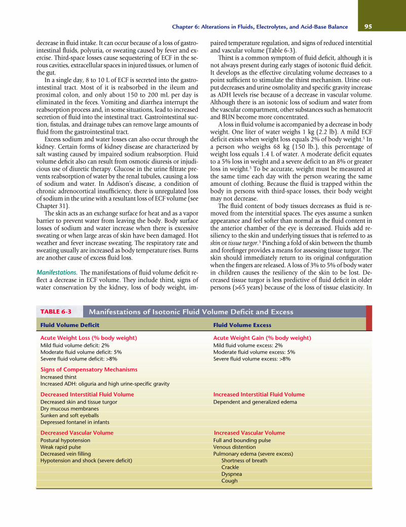
Manifestations. The manifestations of fluid volume deficit re-flect a decrease in ECF volume. They include thirst, signs ofwater conservation by the kidney, loss of body weight, im-
paired temperature regulation, and signs of reduced interstitialand vascular volume (Table 6-3).
Thirst is a common symptom of fluid deficit, although it isnot always present during early stages of isotonic fluid deficit.It develops as the effective circulating volume decreases to apoint sufficient to stimulate the thirst mechanism. Urine out-put decreases and urine osmolality and specific gravity increaseas ADH levels rise because of a decrease in vascular volume.Although there is an isotonic loss of sodium and water fromthe vascular compartment, other substances such as hematocritand BUN become more concentrated.
A loss in fluid volume is accompanied by a decrease in bodyweight. One liter of water weighs 1 kg (2.2 lb). A mild ECFdeficit exists when weight loss equals 2% of body weight.5 Ina person who weighs 68 kg (150 lb.), this percentage ofweight loss equals 1.4 L of water. A moderate deficit equatesto a 5% loss in weight and a severe deficit to an 8% or greaterloss in weight.5 To be accurate, weight must be measured atthe same time each day with the person wearing the sameamount of clothing. Because the fluid is trapped within thebody in persons with third-space losses, their body weightmay not decrease.
The fluid content of body tissues decreases as fluid is re-moved from the interstitial spaces. The eyes assume a sunkenappearance and feel softer than normal as the fluid content inthe anterior chamber of the eye is decreased. Fluids add re-siliency to the skin and underlying tissues that is referred to asskin or tissue turgor.5 Pinching a fold of skin between the thumband forefinger provides a means for assessing tissue turgor. Theskin should immediately return to its original configurationwhen the fingers are released. A loss of 3% to 5% of body waterin children causes the resiliency of the skin to be lost. De-creased tissue turgor is less predictive of fluid deficit in olderpersons (>65 years) because of the loss of tissue elasticity. In
95Chapter 6: Alterations in Fluids, Electrolytes, and Acid-Base Balance
Manifestations of Isotonic Fluid Volume Deficit and ExcessTABLE 6-3
Acute Weight Loss (% body weight) Acute Weight Gain (% body weight)Mild fluid volume deficit: 2% Mild fluid volume excess: 2%Moderate fluid volume deficit: 5% Moderate fluid volume excess: 5%Severe fluid volume deficit: >8% Severe fluid volume excess: >8%
Signs of Compensatory MechanismsIncreased thirstIncreased ADH: oliguria and high urine-specific gravity
Decreased Interstitial Fluid Volume Increased Interstitial Fluid VolumeDecreased skin and tissue turgor Dependent and generalized edemaDry mucous membranesSunken and soft eyeballsDepressed fontanel in infants
Decreased Vascular Volume Increased Vascular VolumePostural hypotension Full and bounding pulseWeak rapid pulse Venous distentionDecreased vein filling Pulmonary edema (severe excess)Hypotension and shock (severe deficit) Shortness of breath
CrackleDyspneaCough
Fluid Volume Deficit Fluid Volume Excess
infants fluid deficit may be evidenced by depression of the an-terior fontanel because of a decrease in cerebrospinal fluid.
Arterial and venous volumes decline during periods of fluiddeficit, as does filling of the capillary circulation. As the volumein the arterial system declines, the blood pressure decreases, theheart rate increases, and the pulse becomes weak and thready.Postural hypotension (a drop in blood pressure upon stand-ing) is an early sign of fluid deficit. On the venous side of thecirculation, the veins become less prominent, and venous refilltime increases. Body temperature may be subnormal becauseof decreased metabolism.5 When volume depletion becomessevere, signs of hypovolemic shock and vascular collapse ap-pear (see Chapter 18).
Diagnosis and Treatment. Treatment of fluid volume deficitconsists of fluid replacement and measures to correct the un-derlying cause. Usually, isotonic electrolyte solutions are usedfor fluid replacement. Acute hypovolemia and hypovolemicshock can cause renal damage; therefore, prompt assessment ofthe degree of fluid deficit and adequate measures to resolve thedeficit and treat the underlying cause are essential.
Isotonic Fluid Volume ExcessFluid volume excess represents an isotonic expansion of theECF compartment with increases in both interstitial and vas-cular volumes. Although increased fluid volume is usually theresult of a disease condition, this is not always true. For ex-ample, a compensatory isotonic expansion of body fluids canoccur in healthy persons during hot weather as a mechanismfor increasing body heat loss.
Causes. Isotonic fluid volume excess almost always resultsfrom an increase in total body sodium that is accompanied bya proportionate increase in body water. Although it can occuras the result of excessive sodium intake, it is most commonlycaused by a decrease in sodium and water elimination by thekidney. Among the causes of decreased sodium and water elim-ination are disorders of renal function, heart failure, liver fail-ure, and corticosteroid excess.
Heart failure produces a decrease in renal blood flow and acompensatory increase in sodium and water retention (Chap-ter 18). Persons with severe congestive heart failure maintain aprecarious balance between sodium and water intake and out-put. Even small increases in sodium intake can precipitate astate of fluid volume excess and a worsening of heart failure.Liver failure impairs aldosterone metabolism and alters renalperfusion, leading to increased salt and water retention. Cor-ticosteroid hormones increase sodium reabsorption by the kid-ney. Persons taking corticosteroid medications and those withCushing’s syndrome (see Chapter 31) often have problemswith sodium retention.
Manifestations. Isotonic fluid volume excess is characterizedby an increase in interstitial and vascular fluids. It is manifestedby weight gain over a short period of time. A mild fluid volumeexcess represents a 2% weight gain; moderate fluid volume ex-cess, a 5% weight gain; and severe fluid volume excess, a weightgain of 8% or more (Table 6-3).5 The presence of edema is char-acteristic of isotonic fluid volume excess. When the excess fluidaccumulates gradually, as often happens in debilitating dis-eases and starvation, edema fluid may mask the loss of tissue
mass. As the vascular volume increases, the neck veins becomedistended, the pulse becomes full and bounding, and the cen-tral venous pressure becomes elevated. The BUN and hemato-crit may be decreased as a result of the expanded plasmavolume. When excess fluid accumulates in the lungs (i.e., pul-monary edema), there is shortness of breath, complaints of dif-ficult breathing, respiratory crackles, and a productive cough(see Chapter 18). Ascites and pleural effusion may occur withsevere fluid volume excess.
Diagnosis and Treatment. The treatment of fluid volume ex-cess focuses on providing a more favorable balance betweensodium intake and output. A sodium-restricted diet is oftenprescribed. Diuretic therapy may be used to increase sodiumelimination.
Alterations in Sodium ConcentrationThe normal serum sodium ranges from 135 to 145 mEq/L(135 to 145 mmol/L). Serum sodium values, expressed inmEq/L, reflect the concentration or dilution of sodium bywater, rather than its absolute value. Because sodium and itsattendant anions account for 90% to 95% of the osmolality ofthe ECF (normal range, 275 to 295 mOsm/kg), changes inserum sodium generally are accompanied by changes in serumosmolality.
HyponatremiaHyponatremia represents a serum sodium concentration below135 mEq/L (135 mmol/L). Because of the effects of other os-motically active particles in the ECF, such as glucose, hypo-natremia may be associated with high or low tonicity.17–19
Hypertonic (translocational) hyponatremia results from anosmotic shift of water from ICF to the ECF, such as occurs withhyperglycemia. In this case, the sodium in the ECF becomes di-luted as water moves out of cells in response to the osmotic ef-fects of the elevated blood glucose level.
Hypotonic (dilutional) is by far the most common form ofhyponatremia. It is caused by water retention and characterizedby a decrease in serum osmolality. Dilutional hyponatremiacan present as a hypervolemic, euvolemic, or hypovolemic con-dition. Hypervolemic hyponatremia involves an increase inECF volume and is seen when hyponatremic conditions are ac-companied by edema-forming disorders such as congestiveheart failure, cirrhosis, and advanced kidney disease.
Euvolemic hyponatremia represents a retention of waterwith dilution of sodium while maintaining the ECF volumewithin a normal range. It is usually the result of inappropriatethirst or SIADH. Hypovolemic hyponatremia occurs whenwater is lost along with sodium but to a lesser extent. It occurswith diuretic use, excessive sweating in hot weather, and vom-iting and diarrhea.
Causes. The most common causes of acute dilutional hypona-tremia in adults are drug therapy (diuretics and drugs that in-crease ADH levels), inappropriate fluid replacement duringheat exposure or after heavy exercise, SIADH, and polydipsia inpersons with psychotic disorder.
Among the causes of hypovolemic hyponatremia are exces-sive sweating in hot weather, particularly during heavy exercise,which leads to loss of salt and water; hyponatremia develops
96 Unit One: Mechanisms of Disease
when water, rather than electrolyte-containing liquids, is usedto replace fluids lost in sweating. Another potential cause ofhypovolemic hyponatremia is the loss of sodium from thegastrointestinal tract caused by repeated tap water enemas orfrequent gastrointestinal irrigations with distilled water. Iso-osmotic fluid loss, such as occurs in vomiting or diarrhea, doesnot usually lower serum sodium levels unless these losses arereplaced with disproportionate amounts of orally ingested orparenterally administered water. Gastrointestinal fluid loss andingestion of excessively diluted formula are common causes ofacute hyponatremia in infants and children.
Hypovolemic hyponatremia is a common complication ofadrenal insufficiency and is attributable to the effects of aldo-sterone and cortisol deficiency (see Chapter 31). A lack of al-dosterone increases renal losses of sodium, and a cortisol defi-ciency leads to increased release of ADH with water retention.
The risk of euvolemic hyponatremia is increased duringthe postoperative period. During this time ADH levels areoften high, producing an increase in water reabsorption bythe kidney (see SIADH). Although these elevated levels usu-ally resolve in about 72 hours, they can persist for as long as 5 days. The hyponatremia becomes exaggerated whenelectrolyte-free fluids (e.g., 5% glucose in water) are used forfluid replacement.
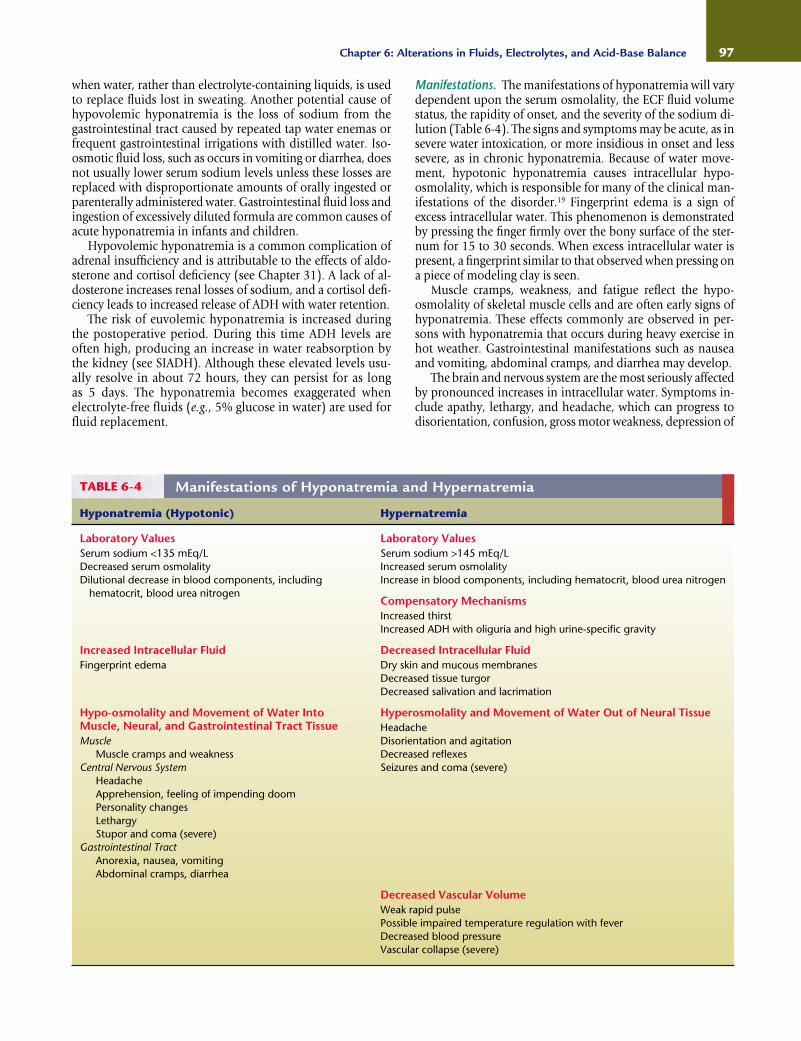
Manifestations. The manifestations of hyponatremia will varydependent upon the serum osmolality, the ECF fluid volumestatus, the rapidity of onset, and the severity of the sodium di-lution (Table 6-4). The signs and symptoms may be acute, as insevere water intoxication, or more insidious in onset and lesssevere, as in chronic hyponatremia. Because of water move-ment, hypotonic hyponatremia causes intracellular hypo-osmolality, which is responsible for many of the clinical man-ifestations of the disorder.19 Fingerprint edema is a sign ofexcess intracellular water. This phenomenon is demonstratedby pressing the finger firmly over the bony surface of the ster-num for 15 to 30 seconds. When excess intracellular water ispresent, a fingerprint similar to that observed when pressing ona piece of modeling clay is seen.
Muscle cramps, weakness, and fatigue reflect the hypo-osmolality of skeletal muscle cells and are often early signs ofhyponatremia. These effects commonly are observed in per-sons with hyponatremia that occurs during heavy exercise inhot weather. Gastrointestinal manifestations such as nauseaand vomiting, abdominal cramps, and diarrhea may develop.
The brain and nervous system are the most seriously affectedby pronounced increases in intracellular water. Symptoms in-clude apathy, lethargy, and headache, which can progress todisorientation, confusion, gross motor weakness, depression of
97Chapter 6: Alterations in Fluids, Electrolytes, and Acid-Base Balance
Manifestations of Hyponatremia and HypernatremiaTABLE 6-4
Laboratory ValuesSerum sodium <135 mEq/LDecreased serum osmolalityDilutional decrease in blood components, including
hematocrit, blood urea nitrogen
Increased Intracellular FluidFingerprint edema
Hypo-osmolality and Movement of Water IntoMuscle, Neural, and Gastrointestinal Tract TissueMuscle
Muscle cramps and weaknessCentral Nervous System
HeadacheApprehension, feeling of impending doomPersonality changesLethargyStupor and coma (severe)
Gastrointestinal TractAnorexia, nausea, vomitingAbdominal cramps, diarrhea
Laboratory ValuesSerum sodium >145 mEq/LIncreased serum osmolalityIncrease in blood components, including hematocrit, blood urea nitrogen
Compensatory MechanismsIncreased thirstIncreased ADH with oliguria and high urine-specific gravity
Decreased Intracellular FluidDry skin and mucous membranesDecreased tissue turgorDecreased salivation and lacrimation
Hyperosmolality and Movement of Water Out of Neural TissueHeadacheDisorientation and agitationDecreased reflexesSeizures and coma (severe)
Decreased Vascular VolumeWeak rapid pulsePossible impaired temperature regulation with feverDecreased blood pressureVascular collapse (severe)
Hyponatremia (Hypotonic) Hypernatremia
deep tendon reflexes. Seizures and coma occur when serumsodium levels reach extremely low levels. These severe effects,which are caused by brain swelling, may be irreversible.19 If thecondition develops slowly, signs and symptoms do not de-velop until serum sodium levels approach 125 mEq/L. Theterm “water intoxication” is often used to describe the neuro-logic effects of acute hypotonic hyponatremia.
Treatment. The treatment of hyponatremia with water excessfocuses on the underlying cause. When hyponatremia iscaused by water intoxication, limiting water intake or dis-continuing medications that contribute to SIADH may be suf-ficient. The administration of saline solution orally or intra-venously may be needed in severe hyponatremia caused bysodium deficiency.
HypernatremiaHypernatremia implies a serum sodium level above 145 mEq/Land a serum osmolality greater than 295 mOsm/kg. Becausesodium is functionally an impermeable solute, it contributes tothe tonicity and movement of water across cell membranes.Hypernatremia is characterized by hypertonicity of the ECF andalmost always causes cellular dehydration.20
Causes. Hypernatremia represents a deficit of water in relationto the body’s sodium levels. It can be caused by net gain ofsodium or net loss of water. Rapid ingestion or infusion ofsodium with insufficient time or opportunity for water inges-tion can produce a disproportionate gain in sodium. A defectin thirst or inability to obtain or drink water can interfere withwater replacement.
Hypernatremia also occurs when there is an excess loss ofbody fluids that have a lower than normal concentration ofsodium so that water is lost in excess of sodium. This can resultfrom increased losses from the respiratory tract during fever orstrenuous exercise, from watery diarrhea, or when osmoticallyactive tube feedings are given with inadequate amounts ofwater. With pure water loss, each body fluid compartment losesan equal percentage of its volume. Because approximately onethird of the water is in the ECF compartment, compared withthe two thirds in the ICF compartment, more actual water vol-ume is lost from the ICF than the ECF compartment.5
Normally, water deficit stimulates thirst and increases waterintake. Therefore, hypernatremia is more likely to occur in in-fants and in persons who cannot express their thirst or obtainwater to drink. With hypodipsia, or impaired thirst, the needfor fluid intake does not activate the thirst response. Hypo-dipsia is particularly prevalent among the elderly. In personswith diabetes insipidus, hypernatremia can develop whenthirst is impaired or access to water is impeded.
Manifestations. The clinical manifestations of hypernatremiacaused by water loss are largely those of ECF fluid loss and cel-lular dehydration (Table 6-4). The severity of signs and symp-toms is greatest when the increase in serum sodium is largeand occurs rapidly. Body weight is decreased in proportion tothe amount of water that has been lost. Because blood plasmais roughly 90% to 93% water, the concentrations of bloodcells, hematocrit, BUN, and other solutes increase as ECF waterdecreases.
Thirst is an early symptom of water deficit, occurring whenwater losses are equal to 0.5% of body water. Urine output isdecreased and urine osmolality increased because of renalwater-conserving mechanisms. Body temperature frequently iselevated, and the skin becomes warm and flushed. As the vas-cular volume decreases, the pulse becomes rapid and thready,and the blood pressure drops. Hypernatremia produces an in-crease in serum osmolality and results in water being pulledout of body cells. As a result, the skin and mucous membranesbecome dry, and salivation and tearing of the eyes are de-creased. The mouth becomes dry and sticky, and the tonguebecomes rough and fissured. Swallowing is difficult. The sub-cutaneous tissues assume a firm, rubbery texture. Most sig-nificantly, water is pulled out of the cells in the CNS, causingdecreased reflexes, agitation, headache, and restlessness. Comaand seizures may develop as hypernatremia progresses.
Treatment. The treatment of hypernatremia includes measuresto treat the underlying cause of the disorder and fluid replace-ment therapy to treat the accompanying dehydration. Replace-ment fluids can be given orally or intravenously. The oral routeis preferable. Oral glucose–electrolyte replacement solutionsare available for the treatment of infants with diarrhea.21 Untilrecently, these solutions were used only early in diarrheal ill-ness or as a first step in re-establishing oral intake after par-enteral replacement therapy. These solutions are now widelyavailable in grocery stores and pharmacies for use in the treat-ment of diarrhea and other dehydrating disorders in infantsand young children.
98 Unit One: Mechanisms of Disease
In summary, body fluids are distributed between the ICFand ECF compartments. Regulation of fluid volume, soluteconcentration, and distribution between the two compart-ments depends on water and sodium balance. Water providesapproximately 90% to 93% of fluid volume and sodium salts,approximately 90% to 95% of extracellular solutes. Bodywater is regulated by thirst, which controls water intake, andADH, which controls urine concentration and renal output.Sodium is ingested in the diet and eliminated by the kidneysunder the influence of the sympathetic nervous system andthe renin-angiotensin-aldosterone system.
Isotonic fluid disorders result from contraction or expan-sion of ECF volume brought about by proportionate losses ofsodium and water. Isotonic fluid volume deficit is characterizedby a decrease in ECF volume. It causes thirst, decreased vascu-lar volume and circulatory function, decreased urine output,and increased urine specific gravity. Isotonic fluid volume ex-cess is characterized by an increase in ECF volume. It is mani-fested by signs of increased vascular volume and edema.
Alterations in extracellular sodium concentration arebrought about by a disproportionate gain (hyponatremia) orloss (hypernatremia) of water. As the major cation in the ECFcompartment, sodium controls the ECF osmolality and its ef-fect on cell volume. Hypotonic hyponatremia is characterizedby water being pulled into the cell from the extracellular com-partment, causing cells to swell. It is manifested by musclecramps and weakness; nausea, vomiting, abdominal cramps,and diarrhea; and CNS signs such as lethargy, headache, de-pression of deep tendon reflexes, and in severe cases seizure
POTASSIUM BALANCE
Potassium is the second most abundant cation in the body andthe major cation in the ICF compartment. Approximately 98%of body potassium is contained within body cells, with anintracellular concentration of 140 to 150 mEq/L.22 The potas-sium content of ECF (3.5 to 5.0 mEq/L) is considerably less.Because potassium is an intracellular ion, total body stores ofpotassium are related to body size and muscle mass. Approx-imately 65% to 75% of potassium is in muscle.23 Thus, totalbody potassium declines with age, mainly as a result of a de-crease in muscle mass.
As the major intracellular cation, potassium is critical tomany body functions. It is involved in a wide range of bodyfunctions, including the maintenance of the osmotic integrityof cells, acid-base balance, and the kidney’s ability to concen-trate urine. Potassium is necessary for growth, and it con-tributes to the intricate chemical reactions that transform car-bohydrates into energy, change glucose into glycogen, andconvert amino acids to proteins.
Potassium also plays a critical role in conducting nerve im-pulses and the excitability of skeletal, cardiac, and smoothmuscle (see Chapter 1). It does this by regulating: (1) the rest-
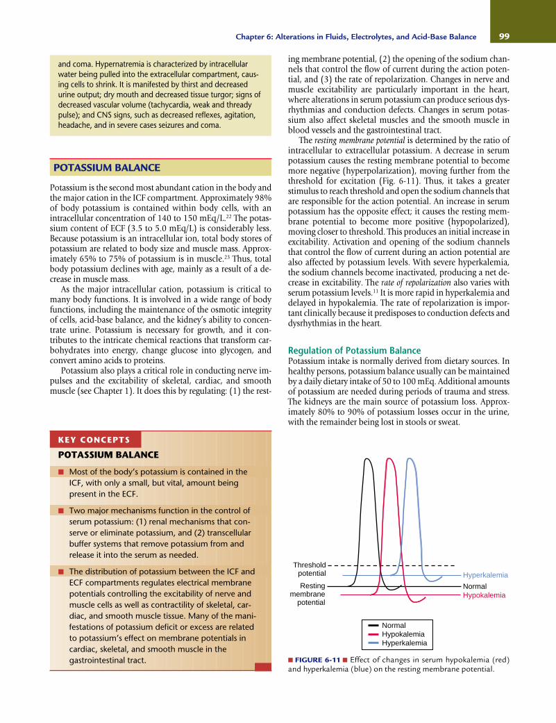
ing membrane potential, (2) the opening of the sodium chan-nels that control the flow of current during the action poten-tial, and (3) the rate of repolarization. Changes in nerve andmuscle excitability are particularly important in the heart,where alterations in serum potassium can produce serious dys-rhythmias and conduction defects. Changes in serum potas-sium also affect skeletal muscles and the smooth muscle inblood vessels and the gastrointestinal tract.
The resting membrane potential is determined by the ratio ofintracellular to extracellular potassium. A decrease in serumpotassium causes the resting membrane potential to becomemore negative (hyperpolarization), moving further from thethreshold for excitation (Fig. 6-11). Thus, it takes a greaterstimulus to reach threshold and open the sodium channels thatare responsible for the action potential. An increase in serumpotassium has the opposite effect; it causes the resting mem-brane potential to become more positive (hypopolarized),moving closer to threshold. This produces an initial increase inexcitability. Activation and opening of the sodium channelsthat control the flow of current during an action potential arealso affected by potassium levels. With severe hyperkalemia,the sodium channels become inactivated, producing a net de-crease in excitability. The rate of repolarization also varies withserum potassium levels.11 It is more rapid in hyperkalemia anddelayed in hypokalemia. The rate of repolarization is impor-tant clinically because it predisposes to conduction defects anddysrhythmias in the heart.
Regulation of Potassium BalancePotassium intake is normally derived from dietary sources. Inhealthy persons, potassium balance usually can be maintainedby a daily dietary intake of 50 to 100 mEq. Additional amountsof potassium are needed during periods of trauma and stress.The kidneys are the main source of potassium loss. Approx-imately 80% to 90% of potassium losses occur in the urine,with the remainder being lost in stools or sweat.
99Chapter 6: Alterations in Fluids, Electrolytes, and Acid-Base Balance
and coma. Hypernatremia is characterized by intracellularwater being pulled into the extracellular compartment, caus-ing cells to shrink. It is manifested by thirst and decreasedurine output; dry mouth and decreased tissue turgor; signs ofdecreased vascular volume (tachycardia, weak and threadypulse); and CNS signs, such as decreased reflexes, agitation,headache, and in severe cases seizures and coma.
KEY CONCEPTS
POTASSIUM BALANCE
■ Most of the body’s potassium is contained in theICF, with only a small, but vital, amount beingpresent in the ECF.
■ Two major mechanisms function in the control ofserum potassium: (1) renal mechanisms that con-serve or eliminate potassium, and (2) transcellularbuffer systems that remove potassium from andrelease it into the serum as needed.
■ The distribution of potassium between the ICF andECF compartments regulates electrical membranepotentials controlling the excitability of nerve andmuscle cells as well as contractility of skeletal, car-diac, and smooth muscle tissue. Many of the mani-festations of potassium deficit or excess are relatedto potassium’s effect on membrane potentials incardiac, skeletal, and smooth muscle in the gastrointestinal tract. ■ FIGURE 6-11 ■ Effect of changes in serum hypokalemia (red)
and hyperkalemia (blue) on the resting membrane potential.
Restingmembrane
potential
Thresholdpotential
NormalHypokalemiaHyperkalemia
Hyperkalemia
HypokalemiaNormal
Mechanisms of Regulation. Normally, the ECF concentrationof potassium is precisely regulated at about 4.2 mEq/ml. Theprecise control is necessary because many cell functions aresensitive to even small changes in ECF potassium levels. An in-crease in serum potassium levels of only maybe 0.3 to 0.4 mEq/Lcan cause serious cardiac dysrhythmias and even death.
Serum potassium levels are largely regulated through twomechanisms: (1) renal mechanisms that conserve or eliminatepotassium, and (2) a transcellular shift of potassium betweenthe ICF and ECF compartments. Normally, it takes 6 to 8 hoursto eliminate 50% of potassium intake.3 To avoid an increase inextracellular potassium levels during this time, excess potas-sium is temporarily shifted into red blood cells and other cellssuch as those of muscle, liver, and bone.
Renal Regulation. The kidney provides the major route forpotassium elimination. Potassium is filtered in the glomerulus,reabsorbed along with sodium and water in the proximaltubule and with sodium and chloride in the thick ascendingloop of Henle, and then secreted into the late distal and corti-cal collecting tubules for elimination in the urine. The lattermechanism serves to “fine-tune” the concentration of potas-sium in the ECF.
Aldosterone plays an essential role in regulating potassiumelimination by the kidney (see Chapter 22). In the presence ofaldosterone, sodium is transported back into the blood andpotassium is secreted into the tubular filtrate for elimination inthe urine. There is also a potassium-hydrogen exchange systemin the collecting tubules of the kidney. When serum potassiumlevels are increased, potassium is secreted into the urine andhydrogen is reabsorbed into the blood, producing a decrease inpH and metabolic acidosis. Conversely, when potassium levelsare low, potassium is reabsorbed and hydrogen is secreted intothe urine, leading to metabolic alkalosis.
Extracellular-Intracellular Shifts. The movement of potassiumbetween the ECF and ICF allows potassium to move into bodycells when serum levels are high and move out when serum lev-els are low. Among the factors that alter potassium distributionbetween the ECF and ICF are insulin, β-adrenergic stimulation,serum osmolality, and acid-base disorders. Both insulin andthe β-adrenergic catecholamines (e.g., epinephrine) increasecellular uptake of potassium. Insulin increases the cellular up-take of potassium after a meal. The potassium content of a sin-gle meal is often as high as 50 mEq; the actions of insulin pre-vent a rise in serum potassium to life-threatening levels. Thecatecholamines, particularly epinephrine, facilitate the move-ment of potassium into muscle tissue during periods of physi-ologic stress.
Extracellular osmolality and pH also influence the move-ment of potassium between the ICF and ECF. Acute increasesin serum osmolality cause potassium to move out of cells.When serum osmolality is increased because of the presence ofimpermeable solutes such as glucose (without insulin), waterleaves the cell. The loss of cell water produces an increase in in-tracellular potassium, causing it to move out of the cell into theECF. Acid-base disorders are often accompanied by a change inserum potassium. Both the hydrogen and potassium ions arepositively charged, and both ions move freely between the ICFand ECF compartments. In metabolic acidosis, hydrogen ionsmove into body cells for buffering; this causes potassium to
leave the cells and move into the ECF. Metabolic alkalosis hasthe opposite effect.
Exercise can also produce compartmental shifts in potas-sium. Repeated muscle contraction releases potassium into theECF. Although the increase usually is small with modest exer-cise, it can be considerable during exhaustive exercise. Even therepeated clenching and unclenching of the fist during a blooddraw can cause potassium to move out of cells and artificiallyelevate serum potassium levels.
HypokalemiaHypokalemia refers to a serum potassium level below 3.5 mEq/L(3.5 mmol/L). Because of transcellular shifts, temporary changesin serum K+ may occur as the result of movement between theICF and ECF compartments.
Causes. The causes of potassium deficit can be grouped intothree categories: (1) inadequate intake; (2) excessive lossesthrough the kidney, skin, and gastrointestinal tract; and (3) re-distribution between the ICF and ECF compartments.
Inadequate intake is a frequent cause of hypokalemia. Apotassium intake of at least 10 to 30 mEq/day is needed tocompensate for obligatory urine losses.4,23 A person on apotassium-free diet continues to lose approximately 5 to 15 mEq of potassium daily. Insufficient dietary intake may re-sult from the inability to obtain or ingest food or from a dietthat is low in potassium-containing foods.
Excessive renal losses can occur with diuretic use, metabolicalkalosis, magnesium depletion, trauma and stress, and in-creased levels of aldosterone. Diuretic therapy, with the excep-tion of potassium-sparing diuretics, is the most common causeof hypokalemia. Both thiazide and loop diuretics increase theloss of potassium in the urine. The degree of hypokalemia is di-rectly related to diuretic dose and is greater when sodium in-take is higher.23 Magnesium depletion causes renal potassiumwasting. Magnesium deficiency often coexists with potassiumdepletion because of diuretic therapy or disease processes suchas diarrhea. Importantly, the ability to correct potassium defi-ciency is impaired when magnesium deficiency is present.
The kidneys do not have the homeostatic mechanismsneeded to conserve potassium during periods of stress or insufficient intake. After trauma and in stress situations, uri-nary losses of potassium are greatly increased, sometimes ap-proaching levels of 150 to 200 mEq/L.24 Renal losses of potas-sium are accentuated by aldosterone and cortisol. Trauma andsurgery produce a stress-related increase in these hormones.Primary aldosteronism, caused by an aldosterone-secretingtumor of the adrenal cortex, can produce severe urinary lossesof potassium. Cortisol binds to aldosterone receptors and exerts aldosterone-like effects on potassium elimination.
Although potassium losses from the gastrointestinal tractand the skin usually are minimal, these losses can become ex-cessive under certain conditions. The gastrointestinal tract isone of the most common sites for acute potassium losses.Vomiting and gastrointestinal suction lead to hypokalemia,partly because of actual potassium losses and because of renallosses associated with metabolic alkalosis (see section on meta-bolic alkalosis under acid-base balance). Because intestinalsecretions are high in potassium content, diarrhea and intes-tinal suction can also cause large losses of potassium. Excess-ive skin losses occur with loss of the protective skin surface
100 Unit One: Mechanisms of Disease
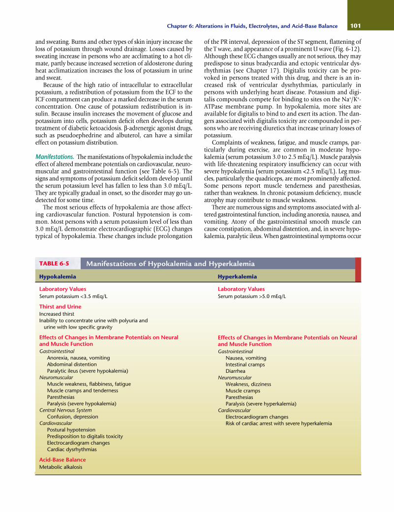
and sweating. Burns and other types of skin injury increase theloss of potassium through wound drainage. Losses caused bysweating increase in persons who are acclimating to a hot cli-mate, partly because increased secretion of aldosterone duringheat acclimatization increases the loss of potassium in urineand sweat.
Because of the high ratio of intracellular to extracellularpotassium, a redistribution of potassium from the ECF to theICF compartment can produce a marked decrease in the serumconcentration. One cause of potassium redistribution is in-sulin. Because insulin increases the movement of glucose andpotassium into cells, potassium deficit often develops duringtreatment of diabetic ketoacidosis. β-adrenergic agonist drugs,such as pseudoephedrine and albuterol, can have a similareffect on potassium distribution.
Manifestations. The manifestations of hypokalemia include theeffect of altered membrane potentials on cardiovascular, neuro-muscular and gastrointestinal function (see Table 6-5). Thesigns and symptoms of potassium deficit seldom develop untilthe serum potassium level has fallen to less than 3.0 mEq/L.They are typically gradual in onset, so the disorder may go un-detected for some time.
The most serious effects of hypokalemia are those affect-ing cardiovascular function. Postural hypotension is com-mon. Most persons with a serum potassium level of less than3.0 mEq/L demonstrate electrocardiographic (ECG) changestypical of hypokalemia. These changes include prolongation
of the PR interval, depression of the ST segment, flattening ofthe T wave, and appearance of a prominent U wave (Fig. 6-12).Although these ECG changes usually are not serious, they maypredispose to sinus bradycardia and ectopic ventricular dys-rhythmias (see Chapter 17). Digitalis toxicity can be pro-voked in persons treated with this drug, and there is an in-creased risk of ventricular dysrhythmias, particularly inpersons with underlying heart disease. Potassium and digi-talis compounds compete for binding to sites on the Na+/K+-ATPase membrane pump. In hypokalemia, more sites areavailable for digitalis to bind to and exert its action. The dan-gers associated with digitalis toxicity are compounded in per-sons who are receiving diuretics that increase urinary losses ofpotassium.
Complaints of weakness, fatigue, and muscle cramps, par-ticularly during exercise, are common in moderate hypo-kalemia (serum potassium 3.0 to 2.5 mEq/L). Muscle paralysiswith life-threatening respiratory insufficiency can occur withsevere hypokalemia (serum potassium <2.5 mEq/L). Leg mus-cles, particularly the quadriceps, are most prominently affected.Some persons report muscle tenderness and paresthesias,rather than weakness. In chronic potassium deficiency, muscleatrophy may contribute to muscle weakness.
There are numerous signs and symptoms associated with al-tered gastrointestinal function, including anorexia, nausea, andvomiting. Atony of the gastrointestinal smooth muscle cancause constipation, abdominal distention, and, in severe hypo-kalemia, paralytic ileus. When gastrointestinal symptoms occur
101Chapter 6: Alterations in Fluids, Electrolytes, and Acid-Base Balance
Manifestations of Hypokalemia and HyperkalemiaTABLE 6-5
Laboratory Values Laboratory ValuesSerum potassium <3.5 mEq/L Serum potassium >5.0 mEq/L
Thirst and UrineIncreased thirstInability to concentrate urine with polyuria and
urine with low specific gravity
Effects of Changes in Membrane Potentials on Neural and Muscle FunctionGastrointestinal
Anorexia, nausea, vomitingAbdominal distentionParalytic ileus (severe hypokalemia)
NeuromuscularMuscle weakness, flabbiness, fatigueMuscle cramps and tendernessParesthesiasParalysis (severe hypokalemia)
Central Nervous SystemConfusion, depression
CardiovascularPostural hypotensionPredisposition to digitalis toxicityElectrocardiogram changesCardiac dysrhythmias
Acid-Base BalanceMetabolic alkalosis
Hypokalemia Hyperkalemia
Effects of Changes in Membrane Potentials on Neuraland Muscle FunctionGastrointestinal
Nausea, vomitingIntestinal crampsDiarrhea
NeuromuscularWeakness, dizzinessMuscle crampsParesthesiasParalysis (severe hyperkalemia)
CardiovascularElectrocardiogram changesRisk of cardiac arrest with severe hyperkalemia
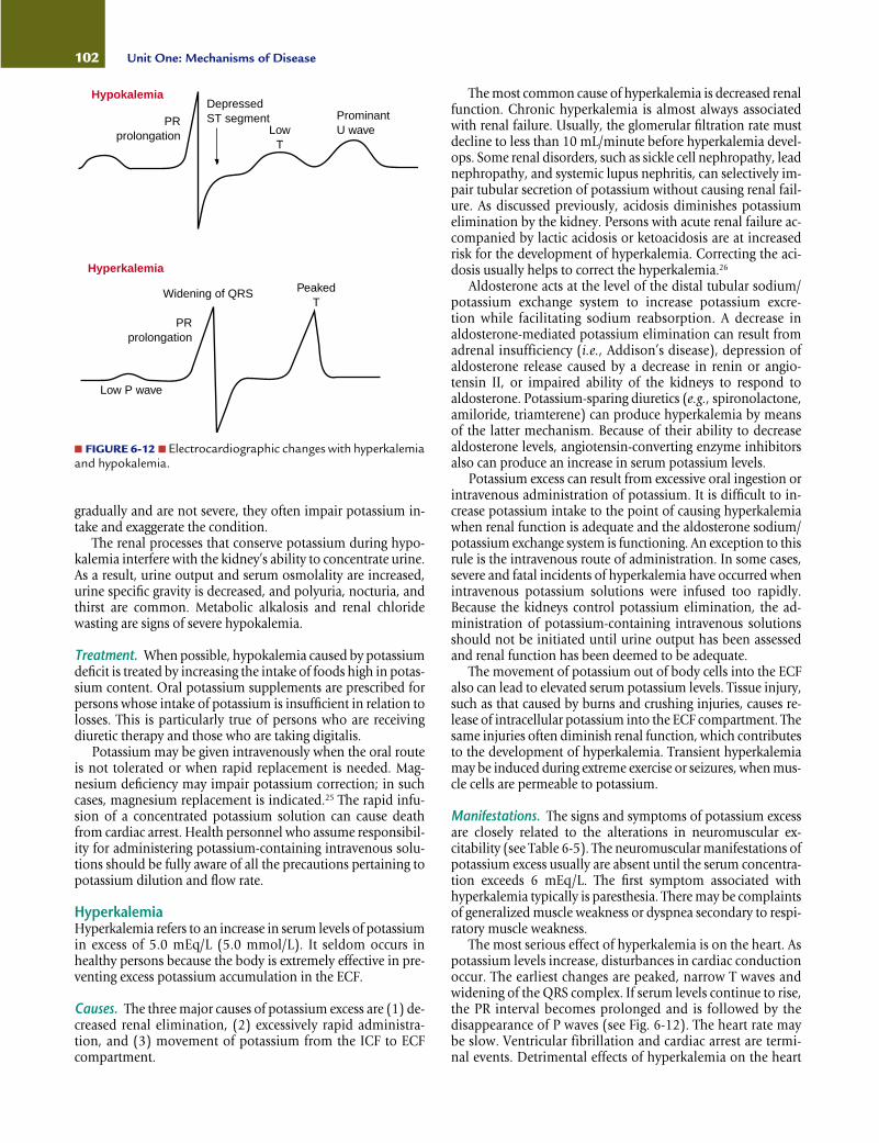
gradually and are not severe, they often impair potassium in-take and exaggerate the condition.
The renal processes that conserve potassium during hypo-kalemia interfere with the kidney’s ability to concentrate urine.As a result, urine output and serum osmolality are increased,urine specific gravity is decreased, and polyuria, nocturia, andthirst are common. Metabolic alkalosis and renal chloridewasting are signs of severe hypokalemia.
Treatment. When possible, hypokalemia caused by potassiumdeficit is treated by increasing the intake of foods high in potas-sium content. Oral potassium supplements are prescribed forpersons whose intake of potassium is insufficient in relation tolosses. This is particularly true of persons who are receivingdiuretic therapy and those who are taking digitalis.
Potassium may be given intravenously when the oral routeis not tolerated or when rapid replacement is needed. Mag-nesium deficiency may impair potassium correction; in suchcases, magnesium replacement is indicated.25 The rapid infu-sion of a concentrated potassium solution can cause deathfrom cardiac arrest. Health personnel who assume responsibil-ity for administering potassium-containing intravenous solu-tions should be fully aware of all the precautions pertaining topotassium dilution and flow rate.
HyperkalemiaHyperkalemia refers to an increase in serum levels of potassiumin excess of 5.0 mEq/L (5.0 mmol/L). It seldom occurs inhealthy persons because the body is extremely effective in pre-venting excess potassium accumulation in the ECF.
Causes. The three major causes of potassium excess are (1) de-creased renal elimination, (2) excessively rapid administra-tion, and (3) movement of potassium from the ICF to ECFcompartment.
The most common cause of hyperkalemia is decreased renalfunction. Chronic hyperkalemia is almost always associatedwith renal failure. Usually, the glomerular filtration rate mustdecline to less than 10 mL/minute before hyperkalemia devel-ops. Some renal disorders, such as sickle cell nephropathy, leadnephropathy, and systemic lupus nephritis, can selectively im-pair tubular secretion of potassium without causing renal fail-ure. As discussed previously, acidosis diminishes potassiumelimination by the kidney. Persons with acute renal failure ac-companied by lactic acidosis or ketoacidosis are at increasedrisk for the development of hyperkalemia. Correcting the aci-dosis usually helps to correct the hyperkalemia.26
Aldosterone acts at the level of the distal tubular sodium/potassium exchange system to increase potassium excre-tion while facilitating sodium reabsorption. A decrease inaldosterone-mediated potassium elimination can result fromadrenal insufficiency (i.e., Addison’s disease), depression ofaldosterone release caused by a decrease in renin or angio-tensin II, or impaired ability of the kidneys to respond to aldosterone. Potassium-sparing diuretics (e.g., spironolactone,amiloride, triamterene) can produce hyperkalemia by meansof the latter mechanism. Because of their ability to decreasealdosterone levels, angiotensin-converting enzyme inhibitorsalso can produce an increase in serum potassium levels.
Potassium excess can result from excessive oral ingestion orintravenous administration of potassium. It is difficult to in-crease potassium intake to the point of causing hyperkalemiawhen renal function is adequate and the aldosterone sodium/potassium exchange system is functioning. An exception to thisrule is the intravenous route of administration. In some cases,severe and fatal incidents of hyperkalemia have occurred whenintravenous potassium solutions were infused too rapidly.Because the kidneys control potassium elimination, the ad-ministration of potassium-containing intravenous solutionsshould not be initiated until urine output has been assessedand renal function has been deemed to be adequate.
The movement of potassium out of body cells into the ECFalso can lead to elevated serum potassium levels. Tissue injury,such as that caused by burns and crushing injuries, causes re-lease of intracellular potassium into the ECF compartment. Thesame injuries often diminish renal function, which contributesto the development of hyperkalemia. Transient hyperkalemiamay be induced during extreme exercise or seizures, when mus-cle cells are permeable to potassium.
Manifestations. The signs and symptoms of potassium excessare closely related to the alterations in neuromuscular ex-citability (see Table 6-5). The neuromuscular manifestations ofpotassium excess usually are absent until the serum concentra-tion exceeds 6 mEq/L. The first symptom associated withhyperkalemia typically is paresthesia. There may be complaintsof generalized muscle weakness or dyspnea secondary to respi-ratory muscle weakness.
The most serious effect of hyperkalemia is on the heart. Aspotassium levels increase, disturbances in cardiac conductionoccur. The earliest changes are peaked, narrow T waves andwidening of the QRS complex. If serum levels continue to rise,the PR interval becomes prolonged and is followed by the disappearance of P waves (see Fig. 6-12). The heart rate maybe slow. Ventricular fibrillation and cardiac arrest are termi-nal events. Detrimental effects of hyperkalemia on the heart
102 Unit One: Mechanisms of Disease
PRprolongation
DepressedST segment
LowT
ProminantU wave
Hypokalemia
Low P wave
PRprolongation
Widening of QRS PeakedT
Hyperkalemia
■ FIGURE 6-12 ■ Electrocardiographic changes with hyperkalemiaand hypokalemia.
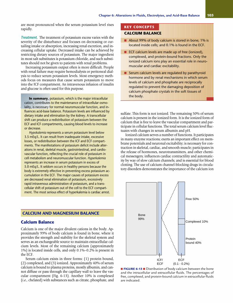
In summary, potassium, which is the major intracellularcation, contributes to the maintenance of intracellular osmo-lality, is necessary for normal neuromuscular function, and in-fluences acid-base balance. Potassium levels are influenced bydietary intake and elimination by the kidney. A transcellularshift can produce a redistribution of potassium between theECF and ICF compartments, causing blood levels to increaseor decrease.
Hypokalemia represents a serum potassium level below 3.5 mEq/L. It can result from inadequate intake, excessivelosses, or redistribution between the ICF and ECF compart-ments. The manifestations of potassium deficit include alter-ations in renal, skeletal muscle, gastrointestinal, and cardio-vascular function, reflecting the crucial role of potassium incell metabolism and neuromuscular function. Hyperkalemiarepresents an increase in serum potassium in excess of 5.0 mEq/L. It seldom occurs in healthy persons because thebody is extremely effective in preventing excess potassium ac-cumulation in the ECF. The major causes of potassium excessare decreased renal elimination of potassium, excessively rapid intravenous administration of potassium, and a trans-cellular shift of potassium out of the cell to the ECF compart-ment. The most serious effect of hyperkalemia is cardiac arrest.
are most pronounced when the serum potassium level risesrapidly.
Treatment. The treatment of potassium excess varies with theseverity of the disturbance and focuses on decreasing or cur-tailing intake or absorption, increasing renal excretion, and in-creasing cellular uptake. Decreased intake can be achieved byrestricting dietary sources of potassium. The major ingredientin most salt substitutes is potassium chloride, and such substi-tutes should not be given to patients with renal problems.
Increasing potassium output often is more difficult. Peoplewith renal failure may require hemodialysis or peritoneal dial-ysis to reduce serum potassium levels. Most emergency meth-ods focus on measures that cause serum potassium to moveinto the ICF compartment. An intravenous infusion of insulinand glucose is often used for this purpose.
sulfate. This form is not ionized. The remaining 50% of serumcalcium is present in the ionized form. It is the ionized form ofcalcium that is free to leave the vascular compartment and par-ticipate in cellular functions. The total serum calcium level fluc-tuates with changes in serum albumin and pH.
Ionized calcium serves a number of functions. It participatesin many enzyme reactions; exerts an important effect on mem-brane potentials and neuronal excitability; is necessary for con-traction in skeletal, cardiac, and smooth muscle; participates inthe release of hormones, neurotransmitters, and other chemi-cal messengers; influences cardiac contractility and automatic-ity by way of slow calcium channels; and is essential for bloodclotting. The use of calcium channel-blocking drugs in circula-tory disorders demonstrates the importance of the calcium ion
103Chapter 6: Alterations in Fluids, Electrolytes, and Acid-Base Balance
CALCIUM AND MAGNESIUM BALANCE
Calcium BalanceCalcium is one of the major divalent cations in the body. Ap-proximately 99% of body calcium is found in bone, where itprovides the strength and stability for the skeletal system andserves as an exchangeable source to maintain extracellular cal-cium levels. Most of the remaining calcium (approximately1%) is located inside cells, and only 0.1%–0.2% is present inthe ECF.
Serum calcium exists in three forms: (1) protein bound,(2) complexed, and (3) ionized. Approximately 40% of serumcalcium is bound to plasma proteins, mostly albumin, and can-not diffuse or pass through the capillary wall to leave the vas-cular compartment (Fig. 6-13). Another 10% is complexed(i.e., chelated) with substances such as citrate, phosphate, and
KEY CONCEPTS
CALCIUM BALANCE
■ About 99% of body calcium is stored in bone; 1% islocated inside cells; and 0.1% is found in the ECF.
■ ECF calcium levels are made up of free (ionized),complexed, and protein-bound fractions. Only theionized calcium ions play an essential role in neuro-muscular and cardiac excitability.
■ Serum calcium levels are regulated by parathyroidhormone and by renal mechanisms in which serumlevels of calcium and phosphate are reciprocallyregulated to prevent the damaging deposition ofcalcium phosphate crystals in the soft tissues of the body.
■ FIGURE 6-13 ■ Distribution of body calcium between the boneand the intracellular and extracellular fluids. The percentages offree, complexed, and protein-bound calcium in extracellular fluidsare indicated.
Bone99%
ICF/ECF
Free 50%
Complexed 10%
Proteinbound 40%
ECF(0.1 - 0.2%)
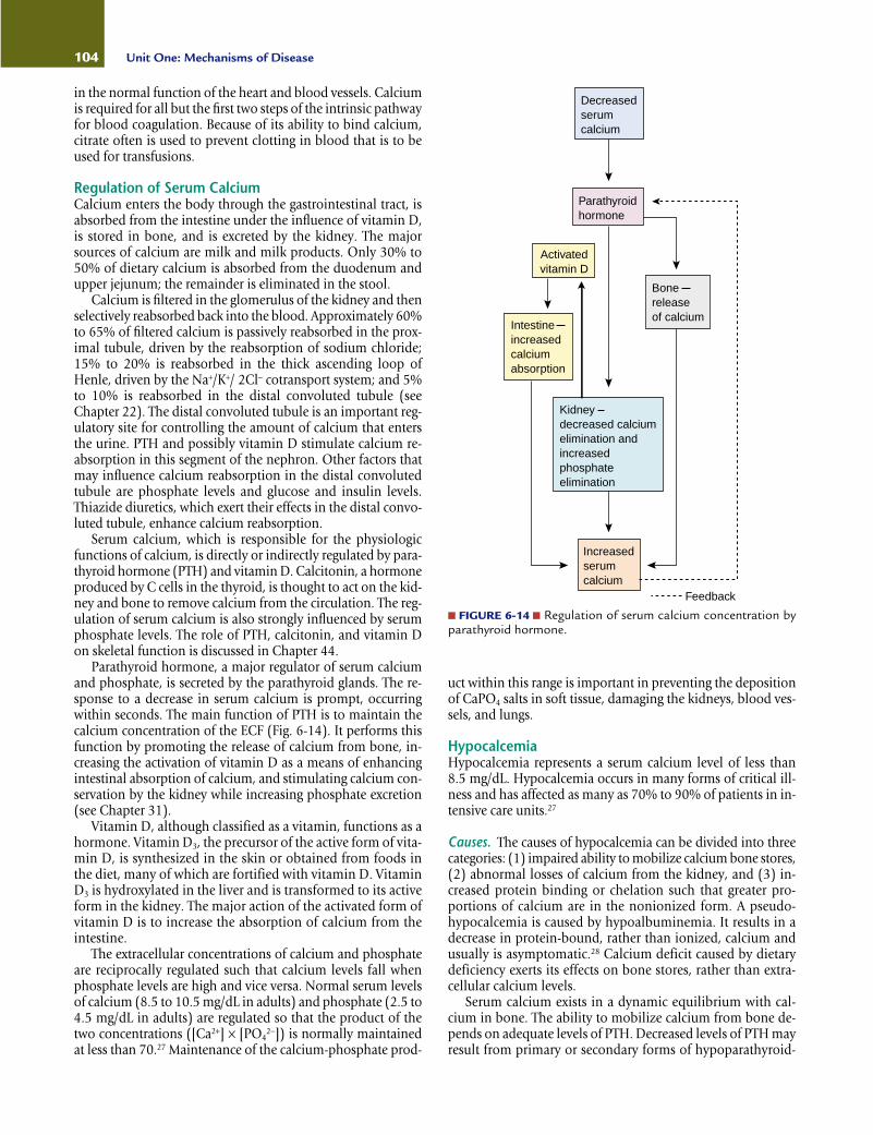
in the normal function of the heart and blood vessels. Calciumis required for all but the first two steps of the intrinsic pathwayfor blood coagulation. Because of its ability to bind calcium,citrate often is used to prevent clotting in blood that is to beused for transfusions.
Regulation of Serum CalciumCalcium enters the body through the gastrointestinal tract, isabsorbed from the intestine under the influence of vitamin D,is stored in bone, and is excreted by the kidney. The majorsources of calcium are milk and milk products. Only 30% to50% of dietary calcium is absorbed from the duodenum andupper jejunum; the remainder is eliminated in the stool.
Calcium is filtered in the glomerulus of the kidney and thenselectively reabsorbed back into the blood. Approximately 60%to 65% of filtered calcium is passively reabsorbed in the prox-imal tubule, driven by the reabsorption of sodium chloride;15% to 20% is reabsorbed in the thick ascending loop ofHenle, driven by the Na+/K+/ 2Cl− cotransport system; and 5%to 10% is reabsorbed in the distal convoluted tubule (seeChapter 22). The distal convoluted tubule is an important reg-ulatory site for controlling the amount of calcium that entersthe urine. PTH and possibly vitamin D stimulate calcium re-absorption in this segment of the nephron. Other factors thatmay influence calcium reabsorption in the distal convolutedtubule are phosphate levels and glucose and insulin levels.Thiazide diuretics, which exert their effects in the distal convo-luted tubule, enhance calcium reabsorption.
Serum calcium, which is responsible for the physiologicfunctions of calcium, is directly or indirectly regulated by para-thyroid hormone (PTH) and vitamin D. Calcitonin, a hormoneproduced by C cells in the thyroid, is thought to act on the kid-ney and bone to remove calcium from the circulation. The reg-ulation of serum calcium is also strongly influenced by serumphosphate levels. The role of PTH, calcitonin, and vitamin Don skeletal function is discussed in Chapter 44.
Parathyroid hormone, a major regulator of serum calciumand phosphate, is secreted by the parathyroid glands. The re-sponse to a decrease in serum calcium is prompt, occurringwithin seconds. The main function of PTH is to maintain thecalcium concentration of the ECF (Fig. 6-14). It performs thisfunction by promoting the release of calcium from bone, in-creasing the activation of vitamin D as a means of enhancingintestinal absorption of calcium, and stimulating calcium con-servation by the kidney while increasing phosphate excretion(see Chapter 31).
Vitamin D, although classified as a vitamin, functions as ahormone. Vitamin D3, the precursor of the active form of vita-min D, is synthesized in the skin or obtained from foods inthe diet, many of which are fortified with vitamin D. VitaminD3 is hydroxylated in the liver and is transformed to its activeform in the kidney. The major action of the activated form ofvitamin D is to increase the absorption of calcium from theintestine.
The extracellular concentrations of calcium and phosphateare reciprocally regulated such that calcium levels fall whenphosphate levels are high and vice versa. Normal serum levelsof calcium (8.5 to 10.5 mg/dL in adults) and phosphate (2.5 to4.5 mg/dL in adults) are regulated so that the product of thetwo concentrations ([Ca2+] × [PO4
2−]) is normally maintainedat less than 70.27 Maintenance of the calcium-phosphate prod-
uct within this range is important in preventing the depositionof CaPO4 salts in soft tissue, damaging the kidneys, blood ves-sels, and lungs.
HypocalcemiaHypocalcemia represents a serum calcium level of less than 8.5 mg/dL. Hypocalcemia occurs in many forms of critical ill-ness and has affected as many as 70% to 90% of patients in in-tensive care units.27
Causes. The causes of hypocalcemia can be divided into threecategories: (1) impaired ability to mobilize calcium bone stores,(2) abnormal losses of calcium from the kidney, and (3) in-creased protein binding or chelation such that greater pro-portions of calcium are in the nonionized form. A pseudo-hypocalcemia is caused by hypoalbuminemia. It results in adecrease in protein-bound, rather than ionized, calcium andusually is asymptomatic.28 Calcium deficit caused by dietarydeficiency exerts its effects on bone stores, rather than extra-cellular calcium levels.
Serum calcium exists in a dynamic equilibrium with cal-cium in bone. The ability to mobilize calcium from bone de-pends on adequate levels of PTH. Decreased levels of PTH mayresult from primary or secondary forms of hypoparathyroid-
104 Unit One: Mechanisms of Disease
Decreasedserumcalcium
Parathyroid hormone
Activatedvitamin D
Bonereleaseof calcium
Intestineincreasedcalciumabsorption
Kidney decreased calcium elimination and increasedphosphateelimination
Increasedserumcalcium
Feedback
■ FIGURE 6-14 ■ Regulation of serum calcium concentration byparathyroid hormone.
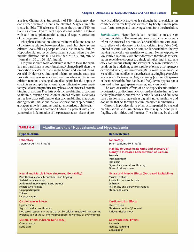
ism (see Chapter 31). Suppression of PTH release may alsooccur when vitamin D levels are elevated. Magnesium defi-ciency inhibits PTH release and impairs the action of PTH onbone resorption. This form of hypocalcemia is difficult to treatwith calcium supplementation alone and requires correctionof the magnesium deficiency.
Phosphate elimination is impaired in renal failure. Becauseof the inverse relation between calcium and phosphate, serumcalcium levels fall as phosphate levels rise in renal failure.Hypocalcemia and hyperphosphatemia occur when the glo-merular filtration rate falls to less than 25 to 30 mL/minute(normal is 100 to 120 mL/minute).
Only the ionized form of calcium is able to leave the capil-lary and participate in body functions. A change in pH alters theproportion of calcium that is in the bound and ionized forms.An acid pH decreases binding of calcium to protein, causing aproportionate increase in ionized calcium, whereas total serumcalcium remains unchanged. An alkaline pH has the oppositeeffect. As an example, hyperventilation sufficient to cause respi-ratory alkalosis can produce tetany because of increased proteinbinding of calcium. Free fatty acids increase binding of calciumto albumin, causing a reduction in ionized calcium. Elevationsin free fatty acids sufficient to alter calcium binding may occurduring stressful situations that cause elevations of epinephrine,glucagon, growth hormone, and adrenocorticotropin levels.
Hypocalcemia is a common finding in a patient with acutepancreatitis. Inflammation of the pancreas causes release of pro-
teolytic and lipolytic enzymes. It is thought that the calcium ioncombines with free fatty acids released by lipolysis in the pan-creas, forming soaps and removing calcium from the circulation.
Manifestations. Hypocalcemia can manifest as an acute orchronic condition. The manifestations of acute hypocalcemiareflect the increased neuromuscular excitability and cardiovas-cular effects of a decrease in ionized calcium (see Table 6-6).Ionized calcium stabilizes neuromuscular excitability, therebymaking nerve cells less sensitive to stimuli. Nerves exposed tolow ionized calcium levels show decreased thresholds for exci-tation, repetitive responses to a single stimulus, and, in extremecases, continuous activity. The severity of the manifestations de-pends on the underlying cause, rapidity of onset, accompanyingelectrolyte disorders, and extracellular pH. Increased neuromuscularexcitability can manifest as paresthesias (i.e., tingling around themouth and in the hands and feet) and tetany (i.e., muscle spasmsof the muscles of the face, hands, and feet). Severe hypocalcemiacan lead to laryngeal spasm, seizures, and even death.
The cardiovascular effects of acute hypocalcemia includehypotension, cardiac insufficiency, cardiac dysrhythmias (par-ticularly heart block and ventricular fibrillation), and failure tohave a response to drugs such as digitalis, norepinephrine, anddopamine that act through calcium-mediated mechanisms.
Chronic hypocalcemia is often accompanied by skeletalmanifestations and skin changes. There may be bone pain,fragility, deformities, and fractures. The skin may be dry and
105Chapter 6: Alterations in Fluids, Electrolytes, and Acid-Base Balance
Manifestations of Hypocalcemia and HypercalcemiaTABLE 6-6
Laboratory LaboratorySerum calcium <8.5 mg/dL Serum calcium >10.5 mg/dL
Inability to Concentrate Urine and Exposure ofKidney to Increased Concentration of CalciumPolyuriaIncreased thirstFlank painSigns of acute renal insufficiencySigns of kidney stones
Neural and Muscle Effects (Increased Excitability) Neural and Muscle Effects (Decreased Excitability)Paresthesias, especially numbness and tingling Muscle weaknessSkeletal muscle cramps Ataxia, loss of muscle toneAbdominal muscle spasms and cramps LethargyHyperactive reflexes Personality and behavioral changesCarpopedal spasm Stupor and comaTetanyLaryngeal spasm
Cardiovascular Effects Cardiovascular EffectsHypotension HypertensionSigns of cardiac insufficiency Shortening of the QT intervalDecreased response to drugs that act by calcium-mediated mechanisms Atrioventricular blockProlongation of the QT interval predisposes to ventricular dysrhythmias
Skeletal Effects (Chronic Deficiency) Gastrointestinal EffectsOsteomalacia AnorexiaBone pain Nausea, vomiting
Constipation
Hypocalcemia Hypercalcemia
scaling, the nails brittle, and hair dry. The development ofcataracts is common. A person with chronic hypocalcemia mayalso present with mild diffuse brain disease mimicking depres-sion, dementia, or psychoses.
Diagnosis and Treatment. Chvostek’s and Trousseau’s testscan be used to assess for an increase in neuromuscular ex-citability and tetany.5 Chvostek’s sign is elicited by tapping theface just below the temple at the point where the facial nerveemerges. Tapping the face over the facial nerve causes spasm ofthe lip, nose, or face when the test result is positive. An inflatedblood pressure cuff is used to test for Trousseau’s sign. The cuffis inflated above systolic blood pressure for 3 minutes. Con-traction of the fingers and hands (i.e., carpopedal spasm) indi-cates the presence of tetany.
Acute hypocalcemia is an emergency situation, requiringprompt treatment. An intravenous infusion containing calciumis used when tetany or acute symptoms are present or antici-pated because of a decrease in the serum calcium level.
Chronic hypocalcemia is treated with oral intake of cal-cium. One glass of milk contains approximately 300 mg of cal-cium. Oral calcium supplements may be used. In some cases,long-term treatment may require the use of vitamin D prepa-rations. The active form of vitamin D is administered when theliver or kidney mechanisms needed for hormone activationare impaired.
HypercalcemiaHypercalcemia represents a total serum calcium concentrationgreater than 10.5 mg/dL. Falsely elevated levels of calcium canresult from prolonged drawing of blood with an excessivelytight tourniquet. Increased plasma proteins (e.g., hyperalbu-minemia, hyperglobulinemia) may elevate the total serum cal-cium but not affect the ionized calcium concentration
Causes. A serum calcium excess (i.e., hypercalcemia) resultswhen calcium movement into the circulation overwhelms thecalcium regulatory hormones or the ability of the kidney to remove excess calcium ions. The most common causes ofhypercalcemia are increased bone resorption caused by neo-plasms or hyperparathyroidism. Hypercalcemia is a commoncomplication of cancer, occurring in approximately 10% to20% of persons with advanced disease.29 A number of malig-nant tumors, including carcinoma of the lungs, have been as-sociated with hypercalcemia. Some tumors destroy the bone,but others produce humoral agents that stimulate osteoclasticactivity, increase bone resorption, or inhibit bone formation.
Less common causes of hypercalcemia are prolonged im-mobilization, increased intestinal absorption of calcium, andexcessive doses of vitamin D. Prolonged immobilization andlack of weight bearing cause demineralization of bone and re-lease of calcium into the bloodstream. Intestinal absorption ofcalcium can be increased by excessive doses of vitamin D or asa result of a condition called the milk-alkali syndrome. The milk-alkali syndrome is caused by excessive ingestion of calcium(often in the form of milk) and absorbable antacids. Becauseof the advent of nonabsorbable antacids, the condition is seenless frequently than in the past, but it may occur in women whoare overzealous in taking calcium preparations for osteoporo-sis prevention.
A variety of drugs elevate calcium levels. The use of lithiumto treat bipolar disorders has caused hypercalcemia and hyper-
parathyroidism. The thiazide diuretics increase calcium re-absorption in the distal convoluted tubule of the kidney.Although the thiazide diuretics seldom cause hypercalcemia,they can unmask hypercalcemia from other causes such asunderlying bone disorders and conditions that increase boneresorption.
Manifestations. The signs and symptoms associated with cal-cium excess originate from three sources: (1) changes in neuralexcitability, (2) alterations in smooth and cardiac muscle func-tion, and (3) exposure of the kidneys to high concentrationsof calcium (see Table 6-6).
Neural excitability is decreased in patients with hypercalce-mia. There may be a dulling of consciousness, stupor, weak-ness, and muscle flaccidity. Behavioral changes may range fromsubtle alterations in personality to acute psychoses.
The heart responds to elevated levels of calcium with in-creased contractility and ventricular dysrhythmias. Digitalis ac-centuates these responses. Gastrointestinal symptoms reflect adecrease in smooth muscle activity and include constipation,anorexia, nausea, and vomiting. Pancreatitis is another poten-tial complication of hypercalcemia and is probably related tostones in the pancreatic ducts.
High calcium concentrations in the urine impair the abilityof the kidneys to concentrate urine by interfering with the ac-tion of ADH. This causes salt and water diuresis and an in-creased sensation of thirst. Hypercalciuria also predisposes tothe development of renal calculi.
Hypercalcemic crisis describes an acute increase in the serumcalcium level.29 Malignant disease and hyperparathyroidism aremajor causes of hypercalcemic crisis. In hypercalcemic crisis,polyuria, excessive thirst, volume depletion, fever, altered lev-els of consciousness, azotemia (i.e., nitrogenous wastes in theblood), and a disturbed mental state accompany other signs ofcalcium excess. Symptomatic hypercalcemia is associated witha high mortality rate; death often is caused by cardiac arrest.
Treatment. The treatment of calcium excess usually is directedtoward rehydration and measures to increase urinary excretionof calcium and inhibit release of calcium from bone. Fluid re-placement is needed in situations of volume depletion. The ex-cretion of sodium is accompanied by calcium excretion.Diuretics and sodium chloride can be administered to increaseurinary elimination of calcium after the ECF volume has beenrestored. Loop diuretics commonly are used, rather than thi-azide diuretics, which increase calcium reabsorption.
The initial lowering of calcium levels is followed by measuresto inhibit bone reabsorption. Drugs that are used to inhibit cal-cium mobilization include bisphosphonates, calcitonin, andthe glucocorticosteroids. The bisphosphonates are a relativelynew group of drugs that act mainly by inhibiting osteoclastic ac-tivity. Calcitonin inhibits osteoclastic activity, thereby decreas-ing resorption. The glucocorticosteroids inhibit bone resorp-tion and are used to treat hypercalcemia associated with cancer.
Magnesium BalanceMagnesium is the second most abundant intracellular cation.The average adult has approximately 24 g of magnesium dis-tributed throughout the body.30 Of the total magnesium con-tent, approximately 50% to 60% is stored in bone, 39% to 49%is contained in the body cells, and the remaining 1% is dis-
106 Unit One: Mechanisms of Disease
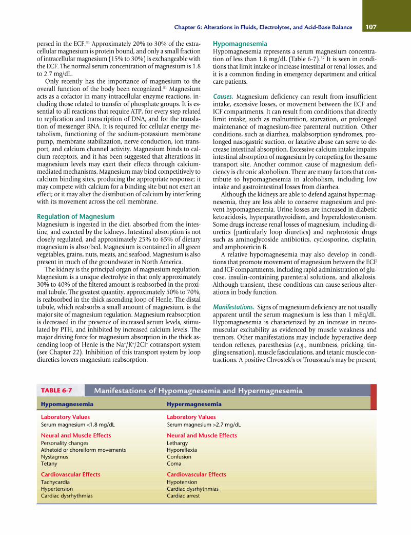
persed in the ECF.31 Approximately 20% to 30% of the extra-cellular magnesium is protein bound, and only a small fractionof intracellular magnesium (15% to 30%) is exchangeable withthe ECF. The normal serum concentration of magnesium is 1.8to 2.7 mg/dL.
Only recently has the importance of magnesium to theoverall function of the body been recognized.31 Magnesiumacts as a cofactor in many intracellular enzyme reactions, in-cluding those related to transfer of phosphate groups. It is es-sential to all reactions that require ATP, for every step relatedto replication and transcription of DNA, and for the transla-tion of messenger RNA. It is required for cellular energy me-tabolism, functioning of the sodium-potassium membranepump, membrane stabilization, nerve conduction, ion trans-port, and calcium channel activity. Magnesium binds to cal-cium receptors, and it has been suggested that alterations inmagnesium levels may exert their effects through calcium-mediated mechanisms. Magnesium may bind competitively tocalcium binding sites, producing the appropriate response; itmay compete with calcium for a binding site but not exert aneffect; or it may alter the distribution of calcium by interferingwith its movement across the cell membrane.
Regulation of MagnesiumMagnesium is ingested in the diet, absorbed from the intes-tine, and excreted by the kidneys. Intestinal absorption is notclosely regulated, and approximately 25% to 65% of dietarymagnesium is absorbed. Magnesium is contained in all greenvegetables, grains, nuts, meats, and seafood. Magnesium is alsopresent in much of the groundwater in North America.
The kidney is the principal organ of magnesium regulation.Magnesium is a unique electrolyte in that only approximately30% to 40% of the filtered amount is reabsorbed in the proxi-mal tubule. The greatest quantity, approximately 50% to 70%,is reabsorbed in the thick ascending loop of Henle. The distaltubule, which reabsorbs a small amount of magnesium, is themajor site of magnesium regulation. Magnesium reabsorptionis decreased in the presence of increased serum levels, stimu-lated by PTH, and inhibited by increased calcium levels. Themajor driving force for magnesium absorption in the thick as-cending loop of Henle is the Na+/K+/2Cl− cotransport system(see Chapter 22). Inhibition of this transport system by loopdiuretics lowers magnesium reabsorption.
HypomagnesemiaHypomagnesemia represents a serum magnesium concentra-tion of less than 1.8 mg/dL (Table 6-7).32 It is seen in condi-tions that limit intake or increase intestinal or renal losses, andit is a common finding in emergency department and criticalcare patients.
Causes. Magnesium deficiency can result from insufficientintake, excessive losses, or movement between the ECF andICF compartments. It can result from conditions that directlylimit intake, such as malnutrition, starvation, or prolongedmaintenance of magnesium-free parenteral nutrition. Otherconditions, such as diarrhea, malabsorption syndromes, pro-longed nasogastric suction, or laxative abuse can serve to de-crease intestinal absorption. Excessive calcium intake impairsintestinal absorption of magnesium by competing for the sametransport site. Another common cause of magnesium defi-ciency is chronic alcoholism. There are many factors that con-tribute to hypomagnesemia in alcoholism, including low intake and gastrointestinal losses from diarrhea.
Although the kidneys are able to defend against hypermag-nesemia, they are less able to conserve magnesium and pre-vent hypomagnesemia. Urine losses are increased in diabeticketoacidosis, hyperparathyroidism, and hyperaldosteronism.Some drugs increase renal losses of magnesium, including di-uretics (particularly loop diuretics) and nephrotoxic drugssuch as aminoglycoside antibiotics, cyclosporine, cisplatin,and amphotericin B.
A relative hypomagnesemia may also develop in condi-tions that promote movement of magnesium between the ECFand ICF compartments, including rapid administration of glu-cose, insulin-containing parenteral solutions, and alkalosis.Although transient, these conditions can cause serious alter-ations in body function.
Manifestations. Signs of magnesium deficiency are not usuallyapparent until the serum magnesium is less than 1 mEq/dL.Hypomagnesemia is characterized by an increase in neuro-muscular excitability as evidenced by muscle weakness andtremors. Other manifestations may include hyperactive deeptendon reflexes, paresthesias (e.g., numbness, pricking, tin-gling sensation), muscle fasciculations, and tetanic muscle con-tractions. A positive Chvostek’s or Trousseau’s may be present,
107Chapter 6: Alterations in Fluids, Electrolytes, and Acid-Base Balance
Manifestations of Hypomagnesemia and HypermagnesemiaTABLE 6-7
Laboratory Values Laboratory ValuesSerum magnesium <1.8 mg/dL Serum magnesium >2.7 mg/dL
Neural and Muscle Effects Neural and Muscle EffectsPersonality changes LethargyAthetoid or choreiform movements HyporeflexiaNystagmus ConfusionTetany Coma
Cardiovascular Effects Cardiovascular EffectsTachycardia HypotensionHypertension Cardiac dysrhythmiasCardiac dysrhythmias Cardiac arrest
Hypomagnesemia Hypermagnesemia
particularly if hypocalcemia is present. Because decreasedserum magnesium increases irritability in nervous tissue, sei-zures may occur. Other manifestations may include ataxia, ver-tigo, disorientation, depression, and psychotic symptoms.
Cardiovascular manifestations include tachycardia, hyper-tension, and ventricular dysrhythmias. There may be ECGchanges such as widening of the QRS complex, appearance ofpeak T waves, prolongation of the PR interval, T-wave inversion,and appearance of U waves. Ventricular dysrhythmias, particu-larly in the presence of digitalis, may be difficult to treat unlessmagnesium levels are normalized.
Magnesium deficiency often occurs in conjunction withhypocalcemia and hypokalemia, producing a number of re-lated neurologic and cardiovascular manifestations. Hypo-calcemia is typical of severe hypomagnesemia. Most personswith hypomagnesemia-related hypocalcemia have decreasedPTH levels, probably as a result of impaired magnesium-dependent mechanisms that control PTH release and synthe-sis. Hypokalemia also is a typical feature of hypomagnesemia.It leads to a reduction in intracellular potassium and impairsthe ability of the kidney to conserve potassium. When hypo-magnesemia is present, hypokalemia is unresponsive to potas-sium replacement therapy.
Treatment. Hypomagnesemia is treated with magnesium re-placement. The route of administration depends on the severityof the condition. Symptomatic, moderate to severe magnesiumdeficiency is treated by parenteral administration. Treatmentmust be continued for several days to replace stored and serumlevels. In conditions of chronic intestinal or renal loss, mainte-nance support with oral magnesium may be required. Patientswith any degree of renal failure must be carefully monitored toprevent magnesium excess. Magnesium often is used therapeu-tically to treat cardiac arrhythmia, myocardial infarct, angina,and pregnancy complicated by preeclampsia or eclampsia.Caution to prevent hypermagnesemia is essential.
HypermagnesemiaHypermagnesemia represents a serum magnesium concentra-tion in excess of 2.7 mg/dL. Because of the ability of the nor-mal kidney to excrete magnesium, hypermagnesemia is rare.
When hypermagnesemia does occur, it usually is related torenal insufficiency and the injudicious use of magnesium-containing medications such as antacids, mineral supplements,or laxatives. The elderly are particularly at risk because theyhave age-related reductions in renal function and tend to con-sume more magnesium-containing medications. Magnesiumsulfate is used to treat toxemia of pregnancy and prematurelabor; in these cases, careful monitoring for signs of hyper-magnesemia is essential.
Hypermagnesemia affects neuromuscular and cardiovas-cular function (see Table 6-7). The signs and symptoms occuronly when serum magnesium levels exceed 4.9 mg/dL.32
Hypermagnesemia diminishes neuromuscular transmission,causing hyporeflexia, muscle weakness, and confusion. Magne-sium decreases acetylcholine release at the myoneural junctionand may cause neuromuscular blockade and respiratory paral-ysis. Cardiovascular effects are related to the calcium channel-blocking effects of magnesium. Blood pressure is decreased,and the ECG shows an increase in the PR interval, a shorteningof the QT interval, T-wave abnormalities, and prolongation of
ACID-BASE BALANCE
Metabolic activities of the body require the precise regulationof acid-base balance, which is reflected by the pH of ECF.Membrane excitability, enzyme systems, and chemical reac-tions depend on pH being regulated within a narrow physio-logic range. Many conditions, pathologic or otherwise, canalter body pH.
Introductory Concepts
Normally, the concentration of body acids and bases is regu-lated so that the pH of extracellular body fluids is maintainedwithin a very narrow range of 7.35 to 7.45. This balance ismaintained through mechanisms that generate, buffer, andeliminate acids and bases.
Acid-Base ChemistryAn acid is a molecule that can release a hydrogen ion (H+),and a base is a molecule that can accept or combine with anH+ ion. Most of the body’s acids and bases are weak acids and
108 Unit One: Mechanisms of Disease
In summary, calcium is one of the major divalent ions inthe body. Approximately 99% of body calcium is found inbone; about 1% is in the ICF and 0.1% to 0.2% is in the ECF.The calcium in bone is in dynamic equilibrium with extracellu-lar calcium. Of the three forms of ECF calcium (i.e., proteinbound, complexed, and ionized), only the ionized form cancross the cell membrane and contribute to cellular function.Ionized calcium has a number of functions. It contributes toneuromuscular function, plays a vital role in the blood clot-ting process, and participates in a number of enzyme reac-tions. Alterations in ionized calcium levels produce neuraleffects; neural excitability is increased in hypocalcemia anddecreased in hypercalcemia.
Magnesium is the second most abundant intracellularcation. It acts as a cofactor in many enzyme reactions andaffects neuromuscular function in the same manner as the cal-cium ion. Magnesium deficiency can result from insufficientintake, excessive losses, or movement between the ECF andICF compartments. The manifestations of hypomagnesemiaare characterized by a decrease in neuromuscular excitabilityas evidenced by paresthesias and hyperactive reflexes. Cardio-vascular effects include tachycardia, hypertension, and ventri-cular dysrhythmias. Hypermagnesemia usually is related torenal insufficiency and the injudicious use of magnesium-containing medications such as antacids, mineral supple-ments, or laxatives. It diminishes neuromuscular transmissionleading to hyporeflexia, muscle weakness, and confusion.
the QRS and PR intervals. Hypotension caused by vasodilationand cardiac dysrhythmias can occur with moderate hypermag-nesemia (≥5 to 10 mg/dL), and confusion and coma can occurwith severe hypermagnesemia (≥10 mg/dL). Very severe hyper-magnesemia (≥15 mg/dL) may cause cardiac arrest.
The treatment of hypermagnesemia includes cessation ofmagnesium administration. Calcium is a direct antagonist ofmagnesium, and intravenous administration of calcium maybe used. Peritoneal dialysis or hemodialysis may be required.
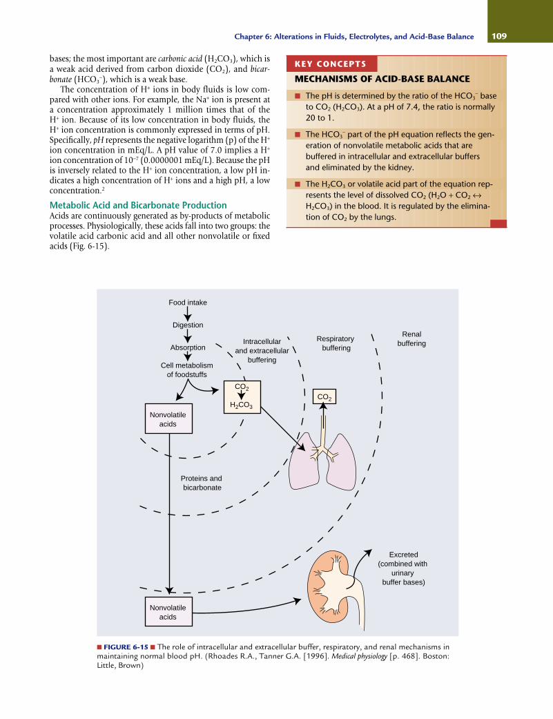
bases; the most important are carbonic acid (H2CO3), which isa weak acid derived from carbon dioxide (CO2), and bicar-bonate (HCO3
−), which is a weak base.The concentration of H+ ions in body fluids is low com-
pared with other ions. For example, the Na+ ion is present at a concentration approximately 1 million times that of the H+ ion. Because of its low concentration in body fluids, the H+ ion concentration is commonly expressed in terms of pH.Specifically, pH represents the negative logarithm (p) of the H+
ion concentration in mEq/L. A pH value of 7.0 implies a H+
ion concentration of 10−7 (0.0000001 mEq/L). Because the pHis inversely related to the H+ ion concentration, a low pH in-dicates a high concentration of H+ ions and a high pH, a lowconcentration.2
Metabolic Acid and Bicarbonate ProductionAcids are continuously generated as by-products of metabolicprocesses. Physiologically, these acids fall into two groups: thevolatile acid carbonic acid and all other nonvolatile or fixedacids (Fig. 6-15).
109Chapter 6: Alterations in Fluids, Electrolytes, and Acid-Base Balance
■ FIGURE 6-15 ■ The role of intracellular and extracellular buffer, respiratory, and renal mechanisms inmaintaining normal blood pH. (Rhoades R.A., Tanner G.A. [1996]. Medical physiology [p. 468]. Boston:Little, Brown)
Food intake
Digestion
Absorption
Cell metabolismof foodstuffs
Intracellularand extracellular
buffering
Respiratorybuffering
Renalbuffering
Proteins and bicarbonate
CO2
Excreted(combined with
urinarybuffer bases)
Nonvolatileacids
CO2
H2CO3
Nonvolatileacids
KEY CONCEPTS
MECHANISMS OF ACID-BASE BALANCE
■ The pH is determined by the ratio of the HCO3− base
to CO2 (H2CO3). At a pH of 7.4, the ratio is normally20 to 1.
■ The HCO3− part of the pH equation reflects the gen-
eration of nonvolatile metabolic acids that arebuffered in intracellular and extracellular buffers and eliminated by the kidney.
■ The H2CO3 or volatile acid part of the equation rep-resents the level of dissolved CO2 (H2O + CO2 ↔H2CO3) in the blood. It is regulated by the elimina-tion of CO2 by the lungs.
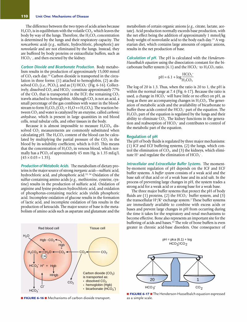
The difference between the two types of acids arises becauseH2CO3 is in equilibrium with the volatile CO2, which leaves thebody by way of the lungs. Therefore, the H2CO3 concentrationis determined by the lungs and their respiratory capacity. Thenoncarbonic acids (e.g., sulfuric, hydrochloric, phosphoric) arenonvolatile and are not eliminated by the lungs. Instead, theyare buffered by body proteins or extracellular buffers, such asHCO3
− , and then excreted by the kidney.
Carbon Dioxide and Bicarbonate Production. Body metabo-lism results in the production of approximately 15,000 mmolof CO2 each day.33 Carbon dioxide is transported in the circu-lation in three forms: (1) attached to hemoglobin, (2) as dis-solved CO2 (i.e., PCO2), and as (3) HCO3
− (Fig. 6-16). Collect-ively, dissolved CO2 and HCO3
− constitute approximately 77%of the CO2 that is transported in the ECF; the remaining CO2
travels attached to hemoglobin. Although CO2 is not an acid, asmall percentage of the gas combines with water in the blood-stream to form H2CO3 (CO2 + H2O ↔ H2CO3). The reaction be-tween CO2 and water is catalyzed by an enzyme, called carbonicanhydrase, which is present in large quantities in red bloodcells, renal tubular cells, and other tissues in the body.
Because it is almost impossible to measure H2CO3, dis-solved CO2 measurements are commonly substituted whencalculating pH. The H2CO3 content of the blood can be calcu-lated by multiplying the partial pressure of the CO2 in theblood by its solubility coefficient, which is 0.03. This meansthat the concentration of H2CO3 in venous blood, which nor-mally has a PCO2 of approximately 45 mm Hg, is 1.35 mEq/L(45 × 0.03 = 1.35).
Production of Metabolic Acids. The metabolism of dietary pro-teins is the major source of strong inorganic acids—sulfuric acid,hydrochloric acid, and phosphoric acid.33, 34 Oxidation of thesulfur-containing amino acids (e.g., methionine, cysteine, cys-tine) results in the production of sulfuric acid. Oxidation ofarginine and lysine produces hydrochloric acid, and oxidationof phosphorus-containing nucleic acids yields phosphoricacid. Incomplete oxidation of glucose results in the formationof lactic acid, and incomplete oxidation of fats results in theproduction of ketoacids. The major source of base is the meta-bolism of amino acids such as aspartate and glutamate and the
metabolism of certain organic anions (e.g., citrate, lactate, ace-tate). Acid production normally exceeds base production, withthe net effect being the addition of approximately 1 mmol/kgbody weight of nonvolatile acid to the body each day.33 A veg-etarian diet, which contains large amounts of organic anions,results in the net production of base.
Calculation of pH. The pH is calculated with the Henderson-Hasselbalch equation using the dissociation constant for the bi-carbonate buffer system (6.1) and the HCO3
− to H2CO3 ratio.
pH = 6.1 + logHCO3
−
H2CO3
The log of 20 is 1.3. Thus, when the ratio is 20 to 1, the pH iswithin the normal range at 7.4 (Fig. 6-17). Because the ratio isused, a change in HCO3
− will have little or no effect on pH, aslong as there are accompanying changes in H2CO3. The gener-ation of metabolic acids and the availability of bicarbonate tobuffer these acids control the HCO3
− part of the equation. TheH2CO3 part of the equation is regulated by the lungs and theirability to eliminate CO2. The kidney functions in the genera-tion and reabsorption of HCO3
− and contributes to control ofthe metabolic part of the equation.
Regulation of pHThe pH of body fluids is regulated by three major mechanisms:(1) ICF and ECF buffering systems, (2) the lungs, which con-trol the elimination of CO2, and (3) the kidneys, which elimi-nate H+ and regulate the elimination of HCO3
− .
Intracellular and Extracellular Buffer Systems. The moment-by-moment regulation of pH depends on the ICF and ECFbuffer systems. A buffer system consists of a weak acid and thebase salt of that acid or of a weak base and its acid salt. In theprocess of preventing large changes in pH, the system trades astrong acid for a weak acid or a strong base for a weak base.
The three major buffer systems that protect the pH of bodyfluids are (1) proteins, (2) the HCO3
− buffer system, and (3)the transcellular H+/K+ exchange system.2 These buffer systemsare immediately available to combine with excess acids orbases and prevent large changes in pH from occurring duringthe time it takes for the respiratory and renal mechanisms tobecome effective. Bone also represents an important site for thebuffering of acids and bases.33 The role of bone buffers is evengreater in chronic acid-base disorders. One consequence of
110 Unit One: Mechanisms of Disease
Red blood cell Tissue cell
H2O
H2OH2CO3
HCO3-
HCO3-
H+
Cl-
Hgb
HHgb
Hgb
CO2 CO2 CO2
CO2
+
+
CA
Carbon dioxide (CO2)is transported as: dissolved CO2 hemoglobin (Hgb) bicarbonate (HCO3
-)
Hgb CO2
■ FIGURE 6-16 ■ Mechanisms of carbon dioxide transport.
pH = pka (6.1) + logHCO /CO
HCO CO3 2
23-
20 1
-
■ FIGURE 6-17 ■ The Henderson-Hasselbalch equation expressedas a simple scale.
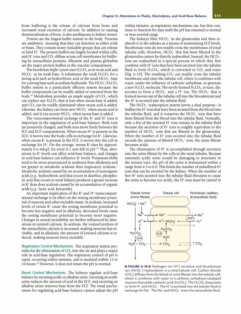
bone buffering is the release of calcium from bone and increased renal excretion of calcium. In addition to causingdemineralization of bone, it also predisposes to kidney stones.
Proteins are the largest buffer system in the body. Proteinsare amphoteric, meaning that they can function as either acidsor bases. They contain many ionizable groups that can releaseor bind H+. The protein buffers are largely located within cells,and H+ ions and CO2 diffuse across cell membranes for buffer-ing by intracellular proteins. Albumin and plasma globulinsare the major protein buffers in the vascular compartment.
The bicarbonate buffer system uses H2CO3 as its weak acid andHCO3
− as its weak base. It substitutes the weak H2CO3 for astrong acid such as hydrochloric acid or the weak HCO3
− basefor a strong base such as sodium hydroxide. The HCO3
−/H2CO3
buffer system is a particularly efficient system because thebuffer components can be readily added or removed from thebody.34 Metabolism provides an ample supply of CO2, whichcan replace any H2CO3 that is lost when excess base is added,and CO2 can be readily eliminated when excess acid is added.Likewise, the kidney can form new HCO3
− when excess acid isadded, and it can excrete HCO3
− when excess base is added.The transcompartmental exchange of the K+ and H+ ions is
important in the regulation of acid-base balance. Both ionsare positively charged, and both ions move freely between theICF and ECF compartments. When excess H+ is present in theECF, it moves into the body cells in exchange for K+. Likewise,when excess K+ is present in the ECF, it moves into the cell inexchange for H+. On the average, serum K+ rises by approxi-mately 0.6 mEq/L for every 0.1 unit fall in pH.33 Thus, alter-ations in K+ levels can affect acid-base balance, and changesin acid-base balance can influence K+ levels. Potassium shiftstend to be more pronounced in acidemia than alkalemia andare greater in metabolic acidosis than respiratory acidosis.5
Metabolic acidosis caused by an accumulation of nonorganicacids (e.g., hydrochloric acid that occurs in diarrhea, phospho-ric acid that occurs in renal failure) produces a greater increasein K+ than does acidosis caused by an accumulation of organicacids (e.g., lactic acid, ketoacids).
An important implication of the K+ and H+ transcompart-mental exchange is its effect on the resting membrane poten-tial of neurons and other excitable tissue. In acidosis, increasedlevels of serum K+ cause the resting membrane potential tobecome less negative and in alkalosis, decreased levels causethe resting membrane potential to become more negative.Changes in neural excitability are further influenced by alter-ations in ionized calcium. In acidosis, the ionized portion ofthe extracellular calcium is increased, making neurons less ex-citable, and in alkalosis, the amount of ionized calcium is re-duced, making neurons more excitable.
Respiratory Control Mechanisms. The respiratory system pro-vides for the elimination of CO2 into the air and plays a majorrole in acid-base regulation. The respiratory control of pH israpid, occurring within minutes, and is maximal within 12 to24 hours.33 However, it does not return the pH to normal.
Renal Control Mechanisms. The kidneys regulate acid-basebalance by excreting acidic or alkaline urine. Excreting an acidicurine reduces the amount of acid in the ECF, and excreting analkaline urine removes base from the ECF. The renal mecha-nisms for regulating acid-base balance cannot adjust the pH
within minutes, as respiratory mechanisms can, but they con-tinue to function for days until the pH has returned to normalor near-normal range.
The kidneys filter HCO3− in the glomerulus and then re-
absorb it in the tubules as a means of maintaining ECF levels.Bicarbonate ions do not readily cross the membranes of renaltubular cells; therefore, HCO3
− that has been filtered in theglomerulus cannot be directly reabsorbed. Instead, the HCO3
−
ions are reabsorbed in a special process in which they firstcombine with H+ ions that have been secreted into the tubularfluid to form H2CO3, which is converted to CO2 and water(Fig. 6-18). The resulting CO2 can readily cross the tubularmembrane and enter the tubular cell, where it combines withwater, under the influence of carbonic anhydrase, to generatea new H2CO3 molecule. The newly formed H2CO3, in turn, dis-sociates to form a HCO3
− and a H+ ion. The HCO3− that is
formed moves out of the tubular cell into the bloodstream andthe H+ is secreted into the tubular fluid.
The HCO3− reabsorption system serves a dual purpose—it
buffers the H+ ions that have been secreted from the blood intothe tubular fluid, and it conserves the HCO3
− ions that havebeen filtered from the blood into the tubular fluid. Normally,only a few of the secreted H+ ions remain in the tubular fluidbecause the secretion of H+ ions is roughly equivalent to thenumber of HCO3
− ions that are filtered in the glomerulus.When the number of H+ ions secreted into the tubular fluidexceeds the amount of filtered HCO3
− ions, the urine filtratebecomes acidic.
The elimination of H+ is accomplished through secretioninto the urine filtrate by the cells in the renal tubules. Becauseextremely acidic urine would be damaging to structures in the urinary tract, the pH of the urine is maintained within arange from 4.5 to 8.0. This limits the number of unbuffered H+
ions that can be excreted by the kidney. When the number offree H+ ions secreted into the tubular fluid threatens to causethe urine to become too acidic, the H+ ions must be carried in
111Chapter 6: Alterations in Fluids, Electrolytes, and Acid-Base Balance
■ FIGURE 6-18 ■ Hydrogen ion (H+) secretion and bicarbonateion (HCO3
−) reabsorption in a renal tubular cell. Carbon dioxide(CO2) diffuses from the blood or urine filtrate into the tubular cell,where it combines with water in a carbonic anhydrase-catalyzedreaction that yields carbonic acid (H2CO3). The H2CO3 dissociatesto form H+ and HCO3
−. The H+ is secreted into the tubular fluid inexchange for Na+. The Na+ and HCO3
− enter the extracellular fluid.
Tubular lumen(Urine filtrate)
Tubular cell Peritubular capillary(Extracellular fluid)
HCO + Na
Na
H
H CO CA
H O + CO
Na
H + HCO
H CO
H O
CO
Na
CO+
CA
- +
+
+
+
+3
2
2
2
2
+
22
2
3
-
33
K+ATP
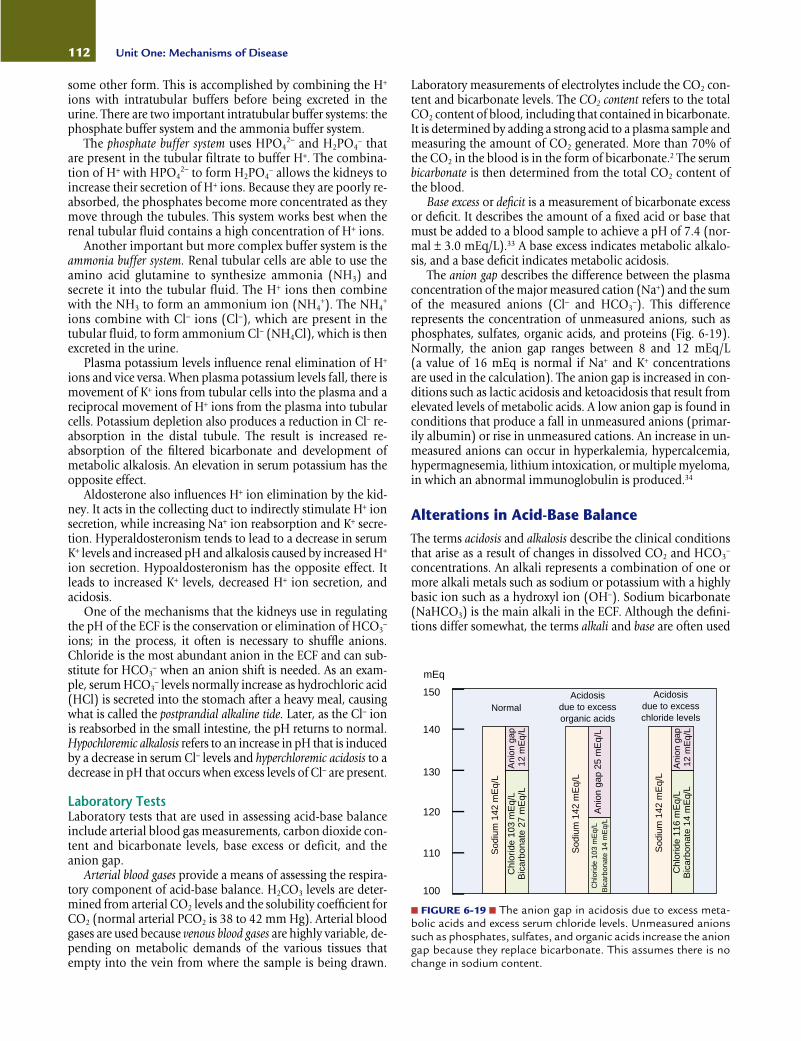
some other form. This is accomplished by combining the H+
ions with intratubular buffers before being excreted in theurine. There are two important intratubular buffer systems: thephosphate buffer system and the ammonia buffer system.
The phosphate buffer system uses HPO42− and H2PO4
− thatare present in the tubular filtrate to buffer H+. The combina-tion of H+ with HPO4
2− to form H2PO4− allows the kidneys to
increase their secretion of H+ ions. Because they are poorly re-absorbed, the phosphates become more concentrated as theymove through the tubules. This system works best when therenal tubular fluid contains a high concentration of H+ ions.
Another important but more complex buffer system is theammonia buffer system. Renal tubular cells are able to use theamino acid glutamine to synthesize ammonia (NH3) andsecrete it into the tubular fluid. The H+ ions then combinewith the NH3 to form an ammonium ion (NH4
+). The NH4+
ions combine with Cl− ions (Cl−), which are present in thetubular fluid, to form ammonium Cl− (NH4Cl), which is thenexcreted in the urine.
Plasma potassium levels influence renal elimination of H+
ions and vice versa. When plasma potassium levels fall, there ismovement of K+ ions from tubular cells into the plasma and areciprocal movement of H+ ions from the plasma into tubularcells. Potassium depletion also produces a reduction in Cl− re-absorption in the distal tubule. The result is increased re-absorption of the filtered bicarbonate and development ofmetabolic alkalosis. An elevation in serum potassium has theopposite effect.
Aldosterone also influences H+ ion elimination by the kid-ney. It acts in the collecting duct to indirectly stimulate H+ ionsecretion, while increasing Na+ ion reabsorption and K+ secre-tion. Hyperaldosteronism tends to lead to a decrease in serumK+ levels and increased pH and alkalosis caused by increased H+
ion secretion. Hypoaldosteronism has the opposite effect. Itleads to increased K+ levels, decreased H+ ion secretion, andacidosis.
One of the mechanisms that the kidneys use in regulatingthe pH of the ECF is the conservation or elimination of HCO3
−
ions; in the process, it often is necessary to shuffle anions.Chloride is the most abundant anion in the ECF and can sub-stitute for HCO3
− when an anion shift is needed. As an exam-ple, serum HCO3
− levels normally increase as hydrochloric acid(HCl) is secreted into the stomach after a heavy meal, causingwhat is called the postprandial alkaline tide. Later, as the Cl− ionis reabsorbed in the small intestine, the pH returns to normal.Hypochloremic alkalosis refers to an increase in pH that is inducedby a decrease in serum Cl− levels and hyperchloremic acidosis to adecrease in pH that occurs when excess levels of Cl− are present.
Laboratory TestsLaboratory tests that are used in assessing acid-base balanceinclude arterial blood gas measurements, carbon dioxide con-tent and bicarbonate levels, base excess or deficit, and theanion gap.
Arterial blood gases provide a means of assessing the respira-tory component of acid-base balance. H2CO3 levels are deter-mined from arterial CO2 levels and the solubility coefficient forCO2 (normal arterial PCO2 is 38 to 42 mm Hg). Arterial bloodgases are used because venous blood gases are highly variable, de-pending on metabolic demands of the various tissues thatempty into the vein from where the sample is being drawn.
Laboratory measurements of electrolytes include the CO2 con-tent and bicarbonate levels. The CO2 content refers to the totalCO2 content of blood, including that contained in bicarbonate.It is determined by adding a strong acid to a plasma sample andmeasuring the amount of CO2 generated. More than 70% ofthe CO2 in the blood is in the form of bicarbonate.2 The serumbicarbonate is then determined from the total CO2 content ofthe blood.
Base excess or deficit is a measurement of bicarbonate excessor deficit. It describes the amount of a fixed acid or base thatmust be added to a blood sample to achieve a pH of 7.4 (nor-mal ± 3.0 mEq/L).33 A base excess indicates metabolic alkalo-sis, and a base deficit indicates metabolic acidosis.
The anion gap describes the difference between the plasmaconcentration of the major measured cation (Na+) and the sumof the measured anions (Cl− and HCO3
−). This differencerepresents the concentration of unmeasured anions, such asphosphates, sulfates, organic acids, and proteins (Fig. 6-19).Normally, the anion gap ranges between 8 and 12 mEq/L (a value of 16 mEq is normal if Na+ and K+ concentrationsare used in the calculation). The anion gap is increased in con-ditions such as lactic acidosis and ketoacidosis that result fromelevated levels of metabolic acids. A low anion gap is found inconditions that produce a fall in unmeasured anions (primar-ily albumin) or rise in unmeasured cations. An increase in un-measured anions can occur in hyperkalemia, hypercalcemia,hypermagnesemia, lithium intoxication, or multiple myeloma,in which an abnormal immunoglobulin is produced.34
Alterations in Acid-Base BalanceThe terms acidosis and alkalosis describe the clinical conditionsthat arise as a result of changes in dissolved CO2 and HCO3
−
concentrations. An alkali represents a combination of one ormore alkali metals such as sodium or potassium with a highlybasic ion such as a hydroxyl ion (OH−). Sodium bicarbonate(NaHCO3) is the main alkali in the ECF. Although the defini-tions differ somewhat, the terms alkali and base are often used
112 Unit One: Mechanisms of Disease
■ FIGURE 6-19 ■ The anion gap in acidosis due to excess meta-bolic acids and excess serum chloride levels. Unmeasured anionssuch as phosphates, sulfates, and organic acids increase the aniongap because they replace bicarbonate. This assumes there is nochange in sodium content.
100
110
120
130
140
150
NormalAcidosis
due to excessorganic acids
Acidosisdue to excesschloride levels
mEq
Sod
ium
142
mE
q/L
Sod
ium
142
mE
q/L
Sod
ium
142
mE
q/L
Chl
orid
e 10
3 m
Eq/
LB
icar
bona
te 2
7 m
Eq/
L
Chl
orid
e 10
3 m
Eq/
LB
icar
bona
te 1
4 m
Eq/
L
Chl
orid
e 11
6 m
Eq/
LB
icar
bona
te 1
4 m
Eq/
L
Ani
on g
ap 2
5 m
Eq/
L
Ani
on g
ap
12 m
Eq/
L
Ani
on g
ap
12 m
Eq/
L

interchangeably. Thus, the term alkalosis has come to mean theopposite of acidosis.
Metabolic Versus Respiratory Acid-Base DisordersThere are two types of acid-base disorders: metabolic and res-piratory. Metabolic disorders produce an alteration in bicar-bonate concentration and result from the addition or loss ofnonvolatile acid or alkali to or from the ECF. A decrease in pHcaused by a decrease in bicarbonate is called metabolic acidosis,and an elevated pH caused by increased bicarbonate levels iscalled metabolic alkalosis. Respiratory disorders involve changesin the CO2, reflecting an increase or decrease in alveolar venti-lation. Respiratory acidosis is characterized by a decrease in pH,reflecting a decrease in ventilation and an increase in CO2.Respiratory alkalosis involves an increase in pH, resulting froman increase in alveolar ventilation and a decrease in CO2.
Primary Versus Compensatory MechanismsAcidosis and alkalosis typically involve a primary or initiatingevent and a compensatory state that results from homeostaticmechanisms that attempt to correct or prevent large changes in
pH. For example, a person may have a primary metabolic aci-dosis as a result of overproduction of ketoacids and respiratoryalkalosis because of a compensatory increase in ventilation (seeTable 6-8).
Compensatory mechanisms adjust the pH toward a morenormal level without correcting the underlying cause of the dis-order. Often, compensatory mechanisms are interim measuresthat permit survival while the body attempts to correct theprimary disorder. Compensation requires the use of mecha-nisms that are different from those that caused the primary dis-order. In other words, the lungs cannot compensate for res-piratory acidosis that is caused by lung disease, nor can thekidneys compensate for metabolic acidosis that occurs becauseof renal failure. Compensatory mechanisms often becomemore effective with time; thus, there are differences betweenthe level of pH change that occurs with acute and chronic acid-base disorders.
Metabolic AcidosisMetabolic acidosis involves a primary deficit in base bicarbon-ate along with a decrease in plasma pH. In metabolic acidosis,the body compensates for the decrease in pH by increasing therespiratory rate in an effort to decrease CO2 and H2CO3 levels.
Causes. Metabolic acidosis can be caused by one of four mech-anisms: (1) increased production of nonvolatile metabolicacids, (2) decreased acid secretion by the kidney, (3) excessiveloss of bicarbonate, or (4) an increase in chloride. Metabolicacids increase when there is an accumulation of lactic acid,overproduction of ketoacids, or ingestion of drugs (e.g., aspirinand salicylates) or chemicals (e.g., methanol or ethylene gly-col) that results in production of metabolic acids or an inabil-ity of the kidneys to excrete metabolic acids or conserve bicar-bonate. The anion gap is often useful in determining the causeof the metabolic acidosis (Chart 6-2). The presence of excessmetabolic acids produces an increase in the anion gap as so-dium bicarbonate is replaced by the sodium salt of the offend-ing acid (e.g., sodium lactate). When acidosis results from in-creased chloride levels (e.g., hyperchloremic acidosis), theanion gap remains within normal levels.
Acute lactic acidosis is one of the most common types of meta-bolic acidosis. Lactic acid is produced by the anaerobic metab-olism of glucose. Lactic acidosis develops when there is excessproduction of lactic acid or diminished lactic acid removal fromthe blood. Most cases of lactic acidosis are caused by inadequateoxygen delivery, as in shock or cardiac arrest. These conditions
113Chapter 6: Alterations in Fluids, Electrolytes, and Acid-Base Balance
KEY CONCEPTS
ACID-BASE DISORDERS
■ The manifestations of acid-base disorders can bedivided into three groups: (1) those due to the pri-mary cause of the imbalance, (2) those due to thechanged pH, and (3) those due to the elicitedcompensatory mechanisms.
■ Metabolic acidosis represents a decrease in HCO3−
that is caused by an excess of nonvolatile acids orloss of bicarbonate, and metabolic alkalosis repre-sents an increase in HCO3
− caused by a decrease innonvolatile acids or increase in HCO3
− intake orgeneration.
■ Respiratory acidosis represents an increase in H2CO3
that is caused by respiratory conditions that interferewith the elimination of CO2, and respiratory alkalosisrepresents a decrease in H2CO3 that is caused byexcess elimination of CO2.
Summary of Acid-Base ImbalancesTABLE 6-8
Metabolic acidosis
Metabolic alkalosis
Respiratory acidosis
Respiratory alkalosis
Acid-Base Imbalance Primary Disturbance Respiratory Compensation Renal Compensation
Decrease in bicarbonate
Increase in bicarbonate
Increase in PCO2
Decrease in PCO2
Hyperventilation to decrease PCO2
Hypoventilation to increase PCO2
None
None
If no renal disease, increased H+ excretionand increased HCO3
− reabsorptionIf no renal disease, decreased H+ excretion
and decreased HCO3− reabsorption
Increased H+ excretion and increasedHCO3
− reabsorptionDecreased H+ excretion and decreased
HCO3− reabsorption
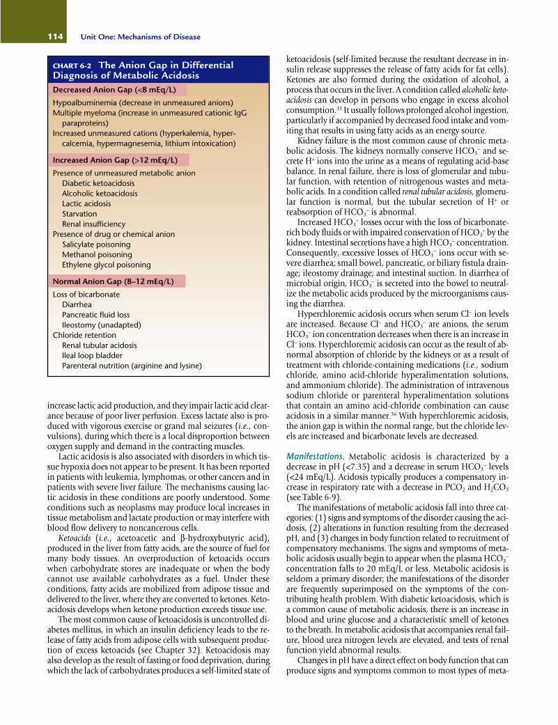
increase lactic acid production, and they impair lactic acid clear-ance because of poor liver perfusion. Excess lactate also is pro-duced with vigorous exercise or grand mal seizures (i.e., con-vulsions), during which there is a local disproportion betweenoxygen supply and demand in the contracting muscles.
Lactic acidosis is also associated with disorders in which tis-sue hypoxia does not appear to be present. It has been reportedin patients with leukemia, lymphomas, or other cancers and inpatients with severe liver failure. The mechanisms causing lac-tic acidosis in these conditions are poorly understood. Someconditions such as neoplasms may produce local increases intissue metabolism and lactate production or may interfere withblood flow delivery to noncancerous cells.
Ketoacids (i.e., acetoacetic and β-hydroxybutyric acid), produced in the liver from fatty acids, are the source of fuel formany body tissues. An overproduction of ketoacids occurswhen carbohydrate stores are inadequate or when the bodycannot use available carbohydrates as a fuel. Under these conditions, fatty acids are mobilized from adipose tissue anddelivered to the liver, where they are converted to ketones. Keto-acidosis develops when ketone production exceeds tissue use.
The most common cause of ketoacidosis is uncontrolled di-abetes mellitus, in which an insulin deficiency leads to the re-lease of fatty acids from adipose cells with subsequent produc-tion of excess ketoacids (see Chapter 32). Ketoacidosis mayalso develop as the result of fasting or food deprivation, duringwhich the lack of carbohydrates produces a self-limited state of
ketoacidosis (self-limited because the resultant decrease in in-sulin release suppresses the release of fatty acids for fat cells).Ketones are also formed during the oxidation of alcohol, aprocess that occurs in the liver. A condition called alcoholic keto-acidosis can develop in persons who engage in excess alcoholconsumption.35 It usually follows prolonged alcohol ingestion,particularly if accompanied by decreased food intake and vom-iting that results in using fatty acids as an energy source.
Kidney failure is the most common cause of chronic meta-bolic acidosis. The kidneys normally conserve HCO3
− and se-crete H+ ions into the urine as a means of regulating acid-basebalance. In renal failure, there is loss of glomerular and tubu-lar function, with retention of nitrogenous wastes and meta-bolic acids. In a condition called renal tubular acidosis, glomeru-lar function is normal, but the tubular secretion of H+ orreabsorption of HCO3
− is abnormal.Increased HCO3
− losses occur with the loss of bicarbonate-rich body fluids or with impaired conservation of HCO3
− by thekidney. Intestinal secretions have a high HCO3
− concentration.Consequently, excessive losses of HCO3
− ions occur with se-vere diarrhea; small bowel, pancreatic, or biliary fistula drain-age; ileostomy drainage; and intestinal suction. In diarrhea ofmicrobial origin, HCO3
− is secreted into the bowel to neutral-ize the metabolic acids produced by the microorganisms caus-ing the diarrhea.
Hyperchloremic acidosis occurs when serum Cl− ion levelsare increased. Because Cl− and HCO3
− are anions, the serumHCO3
− ion concentration decreases when there is an increase inCl− ions. Hyperchloremic acidosis can occur as the result of ab-normal absorption of chloride by the kidneys or as a result oftreatment with chloride-containing medications (i.e., sodiumchloride, amino acid-chloride hyperalimentation solutions,and ammonium chloride). The administration of intravenoussodium chloride or parenteral hyperalimentation solutionsthat contain an amino acid-chloride combination can causeacidosis in a similar manner.36 With hyperchloremic acidosis,the anion gap is within the normal range, but the chloride lev-els are increased and bicarbonate levels are decreased.
Manifestations. Metabolic acidosis is characterized by a decrease in pH (<7.35) and a decrease in serum HCO3
− levels(<24 mEq/L). Acidosis typically produces a compensatory in-crease in respiratory rate with a decrease in PCO2 and H2CO3
(see Table 6-9).The manifestations of metabolic acidosis fall into three cat-
egories: (1) signs and symptoms of the disorder causing the aci-dosis, (2) alterations in function resulting from the decreasedpH, and (3) changes in body function related to recruitment ofcompensatory mechanisms. The signs and symptoms of meta-bolic acidosis usually begin to appear when the plasma HCO3
−
concentration falls to 20 mEq/L or less. Metabolic acidosis isseldom a primary disorder; the manifestations of the disorderare frequently superimposed on the symptoms of the con-tributing health problem. With diabetic ketoacidosis, which isa common cause of metabolic acidosis, there is an increase inblood and urine glucose and a characteristic smell of ketonesto the breath. In metabolic acidosis that accompanies renal fail-ure, blood urea nitrogen levels are elevated, and tests of renalfunction yield abnormal results.
Changes in pH have a direct effect on body function that canproduce signs and symptoms common to most types of meta-
114 Unit One: Mechanisms of Disease
CHART 6-2 The Anion Gap in DifferentialDiagnosis of Metabolic AcidosisDecreased Anion Gap (<8 mEq/L)
Hypoalbuminemia (decrease in unmeasured anions)Multiple myeloma (increase in unmeasured cationic IgG
paraproteins)Increased unmeasured cations (hyperkalemia, hyper-
calcemia, hypermagnesemia, lithium intoxication)
Increased Anion Gap (>12 mEq/L)
Presence of unmeasured metabolic anionDiabetic ketoacidosisAlcoholic ketoacidosisLactic acidosisStarvationRenal insufficiency
Presence of drug or chemical anionSalicylate poisoningMethanol poisoningEthylene glycol poisoning
Normal Anion Gap (8–12 mEq/L)
Loss of bicarbonateDiarrheaPancreatic fluid lossIleostomy (unadapted)
Chloride retentionRenal tubular acidosisIleal loop bladderParenteral nutrition (arginine and lysine)
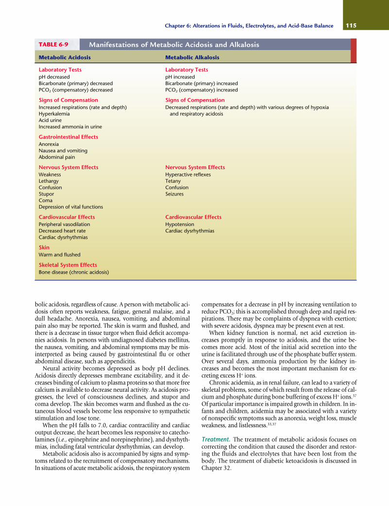
bolic acidosis, regardless of cause. A person with metabolic aci-dosis often reports weakness, fatigue, general malaise, and adull headache. Anorexia, nausea, vomiting, and abdominalpain also may be reported. The skin is warm and flushed, andthere is a decrease in tissue turgor when fluid deficit accompa-nies acidosis. In persons with undiagnosed diabetes mellitus,the nausea, vomiting, and abdominal symptoms may be mis-interpreted as being caused by gastrointestinal flu or otherabdominal disease, such as appendicitis.
Neural activity becomes depressed as body pH declines.Acidosis directly depresses membrane excitability, and it de-creases binding of calcium to plasma proteins so that more freecalcium is available to decrease neural activity. As acidosis pro-gresses, the level of consciousness declines, and stupor andcoma develop. The skin becomes warm and flushed as the cu-taneous blood vessels become less responsive to sympatheticstimulation and lose tone.
When the pH falls to 7.0, cardiac contractility and cardiacoutput decrease, the heart becomes less responsive to catecho-lamines (i.e., epinephrine and norepinephrine), and dysrhyth-mias, including fatal ventricular dysrhythmias, can develop.
Metabolic acidosis also is accompanied by signs and symp-toms related to the recruitment of compensatory mechanisms.In situations of acute metabolic acidosis, the respiratory system
compensates for a decrease in pH by increasing ventilation toreduce PCO2; this is accomplished through deep and rapid res-pirations. There may be complaints of dyspnea with exertion;with severe acidosis, dyspnea may be present even at rest.
When kidney function is normal, net acid excretion in-creases promptly in response to acidosis, and the urine be-comes more acid. Most of the initial acid secretion into theurine is facilitated through use of the phosphate buffer system.Over several days, ammonia production by the kidney in-creases and becomes the most important mechanism for ex-creting excess H+ ions.
Chronic acidemia, as in renal failure, can lead to a variety ofskeletal problems, some of which result from the release of cal-cium and phosphate during bone buffering of excess H+ ions.37
Of particular importance is impaired growth in children. In in-fants and children, acidemia may be associated with a varietyof nonspecific symptoms such as anorexia, weight loss, muscleweakness, and listlessness.33,37
Treatment. The treatment of metabolic acidosis focuses oncorrecting the condition that caused the disorder and restor-ing the fluids and electrolytes that have been lost from thebody. The treatment of diabetic ketoacidosis is discussed inChapter 32.
115Chapter 6: Alterations in Fluids, Electrolytes, and Acid-Base Balance
Manifestations of Metabolic Acidosis and AlkalosisTABLE 6-9
Laboratory Tests Laboratory TestspH decreased pH increasedBicarbonate (primary) decreased Bicarbonate (primary) increasedPCO2 (compensatory) decreased PCO2 (compensatory) increased
Signs of Compensation Signs of CompensationIncreased respirations (rate and depth) Decreased respirations (rate and depth) with various degrees of hypoxia Hyperkalemia and respiratory acidosisAcid urineIncreased ammonia in urine
Gastrointestinal EffectsAnorexiaNausea and vomitingAbdominal pain
Nervous System Effects Nervous System EffectsWeakness Hyperactive reflexesLethargy TetanyConfusion ConfusionStupor SeizuresComaDepression of vital functions
Cardiovascular Effects Cardiovascular EffectsPeripheral vasodilation HypotensionDecreased heart rate Cardiac dysrhythmiasCardiac dysrhythmias
SkinWarm and flushed
Skeletal System EffectsBone disease (chronic acidosis)
Metabolic Acidosis Metabolic Alkalosis
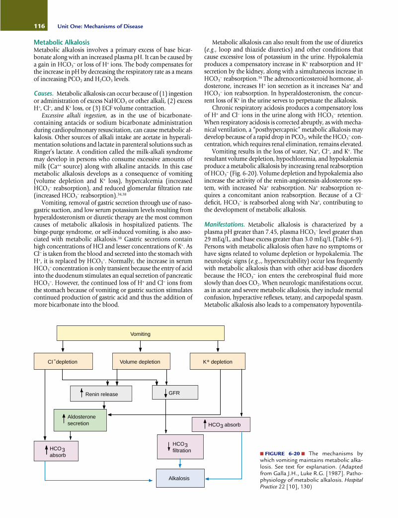
Metabolic AlkalosisMetabolic alkalosis involves a primary excess of base bicar-bonate along with an increased plasma pH. It can be caused bya gain in HCO3
− or loss of H+ ions. The body compensates forthe increase in pH by decreasing the respiratory rate as a meansof increasing PCO2 and H2CO3 levels.
Causes. Metabolic alkalosis can occur because of (1) ingestionor administration of excess NaHCO3 or other alkali, (2) excessH+, Cl−, and K+ loss, or (3) ECF volume contraction.
Excessive alkali ingestion, as in the use of bicarbonate-containing antacids or sodium bicarbonate administrationduring cardiopulmonary resuscitation, can cause metabolic al-kalosis. Other sources of alkali intake are acetate in hyperali-mentation solutions and lactate in parenteral solutions such asRinger’s lactate. A condition called the milk-alkali syndromemay develop in persons who consume excessive amounts ofmilk (Ca++ source) along with alkaline antacids. In this casemetabolic alkalosis develops as a consequence of vomiting(volume depletion and K+ loss), hypercalcemia (increasedHCO3
− reabsorption), and reduced glomerular filtration rate(increased HCO3
− reabsorption).34,38
Vomiting, removal of gastric secretion through use of naso-gastric suction, and low serum potassium levels resulting fromhyperaldosteronism or diuretic therapy are the most commoncauses of metabolic alkalosis in hospitalized patients. Thebinge-purge syndrome, or self-induced vomiting, is also asso-ciated with metabolic alkalosis.38 Gastric secretions containhigh concentrations of HCl and lesser concentrations of K+. AsCl− is taken from the blood and secreted into the stomach withH+, it is replaced by HCO3
−. Normally, the increase in serumHCO3
− concentration is only transient because the entry of acidinto the duodenum stimulates an equal secretion of pancreaticHCO3
−. However, the continued loss of H+ and Cl− ions fromthe stomach because of vomiting or gastric suction stimulatescontinued production of gastric acid and thus the addition ofmore bicarbonate into the blood.
Metabolic alkalosis can also result from the use of diuretics(e.g., loop and thiazide diuretics) and other conditions thatcause excessive loss of potassium in the urine. Hypokalemiaproduces a compensatory increase in K+ reabsorption and H+
secretion by the kidney, along with a simultaneous increase inHCO3
− reabsorption.34 The adrenocorticosteroid hormone, al-dosterone, increases H+ ion secretion as it increases Na+ andHCO3
− ion reabsorption. In hyperaldosteronism, the concur-rent loss of K+ in the urine serves to perpetuate the alkalosis.
Chronic respiratory acidosis produces a compensatory lossof H+ and Cl− ions in the urine along with HCO3
− retention.When respiratory acidosis is corrected abruptly, as with mecha-nical ventilation, a “posthypercapnic” metabolic alkalosis maydevelop because of a rapid drop in PCO2, while the HCO3
− con-centration, which requires renal elimination, remains elevated.
Vomiting results in the loss of water, Na+, Cl−, and K+. Theresultant volume depletion, hypochloremia, and hypokalemiaproduce a metabolic alkalosis by increasing renal reabsorptionof HCO3
− (Fig. 6-20). Volume depletion and hypokalemia alsoincrease the activity of the renin-angiotensin-aldosterone sys-tem, with increased Na+ reabsorption. Na+ reabsorption re-quires a concomitant anion reabsorption. Because of a Cl−
deficit, HCO3− is reabsorbed along with Na+, contributing to
the development of metabolic alkalosis.
Manifestations. Metabolic alkalosis is characterized by aplasma pH greater than 7.45, plasma HCO3
− level greater than29 mEq/L, and base excess greater than 3.0 mEq/L (Table 6-9).Persons with metabolic alkalosis often have no symptoms orhave signs related to volume depletion or hypokalemia. Theneurologic signs (e.g.,, hyperexcitability) occur less frequentlywith metabolic alkalosis than with other acid-base disordersbecause the HCO3
− ion enters the cerebrospinal fluid moreslowly than does CO2. When neurologic manifestations occur,as in acute and severe metabolic alkalosis, they include mentalconfusion, hyperactive reflexes, tetany, and carpopedal spasm.Metabolic alkalosis also leads to a compensatory hypoventila-
116 Unit One: Mechanisms of Disease
Vomiting
CI depletion Volume depletion K depletion
Renin release GFR
Aldosteronesecretion HCO absorb
HCOabsorb
HCOfiltration
Alkalosis
-
33
3
+
■ FIGURE 6-20 ■ The mechanisms bywhich vomiting maintains metabolic alka-losis. See text for explanation. (Adaptedfrom Galla J.H., Luke R.G. [1987]. Patho-physiology of metabolic alkalosis. HospitalPractice 22 [10], 130)
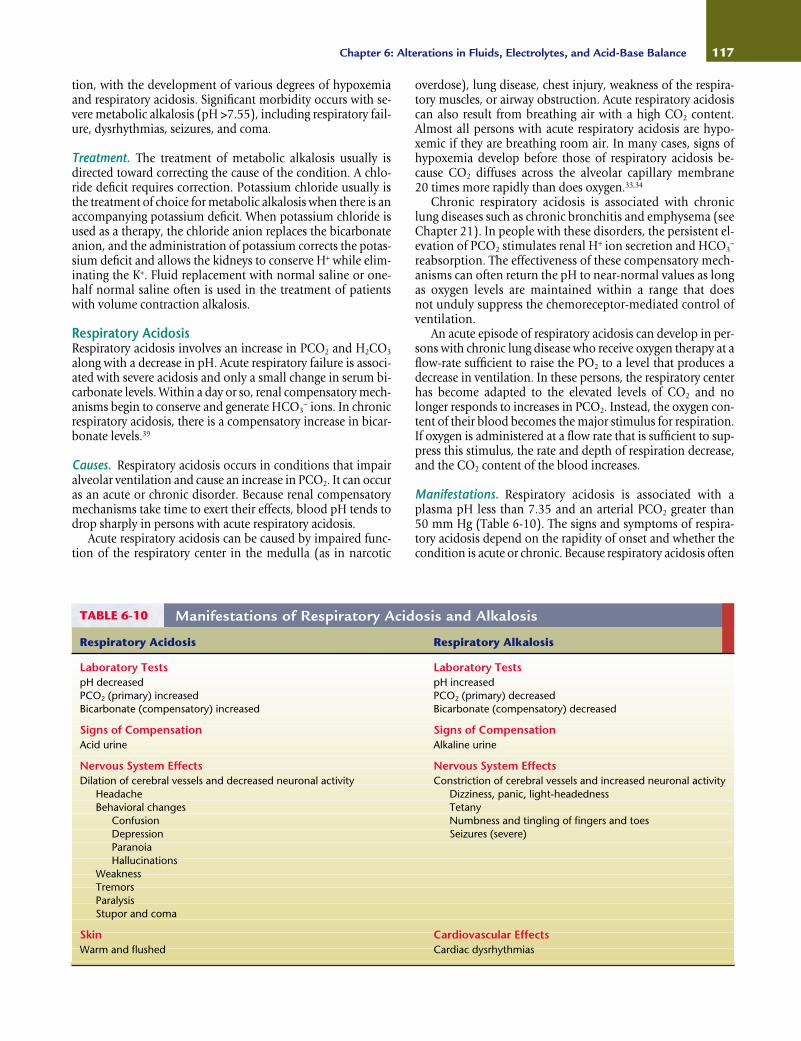
tion, with the development of various degrees of hypoxemiaand respiratory acidosis. Significant morbidity occurs with se-vere metabolic alkalosis (pH >7.55), including respiratory fail-ure, dysrhythmias, seizures, and coma.
Treatment. The treatment of metabolic alkalosis usually isdirected toward correcting the cause of the condition. A chlo-ride deficit requires correction. Potassium chloride usually isthe treatment of choice for metabolic alkalosis when there is anaccompanying potassium deficit. When potassium chloride isused as a therapy, the chloride anion replaces the bicarbonateanion, and the administration of potassium corrects the potas-sium deficit and allows the kidneys to conserve H+ while elim-inating the K+. Fluid replacement with normal saline or one-half normal saline often is used in the treatment of patientswith volume contraction alkalosis.
Respiratory AcidosisRespiratory acidosis involves an increase in PCO2 and H2CO3
along with a decrease in pH. Acute respiratory failure is associ-ated with severe acidosis and only a small change in serum bi-carbonate levels. Within a day or so, renal compensatory mech-anisms begin to conserve and generate HCO3
− ions. In chronicrespiratory acidosis, there is a compensatory increase in bicar-bonate levels.39
Causes. Respiratory acidosis occurs in conditions that impairalveolar ventilation and cause an increase in PCO2. It can occuras an acute or chronic disorder. Because renal compensatorymechanisms take time to exert their effects, blood pH tends todrop sharply in persons with acute respiratory acidosis.
Acute respiratory acidosis can be caused by impaired func-tion of the respiratory center in the medulla (as in narcotic
overdose), lung disease, chest injury, weakness of the respira-tory muscles, or airway obstruction. Acute respiratory acidosiscan also result from breathing air with a high CO2 content.Almost all persons with acute respiratory acidosis are hypo-xemic if they are breathing room air. In many cases, signs ofhypoxemia develop before those of respiratory acidosis be-cause CO2 diffuses across the alveolar capillary membrane 20 times more rapidly than does oxygen.33,34
Chronic respiratory acidosis is associated with chroniclung diseases such as chronic bronchitis and emphysema (seeChapter 21). In people with these disorders, the persistent el-evation of PCO2 stimulates renal H+ ion secretion and HCO3
−
reabsorption. The effectiveness of these compensatory mech-anisms can often return the pH to near-normal values as longas oxygen levels are maintained within a range that does not unduly suppress the chemoreceptor-mediated control ofventilation.
An acute episode of respiratory acidosis can develop in per-sons with chronic lung disease who receive oxygen therapy at aflow-rate sufficient to raise the PO2 to a level that produces adecrease in ventilation. In these persons, the respiratory centerhas become adapted to the elevated levels of CO2 and nolonger responds to increases in PCO2. Instead, the oxygen con-tent of their blood becomes the major stimulus for respiration.If oxygen is administered at a flow rate that is sufficient to sup-press this stimulus, the rate and depth of respiration decrease,and the CO2 content of the blood increases.
Manifestations. Respiratory acidosis is associated with aplasma pH less than 7.35 and an arterial PCO2 greater than 50 mm Hg (Table 6-10). The signs and symptoms of respira-tory acidosis depend on the rapidity of onset and whether thecondition is acute or chronic. Because respiratory acidosis often
117Chapter 6: Alterations in Fluids, Electrolytes, and Acid-Base Balance
Manifestations of Respiratory Acidosis and AlkalosisTABLE 6-10
Laboratory Tests Laboratory TestspH decreased pH increasedPCO2 (primary) increased PCO2 (primary) decreasedBicarbonate (compensatory) increased Bicarbonate (compensatory) decreased
Signs of Compensation Signs of CompensationAcid urine Alkaline urine
Nervous System Effects Nervous System EffectsDilation of cerebral vessels and decreased neuronal activity Constriction of cerebral vessels and increased neuronal activity
Headache Dizziness, panic, light-headednessBehavioral changes Tetany
Confusion Numbness and tingling of fingers and toesDepression Seizures (severe)ParanoiaHallucinations
WeaknessTremorsParalysisStupor and coma
Skin Cardiovascular EffectsWarm and flushed Cardiac dysrhythmias
Respiratory Acidosis Respiratory Alkalosis

is accompanied by hypoxemia, the manifestations of respira-tory acidosis often are intermixed with those of oxygen deficit.
Sudden elevations in arterial CO2 can cause headache,blurred vision, an increase in heart rate and blood pressure,warm and flushed skin, irritability, muscle twitching, and psy-chological disturbances. Carbon dioxide readily crosses theblood-brain barrier, exerting its effects by changing the pH ofbrain fluids. Elevated levels of CO2 produce vasodilation ofcerebral blood vessels. If the condition is severe and prolonged,it can cause an increase in cerebrospinal fluid pressure and pa-pilledema. Impaired consciousness, ranging from lethargy tocoma, develops as the PCO2 rises. Paralysis of extremities mayoccur, and there may be respiratory depression.
Treatment. The treatment of acute and chronic respiratory aci-dosis is directed toward improving ventilation (see Chapter 21).In severe cases, mechanical ventilation may be necessary.
Respiratory AlkalosisRespiratory alkalosis involves a decrease in PCO2 and a pri-mary deficit in carbonic acid (H2CO3) along with an increasein pH. Because respiratory alkalosis can occur suddenly, acompensatory decrease in HCO3
− may not occur before respi-ratory correction has been accomplished. The increase in pHis less in chronic compensated respiratory alkalosis, and thefall in HCO3
− is greater.
Causes. Respiratory alkalosis is caused by hyperventilation ora respiratory rate in excess of that needed to maintain normalPCO2 levels. One of the most common causes of respiratoryalkalosis is the hyperventilation syndrome, which is character-ized by recurring episodes of overbreathing often associatedwith anxiety. Persons experiencing panic attacks frequently pre-sent in the emergency room with acute respiratory alkalosis.Other causes of hyperventilation are fever, hypoxemia, earlysalicylate toxicity, and encephalitis. Hyperventilation can alsooccur during anesthesia or with use of mechanical ventilatorydevices. Hypoxemia exerts its effect through stimulation of theperipheral chemoreceptors. Salicylate toxicity and encephalitisproduce hyperventilation by directly stimulating the respira-tory center in the brain stem.
Manifestations. Respiratory alkalosis is characterized by a pHgreater than 7.45, arterial PCO2 less than 35 mm Hg, andserum HCO3
− levels less than 24 mEq/L (Table 6-10). Thesigns and symptoms of respiratory alkalosis are mainly asso-ciated with hyperexcitability of the nervous system and a de-crease in cerebral blood flow. Alkalosis increases protein bind-ing of serum calcium. This reduces ionized serum calciumlevels, causing an increase in neuromuscular excitability. A de-crease in the CO2 content of the blood causes constriction ofcerebral blood vessels. Because CO2 crosses the blood-brainbarrier rather quickly, the manifestations of acute respiratoryalkalosis often are of sudden onset. The person often experi-ences light-headedness, dizziness, tingling, and numbness ofthe fingers and toes. These manifestations may be accompa-nied by sweating, palpitations, panic, air hunger, and dyspnea.Chvostek’s and Trousseau’s signs may be positive, and tetanyand convulsions may occur. Because CO2 provides the stimulusfor short-term regulation of respiration, short periods of apneamay occur in persons with acute episodes of hyperventilation.
118 Unit One: Mechanisms of Disease
In summary, normal body function depends on the pre-cise regulation of acid-base balance to maintain a pH withinthe narrow physiologic range of 7.35 to 7.45. Metabolicprocesses produce volatile and nonvolatile metabolic acidsthat must be buffered and eliminated from the body. Thevolatile acid H2CO3 is in equilibrium with dissolved CO2,which is eliminated through the lungs. The nonvolatile meta-bolic acids, most of which are excreted by the kidneys, are de-rived mainly from protein metabolism and incomplete carbo-hydrate and fat metabolism. It is the ratio of the bicarbonateion concentration to dissolved CO2 (carbonic acid concentra-tion) that determines body pH. When this ratio is 20�1, thepH is 7.4. The ability of the body to maintain pH within thenormal physiologic range depends on respiratory and renalmechanisms and on intracellular and extracellular buffers; themost important of these is the bicarbonate buffer system. Therespiratory regulation of pH is rapid but does not return pHcompletely to normal. The kidney aids in regulation of pH byeliminating H+ ions or conserving HCO3
− ions. In the processof eliminating H+ ions, it uses the phosphate and ammoniabuffer systems. Body pH is also affected by the distribution ofexchangeable cations (K+ and H+) and anions (Cl− and HCO3
−).Metabolic acidosis is defined as a decrease in HCO3
− , andmetabolic alkalosis as an increase in HCO3
−. Metabolic acidosisis caused by an excessive production and accumulation ofmetabolic acids or an excessive loss of HCO3
−.Metabolic alkalosis is caused by an increase in HCO3
− orfrom a decrease in H+ or Cl−, with a resultant increase in renalreabsorption of HCO3
− . Respiratory acidosis reflects a de-crease in pH that is caused by conditions that producehypoventilation, with a resultant increase in PCO2 levels.Respiratory alkalosis reflects an increase in pH that is causedby conditions that produce hyperventilation, with a resultantdecrease in PCO2 levels.
The signs and symptoms of acidosis and alkalosis reflect al-terations in body function associated with the disorder caus-ing the acid-base disturbance, the effect of the change of pHon body function, and the body’s attempt to correct andmaintain the pH within a normal physiologic range. In gen-eral, neuromuscular excitability is decreased in acidosis andincreased in alkalosis.
Treatment. The treatment of respiratory alkalosis focuses onmeasures to increase the PCO2. Attention is directed towardcorrecting the disorder that caused the overbreathing. Re-breathing of small amounts of expired air (breathing into apaper bag) may prove useful in restoring PCO2 levels in per-sons with anxiety-produced respiratory alkalosis.
REVIEW QUESTIONS■ Describe factors that control fluid exchange between the vas-cular and interstitial fluid compartments and relate them to thedevelopment of edema and third spacing of extracellular fluids.
■ Compare and contrast the causes, manifestations, and treat-ment of isotonic fluid volume deficit, isotonic fluid volume ex-cess, hyponatremia with water excess, and hypernatremia withwater deficit.
■ Characterize the distribution of potassium in the body andexplain how extracellular potassium levels are regulated inrelation to body gains and losses.
■ Persons with severe hyperkalemia are at risk for cardiac arrest.Explain the effects of potassium on membrane potentials thatare responsible for this risk.
■ A person may have hypocalcemia according to laboratoryreports but have no clinical signs of the disorder. Explain.
■ Persons with renal failure experience both hyperphospha-temia and hypocalcemia. Explain.
■ Describe the three forms of carbon dioxide transport andtheir contribution to acid-base balance.
■ Use the Henderson-Hasselbalch equation to calculate pH andcompare compensatory mechanisms for regulating pH.
■ Describe a clinical situation involving an acid-base disorder inwhich primary and compensatory mechanisms might be active.
■ Define metabolic acidosis, metabolic alkalosis, respiratoryacidosis, and respiratory alkalosis.
■ Explain the use of the plasma anion gap in differentiatingtypes of metabolic acidosis.
Visit the Connection site at connection.lww.com/go/porthfor links to chapter-related resources on the Internet.
REFERENCES1. Krieger J.N., Sherrad D.J. (1991). Practical fluid and electrolytes
(pp. 104–105). Norwalk, CT: Appleton & Lange.2. Guyton A., Hall J.E. (2000). Textbook of medical physiology (10th ed.,
pp. 157–171, 264–278, 322–345, 346–363, 820–826). Philadelphia:W.B. Saunders.
3. Cogan M.G. (1991). Fluid and electrolytes (pp. 1, 43, 80–84, 100–111,112–123, 125–130, 242–245). Norwalk, CT: Appleton & Lange.
4. Stearns R.H., Spital A., Clark E.C. (1996). Disorders of water balance.In Kokko J., Tannen R.L. (Eds.), Fluids and electrolytes (3rd ed., pp. 65,69, 95). Philadelphia: W.B. Saunders.
5. Metheney N.M. (2000). Fluid and electrolyte balance (4th ed., pp. 3, 16,18, 43, 47, 56, 162, 256). Philadelphia: Lippincott Williams & Wilkins.
6. Porth C.J.M., Erickson M. (1992). Physiology of thirst and drinking:Implications for nursing practice. Heart and Lung 21, 273–284.
7. Ayus J.C., Arieff A.I. (1996). Abnormalities of water metabolism in theelderly. Seminars in Nephrology 16 (4), 277–288.
8. Kugler J.P., Hustead T. (2000). Hyponatremia and hypernatremia inthe elderly. American Family Physician 61, 3623–3630.
9. Illowsky B.P., Kirch D.G. (1988). Polydipsia and hyponatremia inpsychiatric patients. American Journal of Psychiatry 145, 675–683.
10. Vieweg W.V.R. (1994). Treatment strategies for polydipsia-hyponatremia syndrome. Journal of Clinical Psychiatry 55 (4), 154–159.
11. Rose B.D., Post T.W. (2001). Clinical physiology of acid-base and elec-trolyte disorders (5th ed., pp. 168–178, 822, 835, 858, 909). New York:McGraw-Hill.
12. Berne R.M., Levy M. (2000). Principles of physiology (3rd ed., p. 438).St. Louis: Mosby.
13. Robertson G.L. (1983). Thirst and vasopressin function in normaland disordered states of water balance. Journal of Laboratory and Cli-nical Medicine 101, 351–371.
14. Robertson G.L. (1995). Diabetes insipidus. Endocrinology and Meta-bolic Clinics of North America 24, 549–571.
15. Bendz H., Aurell M. (1999). Drug-induced diabetes insipidus. DrugSafety 21, 449–456.
16. Batchell J. (1994). Syndrome of inappropriate antidiuretic hormone.Critical Care Clinics of North America 69, 687–691.
17. Fried L.F., Palevsky P.M. (1997). Hyponatremia and hypernatremia.Medical Clinics of North America 81, 585–606.
18. Androgue H.J., Madias N.E. (2000). Hyponatremia. New EnglandJournal of Medicine 343, 1581–1589.
19. Oh M.S., Carroll H.J. (1992). Disorders of sodium metabolism:Hypernatremia and hyponatremia. Critical Care Medicine 20, 94–103.
20. Androgue H.J. (2000). Hypernatremia. New England Journal of Med-icine 342, 1493–1499.
21. Behrman R.E., Kliegman R.M., Jenson H.B. (2000). Nelson textbook ofpediatrics (16th ed., pp. 215–218). Philadelphia: W.B. Saunders.
22. Bräxmeyer D.L., Keyes J.L. (1996). The pathophysiology of potassiumbalance. Critical Care Nurse 16 (5), 59–71.
23. Gennari F.J. (1998). Hypokalemia. New England Journal of Medicine339, 451–458.
24. Tannen R.L (1996). Potassium disorders. In Kokko J., Tannen R.L.(Eds.), Fluids and electrolytes (3rd ed., pp. 116–118). Philadelphia:W.B. Saunders.
25. Whang G., Whang G.G., Ryan M.P. (1992). Refractory potassium re-pletion: A consequence of magnesium deficiency. Archives of InternalMedicine 152 (1), 40–45.
26. Clark B.A., Brown R.S. (1995). Potassium homeostasis and hyper-kalemic syndromes. Endocrinology and Metabolic Clinics of North America24, 573–590.
27. Zaloga G.F. (1992). Hypocalcemia in critically ill patients. CriticalCare Medicine 20, 251–262.
28. Yucha C.B., Toto K.H. (1994). Calcium and phosphorous derange-ments. Critical Care Clinics of North America 6, 747–765.
29. Barnett M.L. (1999). Hypercalcemia. Seminars in Oncology Nursing 15,190–201.
30. Workman L. (1992). Magnesium and phosphorus: The neglected elec-trolytes. AACN Clinical Issues 3, 655–663.
31. Swain R., Kaplan-Machlis B. (1999). Magnesium for the next millen-nium. Southern Medical Journal 92, 1040–1046.
32. Toto K., Yucha C.B. (1994). Magnesium: Homeostasis, imbalances,and therapeutic uses. Critical Care Nursing Clinics of North America 6,767–778.
33. Rose B.D., Post T.W. (2001). Clinical physiology of acid-base and elec-trolyte disorders (5th ed., pp. 325, 363, 578–646, 647, 669). New York:McGraw-Hill.
34. Abelow B. (1998). Understanding acid-base (pp. 43–49, 83–93,139–169, 171–188, 189–198, 224–230). Baltimore: Williams &Wilkins.
35. Umpierrez G.E., DiGirolamo M., Tuvlin J.A., et. al. (2000). Dif-ferences in metabolic and hormonal milieu in diabetic and alcohol-induced ketoacidosis. Journal of Critical Care 15 (2), 52–59.
36. Powers F. (1999). The role of chloride in acid-base balance. Journal ofIntravenous Nursing 22, 286–291.
37. Alpern R.J., Sakhaee K. (1997). The clinical spectrum of chronic meta-bolic acidosis: Homeostatic mechanisms produce significant mor-bidity. American Journal of Kidney Diseases 29, 291–302.
38. Galla J.H. (2000). Metabolic alkalosis. Journal of the American Societyof Nephrology 11, 369–375.
39. Adrogue H.J., Madias N.E. (1998). Management of life-threateningacid-base disorders. New England Journal of Medicine 338, 107–111.
119Chapter 6: Alterations in Fluids, Electrolytes, and Acid-Base Balance
