als skills competency verification form
TRANSCRIPT

ALS Skills Competency Verification Form
Page 1 of 3
1a. Name as shown on paramedic license / MICN authorization# 1b. Certificate / license number
1c. Signature of person demonstrating competency 1d. Certifying authority
REMSA
Airway Skills Verification of competency 1. BLS airway adjuncts Affiliation Date
Signature of person verifying competency Printed name Cert / License / Authorization number
2. Continuous Positive Airway Pressure(CPAP)
Affiliation Date
Signature of person verifying competency Printed name Cert / License / Authorization number
3. Laryngoscopy and Magill Forceps Affiliation Date
Signature of person verifying competency Printed name Cert / License / Authorization number
4. Orotracheal Intubation (OTI) Affiliation Date
Signature of person verifying competency Printed name Cert / License / Authorization number
5. Post OTI Confirmation & Monitoring Affiliation Date
Signature of person verifying competency Printed name Cert / License / Authorization number
6. Positive Pressure Ventilation Affiliation Date
Signature of person verifying competency Printed name Cert / License / Authorization number

Page 2 of 3
Cardiac Skills Verification of competency 7. Defibrillation Affiliation Date
Signature of person verifying competency Printed name Cert / License / Authorization number
8. Synchronized Cardioversion Affiliation Date
Signature of person verifying competency Printed name Cert / License / Authorization number
9. Transcutaneous Cardiac Pacing Affiliation Date
Signature of person verifying competency Printed name Cert / License / Authorization number
General Medical Skills Verification of competency 10. Calculating and Preparing Drug Dosages Affiliation Date
Signature of person verifying competency Printed name Cert / License / Authorization number
11. Childbirth with Neonatal Resuscitation Affiliation Date
Signature of person verifying competency Printed name Cert / License / Authorization number
Trauma Care Skills Verification of competency 12. Needle Chest Decompression Affiliation Date
Signature of person verifying competency Printed name Cert / License / Authorization number
13. Tourniquets Affiliation Date
Signature of person verifying competency Printed name Cert / License / Authorization number

Page 3 of 3
For paramedics: a completed ALS Skills Verification Form is required to accompany an initial, and reaccreditation, application for those individuals who are either initially obtaining accreditation, maintaining accreditation without a lapse, or reaccrediting with a lapse of less than one (1) year.
For MICNs: a completed ALS Skills Verification Form is required to accompany an initial authorization application or when challenging MICN authorization.
For reauthorizing MICNs: to meet the additional reauthorization requirements in Policy #1210, a completed ALS SCV form must be included in the reauthorization packet.
1a. Name of Certificate Holder Provide the complete name, last name first, of the paramedic accreditation / MICN authorization holder who is demonstrating skills competency.
1b. Certificate Number Provide the paramedic accreditation / MICN authorization number from the current or lapsed paramedic accreditation / authorization of the paramedic / MICN who is demonstrating competency.
1c. Signature Signature of the paramedic accreditation / MICN authorization holder who is demonstrating competency. By signing this section, the paramedic or MICN is verifying that the information contained on this form is accurate and that the paramedic accreditation / MICN authorization holder has demonstrated competency in the skills listed to a qualified individual.
1d. Certifying Authority Provide the name of the paramedic / MICN certifying authority for which the individual will be accrediting through.
Verification of Competency 1. Affiliation – Provide the name of the EMS service provider or base hospital that the qualified individual who is
verifying competency is affiliated with.
2. Once competency has been demonstrated by direct observation of an actual or simulated patient contact, i.e. skillsstation, the individual verifying competency shall sign the ALS Skills Competency Verification Form for that skill.
3. Qualified individuals who verify skills competency shall be currently licensed or certified as: A paramedic, registerednurse, physician assistant, or physician and shall be either a qualified instructor designated by an EMS approvedtraining program (paramedic training program or continuing education training program) or by a qualified individualdesignated by an EMS service provider. EMS service providers include, but are not limited to, public safety agencies,REMSA authorized private ambulance providers, and other EMS providers.
4. Certification or License Number – Provide the certification or license number for the individual verifyingcompetency. 5.Date – Enter the date that the individual demonstrated competency in each skill.
5. Print name – Print the name of the individual verifying competency in the skill.
Verification of skills competency shall be valid to apply for paramedic reaccreditation or MICN authorization for a maximum of two (2) years from the date of verification.
v91721

Skill Verification Form Effective
April 1, 2021Expires
March 31, 2022 Category I Skill – Low Frequency/High Risk:
BLS Airway Adjuncts Approval: Medical Director
Reza Vaezazizi, MD Signed
Applies To: PSP, EMT, AEMT, EMT-P, MICN, BHP, EMS
System
Approval: EMS Administrator Trevor Douville
Signed
BLS Airway Adjuncts (1/6/21 - 12:54) 1 of 4
Terminal Performance Objective To establish and maintain an open airway for spontaneous respiration or to facilitate effective positive pressure ventilation (PPV).
Before managing a patient’s airway with BLS adjuncts, the PSP, EMT, AEMT, or paramedic must:
1. Methodically complete an assessment of the airway and breathing within 30 seconds.2. Identify inadequate ventilation (minute volume) and/or signs of hypoxia within the first 30 seconds.3. Apply the appropriate, clinically required technique to manually position the head and mandible of the
unconscious patient to open the upper airway.a. Medical – Head tilt/chin liftb. Trauma – Jaw thrust or modified chin lift
4. Clear secretions or other obstructions using appropriate method (manually, log rolling, suctioning, etc.)maintaining spinal stabilization controls as patient’s condition indicates.
5. Utilize the appropriate technique per American Heart Association Standards to insert the appropriate BLS airwaywithin 10 seconds.
a. Nasopharyngeal airway (NPA) is the preferred BLS airway. (PSP exempt; NPA exceeds PSP scope ofpractice)
6. Confirm correct airway placement and immediately initiate PPV with oxygen at 10 – 15 LPMa. For the hypoventilating or apneic patient, initiate PPV with oxygen at 10 – 15 LPM.b. If upper airway management techniques have restored effective spontaneous respiration, apply
supplemental oxygen and closely monitor the patient’s airway and breathing.
While managing a patient’s airway with BLS adjuncts, PSP, EMT, AEMT, or paramedic must: 1. Effectively evaluate the efficacy of PPV following BLS airway insertion.2. Limit suction attempts to 10 secondsi.3. Limit interruption of PPV to 30 seconds4. Consider and treat reversible causes of hypoventilation and hypoxia such as opiate overdose and hypoglycemia5. Rapidly determine the need for Advanced Life Support (ALS) airway adjuncts when airway patency or
ventilations cannot be effectively supported by BLS means.
Critical Success Targets for BLS Airway Management 1. Successful and secure adjunct insertion2. Effective PPV
System Benchmark % of hypoventilating patients that receive effective positive pressure ventilation
Core Competency Requirements to be covered during education/training 1. Patient assessment2. Airway anatomy & physiology3. Airway pathophysiology

BLS Airway Adjuncts (1/6/21 - 12:54) 2 of 4
4. BLS techniques for relief of anatomical or foreign body airway obstruction 5. Manual positioning of the airway 6. Identify the need for use of a BLS airway adjunct 7. Oropharyngeal airway (OPA) indications/ contraindications 8. OPA sizing and insertion 9. NPA indications/ contraindications (FR exempt; NPA exceeds PSP scope of practice) 10. NPA sizing and insertion (FR exempt; NPA exceeds PSP scope of practice) 11. Evaluation of ventilatory efficacy following BLS airway adjunct insertion 12. Identification and correction of complications of BLS airway management 13. Rapid identification of the need for ALS airway and/or medications when BLS airway adjuncts are ineffective
Adjunctive Performance Standards
1. Positive Pressure Ventilation (PPV) 2. Laryngoscopy with FBAO Removal/Magill Forceps (ALS personnel)
Equipment Requirements 1. Airway mannequin 2. OPA(s) 3. NPA(s) 4. Lubricant 5. BVM with reservoir and manometer 6. Stethoscope 7. Supplemental oxygen 8. PPE
Instructor Resource Materials
1. Prehospital Trauma Life Support 2. AHA CPR and BLS Provider Manual 3. 2015 AHA Guidelines for Cardiopulmonary Resuscitation and Emergency Cardiovascular Care 4. NHTSA EMS Educational Instructor Guidelines for EMT and Paramedic

BLS Airway Adjuncts (1/6/21 - 12:54) 3 of 4
BLS Airway Adjuncts Validation PERFORMANCE CRITERIA 100% accuracy required on all items with an * Before managing a patient’s airway with BLS adjuncts, the FR, EMT, AEMT, and paramedic must:
Points Score Performance Steps Additional Information 1 Take or verbalize body substance
isolation Selection: gloves, goggles, mask, gown, booties, N95 PRN
1 Methodically complete an assessment of the airway and breathing within 30 seconds.*
Follow respiratory assessment sequence.
1 Identify inadequate ventilations and/or signs of hypoxia within the first 30 seconds.*
Pale/cyanotic, altered level of consciousness, diaphoresis, increased work of breathing or apnea, poor chest rise and fall
1 Apply the appropriate, clinically required technique to manually position the head and mandible of the unconscious patient to open the upper airway.*
• Medical - Head-tilt/chin lift • Trauma - Jaw thrust or modified chin lift
1 Manually clear blood, vomit, and foreign bodies when present.*
• Clear secretions or other obstructions using appropriate method (manually, log rolling, suctioning, etc.) maintaining spinal stabilization as patient condition indicates.
• Use a rigid pharyngeal tip, if available, for suctioning oropharynx.
1 Utilize appropriate technique per AHA standards to insert the selected airway within 10 seconds. *
• NPA is the preferred BLS airway. (PSP exempt; NPA is optional scope of practice)
• OPA a. Place tip into patient’s mouth with curve facing up toward the nose. b. Advance until you meet resistance and rotate 180 degrees until flange is flush against lips.
• NPA (PSP exempt; NPA is optional scope of practice) a. Lubricate tube using water-soluble lubricant. b. Place tip in nostril with beveled edge against septal wall. c. Gently advance straight back, in direction of patient’s ear, rotating back and forth slightly once resistance is met. d. Continue until flange is resting against outside of nostril.
1 Confirm correct airway placement and immediately initiate PPV with oxygen at 10 – 15 LPM.*
• For the hyperventilating or apneic patient, initiate PPV with oxygen at 10 -15 LPM
• If upper airway management techniques have restored effective spontaneous respiration, apply supplemental oxygen and closely monitor the patient’s airway and breathing.

BLS Airway Adjuncts (1/6/21 - 12:54) 4 of 4
While managing a patient’s airway with BLS adjuncts, the FR, EMT, AEMT, and paramedic must:
Points Score Performance Steps Additional Information 1 Evaluate the efficacy of PPV
following BLS airway insertion.*
Chest rise and fall symmetrically, lung sounds auscultated bilaterally, absent epigastric sounds with ventilations, patient’s skin signs improve.
1 Limit suction attempts to 10 seconds. *
1 Limit interruption of PPV to 30 seconds.*
1 Consider and treat reversible causes of airway obstruction, hypoventilation and hypoxia. *
I.e. opiate overdose, hypoglycemia
1 Rapidly determine the need for Advanced Life Support airway adjuncts when airway patency or ventilations cannot be effectively supported by BLS means.*
Critical Failure Criteria ____Failure to take or verbalize BSI appropriate to the skill prior to performing the skill ____Insertion of an oropharyngeal airway without checking for an intact gag reflex or keeping an oropharyngeal airway in the patient when gag reflex returns ____Failure to properly identify ineffective ventilations ____Any procedure that would have harmed the patient
i AHA

Skill Verification Form Effective
April 1, 2021 Expires
March 31, 2022 Category IV Skill – High Frequency/Low Risk:
Continuous Positive Airway Pressure Approval: Medical Director
Reza Vaezazizi, MD Signed
Applies To:
EMT-P, MICN, BHP, EMS System Approval: EMS Administrator
Trevor Douville Signed
Continuous Positive Airway Pressure (1/6/21 - 12:55) 1 of 5
Terminal Performance Objective Decreased venous return, increased alveolar recruitment, and decreased pulmonary edema resulting in the restoration of adequate cardiac output, gas exchange, and tissue perfusion. Before applying continuous positive airway pressure (CPAP) paramedics must:
1. Assess the patient’s ABC’s 2. Determine the patient is a candidate for CPAP:
a. Confirm the patient is experiencing dyspnea with pertinent findings suggestive of CHF, suspected exacerbation of COPD, asthma or non-fatal drowning
b. Confirm that patient is awake, alert and able to maintain their own airway c. Confirm that assisted ventilation and/or OTI is not indicated d. Recognize contraindications to CPAP:
i. Apneic ii. Unconscious
iii. Unable to maintain own airway iv. Pediatric (appearing to be 14 years of age or less) v. Suspected Pneumothorax
vi. Vomiting vii. Pump failure due to severe bradycardia or non-compensatory tachycardia (treat rate first)
viii. Systolic blood pressure of 90 mmHg or less 3. Place the patient sitting upright with lower extremities dependent to encourage pulmonary function and venous
pooling 4. Provide supplemental oxygen as clinically indicated 5. Provide clinically indicated treatment following the applicable treatment protocol 6. Continue clinically indicated treatment, including pharmacologic interventions, while preparing equipment 7. Explain to the patient what they can expect to experience, while avoiding delays in treatment
While applying continuous positive airway pressure (CPAP) paramedics must:
1. Prepare for application of the Pulmodyne™ O2-MAX™ system a. Select and prepare the appropriate size mask b. Set pressure to 5 cmH2O CPAP c. Prepare the head strap and circuit d. Attach the system to an oxygen supply e. Ensure the availability of suction, airway, and assisted ventilation supplies f. Prepare to increase and titrate pressure in 2.5 – 5 cmH2O increments as clinically indicated
2. Apply the oxygen charged Pulmodyne™ O2-MAX™ system to the patient without securing the head strap 3. Coach the patient to:
a. Hold the mask firmly to their own face b. Inhale through nose and exhale through mouth c. Continue to breathe evenly against the increasing pressure
4. Troubleshoot for leaks and adjust the mask to maintain a complete seal while applying the head strap 5. Confirm the integrity of the circuit, mask seal, and oxygen delivery to the patient 6. Immediately reassess the patient:

Continuous Positive Airway Pressure (1/6/21 - 12:55) 2 of 5
a. Pulmonary assessment including: inspiratory expiratory time ratio (normal adult I:E = 1:2), SpO2, lung sounds, and respiratory rate
b. Cardiovascular assessment including: blood pressure and heart rate 7. Consider the need to medicate for anxiety “Related to CPAP Mask” 8. Increase CPAP in 2.5 – 5 cmH2O increments, up to 15 cmH2O maximum CPAP, as clinically indicated by the
patient’s response to therapy (see “Critical Success Targets for CPAP” below) 9. Immediately re-assess the patient following each change in pressure
a. Pulmonary assessment including: inspiratory expiratory time ratio (normal adult I:E = 1:2), SpO2, lung sounds, and respiratory rate
b. Cardiovascular assessment including: blood pressure and heart rate 10. Titrate CPAP in 2.5 – 5 cmH2O increments to relief of dyspnea while continuously assessing patient’s tolerance
of CPAP 11. Provide clinically indicated treatment following the applicable REMSA Treatment Protocol(s) 12. Consult online medical direction if exceeding 15 cmH2O CPAP is clinically indicated
Critical Success Targets for CPAP
1. Normalizing inspiratory expiratory time ratio (normal adult I:E = 1:2) 2. SpO2 greater than 94% 3. Improved signs of perfusion 4. Improvement in patient’s perceived work of breathing 5. Resolution of, or improvement in, patient’s dyspnea
System Benchmark Percentage of patients receiving CPAP with relief of, or improvement in, dyspnea Core Competency Requirements to be Covered during Education and Training on CPAP
1. Pulmonological and Cardiovascular Anatomy & Physiology 2. Pulmonology and Cardiology – Pathophysiology of congestive heart failure 3. Assessment of respiration and recognition of respiratory instability 4. Assessment of circulation and recognition of hemodynamic instability 5. Identification of need for CPAP and contraindications to its application 6. Proper application of device on patient 7. Patient communication techniques 8. Concurrent medication for anxiety “Related to CPAP Mask” 9. Demonstrates proper technique for use of the Pulmodyne™ O2-RESQ™ system 10. Post application recognition of deterioration and appropriate alternative treatment 11. Reassessment of patient
Equipment Requirements
1. PPE 2. Model patient 3. Stethoscope 4. Blood pressure cuff 5. Oxygen equipment (tank, regulator with DISS, nasal cannula, non-rebreather mask, Pulmodyne™ O2-RESQ™
system) 6. Midazolam, as needed 7. Medication equipment (IV access, IM equipment) 8. Intubation equipment (BVM, suction, laryngoscope with selection of blades, ET tubes, etc.)
Instructor Resource Materials
1. Pulmodyne™ O2-RESQ™ training materials 2. Applicable REMSA Treatment Protocols 3. NHTSA EMS Educational Instructor Guidelines for EMT and Paramedic

Continuous Positive Airway Pressure (1/6/21 - 12:55) 3 of 5
Continuous Positive Airway Pressure Validation PERFORMANCE CRITERIA: 100% accuracy required on all items marked with an * Before applying continuous positive airway pressure, the paramedic must:
Points Score Performance Steps Additional Information 1 Take or verbalize body substance
isolation Selection: gloves, goggles, mask, gown, booties, P100 as needed
1 Assess the patient’s ABC’s * 1 Determine the patient is a
candidate for CPAP: * 1. Confirm the patient is experiencing dyspnea with pertinent
findings suggestive of CHF, suspected exacerbation of COPD, or asthma; or non-fatal drowning
2. Confirm that patient is awake, alert and able to maintain their own airway
3. Confirm that assisted ventilation and/or OTI is not indicated 4. Recognize contraindications to CPAP:
a. Apneic b. Unconscious c. Pediatric (appearing to be 14 years of age or less) d. Suspected Pneumothorax e. Vomiting f. Pump failure due to severe bradycardia or non-
compensatory tachycardia (treat rate first) g. Systolic blood pressure of 90 mmHg or less
1 Place the patient sitting upright with lower extremities dependent to encourage pulmonary function and venous pooling
1 Provide supplemental oxygen as clinically indicated *
1 Provide clinically indicated treatment following the applicable treatment protocol *
1 Continue clinically indicated treatment, including pharmacologic interventions, while preparing equipment
1 Explain to the patient what they can expect to experience, while avoiding delays in treatment
While applying continuous positive airway pressure, the paramedic must:
Points Score Performance Steps Additional Information 1 Prepare for application of the
Pulmodyne™ O2-MAX™ system *
1. Select and prepare the appropriate size mask 2. Set pressure to 5 cmH2O CPAP 3. Prepare the head strap and circuit 4. Attach the system to an oxygen supply 5. Ensure the availability of suction, airway, and assisted
ventilation supplies 6. Prepare to increase and titrate pressure in 2.5 – 5 cmH2O
increments as clinically indicated

Continuous Positive Airway Pressure (1/6/21 - 12:55) 4 of 5
1 Apply the oxygen charged Pulmodyne™ O2-MAX™ system to the patient without securing the head strap *
1 Coach the patient
1. Hold the mask firmly to their own face 2. Inhale through nose and exhale through mouth 3. Continue to breathe evenly against the increasing pressure
1 Troubleshoot for leaks and adjust the mask to maintain a complete seal while applying the head strap
1 Confirm the integrity of the circuit, mask seal, and oxygen delivery to the patient *
1 Immediately reassess the patient *
1. Pulmonary assessment including: inspiratory expiratory time ratio (normal adult I:E = 1:2), SpO2, lung sounds, and respiratory rate
2. Cardiovascular assessment including blood pressure and heart rate
1 Consider Midazolam for anxiety related to CPAP
1 Sequentially increase CPAP in 2.5 – 5 cmH2O increments, up to 15 cmH2O maximum CPAP, as clinically indicated by the patient’s response to therapy *
1. Normalizing inspiratory expiratory time ratio (normal adult I:E = 1:2)
2. SpO2 greater than 94% 3. Improved signs of perfusion 4. Improvement in patient’s perceived work of breathing 5. Resolution of, or improvement in, patient’s dyspnea
1 Immediately re-assess the patient following each change in pressure *
1. Pulmonary assessment including: inspiratory expiratory time ratio (normal adult I:E = 1:2), SpO2, lung sounds, and respiratory rate
2. Cardiovascular assessment including blood pressure and heart rate
1 Titrate CPAP in 2.5 – 5 cmH2O increments to relief of dyspnea while continuously assessing patient’s tolerance of CPAP *
1 Provide clinically indicated treatment following the applicable REMSA Treatment Protocol(s)
1 Consult online medical direction if exceeding 15 cmH2O CPAP is clinically indicated
1 Accurately document all assessment findings, therapeutic treatments, and the patient’s response to therapy
Critical Failure Criteria ____Failure to take or verbalize BSI appropriate to the skill prior to performing the skill ____Failure to assess the patient’s ABC’s ____Failure to determine the patient is a candidate for CPAP ____Failure to provide supplemental oxygen as clinically indicated ____Failure to provide clinically indicated treatment following the applicable treatment protocol ____Failure to prepare for application of the Pulmodyne™ O2-MAX™ system

Continuous Positive Airway Pressure (1/6/21 - 12:55) 5 of 5
____Failure to apply the oxygen charged Pulmodyne™ O2-MAX™ system to the patient ____Failure to confirm the integrity of the circuit, mask seal, and oxygen delivery to the patient ____Failure to immediately reassess the patient ____Failure to sequentially increase CPAP in 2.5 – 5 cmH2O increments as clinically indicated by the patient’s response ____Failure to immediately re-assess the patient following each change in pressure ____Failure to titrate CPAP in 2.5 – 5 cmH2O increments to relief of dyspnea ____Any procedure that would have harmed the patient

Skill Verification Form Effective
April 1, 2021Expires
March 31, 2022 Category I Skill – Low Frequency/High Risk:
Laryngoscopy and Magill Forceps Approval: Medical Director
Reza Vaezazizi, MD Signed
Applies To: EMT-P, MICN, BHP, EMS System
Approval: EMS Administrator Trevor Douville
Signed
Laryngoscopy and Magill Forceps (1/6/21 - 12:56) 1 of 4
Terminal Performance Objective An open unobstructed airway able to support spontaneous respiration or positive pressure ventilation (PPV).
Before performing laryngoscopy and use of Magill forceps, paramedics must: 1. Methodically complete an assessment of the airway and breathing within 30 seconds.2. Identify inadequate ventilations and/or signs of hypoxia within the first 30 seconds.3. Apply appropriate, clinically required technique to manually position the head and mandible of the unconscious
patient to open the upper airway.a. Medical - Head tilt/chin liftb. Trauma - Jaw thrust or modified chin lift
4. Clear secretions or other obstructions using appropriate method (manually, log rolling, suctioning, etc.)maintaining spinal stabilization controls as patient’s condition indicates.
5. Utilize the appropriate technique per the American Heart Association standards to insert the appropriate BLSairway within 10 seconds.
a. NPA is the preferred BLS airway6. Provide positive pressure ventilation (PPV) with oxygen at 10 – 15 LPM.7. Determine the presence of a foreign body airway obstruction (FBAO).8. Perform one (1) cycle of chest compressions and rescue breaths to clear a persistent obstruction.
While performing laryngoscopy and use of Magill forceps, paramedics must: 1. Repeat assessment of airway and breathing to determine if the airway is still obstructed.2. Prepare equipment for laryngoscopy and FBAO removal using Magill forceps within 30 seconds.3. Appropriately position the patient based on presentation and condition (medical vs. trauma).4. Use good technique with the laryngoscope to directly visualize anatomical structures of the airway and minimize
oral trauma.5. Recognize and remove a foreign body with the Magill forceps.6. Recognize persistent airway obstruction with inability to perform PPV and consider the following:
a. Rapid transport to the closest most appropriate hospital7. Reassess airway, breathing and circulation, applying oxygen as clinically indicated (non-rebreather mask or PPV)
or returning to cycles of ventilations / compressions as indicated by patient condition.
Critical Success Targets for laryngoscopy and use of Magill forceps 1. An open unobstructed airway2. Spontaneous respiratory rate within age-appropriate normal limits3. Ability to perform effective positive pressure ventilation4. SpO2 of greater than 95% in the patient with spontaneous circulation
a. In patients with COPD/pulmonary disease, it may not be possible or desirable to attain a SpO2 of 95%.
System Benchmark % of patients with an obstructed airway who receive laryngoscopy with the use of Magill forceps resulting in successful restoration of an open airway.

Laryngoscopy and Magill Forceps (1/6/21 - 12:56) 2 of 4
Core Competency Requirements to be covered during education/training 1. Assessment of mental status 2. Ensure patient is unconscious prior to using Magill forceps or beginning chest compressions 3. Airway / patient positioning 4. Assessment of airway / breathing 5. Relative benefit of NPA over OPA for patients with suspected reversible airway obstruction 6. Suctioning / clearing oral cavity of debris 7. Performance of BLS FBAO maneuvers per current AHA standards 8. Use of laryngoscope to visualize airway and look for obstruction 9. Use of Magill forceps to grasp / remove obstruction 10. Reassessment of airway / breathing status to determine further action(s)
Adjunctive Performance Standards
1. BLS Airway Adjuncts 2. ALS Airways 3. Positive Pressure Ventilation (PPV)
Equipment Requirements 1. Adult advanced airway mannequin 2. Oxygen source 3. Stethoscope 4. Laryngoscope 5. Magill forceps 6. Suction equipment (both rigid and flexible) 7. Personal protective equipment 8. NP/OP Airway 9. BVM w/ manometer and reservoir 10. Pulse oximeter 11. Cardiac monitor
Instructor Resource Materials
1. AHA ACLS Provider Manual 2. AHA PALS Provider Manual 3. Current AHA Guidelines for Cardiopulmonary Resuscitation and Emergency Cardiovascular Care 4. NHTSA EMS Educational Instructor Guidelines for EMT and Paramedic

Laryngoscopy and Magill Forceps (1/6/21 - 12:56) 3 of 4
Laryngoscopy and Magil Forceps Validation PERFORMANCE CRITERIA: 100% accuracy required on all items marked with an * Before performing laryngoscopy and use of Magil forceps, the paramedic must:
Points Score Performance Steps Additional Information 1 Take or verbalize body substance
isolation. Selection: gloves, goggles, mask, gown, booties, N95 PRN
1 Methodically complete an assessment of the airway and breathing within 30 seconds. *
Follow respiratory assessment sequence.
1 Identify inadequate ventilations and/or signs of hypoxia within the first 30 seconds. *
Pale/cyanotic, altered level of consciousness, diaphoresis, increased work of breathing or apnea, poor chest rise and fall
1 Apply appropriate, clinically required technique to manually position the head and mandible of the unconscious patient to open the upper airway.*
• Medical – Head tilt/chin lift • Trauma – Jaw thrust or modified chin lift
1 Manually clear blood, vomit, and foreign bodies when present. *
• Clear secretions or other obstructions using appropriate method (manually, log rolling, suctioning, etc.) maintaining spinal stabilization control as patient condition indicates.
• Use a rigid pharyngeal tip, if available, for suctioning oropharynx.
1 Employ the indicated BLS airway adjunct. *
• NPA is the preferred BLS airway
1 Provide positive pressure ventilation (PPV) with oxygen at 10–15 LPM*
Indicated for the hypoventilating or apneic patient.
1 Determine the presence of a foreign body airway obstruction (FBAO). *
Sudden onset of respiratory distress with coughing, gagging, stridor, or wheezing, inability to ventilate the patient with a BVM despite repositioning of airway
1 Perform one (1) cycle of chest compressions and rescue breaths to clear a persistent obstruction. *
While performing laryngoscopy and use of Magil forceps, the paramedic must:
1 Repeat assessment of airway and breathing to determine if the airway is still obstructed. *
Follow respiratory assessment sequence.
1 Prepare equipment for laryngoscopy and FBAO removal using Magill forceps within 30 seconds. *
Laryngoscope with functioning bulb, Magill forceps, suction, suction catheters (flexible and rigid), stethoscope, BVM with manometer, pulse oximetry
1 Appropriately position the patient based on presentation and condition (medical vs. trauma). *
• Medical – Head/chin lift • Trauma – Jaw thrust or modified chin lift

Laryngoscopy and Magill Forceps (1/6/21 - 12:56) 4 of 4
1 Use good technique with the
laryngoscope to directly visualize anatomical structures of the airway and minimize oral trauma. *
• Do not use the teeth as a fulcrum.
1 Recognize and remove a foreign body with the Magill forceps. *
1 Recognize persistent airway obstruction with inability to perform PPV and consider the following: *
• Rapid transport to the closest most appropriate hospital
Make contact with receiving hospital as soon as possible to allow the receiving hospital time to adequately prepare for the patient’s arrival.
1 Reassess airway, breathing and circulation, applying oxygen via appropriate device (non-rebreather mask or bag-valve-mask) or returning to cycles of compressions/ventilations as indicated by patient condition. *
Critical Failure Criteria ____Failure to take or verbalize BSI appropriate to the skill prior to performing the skill ____Failure to properly identify a FBAO ____Failure to attach oxygen to the BVM ____Failure to appropriately position patient and open airway ____Failure to initiate compressions and ventilations prior to attempting to use Magill forceps ____Using the patient’s teeth as a fulcrum ____Failure to initiate rapid transport once persistent airway obstruction is identified ____Any procedure that would have harmed the patient

Skill Verification Form Effective
April 1, 2021 Expires
March 31, 2022 Category I Skill – Low Frequency/High Risk:
Orotracheal Intubation
Approval: Medical Director Reza Vaezazizi, MD
Signed
Applies To: EMT-P, MICN, BHP, EMS System
Approval: EMS Administrator Trevor Douville
Signed
Orotracheal Intubation (1/6/21 - 12:57) 1 of 8
Terminal Performance Objective Secure placement of an endotracheal tube (ETT) in the trachea to ensure a patent airway for positive pressure ventilation (PPV).
Before performing orotracheal intubation (OTI), paramedics must:
1. Determine BLS airway management is inadequate for effective PPV and confirm the need for Orotracheal Intubation (OTI)1 2.
a. NOTE: OTI is approved only if the patient’s length exceeds the length-based resuscitation tape. A REMSA approved, commercially available and standardized length-based resuscitation tape must be used to identify patient’s height and weight. Clinical assessment should take into account anatomic and genetic conditions that may affect patient stature and weight.
2. Recognize signs of a difficult airway, if present, and select, prepare and employ the appropriate alternative tools and techniques to secure the airway (e.g. ET Introducing Stylet).
a. A difficult airway is defined as the presence of anatomic conditions which preclude direct visualization of the patient’s glottic opening (e.g. airway edema, arthritis or scoliosis of the spine, significant overbite, small mandible, short neck, morbid obesity, cervical spine immobilization, face or neck trauma).
3. Correctly assemble all equipment required for OTI within 60 seconds. 4. Provide optimal ventilation and oxygenation (minute volume) to the patient while OTI equipment is prepared. 5. Test the cuff of the ETT by inflating it with air and ensuring that there is no leak in the cuff.
While performing OTI, paramedics must:
1. Position the patient with their anatomy to best facilitate the intubation. Align the ear canal with the anterior shoulder, pad as clinically indicated. As a last resort. Consider having a team member apply cricoid pressure during intubation attempts.
2. Visualize anatomical structures including the glottic opening (vocal cords) during direct laryngoscopy. a. Use manual percutaneous laryngeal manipulation to assist with visualization of the glottic opening as
needed. 3. Minimize oral trauma during laryngoscopy by utilizing correct technique. 4. Place the appropriately sized ETT securely in the trachea at the correct depth within 30 seconds. 5. Inflate the cuff with enough air to seal the trachea; estimating inflation pressure by palpation of the pilot
balloon.
Immediately after OTI, paramedics must: 1. Confirm placement of the ETT in the trachea by:
a. Direct visualization of the tube passing through the cords. b. IMMEDIATELY attaching a colormetric device to confirm correct placement of the ETT prior to use of
waveform / digital capnography i. Note color change of EtCO2 detector for documentation in the ePCR.
ii. In the event of digital waveform capnography failure, a colormetric device is required for airway patency monitoring.
1 2015 AHA Guidelines for CPR and ECC, Part 8 Adult Advanced Cardiovascular Life Support, pp S730-S735 2 PHTLS , Seventh Edition, Chapter 7 Airway and Ventilation pp 144-145

Orotracheal Intubation (1/6/21 - 12:57) 2 of 8
c. If ETT placement is correct, then attach waveform capnography and commence assisted ventilations. d. Confirm appropriate rectangular waveform is present AND capnography number is present and
consistent with clinical condition. e. Auscultate lung fields and epigastrium, visualize adequate chest rise and fall. f. Print strip of capnogram and retain for documentation. Alternatively, clinicians may mark event in
cardiac monitor to import to ePCR retain for documentation. Capnogram strip is required to be attached to the ePCR.
g. Observe for appropriate chest rise and fall. h. If only right lung sounds heard, carefully adjust ETT as necessary by slowly withdrawing ETT and listening
for onset of left lung sounds. Once bilateral lung sounds occur, secure the tube. i. Remove ETT immediately if esophageal placement suspected.
i. The ETT MUST have a clinically relevant capnography number AND waveform to remain in place. 2. Immediately re-establish PPV at the clinically required rate and tidal volume (minute volume) and oxygen at 10-
15 LPM following ETT placement. 3. Resume BLS PPV within 10 seconds following unsuccessful OTI attempts.
a. Passing the laryngoscope past the teeth with the intent of placing an ETT is considered an intubation attempt.
b. A maximum of two (2) attempts per patient is permitted. 4. Secure the ETT in the trachea at the correct depth with tape or a commercial device. 5. Stabilize the patient’s airway and prevent tube migration by using a device to prevent rotation, flexion, or
extension of patient’s head. 6. Efficiently employ post-OTI diagnostic tools to thoroughly assess overall effectiveness of ventilatory support
throughout the duration of respiratory management efforts, including: a. Visualize symmetrical rise and fall of the chest with PPV. b. Monitor pulse oximetry – the target SpO2 is greater than or equal to 95% if spontaneous circulation is
present. i. In patients with COPD/pulmonary disease, it may not be possible or desirable to attain a SpO2 of
95%, pulse oximetry readings of 88-94% may be clinically adequate, assess thoroughly. c. Monitor EtCO2 for appropriate waveform morphology and target CO2 levels.
i. The target range for EtCO2 level is between 30 – 45 mmHg if spontaneous circulation is present. ii. In cardiac arrest, metabolic derangements will significantly alter EtCO2 values and waveform
morphology. Target range for EtCO2 level is between 15mmHg – 45mmHg during CPR. iii. Recognize that in a patient with traumatic brain injury, EtCO2 less than 35 mmHg due to
hyperventilation may actually cause harm. Minute volume should be adjusted accordingly while maintaining optimal oxygenation, reserving hyperventilation for those patients showing signs of cerebral herniation only.3
d. Monitor ECG for dysrhythmia due to vagal stimulation or other treatable causes. e. Frequent auscultation of lung fields and epigastrium, at minimum after every patient movement. f. Constant evaluation of ventilatory compliance and resistance during PPV
7. Immediately identify malfunctioning equipment, ineffective techniques or changes in post-OTI PPV compliance/resistance and employ alternative measures to achieve effective ventilations.
8. Reconfirm correct ETT placement each time the patient is moved and before transfer of care to hospital staff. a. Record and/or print the waveform EtCO2 strip: after every patient movement, AND just prior to transfer
of care to the hospital staff and attach the recording strip to the completed PCR. 9. Provide direction to personnel that have been delegated management of post-OTI PPV. 10. Maintain effective ventilation and oxygenation throughout the entire prehospital treatment period. 11. Maintain calm and effectively lead a team-based approach to resuscitation under all conditions. 12. Accurately document all assessment findings, diagnostic results, therapeutic treatments and the patient’s
response to therapy.
3 The Brain Trauma Foundation’s Guidelines for Prehospital Management of Severe Traumatic Brain Injury, Second Edition, Sections IV and VI
Orotracheal Intubation (1/6/21 - 12:57) 3 of 8
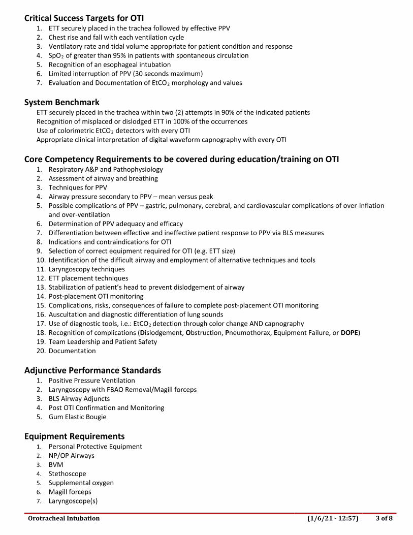
Critical Success Targets for OTI 1. ETT securely placed in the trachea followed by effective PPV 2. Chest rise and fall with each ventilation cycle 3. Ventilatory rate and tidal volume appropriate for patient condition and response 4. SpO2 of greater than 95% in patients with spontaneous circulation 5. Recognition of an esophageal intubation 6. Limited interruption of PPV (30 seconds maximum) 7. Evaluation and Documentation of EtCO2 morphology and values
System Benchmark
ETT securely placed in the trachea within two (2) attempts in 90% of the indicated patients Recognition of misplaced or dislodged ETT in 100% of the occurrences Use of colorimetric EtCO2 detectors with every OTI Appropriate clinical interpretation of digital waveform capnography with every OTI
Core Competency Requirements to be covered during education/training on OTI 1. Respiratory A&P and Pathophysiology 2. Assessment of airway and breathing 3. Techniques for PPV 4. Airway pressure secondary to PPV – mean versus peak 5. Possible complications of PPV – gastric, pulmonary, cerebral, and cardiovascular complications of over-inflation
and over-ventilation 6. Determination of PPV adequacy and efficacy 7. Differentiation between effective and ineffective patient response to PPV via BLS measures 8. Indications and contraindications for OTI 9. Selection of correct equipment required for OTI (e.g. ETT size) 10. Identification of the difficult airway and employment of alternative techniques and tools 11. Laryngoscopy techniques 12. ETT placement techniques 13. Stabilization of patient’s head to prevent dislodgement of airway 14. Post-placement OTI monitoring 15. Complications, risks, consequences of failure to complete post-placement OTI monitoring 16. Auscultation and diagnostic differentiation of lung sounds 17. Use of diagnostic tools, i.e.: EtCO2 detection through color change AND capnography 18. Recognition of complications (Dislodgement, Obstruction, Pneumothorax, Equipment Failure, or DOPE) 19. Team Leadership and Patient Safety 20. Documentation
Adjunctive Performance Standards
1. Positive Pressure Ventilation 2. Laryngoscopy with FBAO Removal/Magill forceps 3. BLS Airway Adjuncts 4. Post OTI Confirmation and Monitoring 5. Gum Elastic Bougie
Equipment Requirements
1. Personal Protective Equipment 2. NP/OP Airways 3. BVM 4. Stethoscope 5. Supplemental oxygen 6. Magill forceps 7. Laryngoscope(s)

Orotracheal Intubation (1/6/21 - 12:57) 4 of 8
8. Laryngoscope Blades (multiple sizes) 9. Appropriate size ET tubes 10. Stylet(s) 11. Pulse oximeter 12. Waveform capnography 13. Suction device (both rigid and flexible catheters) 14. Cardiac monitor 15. Difficult Airway Kit (including just in time training aids)
Instructor Resource Materials
1. Prehospital Trauma Life Support 2. AHA CPR and BLS Provider Manual 3. AHA ACLS Provider Manual 4. AHA PALS Provider Manual 5. Current AHA Guidelines for Cardiopulmonary Resuscitation and Emergency Cardiovascular Care 6. NHTSA EMS Educational Instructor Guidelines for EMT and Paramedic

Orotracheal Intubation (1/6/21 - 12:57) 5 of 8
Orotracheal Intubation Validation
PERFORMANCE CRITERIA: 100% accuracy required on all items with an * Before performing orotracheal intubation, the paramedic must:
Points Score Performance Steps Additional Information 1 Take or verbalize body substance
isolation. Selection: gloves, goggles, mask, gown, booties, N95 PRN
1 Determine BLS airway management is inadequate for effective positive pressure ventilation (PPV) and confirm the need for Orotracheal Intubation (OTI). *
No or inadequate rise and fall of chest, no improvement in patient’s color or condition.
1 Recognize signs of a difficult airway and select, prepare, and employ the appropriate alternative tools and techniques (e.g., ET Introducing Stylet)*
• A difficult airway is defined as the presence of anatomic conditions which preclude direct visualization of the patient’s glottic opening.
• Signs of a difficult airway include, but are not limited to: 1. Airway edema 2. Arthritis or scoliosis of the spine 3. Significant overbite 4. Small mandible 5. Short neck 6. Morbid obesity 7. C-spine immobilization 8. Face or neck trauma
1 Correctly assemble all equipment required for OTI within 60 seconds*
ETT, stylet, laryngoscope with functioning bulb, Magill forceps, suction, suction catheters (flexible and rigid), 10 mL syringe, stethoscope, waveform capnography, pulse oximeter, BVM with manometer
1 Provide optimal ventilation and oxygenation to the patient while OTI equipment is prepared. *
1 Test the cuff of the ETT by inflating it with air, and ensuring that there is not a leak in the cuff*
While performing OTI, the paramedic must:
1 Consider having a team member apply cricoid pressure during intubation attempts.
Apply gentle pressure to the patient’s cricoid cartilage to occlude the esophagus and reduce the patient’s chances of aspirating gastric contents.
1 Properly position the patient for intubation. *
“Sniffing” position, if not a trauma patient
1 Visualize anatomical structures including the glottic opening (vocal cords) during direct laryngoscopy. *
1 Minimize oral trauma during laryngoscopy by utilizing correct technique. *
Do not use the patient’s teeth as a fulcrum.

Orotracheal Intubation (1/6/21 - 12:57) 6 of 8
1 Place the appropriately-sized ETT securely in the trachea at the correct depth within 30 seconds. *
• Adult women typically will take a 7.0 – 7.5 ETT; adult men will typically take 7.5 – 8.0 ETT.
• Appropriate depth is ½ -- 1 inch beyond the vocal cords, usually 22 – 23 cm marking at the teeth
1 Inflate the cuff with enough air to seal the trachea; estimating inflation pressure by palpation of the pilot balloon.*
Immediately after performing OTI, the paramedic must:
1 Immediately re-establish PPV with the appropriate rate, tidal volume and oxygen at 10 – 15 LPM following ETT placement. *
1 Confirm ETT is in the trachea * a. Direct visualization of the tube passing through the cords. b. IMMEDIATELY attach a colormetric device to confirm correct
placement of the ETT prior to use of waveform / digital capnography
c. If ETT placement is correct, then attach waveform capnography and commence assisted ventilations.
d. Confirm appropriate rectangular waveform is present AND capnography number is present and consistent with clinical condition.
e. Auscultate lung fields and epigastrium f. Observe for appropriate chest rise and fall. g. If only right lung sounds heard, carefully adjust ETT as
necessary by slowly withdrawing ETT and listening for onset of left lung sounds. Once bilateral lung sounds occur, secure the tube.
h. Remove ETT immediately if esophageal placement suspected. 1 Immediately re-establish PPV at
the clinically required rate and tidal volume (minute volume) and oxygen at 10 – 15 LPM following ETT placement. *
• Passing the laryngoscope past the teeth with the intent of placing an ETT is considered an intubation attempt.
1 Secure the ETT in the trachea at the correct depth with tape or a commercial device. *
1 Re-implement effective PPV within 10 seconds following unsuccessful OTI attempts.
• Passing the laryngoscope past the teeth with the intent of placing an ETT is considered an intubation attempt.
1 Stabilize the patient’s airway and prevent tube migration by using a device to prevent rotation, flexion, or extension of patient’s head.
1 Efficiently employ post-OTI diagnostic tools to thoroughly assess overall effectiveness of ventilatory support throughout the duration of respiratory management efforts. *
• Symmetrical rise and fall of the chest with PPV • Monitor pulse oximetry – the target SpO2 is greater than or
equal to 95% if spontaneous circulation is present o In patients with COPD/pulmonary disease, it may not be
possible or desirable to attain a SpO2 of 95%. • Monitor EtCO2 for appropriate waveform morphology and
target CO2 levels.

Orotracheal Intubation (1/6/21 - 12:57) 7 of 8
o The target range for EtCO2 level is between 30 – 45 mmHg if spontaneous circulation is present.
o In cardiac arrest, metabolic derangement will significantly alter EtCO2 values and waveform morphology. Target range for EtCO2 levels is between 15 mmHg – 45 mmHg during CPR.
o Recognize that in a patient with traumatic brain injury, EtCO2 less than 35 mmHg due to hyperventilation may actually cause harm. Minute volume should be adjusted accordingly while maintaining optimal oxygenation, reserving hyperventilation for those patients showing signs of cerebral herniation only.
• Monitor ECG for dysrhythmia due to vagal simulation or other treatable causes.
• Frequent auscultation of lung fields and epigastrium. • Constant evaluation of ventilator compliance and resistance
during PPV. 1 Immediately identify
malfunctioning equipment, ineffective techniques, or changes in post-OTI PPV compliance/resistance and employ alternative measures to achieve effective ventilations. *
1 Reconfirm correct ETT placement each time the patient is moved and before transfer of care to hospital staff. *
Record and print the waveform EtCO2 strip prior to transfer of care to the hospital staff and attach the recording strip to the completed PCR.
1 Provide direction to personnel that have been delegated management of post-OTI PPV. *
1 Maintain effective ventilation and oxygenation throughout the entire pre-hospital treatment period. *
Target SpO2is greater than 95%; target EtCO2 is 30 – 45 mmHg in a patient with spontaneous circulation.
1 Maintains calm. Effective in leading a team-based approach to resuscitation in all conditions. *
1 Accurately document all assessment findings, therapeutic treatments, and the patient’s response to therapy.
Critical Failure Criteria ____Failure to take or verbalize BSI appropriate to the skill prior to performing the skill ____Failure to initiate ventilations within 30 seconds after applying gloves or interrupts ventilations for greater than 30 seconds ____Failure to ventilate patient at a rate appropriate to patient age ____Failure to provide adequate tidal volume per breath ____Failure to pre-oxygenate patient prior to intubation attempt ____Failure to successfully intubate within 2 attempts ____Failure to disconnect syringe immediately after inflating cuff of ET tube ____Uses teeth as a fulcrum ____Failure to assure proper tube placement by auscultation over lung fields and epigastrium

Orotracheal Intubation (1/6/21 - 12:57) 8 of 8
____Failure to use either a colorimetric end tidal CO2 cap or waveform capnography ____If used, stylet extends beyond end of tube ____Failure to recognize an esophageal intubation ____Failure to re-check tube placement after each patient movement and before transfer of care to hospital staff ____Any procedure that would have harmed the patient

Skill Verification Form Effective
April 1, 2021 Expires
March 31, 2022 Category I Skill – Low Frequency/High Risk:
ET Introducing Stylet Approval: Medical Director
Reza Vaezazizi, MD Signed
Applies To:
EMT-P, MICN, BHP, EMS System Approval: EMS Administrator
Trevor Douville Signed
ET Introducing Stylet (1/6/21 - 13:01) 1 of 1
PURPOSE This skill verification standard is supplemental to the skill verification standard for adult orotracheal intubation (OTI). Terminal Performance Objective To assist with the secure placement of an endotracheal tube (ETT) in the trachea to ensure a patent airway for positive pressure ventilation (PPV). Before using the ET introducing stylet to assist with intubation, paramedics must:
1. Determine BLS airway management is inadequate for effective PPV and confirm the need for orotracheal intubation (OTI).1 2
2. Recognize signs of a difficult airway characterized by the presence of anatomic conditions which preclude direct visualization of the patient’s glottic opening (i.e., airway edema, arthritis, scoliosis of the spine, significant overbite, small mandible, short neck, morbid obesity, cervical spine immobilization, face or neck trauma)
3. Correctly assemble all equipment required for OTI within 60 seconds. 4. Provide optimal ventilation and oxygenation (minute volume) to the patient while OTI equipment is prepared.
Complications Complications of the ET introducing stylet may include tracheal/esophageal perforation, hemopneumothorax, mediastinal emphysema and / or right-sided pneumothorax
Procedure
1. Consider having a team member apply cricoid pressure while attempting intubation. 2. Perform laryngoscopy as per OTI skill standards and obtain the best possible laryngeal view. 3. While holding the ET introducing stylet in one hand, ensure the angled tip is pointing upward then gently advance
anteriorly (under the epiglottis) to the glottic opening (cords). a. If able to visualize the vocal cords, direct the ET introducing stylet through the cords. b. If unable to visualize the vocal cords, direct the ET introducing stylet to the anatomical area where the
cords should be and feel for a “washboard” sensation as the stylet tip ratchets on the tracheal rings. 4. Gently advance the device until resistance is encountered (at the carina).
a. NEVER force the stylet, as pharyngeal/tracheal perforation can occur. b. If no resistance is encountered and the entire length of the stylet is inserted, the device is in the
esophagus. 5. The stylet is correctly placed when the device can be seen going through the cords, when ratcheting of the tip on
the tracheal rings is felt, and/or when resistance is met after advancing (stylet is at the carina). a. When using a marked stylet, withdraw the stylet back until the black line (or other such mark) is at the
lips prior to advancing the ET tube, indicating distal tip is beyond vocal cords and proximal end has enough length to slide ET tube over it.
6. Once the stylet is positioned, advance the ET tube over the stylet and into the trachea. 7. If resistance is encountered, withdraw the ET tube slightly, rotate 90 degrees and re-attempt. If unsuccessful,
attempt with a smaller tube 8. Once the ET tube is placed, maintain tube placement while removing introducer stylet.
1 2010 AHA Guidelines for CPR and ECC, Part 8 Adult Advanced Cardiovascular Life Support, pp S730-S735 2 PHTLS , Seventh Edition, Chapter 7 Airway and Ventilation pp 144-145

Post-OTI Confirmation and Monitoring (1/6/21 - 12:58) 1 of 5
Skill Verification Form Effective
April 1, 2021 Expires
March 31, 2022 Category I Skill – Low Frequency/High Risk:
Post-OTI Confirmation and Monitoring Approval: Medical Director
Reza Vaezazizi, MD Signed
Applies To: EMT-P, MICN, BHP, EMS System
Approval: EMS Administrator Trevor Douville
Signed
Terminal Performance Objective Secure placement of an endotracheal tube (ETT) in the trachea to ensure a patent open airway allowing effective positive pressure ventilation throughout the entire prehospital period of care.
After placement, paramedics must continuously maintain the ETT within the trachea by performing all of the following:
1. Verify symmetrical rise and fall of the chest with ongoing PPV. 2. Auscultate all lung fields to confirm the presence of airflow to the lower airway during PPV. 3. Auscultate over the epigastrium to confirm the absence of airflow to the stomach during PPV. 4. Monitor EtCO2 for appropriate waveform morphology and target CO2 levels:
a. The target range for EtCO2 level is between 30 – 45 mmHg if spontaneous circulation is present. b. In cardiac arrest, metabolic derangements will significantly alter EtCO2 values and waveform
morphology. Target range for EtCO2 levels is between 15 mmHg – 45 mmHg during CPR. c. Recognize that in a patient with traumatic brain injury, EtCO2 less than 35 mmHg due to hyperventilation
may actually cause harm. Minute volume should be adjusted accordingly while maintaining optimal oxygenation, reserving hyperventilation for those patients showing signs of cerebral herniation only.1
d. If the waveform capnography monitor malfunctions, a colorimetric end tidal CO2 detector shall be used, and the malfunction reported to the organization’s QI Coordinator.
5. Directly visualize ETT placement with laryngoscope blade to ensure tracheal placement as clinically indicated. 6. Utilize pulse oximetry to evaluate for adequate O2 saturation during PPV.
a. Target range is a SpO2 greater than 95% if spontaneous circulation is present. i. In patients with COPD/pulmonary disease, it may not be possible or desirable to attain a SpO2 of
95%. 7. Confirm the absence of gastric contents in the ETT during PPV. 8. Ensure the ETT remains inserted at the correct depth within the trachea during PPV. 9. Stabilize patient’s airway and prevent tube migration by using a device to prevent rotation, flexion, or extension
of patient’s head. 10. Continuously monitor and re-verify tube placement after each and EVERY move, looking for signs of tube
dislodgement and migration out of the trachea. 11. Rapidly identify 100% of the occurrences when an ETT is misplaced or an ETT has migrated out of the trachea
after placement. 12. Once identified as misplaced, or if there is significant doubt of the tube’s placement, remove the tube at once
and provide PPV. 13. Re-intubate or consider insertion of a Rescue Airway if unable to control the airway with BLS adjuncts. 14. Record and print the waveform EtCO2 strip following initial placement, after every patient movement and prior
to transfer of care to the hospital staff and with any change in patient condition. Attach the recording strips to the completed PCR, alternatively clinicians may import the capnograms and electronically attach the images to the ePCR. A procedure to EtCO2 monitoring MUST be created for each capnogram, and capnogram(s) MUST be attached to the ePCR.
1 The Brain Trauma Foundation’s Guidelines for Prehospital Management of Severe Traumatic Brain Injury, Second Edition

Post-OTI Confirmation and Monitoring (1/6/21 - 12:58) 2 of 5
Critical Success Targets for Orotracheal Intubation (OTI) 1. ETT securely placed in the trachea followed by effective PPV 2. Chest rise and fall with each ventilation cycle 3. SpO2 greater than 95% 4. Limited interruption of PPV (30 seconds maximum) 5. Evaluation and Documentation of EtCO2 morphology and values
System Benchmark
ETT securely placed in the trachea within 2 attempts in 90% of the indicated patients. 100% of the misplaced or dislodged ET tubes are identified and corrected. 100% documentation of clinically required waveform capnography procedures 100% attachment of clinically required capnograms to ePCR
Core Competency Requirements to be covered during education/training on post-OTI confirmation and monitoring
1. Rapid assessment of endotracheal tube placement 2. Use of primary verification methods 3. Use of secondary verification methods 4. Positive pressure ventilation 5. Appropriate re-assessment of tube placement after each move 6. Rapid recognition of a misplaced tube 7. Removal of a misplaced tube 8. Alternate techniques for advanced airway management 9. Dislodgement, Occlusion, Pneumothorax, Equipment Failure (DOPE) 10. Consequences, risks and complications of failure to complete post OTI confirmation and monitoring
Adjunctive Performance Standards
1. ALS Airways 2. Positive Pressure Ventilation (PPV)
Equipment Requirements
1. Personal Protective Equipment 2. NP/OP Airways 3. BVM 4. Stethoscope 5. Supplemental oxygen 6. Magill forceps 7. Laryngoscope(s) 8. Laryngoscope blades (multiple sizes) 9. Appropriate-sized ET tubes 10. Stylet(s) 11. Pulse oximeter 12. Waveform capnography 13. Suction device (both rigid and flexible catheters) 14. Cardiac monitor 15. Difficult Airway Kit/Rescue Airway Kit (including just in time training aids)

Post-OTI Confirmation and Monitoring (1/6/21 - 12:58) 3 of 5
Instructor Resource Materials 1. Prehospital Trauma Life Support, Sixth Edition 2. AHA ACLS Provider Manual 3. AHA PALS Provider Manual 4. Current AHA Guidelines for Cardiopulmonary Resuscitation and Emergency Cardiovascular Care 5. NHTSA EMS Educational Instructor Guidelines for EMT and Paramedic

Post-OTI Confirmation and Monitoring (1/6/21 - 12:58) 4 of 5
Post-Orotracheal Intubation Confirmation and Monitoring Validation
PERFORMANCE CRITERIA: 100% accuracy required on all items marked with an *
Points Score Performance Steps Additional Information
1 Take or verbalize body substance isolation.
Selection: gloves, goggles, mask, gown, booties, P100 PRN
1 Verify symmetrical rise and fall of the chest with ongoing PPV. *
1 Auscultate all lung fields to confirm the presence of airflow to the lower airway during PPV. *
1 Auscultate over the epigastrium to confirm the absence of airflow to the stomach during PPV. *
1 Utilize waveform capnography to evaluate for the presence of end tidal carbon dioxide (EtCO2) during PPV. *
Monitor EtCO2 for appropriate waveform morphology and target CO2 levels.
• The target range for EtCO2 level is between 30 – 45 mmHg if spontaneous circulation is present.
• In cardiac arrest, metabolic derangement will significantly alter EtCO2 values and waveform morphology. Target range for EtCO2 levels is between 15 mmHg – 45 mmHg during CPR.
• Recognize that in a patient with traumatic brain injury, EtCO2 less than 35 mmHg due to hyperventilation may actually cause harm. Minute volume should be adjusted accordingly while maintaining optimal oxygenation, reserving hyperventilation for those patients showing signs of cerebral herniation only.
• If the waveform capnography monitor malfunctions, a colorimetric end tidal CO2 detector shall be used, and the malfunction reported to the organization’s QI Coordinator.
1
Utilize pulse oximetry to evaluate for adequate O2 saturation readings during PPV. *
Target range is a SpO2 greater than 95% if spontaneous circulation is present.
• In patient with COPD/pulmonary disease, it may not be possible or desirable to attain a SpO2 of 95%.
1 Confirm the presence of misting of the ETT during PPV. *
1 Confirm the absence of gastric contents in the ETT during PPV. *
1 Ensure the ETT remains inserted at the correct depth within the trachea during PPV. *
1 Stabilize the patient’s head to avoid movement and possible ETT dislodgement during PPV.
Stabilize the patient’s airway and prevent tube migration by using a device to prevent rotation, flexion, or extension of patient’s head.

Post-OTI Confirmation and Monitoring (1/6/21 - 12:58) 5 of 5
1 Continuously monitor and re- verify tube placement after each and EVERY move, looking for signs of tube dislodgement and migration out of the trachea. *
a. After moving the patient from the scene to the back board.
b. After moving the patient into the gurney. c. When the patient is loaded into the ambulance. d. After any significant bumps en route to the hospital. e. When the patient is off-loaded from the ambulance. f. When the patient is moved from the ambulance gurney to
the hospital gurney. g. Any time there is any concern that the tube might have
become displaced. 1 Rapidly identify 100% of the
occurrences when an ETT is misplaced or an ETT has migrated out of the trachea after placement. *
1 Once identified as misplaced or if there is significant doubt of the tube’s placement, remove the tube at once verbalize the need to manage the airway using BLS management techniques *
1 Maintain calm and effectively lead a team-based approach to resuscitation under all conditions. *
1 Accurately document all assessment findings, therapeutic treatments, and the patient’s response to therapy.
Record and print the waveform EtCO2 strip following initial placement and prior to transfer of care to the hospital staff. Attach the recording strips to the completed PCR.
Critical Failure Criteria Failure to take or verbalize BSI appropriate to the skill prior to performing the skill Failure to recognize an esophageal intubation Failure to recognize a tube migrated out of the trachea Failure to re-check the tube placement following each movement Failure to immediately begin PPV following a missed or dislodged ETT Interruption of PPV for greater than 30 seconds maximum Failure to auscultate over all lung fields and epigastrium immediately following intubation Any procedure that would have harmed the patient

Skill Verification Form Effective
April 1, 2021 Expires
March 31, 2022 Category I Skill – Low Frequency/High Risk:
Positive Pressure Ventilation Approval: Medical Director
Reza Vaezazizi, MD Signed
Applies To: PSP*, EMT, AEMT, EMT-P, MICN, BHP, EMS
System
Approval: EMS Administrator Trevor Douville
Signed
Positive Pressure Ventilation (1/6/21 - 12:58) 1 of 6
Terminal Performance Objective To establish and maintain adequate airflow (oxygenation and ventilation) to support gas exchange at the cellular level and prevent or reverse tissue hypoxia. Before performing positive pressure ventilation, the PSP* EMT, AEMT, or paramedic must:
*PSP Providers, PPV is an optional skill and requires REMSA authorization for performance 1. Methodically complete an assessment of the airway and breathing within 30 seconds. 2. Identify inadequate ventilations (minute volume) and/or signs of hypoxia within the first 30 seconds. 3. Apply appropriate, clinically-required technique to manually position the head and mandible of the unconscious
patient to open the upper airway. a. Medical – Head tilt/chin lift b. Trauma – Jaw thrust or modified chin lift.
4. Clear secretions or other obstructions using appropriate method (manually, log rolling, suctioning, etc.) maintaining spinal stabilization controls as patient’s condition indicates.
a. Utilize the two rescuer BVM application technique to maintain an open airway wherever possible. 5. Utilize the appropriate technique per American Heart Association Standards to insert the appropriate BLS airway
within 10 seconds. a. NPA is the preferred BLS airway
6. All ALS Provider agencies MUST have waveform capnography attached to the BVM. While performing positive pressure ventilation, the PSP, EMT, AEMT, or paramedic must:
1. Employ the correct technique to achieve a tight mask seal while maintaining position of the head and mandible to maximize airflow to the lower airway.
a. Utilize the two rescuer BVM application technique to maintain an open airway wherever possible. 2. Initiate ventilatory support using an appropriately-sized BVM with supplemental oxygen at 10 – 15 LPM. 3. Provide the clinically-required ventilatory (minute volume) support for the patient demonstrating the ability to
modify tidal volume and/or ventilation rate to achieve chest/diaphragm expansion and full exhalation with each ventilatory cycle.
4. Ventilate patients with spontaneous circulation (Rescue Breathing) as clinically required : a. Ventilate the adult patient once every 5 to 6 seconds (10 – 12 times per minute) with tidal volume (TV)
sufficient to produce visible chest rise and fall. 1 2 b. Ventilate the pediatric patient once every 3 to 5 seconds (12 – 20 times per minute) with tidal volume
sufficient to produce visible chest rise and fall 3 without hyperinflation or gastric insufflation. c. Ventilate neonatal patients 40-60 times per minute to maintain a heart rate greater than 100. 4
5. Ventilate cardiac arrest patients during CPR as clinically required: a. Ventilate the adult patient without an advanced airway – synchronize 2 ventilations with 30 chest
compressions. Provide ventilations with enough tidal volume to produce visible chest rise and fall during pauses in compression cycles (Class IIa).
1 2015 AHA Guidelines for CPR and ECC, Part 5 Adult Basic Life Support, pp S692-S693. 2 PHTLS, Seventh Edition, Chapter 7 Airway and Ventilation p 164. 3 2015 AHA Guidelines for CPR and ECC, Part 13, Pediatric Basic Life Support, p S868. 4 2015 AHA Guidelines for CPR and ECC, Part 15, Neonatal Resuscitation, p S912.

Positive Pressure Ventilation (1/6/21 - 12:58) 2 of 6
b. Ventilate the adult patient with an advanced airway in place – Provide 8-10 unsynchronized ventilations per minute with enough tidal volume to produce visible chest rise and fall during pauses in compression cycles (Class IIa).
6. Deliver positive pressure ventilations over a minimum of 1 second to avoid hyperinflation and minimize gastric insufflation, high (peak) airway pressures, pulmonary barotrauma and compromise of venous return to the heart (Class IIa).
7. Avoid hyperventilation to minimize high airway pressures, hypocarbia and cerebral vasoconstriction. 5 6 a. Monitor EtCO2 for appropriate waveform morphology and CO2 levels in the non-intubated, intubated
patient/patient with a Rescue Airway: i. The target range for EtCO2 level is between 30 – 45 mmHg if spontaneous circulation is present. ii. In cardiac arrest, metabolic derangements will significantly alter EtCO2 values and waveform
morphology. Target range for EtCO2 levels is between 15 mmHg – 45 mmHg during CPR. iii. Recognize that in a patient with traumatic brain injury, EtCO2 less than 35 mmHg due to
hyperventilation may actually cause harm. Minute volume should be adjusted accordingly while maintaining optimal oxygenation, reserving hyperventilation for those patients showing signs of cerebral herniation only. 7
iv. If the waveform capnography monitor malfunctions, a colorimetric end tidal CO2 detector shall be used, and the malfunction reported to the organization’s QI Coordinator.
8. Differentiate respiratory pathophysiology and modify BVM technique based upon changes in lung compliance and/or airway resistance to maintain therapeutic airway pressure while minimizing gastric insufflations. 8
a. Ensure changes in inspiratory and expiratory time ratio (I:E Ratio) is factored into ventilatory cycles allowing for exhalation of each breath prior to delivery of the next breath.
9. Efficiently employ diagnostic tools such as pulse oximetry (target SpO2 is greater than 95% when spontaneous circulation is present) and auscultation of lung fields to thoroughly assess overall effectiveness of ventilatory support.
a. In patients with COPD/pulmonary disease, it may not be possible or desirable to attain a SpO2 of 95%. 10. Immediately identify malfunctioning equipment or ineffective techniques and employ alternative measures to
achieve effective ventilations. 11. Maintain effective ventilation and oxygenation throughout the entire prehospital period of treatment. 12. Maintain calm and effectively lead a team-based approach to resuscitation. 13. Rapidly determine the need for Advanced Life Support (ALS) airway adjuncts and/or medications when airway
patency or ventilations cannot be effectively supported by BLS means. 14. Document all procedures and patient response to therapy on the PCR.
Critical Success Targets for PPV
1. Tight mask seal 2. Chest rise and fall with each ventilation cycle 3. SpO2 of greater than 95% in patients with spontaneous circulation. 4. Management of secretions and other airway obstructions 5. Minimal gastric distension 6. Evaluation and Documentation of EtCO2 morphology and values
System Benchmark
Number of patients PPV with chest rise and fall, patent airway, signs of adequate oxygenation. Patient arrival at hospital with spontaneous circulation.
5 PHTLS , Seventh Edition, Chapter 7 Airway and Ventilation p 164. 6 2010 AHA Guidelines for CPR and ECC, Part 9 Post–Cardiac Arrest Care, p S773. 7 The Brain Trauma Foundation’s Guidelines for Prehospital Management of Severe Traumatic Brain Injury, Second Edition, Sections IV and VI 8 2010 AHA Guidelines for CPR and ECC, Part 13, Pediatric Basic Life Support, p S868.

Positive Pressure Ventilation (1/6/21 - 12:58) 3 of 6
Core Competency Requirements to be covered during education/training on PPV
1. Respiratory A&P and Pathophysiology 2. Assessment of airway and breathing 3. Differentiation between adequate and inadequate respiration 4. Airway Positioning 5. Suctioning 6. Removal of foreign body obstructions 7. Oropharyngeal Airway selection and insertion 8. Nasopharyngeal Airway selection and insertion 9. BVM selection 10. Induction of supplemental oxygen 11. Hand ventilation with a BVM 12. Assessment of PPV adequacy and efficacy 13. Application and use of waveform capnography with PPV procedure 14. Airway pressure secondary to PPV – mean versus peak 15. Possible complications of PPV – gastric, pulmonary, cerebral, and cardiovascular complications of over-inflation
and over-ventilation 16. Auscultation and diagnostic differentiation of lung sounds 17. Team Leadership and Patient Safety 18. Use of diagnostic tools
Adjunctive Performance Standards
1. Laryngoscopy with FBAO Removal/Magill Forceps (ALS personnel) 2. BLS Airway Adjuncts 3. ALS Airways
Equipment Requirements
1. Mannequin 2. NP Airway 3. OP airway 4. Advanced Airways 5. BVM with manometer 6. Stethoscope 7. Supplemental oxygen 8. Magill forceps 9. Laryngoscope 10. Pulse oximeter 11. Waveform capnography (required for ALS units and CCT units only) 12. Suction device (both hard and flexible)
Instructor Resource Materials
1. Prehospital Trauma Life Support 2. AHA CPR and BLS Provider Manual 3. AHA ACLS Provider Manual 4. AHA PALS Provider Manual 5. Current AHA Guidelines for Cardiopulmonary Resuscitation and Emergency Cardiovascular Care 6. NHTSA EMS Educational Instructor Guidelines for EMT and Paramedic

Positive Pressure Ventilation (1/6/21 - 12:58) 4 of 6
Positive Pressure Ventilation Validation PERFORMANCE CRITERIA: 100% accuracy required on all items with an * Before performing positive pressure ventilation, the PSP*, EMT, AEMT, and paramedic must:
Points Score Performance Steps Additional Information 1 Take or verbalize body substance
isolation. Selection: gloves, goggles, mask, gown, booties, N95 PRN
1 Methodically complete an assessment of the airway and breathing within 30 seconds. *
Follow respiratory assessment sequence.
1 Identify inadequate ventilations and/or signs of hypoxia within the first 30 seconds. *
Pale/cyanotic, altered level of consciousness, diaphoresis, increased work of breathing or apnea, poor chest rise and fall
1 Apply the appropriate, clinically required technique to manually position the head and mandible of the unconscious patient to open the upper airway. *
• Medical – Head tilt/chin lift • Trauma – Jaw trust or modified chin lift
- Utilize the two rescuer BVM technique to maintain an
open airway wherever possible. 1 Manually clear blood, vomit, and
foreign bodies when present. * • Clear secretions or other obstruction using appropriate
method (manually, log rolling, suctioning, etc.) maintaining spinal stabilization control as patient condition indicates.
• Use a rigid pharyngeal tip, if available, for suctioning oropharynx.
1 Utilize the appropriate technique per American Heart Association standards to insert the appropriate BLS airway within 10 seconds. *
NPA is the preferred BLS airway
While performing positive pressure ventilation, the PSP*, EMT, AEMT, and paramedic must:
1 Employ the correct technique to achieve a tight mask seal while maintaining position of the head and mandible to maximize airflow to the lower airway. *
• E –C Clamp technique (one rescuer) • Two rescuers is best for achieving a tight mask seal, with
one rescuer holding the mask against the patient’s face while maintaining head position, and the 2nd rescuer squeezing the bag.
1 Initiate ventilatory support using an appropriately sized BVM with supplemental oxygen at 10 – 15 LPM. *
1 Provide the clinically required ventilatory (minute volume) support for the patient demonstrating the ability to modify tidal volume and/or ventilation rate to achieve chest/diaphragm expansion and full exhalation with each ventilation cycle. *
• Give sufficient volume to cause chest rise.

Positive Pressure Ventilation (1/6/21 - 12:58) 5 of 6
1 Ventilate patients with spontaneous circulation (Rescue Breathing) as clinically required. *
• Ventilate the adult patient once every 5 to 6 seconds (10 - 12 times per minute) with tidal volume (TV) sufficient to produce visible chest rise and fall.
• Ventilate the pediatric patient once every 3 to 5 seconds (12 – 20 times per minute) with tidal volume sufficient to produce visible chest rise and fall without hyperinflation or gastric insufflations.
• Ventilate neonatal patients 40 – 60 times per minute to maintain a heart rate greater than 100.
1 Ventilate cardiac arrest patients during CPR as clinically required. *
• Ventilate the adult patient without an advanced airway-synchronize 2 ventilations with 30 chest compressions. Provide ventilations with enough tidal volume to produce visible chest rise and fall during pauses in compression cycles (Class IIa).
• Ventilate the adult patient with an advanced airway in place-provide 8 – 10 unsynchronized ventilations per minute with tidal volume sufficient to achieve rise and fall of the chest (Class IIa).
1 Deliver positive pressure ventilations over a minimum of 1 second. *
This is to avoid hyperinflation and minimize gastric insufflations, high (peak) airway pressures, pulmonary barotraumas and compromise of venous return to the heart (Class IIa).
1 Avoid hyperventilation to minimize high airway pressures, hypocarbia, and cerebral vasoconstriction. *
Monitor EtCO2 for appropriate waveform morphology and target CO2 levels in the intubated patient/patient with a Rescue Airway:
• The target range for EtCO2 level is between 30 – 45 mmHg if spontaneous circulation is present.
• In cardiac arrest, metabolic derangement will significantly alter EtCO2 values and waveform morphology. Target ranges for EtCO2 levels are between 15 mmHg – 45 mmHg during CPR.
• Recognize that in a patient with traumatic brain injury, EtCO2 less than 35 mmHg due to hyperventilation may actually cause harm. Minute volume should be adjusted accordingly while maintaining optimal oxygenation, reserving hyperventilation for those patients showing signs of cerebral herniation only.
• If the waveform capnography monitor malfunctions, a colorimetric end tidal CO2 detector shall be used, and the malfunction reported to the organization’s QI Coordinator.
1 Differentiate respiratory pathophysiology and modify BVM technique based upon changes in lung compliance and/or airway resistance to maintain therapeutic airway pressure while minimizing gastric insufflation. *
Ensure changes in inspiratory and expiratory time ratio (I: E ratio) is factored into ventilatory cycles allowing for exhalation of each breath prior to delivery of the next breath.
1 Efficiently employ diagnostic tools such as pulse oximetry and auscultation of appropriate lung fields to thoroughly assess overall effectiveness of ventilatory support. *
SpO2 target is greater than 95% when spontaneous circulation is present.
• In patients with COPD/pulmonary disease, it may not be possible or desirable to attain a SpO2 of 95%.

Positive Pressure Ventilation (1/6/21 - 12:58) 6 of 6
1 Immediately identify malfunctioning equipment or ineffective techniques and employ alternative measures to achieve effective ventilations. *
1 Maintain effective ventilation and oxygenation throughout the entire pre-hospital period of treatment. *
1 Maintain calm and effectively lead a team based approach to resuscitation. *
1 Rapidly determine the need for Advanced Life Support (ALS) airway adjuncts and/or medications when airway patency or ventilations cannot be effectively supported by BLS means.*
1 Document all procedures and patient response to therapy on Patient Care Report (PCR).
Critical Failure Criteria ____Failure to take or verbalize BSI appropriate to the skill prior to performing the skill ____Failure to achieve and maintain a tight mask seal ____Failure to properly identify ineffective ventilations ____Any procedure that would have harmed the patient

Skill Verification Form Effective
April 1, 2021 Expires
March 31, 2022 Category I Skill – Low Frequency/High Risk:
Defibrillation Approval: Medical Director
Reza Vaezazizi, MD Signed
Applies To:
EMT-P, MICN, BHP, EMS System Approval: EMS Administrator
Trevor Douville Signed
Defibrillation (1/6/21 - 12:55) 1 of 4
Terminal Performance Objective Termination of pulseless ventricular tachycardia or ventricular fibrillation using electrical therapy. Before performing defibrillation, paramedics must:
1. Determine pulselessness within ten (10) seconds 2. Follow applicable REMSA protocols for CPR and resuscitation 3. Confirm that the patient is in a shockable rhythm (pulseless ventricular tachycardia or ventricular fibrillation)
a. Confirm ECG monitor leads have been placed appropriately 4. Explain to patient’s family what they can expect to see while avoiding delays in treatment.
While performing defibrillation, paramedics must:
1. Select and prepare the appropriate sites for application of the ECG monitor/defibrillator multifunction pads (MFPs).
a. Proper pad placement on the patient’s cleaned, dry skin is essential to maximize current conduction; the better the contact, the more effective attempts at defibrillation will be
2. Apply the MFPs firmly to the patient’s clean, bare skin in the correct anatomical locations for maximum electrical current flow through the heart
3. Identify a patient with a pacemaker or automatic internal cardiac defibrillator (AICD) and place the MFP(s) in alternate position(s) to minimize damage to the device(s) and to avoid disruption of current flow through the heart
4. Ensure that the ECG monitor/defibrillator is not in the synchronize mode 5. Select the correct energy setting on the ECG monitor/defibrillator
a. Per the REMSA Treatment Protocol: Calculation Chart 6. Assure everyone is clear from the patient and all possible energy conducting surfaces/contacts 7. Discharge the defibrillator 8. Immediately resume chest compressions for two (2) minutes without checking for a pulse 9. Treat subsequent rhythms per REMSA protocols
Critical Success Targets for Defibrillation
1. Successful delivery of electrical current at the proper setting to the heart of a patient in pulseless ventricular tachycardia or ventricular fibrillation
2. Safe and proficient use of the ECG monitor/defibrillator including lead and MFP placement System Benchmark % of patients receiving defibrillation with restoration of a stable perfusing rhythm Core Competency Requirements to be covered during education/training on defibrillation
1. Cardiovascular A & P 2. Cardiology – pathophysiology of malignant ventricular dysrhythmias 3. Assessment of circulation and recognition of hemodynamic instability 4. ECG rhythm interpretation – ventricular tachycardia and ventricular fibrillation 5. Indications and contraindications for defibrillation 6. Proper placement of ECG electrodes on patient

Defibrillation (1/6/21 - 12:55) 2 of 4
7. Proper placement of multifunction pads on patient 8. Knowledge of the monitor/defibrillator 9. Joules used in each defibrillation 10. Methods to help eliminate impedance from chest walls 11. Early defibrillation in pulseless ventricular tachycardia and ventricular fibrillation 12. Defibrillation with a implantable cardioverter defibrillator
Adjunctive Performance Standards Currently no adjunctive performance standards have been created Equipment Requirements
1. Monitor/defibrillator 2. ECG electrodes 3. Multifunction pads 4. Razor 5. 4 x 4 gauze pads 6. Alcohol preps
Instructor Resource Materials
1. REMSA Protocol Manual a. REMSA Calculation Chart
2. ECG monitor/defibrillator manufacturer guidelines 3. Paramedic Care Principles & Practice, Third Edition, Bledsoe, Porter and Cherry 4. 2010 American Heart Association Guidelines for ECC and CPR

Defibrillation (1/6/21 - 12:55) 3 of 4
Defibrillation Validation PERFORMANCE CRITERIA: 100% accuracy required on all items marked with an *
Before defibrillating, paramedics must: Pts Score Performance Steps Additional Information 1 Take or verbalize body substance
isolation Selection: gloves, goggles, mask, gown, booties, P100 PRN
1 Determine pulselessness within 10 seconds *
1 Follow applicable REMSA protocols for CPR and resuscitation
1 Confirm that the patient is in a shockable rhythm (pulseless ventricular tachycardia or ventricular fibrillation) *
Confirm ECG monitor leads have been placed appropriately.
1 Confirm the ECG monitor leads have been placed appropriately *
1 Explain to patient’s family what they can expect to see while avoiding delays in treatment
While defibrillating, paramedics must: Pts Score Performance Steps Additional Information 1 Apply the ECG
monitor/defibrillator MFPs firmly to the patient’s clean, bare skin in the correct anatomical locations for maximum electrical current flow through the heart *
• Anterior-posterior placement is recommended, if possible• Proper pad placement on the patient’s cleaned, dry skin is
essential to maximizing conduction; the better the contact,the more effective attempts at defibrillation will be
1 Identify a patient with a pacemaker or automatic internal cardiac defibrillator (AICD) and place the MFP(s) in alternate position(s) to minimize damage to the device and to avoid disruption of current flow through the heart *
1 Ensure that the ECG monitor/defibrillator is not in the synchronize mode *
1 Select the correct energy setting on the ECG monitor/defibrillator *
Per the REMSA Treatment Protocol: Calculation Chart
1 Assure everyone is clear from the patient and all possible energy conducting surfaces/contacts *
1 Discharge the defibrillator for unsynchronized delivery of electrical current *

Defibrillation (1/6/21 - 12:55) 4 of 4
1 Immediately resume chest compressions for 2 minutes without checking for a pulse *
1 Maintain calm and effectively lead a team-based approach to resuscitation under all conditions *
1 Provide treatment based upon reassessment findings
1 Accurately document all assessment findings, therapeutic treatments, and the patient’s response to therapy *
Critical Failure Criteria ____Failure to take or verbalize BSI appropriate to the skill prior to performing the skill. ____Failure to identify indications for procedure. ____Failure to ensure the functionality of cardiac monitor and availability of equipment. ____Failure to assure that everyone is clear prior to discharging the defibrillator. ____Failure to immediately begin chest compressions following defibrillation without checking for a pulse. ____Any procedure that would have harmed the patient.

Skill Verification Form Effective
April 1, 2021Expires
March 31, 2022 Category I Skill – Low Frequency/High Risk:
Synchronized Cardioversion Approval: Medical Director
Reza Vaezazizi, MD Signed
Applies To: EMT-P, MICN, BHP, EMS System
Approval: EMS Administrator Trevor Douville
Signed
Synchronized Cardioversion (1/6/21 - 12:58) 1 of 4
Terminal Performance Objective Termination of hemodynamically significant tachycardia resulting in restoration of adequate cardiac output and tissue perfusion.
Before performing synchronized cardioversion, paramedics must: 1. Methodically assess the patient’s ABC’s within 30 seconds.2. Determine the patient is hemodynamically unstable due to idiopathic (non-compensatory) tachycardia and is a
candidate for immediate cardioversion:a. Confirm the patient is exhibiting signs and symptoms of systemic poor perfusion (including but not
limited to hypotension, altered mental status, chest pain, dyspnea/tachypnea, diaphoresis, pale/coolskin).
b. Confirm tachycardia (HR greater than 150 in adults, greater than 180 in children, greater than 220 ininfants) is present on the ECG.
c. Confirm underlying causes of the dysrhythmia have been considered and reversible causes have beentreated.
3. Provide supplemental oxygen in high concentration (10 – 15 LPM).4. Confirm the ECG monitor leads have been placed appropriately.5. Differentiate between wide and narrow complex tachycardia.
a. Print Lead II strip prior to performing any medical treatment as this could appear to be a wide complexrhythm when in fact it is a paced rhythm (some monitors do not show pacer spikes).
b. Consider performing a 12 Lead ECG prior to cardioversion if such delay does not cause harm to thepatient.
6. Strongly consider Versed for sedation/amnesic affect for alert patients while preparing cardioversionequipment, but do not delay cardioversion in an unstable patient presenting with signs and symptoms of poorperfusion (hypotension, decreased LOC, chest pain, dyspnea/tachypnea, diaphoresis, pale/cool skin).
a. If IV access is delayed, consider faster alternate routes of administration for Versed (IN/IM).7. Explain to patient/family what they can expect to feel and to see while avoiding delays in treatment.
While performing synchronized cardioversion, paramedics must: 1. Select and prepare the appropriate sites for application of the ECG monitor/defibrillator multifunction pads.
a. Proper pad placement on the patient’s cleaned, dry skin is essential to minimize pain (heat generatedfrom passage of current through the skin) and maximize current conduction. The better the contact,the more effective attempts at cardioversion will be.
2. Apply the ECG monitor/defibrillator multifunction pads (MFP) firmly to the patient’s clean, bare skin in thecorrect anatomical locations for maximum electrical current flow through the heart.
3. Identify a patient with a pacemaker or automatic internal cardiac defibrillator (AICD) and place the MFP(s) inalternate position(s) to minimize damage to the device(s) and to avoid disruption of current flow through theheart.
4. Correctly place the ECG monitor/defibrillator in synchronize mode.5. Confirm the monitor is tracking the R wave for delivery of synchronized current.6. Select the correct energy setting on the ECG monitor/defibrillator.
a. Per REMSA protocols7. Assure everyone is clear from the patient and all possible energy conducting surfaces/contacts.8. Discharge the defibrillator for synchronized delivery of electrical current.

Synchronized Cardioversion (1/6/21 - 12:58) 2 of 4
9. Immediately re-assess the patient. 10. Perform and print a 12 Lead ECG and attach to PCR. 11. Provide treatment based upon re-assessment findings.
Critical Success Targets for Synchronized Cardioversion
1. Improvement in patient level of consciousness 2. Improved signs of perfusion 3. Resolution of patient’s tachycardia-related signs and symptoms (chest pain) 4. ECG return to normal sinus rhythm or sinus tachycardia 5. Proficient use of the ECG monitor/defibrillator including lead and MFP placement
System Benchmark
% of patients receiving cardioversion with restoration of a stable perfusing rhythm Core Competency Requirements to be covered during education/training on synchronized cardioversion
1. Cardiovascular A & P 2. Cardiology – Pathophysiology of tachycardias 3. Assessment of circulation and recognition of hemodynamic instability 4. Identification and contraindications for synchronized cardioversion 5. Proper placement of ECG electrodes on patient 6. Proper placement of multi-function pads on patient 7. Patient communication techniques 8. Pre-cardioversion Versed for sedation and amnesic effect 9. Demonstrates proper technique for use of the ECG monitor/defibrillator for cardioversion 10. Post-cardioversion cardiac monitoring / rhythm recognition and treatment 11. Reassessment of patient
Adjunctive Performance Standards
Patient Assessment
Equipment Requirements 1. PPE 2. CPR mannequin(s) 3. Stethoscope 4. Cardiac monitor/ECG/Defibrillator 5. ECG Rhythm Generator 6. ECG electrodes 7. Defibrillation/Multifunction Pads 8. Versed 9. Pre-medication equipment (IV access, IN equipment, IM equipment)
Instructor Resource Materials
1. AHA ACLS Provider Manual 2. AHA PALS Provider Manual 3. Current AHA Guidelines for Cardiopulmonary Resuscitation and Emergency Cardiovascular Care 4. NHTSA EMS Educational Instructor Guidelines for EMT and Paramedic

Synchronized Cardioversion (1/6/21 - 12:58) 3 of 4
Synchronized Cardioversion Validation PERFORMANCE CRITERIA: 100% accuracy required on all items marked with an * Before performing synchronized cardioversion, paramedics must:
Points Score Performance Steps Additional Information 1 Take or verbalize body substance
isolation. Selection: gloves, goggles, mask, gown, booties, P100 PRN
1 Methodically assess the patient’s ABC’s within 30 seconds. *
1 Determine the patient is hemodynamically unstable due to idiopathic (non-compensatory) tachycardia and is a candidate for immediate cardioversion. *
• Confirm the patient is exhibiting signs and symptoms of systemic poor perfusion:
o Hypotension o Altered mental status o Chest pain o Dyspnea/tachypnea o Diaphoresis o Pale/cool skin
• Confirm tachycardia is present on the ECG o Heart rate greater than 150 in adults o Heart rate greater than 180 in children o Heart rate greater than 220 in infants
• Confirm underlying causes of the dysrhythmia have been considered and reversible causes have been treated.
1 Provide supplemental oxygen in high concentration (10 – 15 LPM)
1 Confirm the ECG monitor leads have been placed appropriately. *
1 Differentiate between wide and narrow complex tachycardia. *
• Print a Lead II strip prior to performing any medical treatment as this could appear to be wide complex rhythm when in fact it is a paced rhythm (some monitors do not show pacer spikes)
• Consider performing a 12 Lead ECG prior to cardioversion if such delay does not cause harm to the patient.
1 Strongly consider Versed for sedation/amnesic effect while preparing cardioversion equipment.
• Do not delay cardioversion in an unstable patient presenting with signs and symptoms of poor perfusion.
• Use IN/IM route Versed for sedation/amnesic effect if IV access is poor.
1 Explain to the patient/family what they can expect to feel and to see.
Do not delay immediately needed treatment.
While performing synchronized cardioversion, paramedics must:
1 Apply the ECG monitor/defibrillator multifunction pads (MFP) firmly to the patient’s clean, bare skin in the correct anatomical locations for maximum electrical current flow through the heart. *
• Anterior-posterior placement is recommended, if possible.
• Proper pad placement on the patient’s cleaned, dry skin is essential to minimize pain (heat generated from passage of current through the skin) and maximize current conduction. The better the contact, the more effective conduction will be.

Synchronized Cardioversion (1/6/21 - 12:58) 4 of 4
1 Identify a patient with a pacemaker or automatic internal cardiac defibrillator (AICD) and place the MFP(s) in alternate position(s) to minimize damage to the device(s) and to avoid disruption of current flow through the heart *
1 Correctly place the ECG monitor/defibrillator in synchronize mode. *
1 Confirm the monitor is tracking the R wave for delivery of synchronized current *
1 Select the correct energy setting on the ECG monitor / defibrillator. *
See the REMSA Calculation Chart for patient specific energy settings for both initial and subsequent shocks
1 Assure everyone is clear from the patient and all possible energy conducting surfaces/contacts. *
1 Discharge the defibrillator for synchronized delivery of electrical current. *
Immediately reassess the patient. *
Perform a 12 Lead ECG and print a rhythm strip.
Attach the rhythm strip to your PCR.
Provide treatment based upon reassessment findings.
Maintain calm and effectively lead a team-based approach to resuscitation under all conditions. *
Accurately document all assessment findings, therapeutic treatments, and the patient’s response to therapy. *
Critical Failure Criteria ____Failure to take or verbalize BSI appropriate to the skill prior to performing the skill ____Failure to identify indications for procedure ____Failure to ensure the functionality of cardiac monitor and availability of equipment ____Failure to assure that everyone is clear from the patient and all possible energy conducting surfaces/contacts. ____Failure to confirm efficacy of intervention ____Any procedure that would have harmed the patient

Skill Verification Form Effective
April 1, 2021 Expires
March 31, 2022 Category I Skill – Low Frequency/High Risk:
Transcutaneous Cardiac Pacing Approval: Medical Director
Reza Vaezazizi, MD Signed
Applies To: EMT-P, MICN, BHP, EMS System
Approval: EMS Administrator Trevor Douville
Signed
Transcutaneous Cardiac Pacing (3/3/21 - 15:14) 1 of 4
Terminal Performance Objective Electrical capture and control of the mechanical contraction of the heart resulting in adequate cardiac output and tissue perfusion.
Before performing transcutaneous cardiac pacing (TCP), paramedics must: 1. Methodically assess patient’s ABC’s within 30 seconds. 2. Determine the patient is hemodynamically unstable due to bradycardia and is a candidate for immediate TCP by
confirming all of the following are present: a. The patient is exhibiting signs and symptoms of systemic poor perfusion and b. Significant bradycardia (HR less than 60) is present on the ECG and c. Underlying causes of the dysrhythmia have been considered and reversible causes have been treated
and d. Confirm no contraindications to TCP are present and e. The cardiac monitoring equipment is placed correctly on patient and a baseline rhythm strip is printed.
i. Consider performing a 12 Lead ECG prior to initiating TCP if such delay does not cause harm to the patient.
3. Identify contraindications for TCP: a. Children less than or equal to 12 years old (bradydysrhythmias in children are usually respiratory-
related) b. Asystolic arrest, unless approved by base hospital
4. Prepare for TCP a. Administer two (2) doses of Atropine while preparing pacer. Do not delay TCP if there is difficulty
establishing an IV. b. Use TCP without delay for high-degree block (type II second-degree block or third-degree block)
5. Explain to patient/family what they can expect to feel and to see but do not delay immediately needed treatment.
6. While setting up for TCP, strongly consider Versed for sedation and amnesic effect of conscious patient; and Fentanyl for pain control if needed.
a. Use IN/IM route for sedation if IV access is poor and would delay TCP. While performing transcutaneous cardiac pacing (TCP), paramedics must:
1. Apply the ECG monitor/pacer multifunction pads (MFP) firmly to the patient’s clean, bare skin in the correct anatomical locations for maximum electrical current flow through the heart.
a. Proper pad placement on the patient’s cleaned, dry skin is essential to minimize pain (heat generated from passage of current through the skin) and maximize current conduction. The better the contact, the more effective pacing will be.
2. Identify a patient with a pacemaker or automatic internal cardiac defibrillator (AICD) and place the MFP(s) in alternate position(s) to minimize damage to the device(s) and disruption of current flow through the heart.
3. Correctly place the ECG monitor into pacing mode. 4. Turn Pacer on and accurately set initial Rate and Current values for procedure (70 ppm and 20 mA). Gradually
increase current until electrical capture is gained. (i.e. Pacer spike generates a QRS complex on the ECG) 5. Accurately determine and utilize minimum electrical current needed to maintain capture (i.e. decrease current
by 5 mA increments until pulses/capture lost, increase current by 5 mA increments until capture/pulses regained).

Transcutaneous Cardiac Pacing (3/3/21 - 15:14) 2 of 4
6. Confirm mechanical capture by palpating pulses that match pacemaker (70 bpm). 7. Evaluate the effectiveness of TCP by assessing the patient’s level of consciousness and vital signs for
improvement. 8. Identify continuing signs and symptoms of poor perfusion (including but not limited to hypotension, altered
level of consciousness, chest pain, dyspnea/tachypnea, diaphoresis, pale, cool skin) despite effective mechanical capture and increase TCP rate in increments of 10 bpm to a max of 100 bpm to increase cardiac output.
9. Provide Versed for sedation and amnesic effect as clinically required per protocol. 10. Continuously re-assesses the patient’s vital signs and level of consciousness throughout the prehospital period
of treatment. 11. Contact the base hospital if signs and symptoms of poor perfusion persist. 12. Properly document procedure, printing paced rhythm strip and attach it to the PCR.
Critical Success Targets for TCP
1. Electrical and mechanical capture 2. Resolution of patient’s bradycardia related signs and symptoms (hypotension, skin signs, level of consciousness,
dyspnea/tachypnea, chest pain) System Benchmark
% of patients that experience mechanical capture with signs of improved cardiac output (i.e. Improved level of consciousness/mentation, peripheral pulses, BP, skin signs)
Core Competency Requirements to be covered during education/training on TCP
1. Assessment of patient to determine if appropriate indications are present (hemodynamically unstable bradycardia or AV blocks with wide complexes), and contraindications (patients under age 12 or asystole)
2. Proper placement of ECG electrodes on patient 3. Proper identification of cardiac dysrhythmia(s) requiring TCP 4. Proper placement of multi-function pads on patient 5. Assessment for and recognition of hemodynamic instability 6. Verbalize possible treatments for hemodynamically unstable bradycardias other than TCP 7. Explain procedure to patient (where applicable) / pre-medicate patient (where applicable) 8. Demonstrate proper technique for setting rate and current 9. Demonstrate proper technique for gaining electrical capture with minimum required current 10. Demonstrate/explain concept of mechanical capture 11. Describe how to obtain mechanical capture if not gained with initial electrical capture 12. Cardiac monitoring / rhythm recognition and treatment
Equipment Requirements
1. PPE 2. CPR mannequin(s) 3. Stethoscope 4. Cardiac monitor/ECG/Defibrillator 5. ECG Rhythm Generator 6. ECG electrodes 7. Defibrillation/Multifunction Pads 8. Versed 9. Pre-medication equipment (IV access, IN equipment, IM equipment)
Instructor Resource Materials
1. AHA ACLS Provider Manual 2. AHA PALS Provider Manual 3. Current AHA Guidelines for Cardiopulmonary Resuscitation and Emergency Cardiovascular Care 4. NHTSA EMS Educational Instructor Guidelines for EMT and Paramedic

Transcutaneous Cardiac Pacing (3/3/21 - 15:14) 3 of 4
Transcutaneous Cardiac Pacing Validation PERFORMANCE CRITERIA: 100% accuracy required on all items marked with an * Before performing transcutaneous cardiac pacing, paramedics must:
Points Score Performance Steps Additional Information 1 Take or verbalize body substance
isolation. Selection: gloves, goggles, mask, booties, gown, N95 PRN
1 Methodically assess patient’s ABC’s within 30 seconds. *
1 Determine the patient is hemodynamically unstable due to bradycardia and is a candidate for immediate Transcutaneous Pacing (TCP).*
Confirm that all of the following are present: • The patient is exhibiting signs and symptoms of systemic
poor perfusion; and • Significant bradycardia (HR less than 60) is present on the
ECG; and • Underlying causes of the dysrhythmia have been
considered and reversible causes have been treated; and • No contraindications of TCP are present.
1 Place cardiac monitoring equipment on the patient correctly and print a baseline rhythm strip.
Consider performing a 12 Lead ECG prior to initiating TCP, if such delay does not cause harm to the patient.
1 Identify contraindications for TCP. • Children less than or equal to 12 years old (bradydysrhythimias in children are usually respiratory related)
• Asystolic arrest, unless approved by base hospital 1 Prepare for TCP. • Administer two (2) doses of Atropine while preparing
pacer. o Do not delay TCP if there is difficulty establishing
an IV. • Use TCP without delay for high-degree block (type II
second-degree block or third-degree block) 1 Explain to patient/family what
they can expect to feel and to see. *
Do not delay immediately needed treatment.
1 Strongly consider Versed for sedation/amnesic effect while preparing TCP equipment.
Use IN/IM route for Versed for sedation/amnesic effect if IV access is poor and would delay TCP.
While performing transcutaneous cardiac pacing, paramedics must:
1 Apply the ECG monitor/pacer multifunction pads (MFP) firmly to the patient’s clean, bare skin in the correct anatomical locations for maximum electrical current flow through the heart. *
• Anterior-posterior placement is recommended, if possible.
• Proper pad placement on the patient’s cleaned, dry skin is essential to minimize pain (heat generated from passage of current through the skin) and maximize current conduction. The better the contact, the more effective pacing will be.

Transcutaneous Cardiac Pacing (3/3/21 - 15:14) 4 of 4
1 Identify a patient with a pacemaker or automatic internal cardiac defibrillator (AICD) and place the MFP(s) in alternate position(s) to minimize damage to the device(s) and disruption of current flow through the heart *
1 Correctly place the ECG monitor into pacing mode. *
1 Turn the pacer on and accurately set initial rate and current values for procedure. *
70 beats per minute (bpm) and 20 mA
1 Gradually increase current until electrical capture is gained *
i.e., a Pacer spike generates a QRS complex on the ECG
1 Accurately determine and utilize minimum electrical current needed to maintain capture. *
i.e. decrease current by 5 mA increments until pulses/capture lost, increase current by 5 mA increments until capture/pulses regained
1 Confirm mechanical capture by palpating pulses that match pacemaker (70 bpm). *
1 Evaluate effectiveness of TCP * Assess the patient’s mentation and vital signs for improvement. 1 Identify continuing signs and
symptoms of poor perfusion despite effective mechanical capture and increase TCP rate in increments of 10 bpm to a maximum of 100 bpm to increase cardiac output.
Signs and symptoms of poor perfusion include but are not limited to:
• Hypotension • Altered level of consciousness • Chest pain • Dyspnea/tachypnea • Diaphoresis • Pale/cool skin
1 Provide Versed for sedation/amnesic effect and Fentanyl for pain as clinically required per protocol.
If not done before, or if ineffective
1 Continuously reassess vital signs and level of consciousness throughout the prehospital period of treatment. *
Contact the base hospital if signs and symptoms of poor perfusion persist.
1 Maintain calm and effectively lead a team-based approach to resuscitation in all conditions. *
1 Accurately document all assessment findings, therapeutic treatments, and the patient’s response to therapy.
Print paced rhythm strip and attach it to the PCR.
Critical Failure Criteria ____Failure to take or verbalize BSI appropriate to the skill prior to performing the skill ____Failure to identify indications/contraindications for procedure ____Failure to ensure the functionality of cardiac monitor and availability of equipment ____Failure to adjust current and rate appropriately ____Failure to confirm efficacy of intervention – using electrical and mechanical capture ____Any procedure that would have harmed the patient
Skill Verification Form Effective
April 1, 2021 Expires
March 31, 2022 Category I Skill – Low Frequency/High Risk: Calculating and Preparing Medication Dosage
Approval: Medical Director Reza Vaezazizi, MD
Signed
Applies To: PSP, EMT, AEMT, EMT-P, MICN, BHP, EMS
System
Approval: EMS Administrator Trevor Douville
Signed
Calculating and Preparing Medication Dosage (1/6/21 - 12:56) 1 of 6
Terminal Performance Objective To correctly calculate, prepare and administer medication(s) to a patient. Before administering a medication, the PSP*, EMT, AEMT, or paramedic must: *applies to REMSA authorized PSP provider agencies and personnel only
a. Calculate the patient’s weight in kg 1. The patient may be able to verbalize his or her weight when awake, alert and oriented
b. Review the six patient rights1: 1. Right Medication 2. Right Patient 3. Right Dose 4. Right Time 5. Right Route 6. Right Documentation
c. Some additional rights to consider: 1. Right to refuse 2. Right to be informed, including:
i. Name of medication ii. Goal of administration
iii. Possible results, both positive and negative 3. Right indication
d. Ascertain the patient’s allergies, including food, latex and medications e. Medication Calculations
1. Calculate patient’s weight in kg (1 kg = 2.2 pounds) 2. Ensure that all medications are in the same system of measurement (ex: gm, mg, mcg or mL)
Basic medication calculation: Desired X quantity on hand = dose Have
IV medication calculation: Drip rate = Total mL Total minutes X drip factory (in drops / mL)
f. Check medication for right Drug, Dose, Integrity, Clarity, and Expiration date (DDICE)2 three (3) times:
1. When you select the medication 2. As you are drawing it up 3. Just before administering medication
1 Paramedic Care Principles & Practice, Volume 1, Third Edition, Bledsoe, Porter, and Cherry 2 Paramedic Care Principles & Practice, Volume 1, Third Edition, Bledsoe, Porter, and Cherry

Calculating and Preparing Medication Dosage (1/6/21 - 12:56) 2 of 6
While administering a medication, the PSP, EMT, AEMT, or paramedic should:
1. Utilize appropriate body substance isolation 2. Maintain an aseptic environment
a. If administering a parenteral medication, cleanse the site with an antiseptic, such as alcohol wipes or betadine
b. Recap needles only as a last resort i. Never use two hands to recap; lay the cap on a flat surface, and aim the syringe with the open
needle attached into the cap 3. Use the appropriate route of medication administration:
a. Medication routes for the PSP: intranasal (as authorized by REMSA) b. Medication routes for the EMT, AEMT, or paramedic:
i. Enteral 1. Oral (PO)
ii. Parenteral 1. Intramuscular (IM) 2. Autoinjection(s) in vastus lateralis
c. Additional medication routes for the AEMT, or paramedic: i. Enteral
1. Oral (PO) 2. Sublingual (SL)
ii. Parenteral 1. Intravenous (IV) 2. Push (IVP) (Always flush with at least 3 mL of normal saline after administering a medication
IV/IO push and immediately following the infusion of an IVPB/IOPB medication 3. Intranasal (IN)
i. Adult maximum volume: 1 mL per nostril ii. Pediatric maximum volume: 0.5 mL per nostril
4. Intramuscular (IM) i. 1 mL maximum in deltoid
ii. 2.5 mL maximum in gluteus iii. 2 mL maximum in vastus lateralis iv. Needle gauge and length:
a. 20 – 23 ga, 1 – 1.5 inch 5. Inhalation/Nebulized
d. Additional medication routes for the paramedic: i. Enteral
1. Buccal ii. Parenteral
1. Intravenous (IV) 2. Intravenous piggyback (IVPB)* 3. Intraosseous (IO)
i. Push (IOP)* 4. Intramuscular (IM)
i. 5 mL maximum in gluteus or vastus lateralis in true emergencies such as when giving Magnesium Sulfate by the IM route for eclampsia
ii. Needle gauge and length: a. 20 – 23 ga, 1 – 1.5 inch
5. Topical

Calculating and Preparing Medication Dosage (1/6/21 - 12:56) 3 of 6
4. Reassess the patient after administration for response to medication 5. Documentation
a. Document all information concerning the patient and the medication including: i. Indication for medication administration
ii. Dosage and route delivered iii. Patient response to the medication; positive, negative, or none iv. Patient’s condition before and after medication administration v. Vital signs before and after medication administration
Critical Success Factors Appropriate dose and volume of the correct medication is administered. Desired effect of the medication is achieved. Adverse effects are identified immediately and addressed appropriately. System Benchmark
1. Appropriate dose and volume of the correct medication is administered 100% of the time 2. Adverse effects of medication are identified and addressed appropriately 100% of the time
Core Competency Requirements
1. Differentiate among the chemical, generic, official, and trade names of a medication 2. Six patient “rights” of medication administration 3. Special considerations: pediatric patients, pregnant patients, geriatric patients 4. Medication routes (IVP, IVPB, IM, IO, topical, nebulized or inhaled, nasal atomizer device) 5. Appropriate volumes for each route 6. Standard Precautions 7. Calculating IV drip rates 8. Complications of peripheral IV access 9. Conversion to metric system & metric equivalents 10. Calculating dosages for medications
Equipment Requirements
1. PPE 2. Needles, syringes, (exact sizes), nebulizers, Nasal Atomizer Device (NAD), measurement paper (for Nitroglycerin
paste) Instructor Resource Materials Paramedic Care Principles & Practice, Volume 1, Third Edition, Bledsoe, Porter, and Cherry

Calculating and Preparing Medication Dosage (1/6/21 - 12:56) 4 of 6
Calculating and Preparing Medication Dosage Validation PERFORMANCE CRITERIA: 100% accuracy required on all items marked with an * Before administering a medication the PSP, EMT, AEMT, or paramedic must:
Points Score Performance Steps Additional Information 1 Calculate the patient’s weight in
kilograms (kg) * • The patient may be able to verbalize his or her weight when
awake, alert and oriented • Divide the patient’s weight in pounds by 2.2 to arrive at kg
1 Review the six patient rights * 1. Right medication 2. Right patient 3. Right Dose 4. Right Time 5. Right Route 6. Right Documentation
Additional “rights” to consider: • Right to refuse • Right to be informed of medication
o Name of medication o Goal of medication administration o What patient can expect from medication, positive and
negative • Right indication
1 Ascertain the patient’s allergies, including food, latex and medications *
1 Calculate dosage * a. Calculate patient’s weight in kg (1 kg = 2.2 pounds) b. Ensure that all medications are in the same system of
measurement (ex: gm, mg, mcg, or mL) c. Basic medication calculation as in standard above d. IV medication calculation as in standard above
1 Check medication for Drug, Dose, Integrity, Clarity, and Expiration date (DDICE) 3 times *
a. When you select the medication b. As you are drawing it up c. Just before administering medication
While administering a medication the PSP, EMT, AEMT, or paramedic must:
Points Score Performance Steps Additional Information 1 Take or verbalize body substance
isolation * Selection: gloves, goggles, mask, gown, booties, P100 PRN
1 Maintain an aseptic environment *
1. If administering a parenteral medication, cleanse the site with an antiseptic, such as alcohol wipes or betadine
2. Recap needles only as a last resort a. Never use two hands to recap; lay the cap on a flat
surface, and aim the syringe with the open needle attached into the cap
1 Use the appropriate route of medication administration *
Medication routes for the EMT, AEMT, or paramedic: i. Enteral
1. Oral (PO) ii. Parenteral
1. Intramuscular (IM) 2. Autoinjection(s) in vastus lateralis

Calculating and Preparing Medication Dosage (1/6/21 - 12:56) 5 of 6
Additional medication routes for the AEMT, or paramedic:
i. Enteral 1. Oral (PO) 2. Sublingual (SL)
ii. Parenteral 1. Intravenous (IV) 2. Push (IVP) (Always flush with at least 3 mL of
normal saline after administering a medication IV/IO push and immediately following the infusion of an IVPB/IOPB medication
3. Intranasal (IN) i. Adult maximum volume: 1 mL per nostril
ii. Pediatric maximum volume: 0.5 mL per nostril
4. Intramuscular (IM) i. 1 mL maximum in deltoid
ii. 2.5 mL maximum in gluteus iii. 2 mL maximum in vastus lateralis iv. Needle gauge and length:
a. 20 – 23 ga, 1 – 1.5 inch 5. Inhalation/Nebulized
Additional medication routes for the paramedic:
i. Enteral 1. Buccal
ii. Parenteral 1. Intravenous (IV) 2. Intravenous piggyback (IVPB)* 3. Intraosseous (IO)
i. Push (IOP)* 4. Intramuscular (IM)
i. 5 mL maximum in gluteus or vastus lateralis in true emergencies such as when giving Magnesium Sulfate by the IM route for eclampsia
ii. Needle gauge and length: a. 20 – 23 ga, 1 – 1.5 inch
5. Topical 1 Reassess the patient after
administration for response to medication *
1 Perform documentation * Document all information concerning the patient and the medication including:
1. Indication for medication administration 2. Dosage and route delivered 3. Patient response to the medication; positive, negative, or
none 4. Patient’s condition before and after medication
administration 5. Vital signs before and after medication administration

Calculating and Preparing Medication Dosage (1/6/21 - 12:56) 6 of 6
Critical Failure Criteria ____Failure to calculate the patient’s weight in kilograms (kg). ____Failure to review the six patient rights. ____Failure to ascertain the patient’s allergies. ____Failure to calculate dosage. ____Failure to check medication (DDICE) three times. ____Failure to take or verbalize BSI appropriate to the skill prior to performing the skill. ____Failure to maintain an aseptic environment. ____Failure to use the appropriate route of medication administration. ____Failure to reassess the patient after administration for response to medication. ____Failure to perform documentation. ____Any procedure that would have harmed the patient.

National Registry of Emergency Medical Technicians®
Paramedic Psychomotor Competency Portfolio Manual
© 2015 National Registry of Emergency Medical Technicians, Inc., Columbus, OH
All materials subject to this copyright may be photocopied for the non-commercial purpose of educational or scientific advancement. p471/01-15
ABNORMAL DELIVERY WITH NEWBORN CARE SKILLS LAB
Student Name: ________________________________________ Date: _________________________
Instructor Evaluator: _________________________ Student Evaluator: _________________________ Signature Signature
SCORING
N/A Not applicable for this patient
0 Unsuccessful; required critical or excessive prompting; inconsistent; not yet competent
1 Not yet competent, marginal or inconsistent, this includes partial attempts
2 Successful; competent; no prompting necessary
Actual Time Started: __________ SCORE
Takes appropriate PPE precautions
Obtains a history relevant to the pregnancy
Estimated date of confinement
Frequency of contractions
Duration of contractions
Intensity of contractions
Rupture of amniotic sac (time and presence of meconium)
Previous pregnancies and deliveries (complications, vaginal delivery, C-section)
Pre-existing medical conditions (HTN, DM, seizure, cardiac)
Medications taken prior to labor
Prenatal care (identified abnormalities with pregnancy)
Vaginal bleeding
Abdominal pain
Assessment
Vital signs (BP, P, R, Temperature)
Evidence of imminent delivery (crowning, contractions, urge to push, urge to defecate)
Prepares for delivery
Prepares appropriate delivery area
Removes patient’s clothing
Opens and prepares obstetric kit
Places clean pad under patient
Prepares bulb syringe, cord clamps, towels, newborn blanket
Delivers newborn
During contractions, urges patient to push
Delivers and supports the emerging fetal presenting part if not the head
Recognizes abnormal presentation that requires immediate care and transport
(prolapsed cord, hand, foot, shoulder dystocia)
Delivers legs and body if possible and continues to support fetus
Delivers head
If fetal head is not promptly delivered, inserts gloved fingers/hand to establish a
space for breathing/relieve pressure on umbilical cord
Assesses for and notes the presence of meconium
Initiates rapid transport
Delivers the shoulders if not previously delivered
Delivers the remainder of the body if not previously delivered

© 2015 National Registry of Emergency Medical Technicians, Inc., Columbus, OH
All materials subject to this copyright may be photocopied for the non-commercial purpose of educational or scientific advancement. p471/01-15
Critical Criteria
___ Failure to take or verbalize appropriate PPE precautions
___ Failure to identify or appropriately manage an abnormal presentation
___ Performs any dangerous activity during delivery (pulls on fetus, places fetus in a dangerous position,
pulls on umbilical cord to deliver placenta, handles newborn inappropriately)
____Failure to provide appropriate newborn care (correct sequence and within recommended time limits)
___ Failure to manage the patient as a competent EMT
___ Exhibits unacceptable affect with patient or other personnel
___ Uses or orders a dangerous or inappropriate intervention
___ Failure to receive a total score of 74 or greater
STUDENT SELF-EVALUATION (The examiner is to ask the student to reflect on his/her performance
and document his/her response to the following question:)
Were you successful or unsuccessful in this skill? Successful
Unsuccessful
Places newborn on mother’s abdomen or level with mother’s uterus
Notes the time of birth
Controls hemorrhage as necessary
Reassesses mother’s vital signs
Newborn care (Birth – 30 seconds postpartum):
Warm, dry, and stimulate the newborn
Clears airway if obvious obstruction to spontaneous breathing or requires PPV
Wraps newborn in blanket or towels to prevent hypothermia
Newborn care (30 – 60 seconds postpartum):
If heart rate is less than 100, gasping or apneic:
Provides PPV without supplemental oxygen
Monitors SpO2 in neonate
Clamps and cuts umbilical cord
Places on mother’s chest to retain warmth (if not actively resuscitating the neonate)
Determines 1 minute APGAR score
Newborn care (after 1 minute postpartum):
If heart rate is less than 100:
Takes ventilation corrective steps and continues PPV with supplemental oxygen
If heart rate is less than 60:
Considers intubation if no chest rise with PPV
Begins chest compressions
If heart rate remains less than 60 after chest compressions and PPV:
Administers epinephrine IO
Determines 5 minute APGAR score
Affective
Accepts evaluation and criticism professionally
Shows willingness to learn
Interacts with simulated patient and other personnel in professional manner
Actual Time Ended: __________
TOTAL /98

Skill Verification Form Effective
April 1, 2021 Expires
March 31, 2022 Category I Skill – Low Frequency/High Risk:
Needle Chest Decompression Approval: Medical Director
Reza Vaezazizi, MD Signed
Applies To: EMT-P, MICN, BHP, EMS System
Approval: EMS Administrator Trevor Douville
Signed
Needle Chest Decompression (1/6/21 - 12:57) 1 of 6
Terminal Performance Objective Relieve intrathoracic pressure due to tension pneumothorax to improve cardiac output, ventilation and oxygenation. Prior to Needle Chest Decompression
Assess the patient • Be suspicious of tension pneumothorax in the context of known or suspected torso trauma • Be suspicious of spontaneous tension pneumothorax • Be suspicious of tension pneumothorax during prolonged artificial ventilation • Must have signs of compromised cardiac output (i.e. hypotension, hypoxemia, etc.)
Recognize and differentiate the signs and symptoms of tension pneumothorax:
• Hypotension • Chest pain • Air hunger • Respiratory distress • Tachycardia • Neck vein distension • Tracheal deviation away from the side of the injury • Unilateral absence of breath sounds • Elevated hemithorax without respiratory movement • Cyanosis (late manifestation)
Treat hypoxemia and inadequate ventilation:
• Position the patient as clinically indicated to meet physiologic requirements • Assist breathing as clinically indicated:
o Give only sufficient volume to cause chest rise o Monitor the manometer during bag-valve-mask (BVM) ventilation
• Use minimum titratable oxygen to reach 95% SpO2 Recognize and correct confounding factors:
• Occlusive dressing of open pneumothorax • Misplaced endotracheal tube
Confirm the indication for unilateral needle chest decompression:
• Signs and symptoms of tension pneumothorax with compromised cardiac output AND rapidly progressing respiratory distress unrelieved by less invasive means
Confirm one of the indications for bilateral needle chest decompression:
• Cardiac arrest with known/suspected torso trauma • Cardiac arrest with a presentation suggesting spontaneous pneumothorax
Assemble equipment required for needle chest decompression

Needle Chest Decompression (1/6/21 - 12:57) 2 of 6
Identify and aseptically mark the appropriate side(s), approach(es), and insertion site(s):
• Left, right, or bilateral o The side(s) requiring needle chest decompression
• Anterior approach:
o Second intercostal space at the midclavicular line immediately above the third rib (2 ICS @ MCL)
o Third intercostal space at the midclavicular line immediately above the fourth rib (3 ICS @ MCL)
• Anterolateral approach: o Fourth intercostal space at the anterior axillary line immediately above the fifth rib
(4 ICS @ AAL) o Fifth intercostal space at the anterior axillary line immediately above the sixth rib
(5 ICS @ AAL)
• Lateral approach: o Fourth intercostal space at the midaxillary line immediately above the fifth rib
(4 ICS @ MAL) o Fifth intercostal space at the midaxillary line immediately above the sixth rib
(5 ICS @ MAL)
Note: Inability to positively identify the insertion site is a contraindication to needle chest decompression. Prepare the insertion site:
• Use aseptic technique • Swab the site with alcohol, povidone iodine, and/or ChloraPrep • Confirm use of the clinically indicated personal protective equipment (PPE) • Remove the Luer lock or slip tip fitting from the end of the IV needle-catheter set
o Alternatively attach a syringe partially filled with Normal Saline Needle Chest Decompression
Perform needle chest decompression: • Insert the IV needle-catheter set at a 45° angle, bevel up, just superior to the inferior rib • Advance the IV needle-catheter set into the intercostal space while rapidly transitioning to a 90° angle • Advance the needle-catheter set through the parietal pleura while listening for a rush of air
o Alternatively while watching for a rush of bubbles in the partially filled syringe • If air is released:
o Withdraw the needle while leaving the catheter in place o Secure the catheter as needed using bandages and tape o Reassess signs and symptoms of tension pneumothorax o Monitor patency of catheter
• If air is not released: o Advance the needle-catheter set up to approximately 5 mm and stop o Withdraw the needle-catheter set and dress the insertion site with a petrolatum gauze dressing o Reassess signs and symptoms of tension pneumothorax o Monitor for iatrogenic pneumothorax
Note: If air is released and signs and symptoms improve this indicates temporary relief of the tension pneumothorax. Repetition of the procedure may be indicated. Note: If air is not released repetition of the procedure may be indicated.

Needle Chest Decompression (1/6/21 - 12:57) 3 of 6
Critical Success Targets • Improved cardiac output; including return of pulses, improved skin color, improved BP, improved level of
consciousness • Chest rise and fall with each breathe or ventilation • SpO2 of 95% • Improved EtCO2
System Benchmark
The percentage of patients with improved tissue perfusion, ventilation and oxygenation following needle chest decompression.
Competency & Proficiency Standards
• Team leadership, patient safety, and use of diagnostic tools • Respiratory anatomy, physiology, and pathophysiology • Assessment of airway, breathing, and circulation • Differentiation between adequate and inadequate breathing/ventilation • Auscultation and diagnostic differentiation of lung sounds • Differentiation between a simple pneumothorax and a tension pneumothorax • Needle chest decompression indications and contraindications • The procedure for needle chest decompression
Equipment Requirement
The following required equipment is included in the REMSA Drug and Equipment List: • Hand Sanitizer • Multiple-Use Eye Protection • Respiratory Protection • Barrier Garment • Medical Exam Glove • Stethoscope • Monitoring and Resuscitation Equipment • Alcohol, Povidone Iodine, and/or ChloraPrep Swab • Syringe and Hypodermic Needle • Normal Saline 0.9% — 10 mL Prefilled Syringe or Vial • 3.25 inch 14 ga IV Catheter(s) for Needle Chest Decompression
3.25 inch 16 ga IV Catheter(s) for Needle Chest Decompression (optional) • 1.75-2 inch 18 ga IV Catheter(s) for Needle Chest Decompression • Portable Sharps Container • Gauze Sponge, Pad, Bandage, and/or Dressing • Tape • Petrolatum Gauze Dressing • Biohazard Bag • Waste Bag
Instructor Resource Materials 1. Advanced Trauma Life Support, 9th Edition 2. Prehospital Trauma Life Support 7th Edition 3. NHTSA EMS Educational Instructor Guidelines for EMT and Paramedic

Needle Chest Decompression (1/6/21 - 12:57) 4 of 6
Validation of Needle Chest Decompression PERFORMANCE CRITERIA: 100% accuracy required on all items marked with an * Before performing needle chest decompression, the paramedic must:
Points Score Performance Steps Additional Information 1 Use standard, contact, and
droplet precautions Personal protective equipment includes multiple-use eye protection, respiratory protection, barrier garment, and medical exam gloves
1 Assess the patient • Be suspicious of tension pneumothorax in the context of known or suspected torso trauma
• Be suspicious of spontaneous tension pneumothorax 1 Recognize and differentiate the
signs and symptoms of tension pneumothorax *
• Chest pain • Air hunger • Respiratory distress • Tachycardia • Hypotension • Tracheal deviation away from the side of the injury (late
manifestation) • Unilateral absence of breath sounds • Elevated hemithorax without respiratory movement • Neck vein distension • Cyanosis (late manifestation)
1 Treat hypoxemia and inadequate ventilation *
• Position the patient as clinically indicated to meet physiologic requirements
• Assist breathing as clinically indicated: o Give only sufficient volume to cause chest rise o Monitor the manometer during bag-valve-mask
(BVM) ventilation • Use minimum titratable oxygen to reach 95% SpO2
1 Recognize and correct confounding factors *
• Tension pneumothorax due to occlusive dressing of open pneumothorax
• Misplaced endotracheal tube 1 Confirm the indication for needle
chest decompression * Unilateral:
• Signs and symptoms of tension pneumothorax with rapidly progressing respiratory distress unrelieved by less invasive means with s/s compromised cardiac output
Bilateral:
• Cardiac arrest with known/suspected torso trauma • Cardiac arrest with a presentation suggesting
spontaneous pneumothorax 1
Assemble equipment required for needle chest decompression
• Hand Sanitizer • Multiple-Use Eye Protection • Respiratory Protection • Barrier Garment • Medical Exam Glove • Stethoscope • Monitoring and Resuscitation Equipment • Alcohol, Povidone Iodine, and/or ChloraPrep Swab

Needle Chest Decompression (1/6/21 - 12:57) 5 of 6
• Syringe and Hypodermic Needle • Normal Saline 0.9% — 10 mL Prefilled Syringe or Vial • 3.25 inch 14 ga IV Catheter(s) for Needle Chest
Decompression o 3.25 inch 16 ga IV Catheter(s) for Needle Chest
Decompression (optional) • 1.75-2 inch 18 ga IV Catheter(s) for Needle Chest
Decompression (optional) • Portable Sharps Container • Gauze Sponge, Pad, Bandage, and/or Dressing • Tape • Petrolatum Gauze Dressing • Biohazard Bag • Waste Bag
1 Identify and aseptically mark the appropriate side(s), approach(es), and insertion site(s) *
Left, right, or bilateral • The side(s) requiring needle chest decompression
Anterior approach: • Second intercostal space at the midclavicular line immediately
above the third rib (2 ICS @ MCL) • Third intercostal space at the midclavicular line immediately
above the fourth rib (3 ICS @ MCL) Anterolateral approach: • Fourth intercostal space at the anterior axillary line
immediately above the fifth rib (4 ICS @ AAL) • Fifth intercostal space at the anterior axillary line immediately
above the sixth rib (5 ICS @ AAL) Lateral approach: • Fourth intercostal space at the midaxillary line immediately
above the fifth rib (4 ICS @ MAL) • Fifth intercostal space at the midaxillary line immediately
above the sixth rib (5 ICS @ MAL) Note that the inability to positively identify the insertion site is a contraindication to needle chest decompression.
1 Prepare the insertion site • Use aseptic technique • Swab the site with alcohol, povidone iodine, and/or
ChloraPrep • Confirm use of the clinically indicated personal protective
equipment (PPE) • Remove the Luer lock or slip tip fitting from the end of the IV
needle-catheter set o Alternatively attach a syringe partially filled with
Normal Saline

Needle Chest Decompression (1/6/21 - 12:57) 6 of 6
While performing needle chest decompression, the paramedic must:
1 Perform needle chest decompression *
• Insert the IV needle-catheter set at a 45° angle, bevel up, just superior to the inferior rib
• Advance the IV needle-catheter set into the intercostal space while rapidly transitioning to a 90° angle
• Advance the needle-catheter set through the parietal pleura while listening for a rush of air
o Alternatively while watching for a rush of bubbles in the partially filled syringe
• If air is released: o Withdraw the needle while leaving the catheter in
place o Secure the catheter as needed using bandages and
tape o Reassess signs and symptoms of tension
pneumothorax o Monitor patency of catheter
• If air is not released: o Advance the needle-catheter set up to approximately
5 mm and stop o Withdraw the needle-catheter set and dress the
insertion site with a petrolatum gauze dressing o Reassess signs and symptoms of tension
pneumothorax o Monitor for iatrogenic pneumothorax
Critical Failure Criteria ____Failure to use standard, contact, and droplet precautions ____Failure to recognize and differentiate the signs and symptoms of tension pneumothorax ____Failure to treat hypoxemia and inadequate ventilation ____Failure to recognize and correct confounding factors ____Failure to confirm the indication for needle chest decompression (must have compromised cardiac output) ____Failure to identify and aseptically mark the appropriate side(s), approach(es), and insertion site(s) ____Failure to prepare the insertion site ____Failure to perform clinically indicated needle chest decompression ____Failure to reassess and monitor ____Any procedure that would have harmed the patient

Skill Verification Form Effective
April 1, 2021 Expires
March 31, 2022 Category I Skill – Low Frequency/High Risk:
Tourniquets Approval: Medical Director
Reza Vaezazizi, MD Signed
Applies To:
PSP, EMT, AEMT, EMT-P, MICN, BHP, EMS System
Approval: EMS Administrator Trevor Douville
Signed
Tourniquets (1/6/21 - 12:53) 1 of 4
Terminal Performance Objective Hemorrhage control via the appropriate use, and correct application, of an extremity arterial tourniquet. Tourniquets for use include those approved by the Co-TCCC and the SWAT-T, follow specific manufacturers recommendations as clinically indicated. Before applying an arterial tourniquet:
1. Determine presence of probable life-threatening bleeding extremity injury(ies). 2. Immediately attempt to control bleeding using dressings and direct pressure. 3. Tourniquets are only to be used if direct pressure does not control the hemorrhage. 4. Remove any other articles of clothing and jewelry from the affected extremity(ies).
Application of an arterial tourniquet must be accomplished as soon as possible—blood lost cannot be replaced:
1. Do not delay tourniquet application to extricate/load patient, establish IVs, or other treatments. 2. Place tourniquet at least 2”-4” proximal to site of hemorrhage. 1
a. Tourniquets that are narrow and band-like should be avoided. Wider tourniquets are more effective at controlling bleeding, and they control hemorrhage at a lower pressure. 2
b. Avoid obvious fractures—place tourniquet proximally to avoid creating further injury/pain. c. If joint is closer than 4” to hemorrhage site, apply tourniquet 2”- 4” proximal to that joint.
3. Tighten tourniquet until bleeding is stopped, then secure it in place. 4. Leave the tourniquet uncovered so that the site can be monitored for recurrent hemorrhage. 5. Pain management should be considered unless clinically contraindicated. 6. Note the time of tourniquet application.
a. Clearly identify location of tourniquet(s) on turnover to other care providers. b. Document the time of tourniquet application and results on the PCR/ePCR. c. In multi-patient situations (i.e. MCI) clearly mark triage tags with a “T” (for tourniquet) and the time
using 24-hour format, e.g. T-13:40 Aug. 6, 2011. Label or otherwise identify the tourniquet(s) the same way.
7. Provide oxygen at 10-15 LPM. 8. Initiate transport to a trauma center or as otherwise directed by base hospital. 9. If a tourniquet was applied appropriately prior to the arrival of ALS providers, keep the tourniquet in place.
a. If bleeding control has not been achieved, ALS providers should be prepared to apply a tourniquet proximal to existing tourniquet.
Critical Success Targets for Application of an Arterial Tourniquet
1. Recognize probable life-threatening extremity hemorrhage. 2. Bleeding control is not possible via any other methods (i.e., direct pressure). 3. Successful hemorrhage control within two (2) minutes is achieved using an arterial tourniquet.
1Www.ArmyStudyGuide.com, retrieved 11/09/11 2 PHTLS , Seventh Edition, Chapter 8 Shock p 200

Tourniquets (1/6/21 - 12:53) 2 of 4
System Benchmark % of patients whose critical extremity hemorrhage is controlled via treatment with arterial tourniquet. Core Competency Requirements to be covered during education/training on tourniquets
1. Hemorrhagic shock—recognizing critical patients requiring immediate bleeding control. 2. Definition of arterial vs. constricting band vs. venous tourniquets. 3. Indications/contraindications for arterial tourniquet application. 4. Review of current research on tourniquets. 5. Proper application of tourniquets. 6. Importance of complete bleeding control. 7. Importance of securing the windlass.
Equipment Requirements
1. PPE 2. Commercially available tourniquets (e.g. CAT) or blood pressure cuff 3. Trauma dressings
Instructor Resource Materials
1. Applicable manufacturer’s guidelines 2. Applicable REMSA Treatment Protocols 3. Prehospital Trauma Life Support (PHTLS), seventh edition

Tourniquets (1/6/21 - 12:53) 3 of 4
Application of Tourniquets Validation PERFORMANCE CRITERIA: 100% accuracy required on all items marked with an * Before applying a tourniquet, the PSP, EMT, AEMT, or paramedic must:
Points Score Performance Steps Additional Information 1 Take or verbalize body substance
isolation. * Selection: gloves, goggles, mask, gown, booties, P100 PRN
1 Determine the presence of probable life-threatening bleeding extremity injury(ies). *
1 Immediately attempt to control bleeding using dressings and direct pressure. *
Tourniquets are only to be used if direct pressure does not control the hemorrhage.
1 Remove any other articles of clothing and jewelry from the affected extremity(ies).
1 Do not delay tourniquet application to extricate/load patient, establish IVs, or other treatments.
While applying a tourniquet, the PSP, EMT, AEMT, or paramedic must:
Points Score Performance Steps Additional Information 1 Place tourniquet at least 2” – 4”
proximal to site of hemorrhage. * • Tourniquets that are narrow and band like should be avoided.
Wider tourniquets are more effective at controlling bleeding, and they control hemorrhage at a lower pressure.
• Avoid obvious fractures – place tourniquet proximally to avoid creating further injury/pain.
• If joint is closer than 4” to hemorrhage site, apply tourniquet 2” – 4” proximal to that joint.
1 Tighten tourniquet until bleeding is stopped then secure it in place. *
1 Leave the tourniquet uncovered so that the site can be monitored for recurrent hemorrhage. *
1 Pain management should be considered unless clinically contraindicated.
1 Note the time of tourniquet application. *
• Clearly identify location of tourniquet(s) on turnover to other care providers.
• Document the time of tourniquet application and results on the PCR
• In multi-patient situations (i.e. MCI) clearly mark triage tags with a “T” (for tourniquet) and the time using 24-hour format, e.g. T-13:40 Aug. 6, 2011. Label or otherwise identify the tourniquet(s) the same way.
1 Provide oxygen at 10 – 15 LPM.
Tourniquets (1/6/21 - 12:53) 4 of 4
1 Initiate transport to a trauma center or as otherwise directed by base hospital.
1 If a tourniquet was applied appropriately prior to the arrival of ALS provides, keep the tourniquet in place. *
If bleeding control has not been achieved, ALS providers should be prepared to apply a tourniquet proximal to existing tourniquet.
Critical Failure Criteria ____Failure to take or verbalize BSI appropriate to the skill prior to performing the skill. ____Failure to recognize probably life-threatening extremity hemorrhage. ____Failure to attempt to control hemorrhage via the use of dressings and direct pressure prior to use of tourniquet. ____Any procedure that would have harmed the patient.