alkylphosphocholines: new drugs in cancer therapy · pdf filealkylphosphocholines: new drugs...
TRANSCRIPT

Alkylphosphocholines: New Drugs in Cancer Therapy
Volume Editors H. Eibi Gottingen P. Hilgard, Frankfurt C. linger, Gottingen
85 figures and 32 tables, 1992
KARGER Basel • Miinchen • Paris • London • New York • New Delhi • Bangkok • Singapore • Tokyo • Sydney


Eibl H, Hilgard P, Unger C (eds): Alkylphosphocholines: New Drugs in Cancer Therapy. Prog Exp Tumor Res. Basel, Karger, 1992, vol 34, pp 160 169
Topical Application of Hexadecylphosphocholine in Patients with Cutaneous Lymphomas
Reinhard DummerdX Jurgen Roger*, Thomas Vogt*, Jiirgen Becker'", Hiltrud Hefner*, Herbert Sindermannb, Giinter BurgdX
''Department of Dermatology, University of Wurzburg Medical School, Wiirzburg; bClinical Cancer Research, ASTA Pharma A G , Frankfurt, F R G ; ^Department of Dermatology, University of Zurich Medical School, Zurich, Switzerland
Introduction
Cutaneous lymphomas comprise a heterogeneous group of clonal lymphoproliferative disorders originating in the skin. They are differentiated analogous to the structure of the lymphatic systems in cutaneous B and T cell lymphomas [1,2]. Recently, the development of immunhisto-chemistry allowed a more sophisticated classification of these neoplasms (table 1) [3].
Another disease closely related to cutaneous T cell lymphomas is lymphomatoid papulosis. The cutaneous lesions of the latter lymphoproliferative disorder consist of CD-30-positive blastic T lymphocytes. The papulonodular eruptions resemble pityriasis lichenoides et varioliformis acuta (Mucha-Haberman disease) clinically, but lymphoma histologically [4]. A clonal rearrangement of the T cell receptor beta-chain had been demonstrated [5]. The clinical course is rather benign. The disease is recurrent, but sometimes self-healing. However, there is an as yet undefined relationship to Hodgkin's lymphoma [6].
Low-grade peripheral T cell lymphomas represent the majority of cutaneous lymphomas. The most common diseases in this group include mycosis fungoides and its leukemic counterpart, the Sezary syndrome. The first symptoms of these cutaneous lymphomas appear in the skin. In the early stages, flat lesions such as macules and patches or slightly elevated lesions such as papules or plaques are dominating, whereas in the more advanced stages also larger lesions such as nodules or tumors develop [1,2].
The early stage skin lesions are often misdiagnosed as chronic contact dermatitis or eczema, because they present only nonspecific histological features such as spongiosis and sparse infiltrate of mononuclear cells.

HePC in Patients with Cutaneous Lymphomas
Table 1. Histological classification of malignant cutaneous lymphomas
Cutaneous T cell lymphoma A Prethymic and thymic T cell lymphoma
Lymphoblastic B Peripheral T cell lymphoma I Low grade
1 Chronic lymphatic or polymorphocytic leukemia 2 Small cerebriform (mycosis fungoides, Sezary syndrome) 3 Lymphoepitheloid (Lennert's) lymphoma 4 Angioimmunoblastic lymphoma 5 T-zone lymphoma 6 Pleomorphic, small cell lymphoma
II High-grade 1 Pleomorphic, medium and large cell lymphoma 2 Immunoblastic lymphoma 3 Large cell anaplastic lymphoma
III Unclassifiable
Cutaneous B cell lymphoma I Low grade
1 Lymphocytic lymphoma 2 Lymphoplasmacytoid lymphoma 3 Centrocytic lymphoma 4 Centroblastic-centrocytic lymphoma
II High-grade 1 Centroblastic lymphoma 2 Immunoblastic lymphoma 3 Lymphoblastic lymphoma
III Unclassifiable Lymphomatoid papulosis
However, repeated biopsies may help to identify characteristic histological criteria for the diagnosis of cutaneous T cell lymphoma. They include an epidermotropic, band-like mononuclear infiltrate, or epidermal collections of lymphatic cells (Pautrier's microabscess) [1,2].
Cutaneous lymphomas are usually slowly progressing. There are often years between the first appearance of skin lesions and the definite diagnosis. Lymph nodes and other organs are involved in advanced stages, after the T cells have lost their epidermotropic distribution pattern and start to form nodules or tumors.
There have been attempts to apply aggressive treatment modalities such as high-dose chemotherapy or radiation. The long-term results, however, were disappointing. A recently published randomized clinical trial failed to show an increased survival for a high-dose chemotherapy plus

Dummer et al. 162
radiotherapy compared to stage-adapted therapy [7]. As a consequence, the treatment of cutaneous lymphomas is palliative. A topical therapy is preferred as long as possible. For the early stages, local application of steroids such as betametasone or photochemotherapy (psoralen plus U V -A) are used [8]. Advanced stages require the topical application of cytotoxic drugs such as carmustine ( B C N U ) or mechlorethamine (NH2) [9]. Interferon-alpha seems to be an effective cytokine for the therapy of cutaneous T cell lymphoma [10]. Systemic involvement is treated by chemotherapy or radiation in case of single lesions.
However, long-term treatment with these drugs results in various dose-limiting side effects. Steroids induce atrophy of the dermis and epidermis [11]. Photochemotherapy is associated with an increased incidence of epidermal neoplasia such as basal cell carcinoma [12]. Finally, cutaneous T cell lymphomas often lose their responsiveness to these topical drugs. Therefore, therapeutic alternatives are required for cutaneous lymphomas. Epidermotropism and long-lasting restriction to the skin are pathophysiological properties which suggest further development of topical therapy. Since lymphatic tumor cells are suitable targets for phospholipid derivates [13, 14], this new group of cytotoxic drugs is a candidate for the therapy of these lymphoproliferative disorders.
The ointment preparation of hexadecylphosphocholine [15], an alkylphosphocholine, allowed us to explore this class of new drugs in a phase I/I I clinical trial in patients with cutaneous lymphomas.
Patients, Materials and Methods
Patients
The study protocol was approved by the ethical committee of the University of Wurzburg. Written informed consent was obtained from each patient. Patients with histologically proven cutaneous lymphoma were included if they did not require systemic therapy. There had to be measurable or evaluable involvement of the skin which was documented by photography. Patients with leukopenia, anemia, severe cardiac, hepatic or renal disease were excluded. No systemic chemotherapy was allowed during the trial and 4 weeks prior to the entry of the study. Topical steroids and photochemotherapy (PUVA) of indicator lesions were not allowed for a period of at least 2 weeks before study entry. Prior to and during the application of topical hexadecylphosphocholine standard laboratory parameters were monitored. In addition, serum was collected for determination of circulating levels of hexadecylphosphocholine. In the case of a clinical complete response, a biopsy was performed to verify the histological appearance of hexadecylphosphocholine induced tumor regression.
Hexadecylphosphocholine Ointment (Miltefosine) The ointment used in this trial was supplied by ASTA Pharma A G , Frankfurt, F R G . It
contains 6% of hexadecylphosphocholine and has been used for topical treatment of skin metastasis of breast cancer in prior studies [15].

Area of Treatment For the first 6 patients the treated area was limited to 200 cm2. In subsequent patients,
the treated area was extended stepwise up to a maximum area of 3,200 cm 2 in the present group of patients.
Dosage and Duration of Therapy During the first week the ointment was applied once daily, in the following 7 weeks
twice daily in the defined area. The dosage was approximately one drop per 10 cm 2 lesion area. The initial duration of therapy was 8 weeks. In the case of partial or minor response continuation for additional 4 weeks was allowed.
Criteria of Response A complete response (CR) is defined as the complete disappearance of all lesions in the
treated area for at least 4 weeks. A partial response (PR) is defined as equal to or greater than a 50% decrease of all lesions in the treated area, lasting for at least 4 weeks, without any appearance of new lesions. Minor response (MR) is a regression of cutaneous lesions by at least 26% and maximal 49%. Stable disease (SD) is defined as no increase or decrease of cutaneous lesions by more than 25%. Progressive disease (PD) is at least a 26% increase of measured lesions. In case of clinically diagnosed CR, histological verification was requested.
Histological Monitoring In each patient, a biopsy was taken to confirm the clinical diagnosis. Standard
histological staining (HE, PAS, Giemsa) was used in formalin-embedded tissues. Snap-frozen biopsy specimens were stained with a panel of monoclonal antibodies (APAAP technique). An additional biopsy was taken in patients demonstrating a complete clearing of treated cutaneous lesions.
Results
Response Fifteen patients with histologically proven cutaneous lymphomas were
treated. Nine were male, 6 were female. The mean age was 60 years (range 32-86 years). Eight patients presented with mycosis fungoides of different stages (table 2a). Five patients had cutaneous B cell lymphoma (table 2b); staging revealed no extracutaneous tumor manifestation in 4 of them. One patient with low-grade B cell lymphoma (lymphocytic immunocytoma) presented nodular infiltrates of a chronic lymphocytic B cell leukemia without anemia or thrombocytopenia (fig. 2a). In addition, 2 patients with lymphomatoid papulosis were treated.
In the group of cutaneous T cell lymphoma patients, 7 patients were evaluable for response. Two complete (fig. la/b), two partial remission, two stable disease and one progression were observed, clinically (table 2a). In the 5 patients with B-cell lymphoma, one complete (fig. 2a, b), three partial remissions and one stable disease were seen (table 2b). Both patients with lymphomatoid papulosis (table 2c) showed a complete clearing of the lesions (complete remission).

Dummer et al.
Table 2. Patients' characteristics
Patient Age Years
Sex Diagnosis Stage [3] Response
a Cutaneous T cell lymphoma patients K.J. G.A. G.C. T .H. S.J. N.E. L .H. Z .G.
53 76 86 60 32 50 32 54
b B cell lymphomas R.P. 77 K . H . 79 S.M. 77 W.M. 41 S.A. 74 c Lymphomatoid papulosis L.A. 77 W.E. 36
M M M M M M M F
M M F F F
M F M F M F M F M F M F M F M F
cb-cc cc cb cb-cc cb-cc
LP LP
IA IVA IA IIA IA IA IA IIB
SD SD CR PD n.e. PR CR PR
PR CR SD PR PR
CR CR
M F = Mycosis fungoides; lymphomatoid papulosis.
n.e. = not evaluable; cb = centroblastic; cc = centrocytic; LP =
Although the calculation of an overall response rate is critical due to limited number and the heterogeneous group of patients, we indicate an objective response rate (partial and complete response) of 71% (10/14) for the cutaneous lymphoma patients treated in this study.
One patient presented a keloid at a biopsy site before starting hexadecylphosphocholine therapy. In this patient, an impressive regression of the keloid was observed during the topical treatment.
Toxicity The topical application was tolerated without any local or systemic
side effects in 4 patients. A slight erythema with fine scaling and a discrete atrophy of the skin was a common finding during the last 4 weeks of therapy in 9 patients. However, 2 patients presented a striking, sharply demarcated erythema in the treated area. They reported local itching and 'burning'. In these patients, the therapy was discontinued. These side effects were observed in the intertriginous areas (elbow and groin) in both patients suggesting an occlusive effect on the topical medication. Symptoms re-

1 1 •
1 • Fig. la. h. Clinical appearance of a mycosis fungoides lesion prior to and after 8 weeks
of miltefosine treatment (complete response) which shows wrinkling and the thin, almost transparent aspect of the skin with postinflammatory hypcrpigmentation posttreatment (h).
solved within a few days while the local inflammation was treated by topical steroids. It should be emphasized that the local application of hexadecylphosphocholine did not have any impact on hematopoesis, renal or hepatic function documented by standard laboratory tests. Furthermore, none of the patients noted any subjective complaints.
Histological Monitoring In all patients treated in this study, pretreatment biopsies were taken
to confirm the diagnosis. In 5 patients with complete remission and 1 patient with stable disease, a second biopsy was taken from the treated area after 8 weeks of treatment. In all biopsies taken from a treated area, a thinning of the epidermis was observed suggesting an inhibitory effect on epidermal cell proliferation.
In the patients presenting a complete response clinically, the histological section showed a significant decrease of the number of infiltrating lymphocytes in the epidermis and the upper dermis. However, the infiltrates
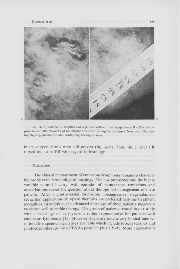
Fig. 2ci. b. Cutaneous eruptions of a patient with chronic lymphocytic B cell leukemia prior to and after 8 weeks of miltefosine treatment (complete response). Note postinflammatory hyperpigmentation and continuing teleangiectasias.
in the deeper dermis were still present (fig. 3a/b). Thus, the clinical C R turned out to be PR with regard to histology.
Discussion
The clinical management of cutaneous lymphoma remains a challenging problem in dermatological oncology. The low prevalence and the highly variable natural history, with episodes of spontaneous remissions and exacerbations raised the question about the optimal management of these patients. After a controversial discussion, nonaggressive, stage-adapted, sequential application of topical therapies are preferred first-line treatment modalities. In addition, the advanced mean age of these patients suggests a moderate well-tolerable therapy. The group of patients treated in our study with a mean age of sixty years is rather representative for patients with cutaneous lymphoma [ 16]. However, there are only a very limited number of mild therapeutic alternatives available which include topical steroids and photochemotherapy with P U V A (psoralen plus U V - A ) . More aggressive is

Fig. 3a, b. Histology ( H E ) of a mycosis fungoides lesion prior to (a) and after S weeks of miltefosine treatment (/>). Epidermotropic mononuclear cells (a\ arrows) have cleared. Mononuclear cells are still detectable in the deeper dermis (h: arrows).

Dummer ct al. 168
the topical application of cytotoxic drugs such as carmustine, but systemic side effects such as thrombocytopenia are common [5]. New promising approaches include interferon-alpha or desoxycoformycin [10, 17]. The clinical efficacy of these drugs remains to be determined.
Therefore, it is necessary to search for new agents with low toxicity. Phospholipids are effective against a broad variety of tumor cells in vitro [13, 14, 18, 19,20]. In the case of hexadecylphosphocholine, which was introduced as antineoplastic agent by Eibl and Unger [21], this compound shows strong and selective antitumoral activity. The topical application is feasible and provides high tolerabililty [15]. We observed no systemical side effects during the topical application which was limited, however, to an area of 20 40 cm 2 in most of the patients.
In 2 patients (13%), local inflammatory reactions caused the discontinuation of the therapy. Both events occurred early (3 and 4 weeks after initiation) during the study period in intertriginous areas. This point suggests a drug-related toxicity which was probably enhanced by an occlusive effect due to the localization of the lesions. Allergologic testing to exclude sensitization, however, was not performed.
Histological demonstration of residual infiltrates in the deeper dermis of clinically responding lesions indicates that cytotoxicity against the infiltrate seems to be restricted to the epidermis and the upper dermis. Limited skin penetration, clearance of the drug via dermal blood vessels, and local metabolic deactivation might explain this restriction of activity.
Formulations with increased skin penetration may be expected to enhance local bioavailability and to facilitate cytotoxicity against deeper infiltrates. The determination of hexadecylphosphocholine serum levels will cast light on this point, especially during treatment of larger areas.
The preliminary results reported here are promising. Miltefosine appears as an active agent against the infiltrating cells in cutaneous lymphomas. Due to the biological property of epidermotropism of these lymphoproliferative disorders, a topical application of cytotoxic drugs is suitable. This study is ongoing and it is reasonable to assume that it will provide data to understand in more detail the therapeutic efficacy of miltefosine in cutaneous lymphomas as well as in other neoplasias.
References
1 Lcnncrt K, Feller AC: Histopathologic der Non-Hodgkin-Lymphome. Berlin, Springer, 1990, pp 162 167.
2 Burg G . Braun-Falco O: Cutaneous Lymphomas, Pseudolymphomas and Related Disorders. Berlin. Springer, 1983.
3 Burg G, Sterry W (eds): Recommendations for Staging and Therapy of Cutaneous Lymphomas. E O R T C B M F T Project Group 1987, pp 1 14.

4 Wood GS, Strickler JG, Deneau DG, Egbert B, Warnke RA: Lymphomatoid papulosis expresses immunophenotypes associated with T cell lymphoma but not inflammation. J Am Acad Dermatol 1986:15:444 458.
5 Weiss L M . Wood GS, Trela M . Warnke RA. Sklar J: Clonal T-cell population in lymphomatoid papulosis: Evidence of a lymphoproliferative origin for a clinically benign disease. N Engl J Med 1986:315:475 479.
6 Macaulay WL: Lymphomatoid papulosis update. Arch Dermatol 1989:125:1387 1389. 7 Kaye FJ. Bunn PA, Steinberg SM, Stocker JL, Ihde DC. Fischmann AB, Glatstein EJ,
Schechter GP, Phelbs R M . Foss F M , Parlette HL, Anderson MJ, Sausvillc EA: A randomized trial comparing combination electron-beam radiation and chemotherapy with topical therapy in the initial treatment of mycosis fungoides. N Engl J Med 1989:321:1784 1790.
8 Edelson RL: Treatment of Cutaneous T Cell Lymphoma: in van Vloten WA, Willemze R, Vejlsgaard G L , Thomsen K (eds): Current Problems in Dermatology, vol 19, Basel, Karger. 1990. pp 226 237.
9 Zackheim HS: Topical mechlorcthamine and carmustine for cutaneous T-cell lymphoma. Semin Dermatol 1983:2:307 318.
10 Thestrup-Pederson K: Interferon therapy in cutaneous T-cell lymphoma: in van Vloten WA. Willemze R. Vcjlsgaars G L . Thomsen K (eds): Current Problems in Dermatology, vol 19. Basel, Karger. 1990, pp 258 263.
11 Kirby JD, Munro DD: Steroid induced atrophy in an animal and human model. Br J Dermatol 1976;94(suppl 12): 111.
12 Roenigk HH Jr. Cam WA: Skin cancer in the PUVA-48 cooperative study. J Am Acad Dermatol 1981:4:319.
13 Fleer E A M , Kim DJ. Eibl H, Ungcr C: Cytotoxic activity of lysophosphatidylcholinc analogues on human lymphoma Raji cells. Onkologie 1990:13:295 300.
14 Schick HD, Berdel WE, Fromm M , Fink U, Jehn U, Ulm K, Rcichcrt A, Eibl H, Unger C, Rastetter J: Cytotoxic effects of ether lipids and derivatives in human nonneoplastic bone marrow cells and leukemic cells in vitro. Lipids 1987:22:904 910.
15 Unger C. Eibl H, Nagcl G A , von Hcydcn HW, Breiser A, Engel J, Stekar J, Peukert M , Hilgard P, Bcrger M: Hexadecylphosphocholinc in the topical treatment of skin metastases: A phase I trial. Contrib Oncol 1989:37:219 223.
16 Weinstock MA, Horm JW: Population-based estimate of survival and determinants of prognosis in patients with mycosis fungoides. Cancer 62:1658 1661.
17 Dang-Vu AP, Olsen EA, Vollmcr RT, Grcenbcrg M L , Hcrshficld MS: Treatment of cutaneous T-cell lymphoma with 2'-dcsoxycoformycin (pentostatin). J Am Acad Dermatol 1988:19:692 698.
18 Fleer E A M , Unger C, Kim DJ, Eibl H: Metabolism of ether phospholipids and analogs in neoplastic cells. Lipids 1987:22:856 861.
19 Kosano H, Takatani O: Inhibition by an alkyl-lysophospholipid of the uptake of epidermal growth factor in human breast cancer cell lines in relation to epidermal growth factor internalization. Cancer Res 1989:49:2868 2870.
20 Verdonck L F , Witteveen EO, vanHeugten H G , Rozemuller E, Rijksen G: Selective killing of malignant cells from leukemic patients by alkyllysophospholipid. Cancer Res 1990:50:4020 4025.
21 Eibl H. Unger C: Hexadecylphosphocholinc, a new and selective antitumor drug. Cancer Treat Rev 1990:17:233 242.
Reinhard Dummer, Department of Dermatology. University of Zurich Medical School. Gloriastrasse 31, C H 8091 Zurich (Switzerland)