algorithm: outpatient/ed management x pathway page 1 of 12 viral croup algorithm: outpatient/ed...
TRANSCRIPT
CLINICAL PATHWAY
Page 1 of 12
VIRAL CROUP
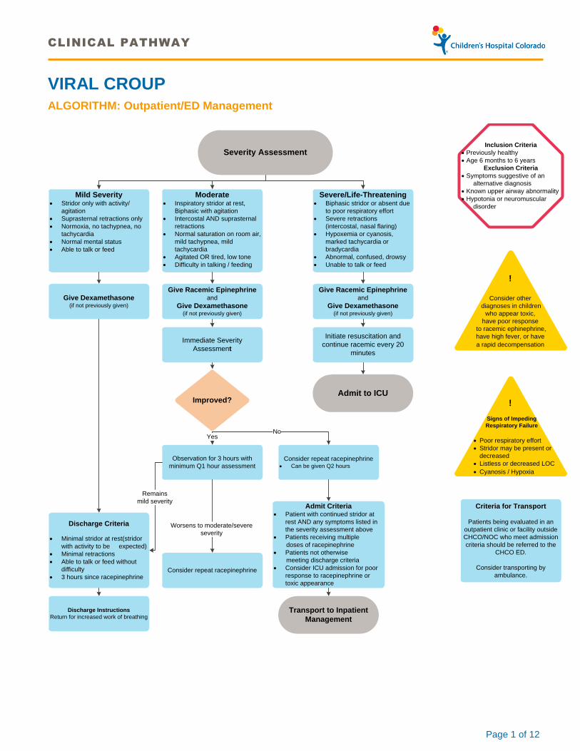
ALGORITHM: Outpatient/ED Management
Inclusion Criteria
· Previously healthy
· Age 6 months to 6 years
Exclusion Criteria
· Symptoms suggestive of an
alternative diagnosis
· Known upper airway abnormality
· Hypotonia or neuromuscular
disorder
!
Consider other
diagnoses in children
who appear toxic,
have poor response
to racemic ephinephrine,
have high fever, or have
a rapid decompensation
Severity Assessment
Mild Severity· Stridor only with activity/
agitation
· Suprasternal retractions only
· Normoxia, no tachypnea, no
tachycardia
· Normal mental status
· Able to talk or feed
Moderate· Inspiratory stridor at rest,
Biphasic with agitation
· Intercostal AND suprasternal
retractions
· Normal saturation on room air,
mild tachypnea, mild
tachycardia
· Agitated OR tired, low tone
· Difficulty in talking / feeding
Severe/Life-Threatening· Biphasic stridor or absent due
to poor respiratory effort
· Severe retractions
(intercostal, nasal flaring)
· Hypoxemia or cyanosis,
marked tachycardia or
bradycardia
· Abnormal, confused, drowsy
· Unable to talk or feed
Give Dexamethasone(if not previously given)
Give Racemic Epinephrine and
Give Dexamethasone(if not previously given)
Immediate Severity
Assessment
Give Racemic Epinephrine and
Give Dexamethasone(if not previously given)
Initiate resuscitation and
continue racemic every 20
minutes
Discharge Criteria
· Minimal stridor at rest(stridor
with activity to be expected)
· Minimal retractions
· Able to talk or feed without
difficulty
· 3 hours since racepinephrine
Improved?
Observation for 3 hours with
minimum Q1 hour assessment
Consider repeat racepinephrine
Consider repeat racepinephrine · Can be given Q2 hours
Admit to ICU
Admit Criteria· Patient with continued stridor at
rest AND any symptoms listed in
the severity assessment above
· Patients receiving multiple
doses of racepinephrine
· Patients not otherwise
meeting discharge criteria
· Consider ICU admission for poor
response to racepinephrine or
toxic appearance
Yes
Worsens to moderate/severe
severity
No
Remains
mild severity
Discharge Instructions
Return for increased work of breathingTransport to Inpatient
Management
Criteria for Transport
Patients being evaluated in an
outpatient clinic or facility outside
CHCO/NOC who meet admission
criteria should be referred to the
CHCO ED.
Consider transporting by
ambulance.
!
Signs of Impeding
Respiratory Failure
· Poor respiratory effort
· Stridor may be present or
decreased
· Listless or decreased LOC
· Cyanosis / Hypoxia

CLINICAL PATHWAY
Page 2 of 12
ALGORITHM: Inpatient Management
Inclusion Criteria
·Previously healthy
· Age 6 months to 6 years
Exclusion Criteria
· Symptoms suggestive of an
alternative diagnosis
· Known upper airway abnormality
· Hypotonia or neuromuscular
disorder
· Patient in PICU
!
Consider other
diagnoses in children
who appear toxic,
have poor response
to racemic ephinephrine,
have high fever, or have
a rapid decompensation
Severity Assessment
!
Signs of Impeding
Respiratory Failure
· Poor respiratory effort
· Stridor may be present or
decreased
· Listless or decreased LOC
· Cyanosis / Hypoxia
Mild Severity· Stridor only with activity/
agitation
· Suprasternal retractions only
· Normoxia, no tachypnea, no
tachycardia
· Normal mental status
· Able to talk or feed
Moderate· Inspiratory stridor at rest,
Biphasic with agitation
· Intercostal AND suprasternal
retractions
· Normal saturation on room air,
mild tachypnea, mild
tachycardia
· Agitated OR tired, low tone
· Difficulty in talking / feeding
Severe/Life-Threatening· Biphasic stridor or absent due
to poor respiratory effort
· Severe retractions
(intercostal, nasal flaring)
· Hypoxemia or cyanosis,
marked tachycardia or
bradycardia
· Abnormal, confused, drowsy
· Unable to talk or feed
Give Dexamethasone
(if not previously given)
Give Racemic Epinephrine
Give Dexamethasone(if not previously given)
Immediate Severity
Assessment
Discharge Criteria
· Minimal stridor at rest(stridor
with activity to be expected)
· Minimal retractions
· Able to talk or feed without
difficulty
· 3 hours since racepinephrine
Improved?
Observation Assess severity Q1 hr x 2 using
severity assessment
· If patient worsens, consider
repeat racepinephrine
· Can be given Q2 hrs
Can give racemic
epinephrine every 1 hour if
MD at bedside and RRT
called· Consider alternative diagnosis
(see page 5 for differential
diagnosis and
recommendations)
· Consider blood gas
· Consider ICU transfer
· Consider ENT consult
Call code and continue
racemic every 20 minutes
Yes
No
Discharge Instructions
Return for increased work of
breathing
Recommendations
1. Consider ENT consultation for direct laryngoscopy in
patients with 2 or more episodes of croup AND one of
the following:
- history of intubation
- age less than 36 months
- prolonged severe disease requiring
inpatient management
2. Consider evaluation for GERD and initiation of anti-
reflux medications in patients with prolonged or
recurrent coup
3. Consider evaluation and treatment for allergies
!
Hypoxia is
uncommon
in Croup
Indicates severe disease,
alternate diagnosis or lower
respiratory tract disease
Not Routinely Recommended
(No evidence supporting the use of)
Viral PCR
Radiographs
Repeat Dexamethasone
Cool Mist
Observation Complete severity assessment Q4 hr
until patient meets discharge criteria
Observe
Meets discharge criteria
Evaluate criteria for racemic epinephrine
≥3 doses racemic epinephrine
Consider further workup,
and/or RRT evaluation
(see page 5 for differential
diagnosis)

CLINICAL PATHWAY
Page 3 of 12
TABLE OF CONTENTS
Algorithm: Outpatient/ED Management
Algorithm: Inpatient Management
Target Population
Prevention
Outpatient Telephone Triage
Clinical Management
Laboratory Studies | Imaging
Therapeutics
Disposition
Follow-Up | Discharge Instructions
Education
Etiology of Croup
References
Clinical Improvement Team
TARGET POPULATION
Inclusion Criteria
· First or repeat episode
· Age 6 months to 6 years
· Principle diagnoses: croup (laryngotracheitis)
Exclusion Criteria
· Suspicion of bacterial tracheitis, epiglottitis, upper-airway abscess (peritonsillar or retropharyngeal), or other serious bacterial infection
· Severe or life-threatening disease requiring PICU admission
· Chronic lung disease (bronchopulmonary dysplasia, cystic fibrosis, pulmonary artery hypertension)
· Known upper airway abnormalities (for example: laryngomalacia, tracheomalacia, subglottic stenosis)
· Recent airway instrumentation
· Foreign body aspiration or ingestion
· Neuromuscular disorder or hypotonia
· Allergic reaction
· Angioedema
· Active varicella or tuberculosis (TB)
· Congenital or acquired heart disease
CLINICAL PATHWAY
Page 4 of 12
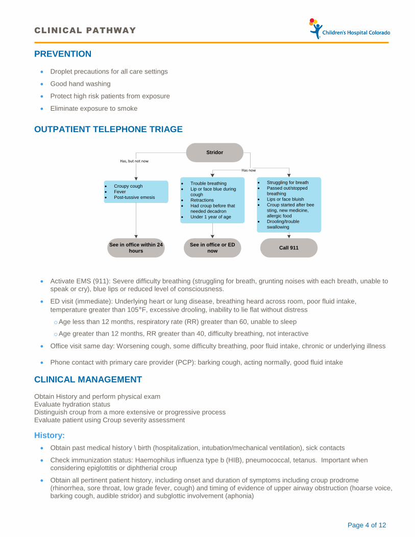
PREVENTION
· Droplet precautions for all care settings
· Good hand washing
· Protect high risk patients from exposure
· Eliminate exposure to smoke
OUTPATIENT TELEPHONE TRIAGE
Has, but not now
Stridor
Has now
· Croupy cough
· Fever
· Post-tussive emesis
· Trouble breathing
· Lip or face blue during
cough
· Retractions
· Had croup before that
needed decadron
· Under 1 year of age
· Struggling for breath
· Passed out/stopped
breathing
· Lips or face bluish
· Croup started after bee
sting, new medicine,
allergic food
· Drooling/trouble
swallowing
See in office within 24
hours
See in office or ED
nowCall 911
· Activate EMS (911): Severe difficulty breathing (struggling for breath, grunting noises with each breath, unable to speak or cry), blue lips or reduced level of consciousness.
· ED visit (immediate): Underlying heart or lung disease, breathing heard across room, poor fluid intake,
temperature greater than 105 F, excessive drooling, inability to lie flat without distress
o Age less than 12 months, respiratory rate (RR) greater than 60, unable to sleep
o Age greater than 12 months, RR greater than 40, difficulty breathing, not interactive
· Office visit same day: Worsening cough, some difficulty breathing, poor fluid intake, chronic or underlying illness
· Phone contact with primary care provider (PCP): barking cough, acting normally, good fluid intake
CLINICAL MANAGEMENT
Obtain History and perform physical exam Evaluate hydration status Distinguish croup from a more extensive or progressive process Evaluate patient using Croup severity assessment
History:
· Obtain past medical history \ birth (hospitalization, intubation/mechanical ventilation), sick contacts
· Check immunization status: Haemophilus influenza type b (HIB), pneumococcal, tetanus. Important when considering epiglottitis or diphtherial croup
· Obtain all pertinent patient history, including onset and duration of symptoms including croup prodrome (rhinorrhea, sore throat, low grade fever, cough) and timing of evidence of upper airway obstruction (hoarse voice, barking cough, audible stridor) and subglottic involvement (aphonia)

CLINICAL PATHWAY
Page 5 of 12
· Inquire regarding history of congenital or acquired heart disease, congenital or acquired subglottic stenosis, tracheomalacia, tracheal webs, choanal narrowing or atresia, micrognathia, macroglossia
· Check current medications and time and dose of last antipyretic and recent steroid use.
Clinical Symptoms of Croup:
· Symptoms increase at night and improve during day
o Hoarse voice
o Barking cough (often described as a “barking seal”)
o Stridor (variable, usually inspiratory)
· Respiratory distress (variable):
o Retractions (suprasternal, intercostal)
o Tachypnea
o Tachycardia
Clinical Progress of Croup:
Day 1 to 3 Rhinorrhea Day 3 to 7 Onset symptoms of upper airway inflammation Sore throat Hoarseness Low grade fever Barking cough Mild cough Stridor (variable)
Respiratory distress (variable)
Clinical symptoms that suggest Croup is not the diagnosis:
· Bacterial tracheitis should be considered if patients have a toxic appearance, poor response to racemic epinephrine, high fever, or have a rapid decomposition
· Hypoxemia is uncommon in croup and indicates severe disease, an alternate diagnosis, or lower respiratory tract disease
Differential Diagnosis:
· Distinguish croup from a more extensive or progressive process
· Conditions mimicking croup:
Increased Work of Breathing
Barking cough/
voice hoarseness
High fever/
barking cough/
voice
hoarseness/rapid
deterioration
History of
choking/dyspagia
High fever/sore
throat/dysphagia/
muffled voice
High fever/sore
throat/torticollis/
limitation of neck
movements
Viral or
spasmodic croup
Bacterial
tracheitis
Foreign body
aspirationEpiglottitis
Retro-/para-
pharyngeal
abscess
Imaging with radiographs or CT scan may aid diagnosis. Choice of imaging
depends on clinical situation. Consult radiology for recommendations.
Stridor not
prominentStridor
CLINICAL PATHWAY
Page 6 of 12
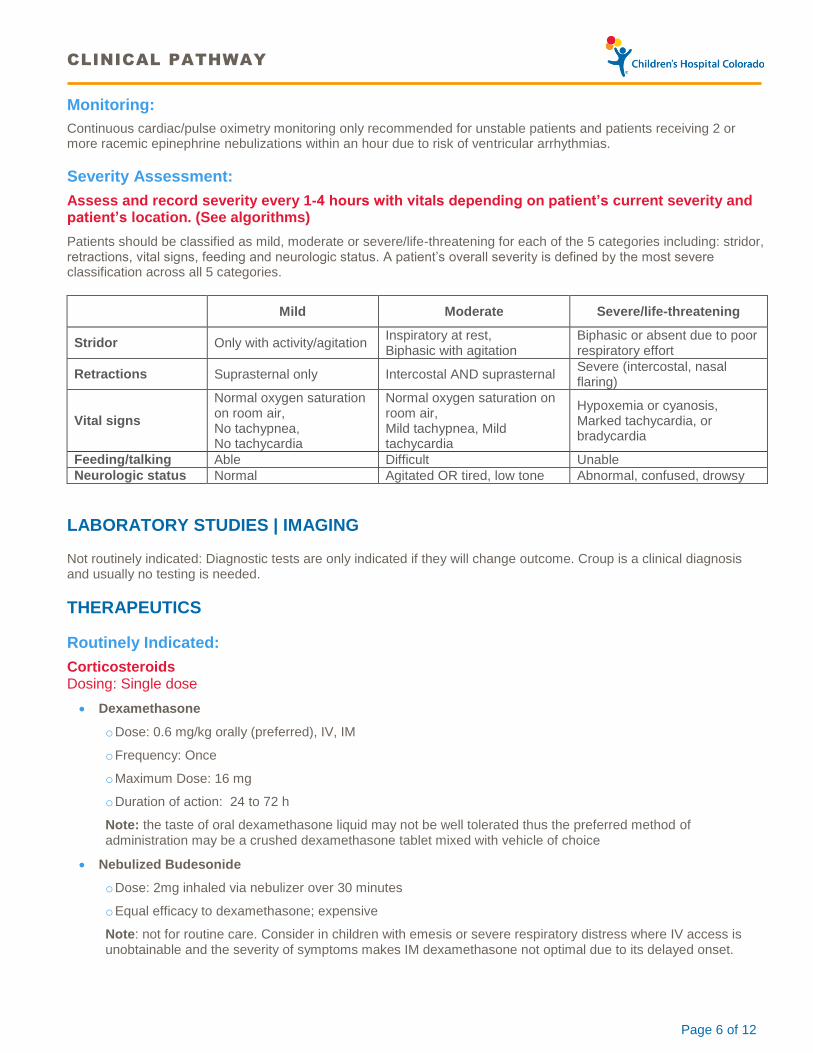
Monitoring:
Continuous cardiac/pulse oximetry monitoring only recommended for unstable patients and patients receiving 2 or more racemic epinephrine nebulizations within an hour due to risk of ventricular arrhythmias.
Severity Assessment:
Assess and record severity every 1-4 hours with vitals depending on patient’s current severity and patient’s location. (See algorithms)
Patients should be classified as mild, moderate or severe/life-threatening for each of the 5 categories including: stridor, retractions, vital signs, feeding and neurologic status. A patient’s overall severity is defined by the most severe classification across all 5 categories.
Mild Moderate Severe/life-threatening
Stridor Only with activity/agitation Inspiratory at rest, Biphasic with agitation
Biphasic or absent due to poor respiratory effort
Retractions Suprasternal only Intercostal AND suprasternal Severe (intercostal, nasal flaring)
Vital signs
Normal oxygen saturation on room air, No tachypnea, No tachycardia
Normal oxygen saturation on room air, Mild tachypnea, Mild tachycardia
Hypoxemia or cyanosis, Marked tachycardia, or bradycardia
Feeding/talking Able Difficult Unable
Neurologic status Normal Agitated OR tired, low tone Abnormal, confused, drowsy
LABORATORY STUDIES | IMAGING
Not routinely indicated: Diagnostic tests are only indicated if they will change outcome. Croup is a clinical diagnosis and usually no testing is needed.
THERAPEUTICS
Routinely Indicated:
Corticosteroids Dosing: Single dose
· Dexamethasone
o Dose: 0.6 mg/kg orally (preferred), IV, IM
o Frequency: Once
o Maximum Dose: 16 mg
o Duration of action: 24 to 72 h
Note: the taste of oral dexamethasone liquid may not be well tolerated thus the preferred method of administration may be a crushed dexamethasone tablet mixed with vehicle of choice
· Nebulized Budesonide
o Dose: 2mg inhaled via nebulizer over 30 minutes
o Equal efficacy to dexamethasone; expensive
Note: not for routine care. Consider in children with emesis or severe respiratory distress where IV access is unobtainable and the severity of symptoms makes IM dexamethasone not optimal due to its delayed onset.

CLINICAL PATHWAY
Page 7 of 12
Nebulized Epinephrine
Racemic Epinephrine (1:1 mixture of & -isomers)
· isomers epinephrine)
o Dose: 0.05 mL/kg/dose of 2.25% solution in 2.5mL normal saline (NS) via nebulizer over 15 minutes
o Frequency: as needed based on severity (see algorithms)
o Maximum single dose: 0.5 mL
o Duration of action: less than or equal to 2 hours
Note: If a patient requires 2 or more nebulizations within an hour, cardiac monitoring is recommended due to risk of ventricular arrhythmias.
The term ‘rebound phenomenon’ is a misnomer. Epinephrine doesn’t change the duration of croup. Benefits lasts up to 2 hours. It is safe to send children home from the ED after receiving racemic epinephrine if they have been observed for a minimum of 3 hours post therapy.
Recommended in some patients:
In patients requiring 3 or more doses of racemic epinephrine consider ENT evaluation, additional work-up for alternative diagnoses and/or a Rapid Response Team (RRT) evaluation.
· Consider ENT consult for laryngoscopy in patients with 2 more episodes of croup AND one of the following:
o History of intubation
o Age less than 36 months
o Prolonged or severe disease requiring inpatient management
· Consider evaluation for GERD and initiation of anti-reflux medications with prolonged or recurrent croup
· Consider evaluation and treatment for allergies
Not Routinely Indicated:
Oxygen
· The presence of hypoxemia or intermittent desaturations is a sign of impending respiratory failure in croup and other central airway obstruction. Oxygen can be used to normalize SpO2, but further diagnostic evaluation and therapies may be needed. If hypoxemia is present, a blood gas may be useful to assess for hypercarbia.
Other Therapies
· Mist: Humidified air with or without oxygen is not indicated
· Antitussive or decongestant medications are not indicated.
· Antibiotics
o No role in viral croup

CLINICAL PATHWAY
Page 8 of 12
DISPOSITION
Begin discharge planning at time of initial presentation
· Assess caretaker ability to provide home care
· Assess home resources adequate to support care
· Confirm transportation and telephone
· Confirm follow-up PCP/designee in specified time frame
· Complete croup teaching
· Provide verbal and written instructions to caretakers
· Assure family awareness indications return
· Provide 24-hour contact number for PCP or designee
· Assure chart faxed to PCP or designee
Discharge Home
· Croup severity mild
· Minimal Stridor at rest (stridor with activity to be expected)
· Normal saturation on room air
· Able to talk and feed without difficulty
· Minimal or no retractions (mild suprasternal acceptable)
· 3 hours since racepinephrine
Note: Patients who have received nebulized epinephrine may be discharged home from the outpatient/ED/UC setting after a minimum of 3 hours if no stridor at rest. Consider additional monitoring or work-up prior to discharge in inpatients requiring repeated doses of racemic epinephrine (see algorithm)
Admit to Inpatient/ Observation
· Moderate severity despite treatment with corticosteroids
· Inadequate hydration
· Require supplemental oxygen and are proven not to be in acute or impending respiratory failure
· Condition deteriorates or does not improve with therapy
· Patients receiving multiple doses of racepinephrine
· Patients not otherwise meeting discharge criteria
Admit to ICU
· Severe or life-threatening severity
· Acute respiratory acidosis
· Bradypnea suggesting respiratory muscle fatigue and impending respiratory failure
· Lack of response to steroids and racemic epinephrine as characterized by persistent moderate-severe retractions, hypoxemia, severely decreased air entry, altered level of consciousness, difficulty feeding/talking, or difficulty controlling oral secretions

CLINICAL PATHWAY
Page 9 of 12
FOLLOW-UP | DISCHARGE INSTRUCTIONS
With PCP or designee as scheduled
If patient evaluated and discharged from the ED: PCP phone follow-up within 24 hours
If seen in PCP office: Parent/guardian to call back if patient worsens
If admitted: PCP phone follow-up within 24 hours of discharge and PCP office visit within 2 days.
Note: If patients received multiple doses of steroids while hospitalized, consider more than one outpatient follow-up visit due to long half-life of dexamethasone.
EDUCATION
Parent | Caregiver Education
· Expected clinical course less than seven days
· Educate to return for respiratory distress
· Smoking cessation counseling
· Provide parent with patient education materials
Knowledge Base
Viral croup is an acute inflammatory process in response to a viral infection that causes upper airway obstruction (primarily of the subglottic region) resulting in inspiratory stridor, barky cough and in more severe cases respiratory distress. Infection begins in the nasopharynx and spreads to the respiratory epithelium of larynx & trachea. Inflammation and edema of the vocal folds causes hoarseness.
Age: 6 months to 6 yrs (Mean = 18 mos)
Duration: 2 to 7 days
Morbidity: Highest first year of life
Epidemiology: Year round; most common fall and winter
ETIOLOGY OF CROUP
· Parainfluenza type 1(most common) 2, 3
· Influenza A & B
· Human metapneumovirus (hMPV)
· Respiratory syncitial virus (RSV)
· Rhinovirus
· Mycoplasma pneumoniae
· Enteroviruses
· Herpes Simplex viruses
· Adenovirus
· Measles virus

CLINICAL PATHWAY
Page 10 of 12
REFERENCES
1. Geelhoed G, Macdonald W. Oral and inhaled steroids in croup: a randomized, placebo-controlled trial. Pediatr Pulmonol 1995; 20: 355–61.
2. Geelhoed GC, Turner J, Macdonald WB. Efficacy of a small single dose of oral dexamethasone for outpatient croup: a double blind placebo controlled clinical trial [see comments]. BMJ 1996; 313: 140–2.
3. Klassen TP, Rowe PC. The croup score as an evaluative instrument in clinical trials. Arch Pediatr Adolesc Med 1995; 149: 60–1.
4. Jacobs S, Shortland G, Warner J, Dearden A, Gataure PS, Tarpey J. Validation of a croup score and its use in triaging children with croup. Anaesthesia 1994; 49: 903–6
5. Chan, A., J. Langley, et al. (2001). "Interobserver variability of croup scoring in clinical practice." Paediatr Child Health 6(6): 347-351.
6. Westley, C. R., E. K. Cotton, et al. (1978). "Nebulized racemic epinephrine by IPPB for the treatment of croup: a double-blind study." Am J Dis Child 132(5): 484-487.
7. Brown, J.C., The management of croup. Br Med Bull, 2002. 61: p. 189-202.
8. Johnson DW. Croup. BMJ Clinical Evidence. 2009;2009:0321.
9. Kristjánsson S1, Berg-Kelly K .Inhalation of racemic adrenaline in the treatment of mild and moderately severe croup. Clinical symptom score and oxygen saturation measurements for evaluation of treatment effects. Acta Paediatr. 1994 Nov;83(11):1156-60.
10. Downes JJ, Raphaely RC. Pediatric intensive care. Anesthesiology 1975;43:238-50.
11. Petrocheilou, A, Tanou, K, Kalampouka, E, Malakasioti, G, Ciannios, C, Kaditis, A. Viral Croup: Diagnosis and a Treatment Algorithm. Pediatric Pulmonlogy 2014; 49: 421-429.

CLINICAL PATHWAY
Page 11 of 12
Clinical pathways are intended for informational purposes only. They are current at the date of publication and are reviewed on a regular basis to align with the best available evidence. Some information and links may not be available to external viewers. External viewers are encouraged to consult other available sources if needed to confirm and supplement the content presented in the clinical pathways. Clinical pathways are not intended to take the place of a physician’s or other health care provider’s advice, and is not intended to diagnose, treat, cure or prevent any disease or other medical condition. The information should not be used in place of a visit, call, consultation or advice of a physician or other health care provider. Furthermore, the information is provided for use solely at your own risk. CHCO accepts no liability for the content, or for the consequences of any actions taken on the basis of the information provided. The information provided to you and the actions taken thereof are provided on an “as is” basis without any warranty of any kind, express or implied, from CHCO. CHCO declares no affiliation, sponsorship, nor any partnerships with any listed organization, or its respective directors, officers, employees, agents, contractors, affiliates, and representatives.
CLINICAL IMPROVEMENT TEAM MEMBERS
Amy Tyler, MD | Hospitalist
Oren Kupfer, MD | Pulmonology
Todd Carpenter, MD | Critical Care
Ryan Caltagirone, MD | Emergency Department
Leigh Anne Bakel, MD | Hospitalist
David Fox, MD | Special Care Clinic
Leana May, DO, MPH | Emergency Medicine
Melissa Scholes, MD | ENT
Sarah Parker, MD | Infectious Disease
Jason Child, MD | Infectious Disease
Christina Suh, MD | Child Health Clinic
Suzanne Cooper, MD | Primary Care Provider
Don Traver, MD | Primary Care Provider
Marc Avner, MD | Primary Care Provider
Sunit Gill, MD | Primary Care Provider
Kelly Newgent, MD | Primary Care Provider
Joyce Baker, RT | Respiratory Therapy
Chris Poppy | Solutions Architect
Heather Hewitt, MD | CPC |
Bethany Smith, RN | Clinical Nurse
Michael Barberio, PharmD | Clinical Pharmacist
Ron Guittar | Clinical Data Analyst
Angela Stowe, MS | Director, Clinical Effectiveness
Paige Krack, MBA | Process Improvement Lead
APPROVED BY
Pharmacy & Therapeutics Committee – December 1, 2016
Clinical Care Guideline and Measures Review Committee – December 13, 2016
MANUAL/DEPARTMENT Clinical Care Guidelines/Quality
ORIGINATION DATE August 11, 2011
LAST DATE OF REVIEW OR REVISION December 13, 2016
APPROVED BY
Lalit Bajaj, MD, MPH
Medical Director, Clinical Effectiveness
REVIEW REVISION SCHEDULE
Scheduled for full review on December 13, 2020

CLINICAL PATHWAY
Page 12 of 12