aldose reductase acts as a selective derepressor of ... reports, volume 15 supplemental information...
TRANSCRIPT

Cell Reports, Volume 15
Supplemental Information
Aldose Reductase Acts as a Selective Derepressor
of PPARg and the Retinoic Acid Receptor
Devi Thiagarajan, Radha Ananthakrishnan, Jinghua Zhang, KarenM. O'Shea, NosirudeenQuadri, Qing Li, Kelli Sas, Xiao Jing, Rosa Rosario, Subramaniam Pennathur, Ann MarieSchmidt, and Ravichandran Ramasamy
Supplemental Figures and Figure Legends
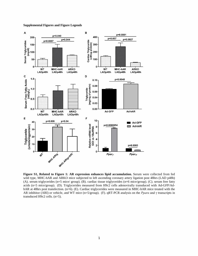
Figure S1, Related to Figure 1: AR expression enhances lipid accumulation. Serum were collected from fed wild type, MHC-hAR and ARKO mice subjected to left ascending coronary artery ligation post 48hrs (LAD p48h) (A). serum triglycerides (n=5 mice/ group). (B). cardiac tissue triglycerides (n=6 mice/group). (C). serum free fatty acids (n=5 mice/group). (D). Triglycerides measured from H9c2 cells adenovirally transduced with Ad-GFP/Ad-hAR at 48hrs post transfection. (n=6). (E). Cardiac triglycerides were measured in MHC-hAR mice treated with the AR inhibitor (ARI) or vehicle, and WT mice (n=5/group). (F). qRT-PCR analysis on the Pparα and γ transcripts in transduced H9c2 cells. (n=5).
1

Figure S2, Related to Figure 2: PPARγ is activated in AR expressing cells. Lysates from adenovirally transduced H9c2 cells with GFP/hAR were used post 48hrs transduction. (A). HDAC activity was measured from the nuclear fraction. (n=6). (B). Acetylated histone H3 modifications normalized against total H3. (C). Acetylated histone H4
2

modifications normalized against total H4. (D). Profiling of histone deacetylases, normalized against β-actin. (E). Western blot analysis of total cell lysates using HDAC3, AR and PPARγ. β-actin used as loading control. B-E. (n=3). (F). AR expression in the cardiac tissue lysates from wild type, MHC-hAR and ARKO mice. (n=6). (G). Nuclear and cytoplasmic fractions were tested for purity using VCAM1 and TBP as cytoplasmic and nuclear specific proteins by western blot. (H). qRT-PCR analysis of PPARγ target genes in adenovirally transduced H9c2 cells. (n=6).
3

Figure S3, Related to Figure 2: AR activates PPARγ in cells. Lysates from Ad-GFP/Ad-hAR cells, 48hrs post transduction were used for (A). Western blot analysis and quantification of CD36, normalized to β-actin. (n=3). (B). ChIP analysis done using PPARγ antibody and RT-PCR were done using Sybrgreen with primers spanning the PPAR response element (PPRE) in the Cd36 promoter region. Upstream to that region, devoid of PPRE, was used as negative control. (n=3). (C). qRT-PCR analysis on expression of Hdac3 and nuclear corepressors. (D). Overlapping of secondary structures of HDAC3-DAD complex and AR using Pymol program. Alignment of protein sequences of HDAC3 and AR, within the overlapping region. Red- HDAC3 (9-49 aa); Yellow- SMRT-DAD; White- AR (266-312 aa). (E). qRT‐PCR analysis performed on retrovirally transduced H9c2 cells (n=5/group). (F).Triglycerides were measured in H9c2 cells retrovirally transduced with mock/hAR/L289A (n=3/group).
4

Figure S4, Related to Figure 6: AR expression hyper-activates RAR target genes. (A). serum T3 and T4 measurements made from wild type and MHC-hAR mice. (n=6 mice/group). (B). qRT-PCR analysis from the liver tissues of wild type and MHC-hAR mice. (C). qRT-PCR analysis for Cyp1a1 and Cyp26b1 in transduced H9c2 cells treated with/without at RA (all-trans retinoic acid) (n=3). (D). qRT-PCR analysis for Tnf-α in cardiac tissue (n=6 mice/group).E.PCR array results for genes involved in the Circadian rhythm on RNA isolated from MHC-hAR and WT hearts (n=3/group).
5
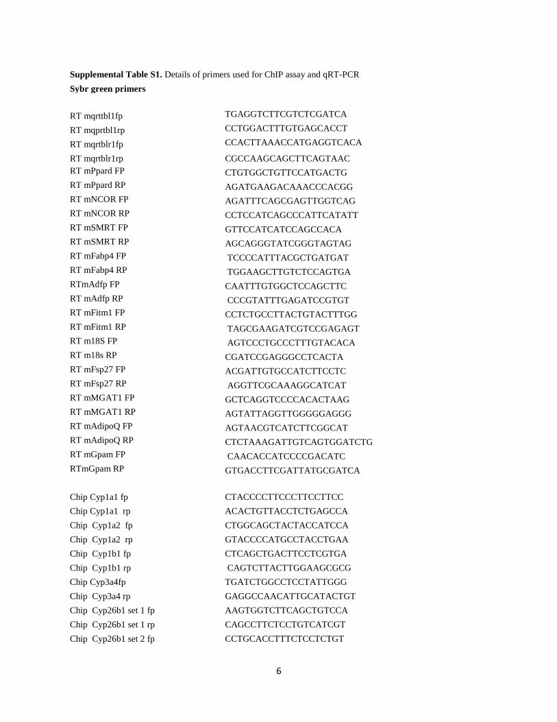
Supplemental Table S1. Details of primers used for ChIP assay and qRT-PCR Sybr green primers
RT mqrttbl1fp TGAGGTCTTCGTCTCGATCA
RT mqprtbl1rp CCTGGACTTTGTGAGCACCT
RT mqrtblr1fp CCACTTAAACCATGAGGTCACA
RT mqrtblr1rp CGCCAAGCAGCTTCAGTAAC RT mPpard FP CTGTGGCTGTTCCATGACTG RT mPpard RP AGATGAAGACAAACCCACGG RT mNCOR FP AGATTTCAGCGAGTTGGTCAG RT mNCOR RP CCTCCATCAGCCCATTCATATT RT mSMRT FP GTTCCATCATCCAGCCACA RT mSMRT RP AGCAGGGTATCGGGTAGTAG RT mFabp4 FP TCCCCATTTACGCTGATGAT RT mFabp4 RP TGGAAGCTTGTCTCCAGTGA RTmAdfp FP CAATTTGTGGCTCCAGCTTC RT mAdfp RP CCCGTATTTGAGATCCGTGT RT mFitm1 FP CCTCTGCCTTACTGTACTTTGG RT mFitm1 RP TAGCGAAGATCGTCCGAGAGT RT m18S FP AGTCCCTGCCCTTTGTACACA RT m18s RP CGATCCGAGGGCCTCACTA RT mFsp27 FP ACGATTGTGCCATCTTCCTC RT mFsp27 RP AGGTTCGCAAAGGCATCAT RT mMGAT1 FP GCTCAGGTCCCCACACTAAG RT mMGAT1 RP AGTATTAGGTTGGGGGAGGG RT mAdipoQ FP AGTAACGTCATCTTCGGCAT RT mAdipoQ RP CTCTAAAGATTGTCAGTGGATCTG RT mGpam FP CAACACCATCCCCGACATC RTmGpam RP GTGACCTTCGATTATGCGATCA
Chip Cyp1a1 fp CTACCCCTTCCCTTCCTTCC Chip Cyp1a1 rp ACACTGTTACCTCTGAGCCA Chip Cyp1a2 fp CTGGCAGCTACTACCATCCA Chip Cyp1a2 rp GTACCCCATGCCTACCTGAA Chip Cyp1b1 fp CTCAGCTGACTTCCTCGTGA Chip Cyp1b1 rp CAGTCTTACTTGGAAGCGCG Chip Cyp3a4fp TGATCTGGCCTCCTATTGGG Chip Cyp3a4 rp GAGGCCAACATTGCATACTGT Chip Cyp26b1 set 1 fp AAGTGGTCTTCAGCTGTCCA Chip Cyp26b1 set 1 rp CAGCCTTCTCCTGTCATCGT Chip Cyp26b1 set 2 fp CCTGCACCTTTCTCCTCTGT
6
Chip Cyp26b1 set 2 rp AAGCCTGCCTTCCTCAAGTT chip sercafp TCTGGTCCTCAGGGATCATT chip sercarp GACCTCCTGTGGTGAGTGCT ChiP NCX FP TGAAGCCATACTGGGGAAAG ChiP NCX RP TTGCTCTGCTGGAGAATGAA
Taqman primers: Mouse specific Taqman primers
mGps2 Mm00517238_g1 mMyh6 Mm00440359_m1 mMyh7 Mm01319006_g1 mSlc8a1 Mm01232254_m1 mSerca2a Mm01201431_m1 mLXRa Mm00443451_m1
mSrebf1 Mm00550338_m1
mAbca1 Mm00442646_m1
mAbcg1 Mm00437390_m1
mCyp1a1 Mm00487218_m1
mCyp1a2 Mm00487224_m1
mCyp1b1 Mm00487229_m1
mCyp26b1 Mm00558507_m1
mRara Mm01296312_m1
mSlc27a1 Mm01232254_m1
mCpt1a Mm01231183_m1
mCpt2 Mm00487205_m1
mCd36 Mm01135198_m1
mAcsl1 Mm04207567_g1
mPPara Mm00440939_m1
mPParg Mm01184322_m1
Rat specific Taqman primers
rCd36 Rn01442638_m1
rFatp1 Rn00585821_m1
rSlc2a4 Rn01752377_m1
rFasn Rn01463550_m1
rSirt3 Rn01501410_m1
rSrebf1 Rn01495769_m1
rAdipoq Rn00595250_m1
rNcor1 Rn01399460_m1
7
Supplemental Experimental Procedures: Surgical procedures related to coronary artery ligation (LAD) were done as previously described (Hwang et al., 2004). LAD was done on 4 months old mice and was sacrificed 48 hrs post-surgery. For the experiments conducted at basal level, 2 months old mice were used. For aging studies, young (4 months) and old (24 months) wild type C57B/6 mice were used. Diabetic mice were generated using STZ injection as described earlier (Vedantham et al., 2014). 6 months old diabetic mice with blood glucose >250mg/dl at time of experiment, along with age matched controls were used. For cell culture experiments, cells were harvested post 48hrs after transduction. Chemicals including MG132 (5uM), bortezomib (1uM) and epoxomicin (1uM) were purchased from Sigma chemicals and cells were treated for overnight. For retroviral transduction, human NCOR1 cDNA (Transomic) was subcloned into retroviral vector pQXCIN (Clontech) and clones were verified through DNA sequencing. pQXCIN-NCOR1 plasmid was transfected using Lipofectamine 2000 (Invitrogen) in HEK293T cells. Supernatant media was collected for 3 days and were transduced in H9c2 using polybrene. 3 days after transfection, cells were selected with G418 for 15 days. The stably expressing NCOR1 cells were then adenovirally transduced with GFP or hAR. Western Blot analysis: Total lysates from heart tissues or cultured cells were prepared using lysis buffer (Cell Signaling) for the detection of protein using the following antibodies: HDAC3 (Activemotif (N-terminal specific ab), Biovision (C-terminal specific ab), Abcam (N-terminal specific ab), PPARγ (Caymen Chemicals), SMRT (Santacruz), NCOR1 (Abcam), CD36 (Novus Biologicals), Smooth muscle alpha Actin, β-actin (Sigma) TBP (Abcam), Acetyl histone H3 and acetyl histone H4 antibody sampler kit (Cell signaling). qPcr: cDNA was prepared using iScriptcDNA synthesis kit (BioRad). Taqman probes (Life Tech) were used for quantification and the data were normalized using 18s rRNA. For TR, LXR and RAR target genes, primers were obtained from IDT. Sybr green quantification was done using Fast Sybr green master mix (Applied Biosystems) and normalized using 18s rRNA. Chromatin immunoprecipitation: For chromatin immunoprecipitation, ChIP grade HDAC3 and PPARγ antibodies were purchased from Abcam.ChIP was performed using ChIP-IT kit (ActiveMotif). Briefly, heart tissue was homogenized and crosslinked with 1% formaldehyde for 10min at room temperature and chromatin was obtained as per protocol. 150ug of chromatin was immunoprecipitated using 15ug HDAC3 antibody. Primers were designed using the nuclear receptors binding site data available from Applied Biosciences website and Primer3 software. The details of the primers were given in the Table S1. For ChIP in cells, primers were designed in the region containing PPRE and 2 Kb upstream regions were used as Negative PPRE. For HDAC3 ChIP, primer sequences corresponding to Serca2a and Ncx.1 were used as negative control. Real time PCR were setup using Sybrgreen (Applied Biosystems) and results were normalized using input DNA. Data were represented as % of total input and fold change.
Supplemental References Hwang, Y.C., Kaneko, M., Bakr, S., Liao, H., Lu, Y., Lewis, E.R., Yan, S., Ii, S., Itakura, M., Rui, L., et al. (2004). Central role for aldose reductase pathway in myocardial ischemic injury. FASEB J 18, 1192-1199. Vedantham, S., Thiagarajan, D., Ananthakrishnan, R., Wang, L., Rosario, R., Zou, Y.S., Goldberg, I., Yan, S.F., Schmidt, A.M., and Ramasamy, R. (2014). Aldose reductase drives hyperacetylation of Egr-1 in hyperglycemia and consequent upregulation of proinflammatory and prothrombotic signals. Diabetes 63, 761-774.
8