aice biology cellular biology and microscopy. ultrastructure the fine or detailed structure of a...
TRANSCRIPT

AICE Biology
CELLULAR BIOLOGY AND MICROSCOPY

ULTRASTRUCTURE• The fine or detailed structure of a cell that is revealed by an electron
microscope
• Compartmentalisation and division of labour in a eukaryotic is obvious with an electron microscope than with a light microscope

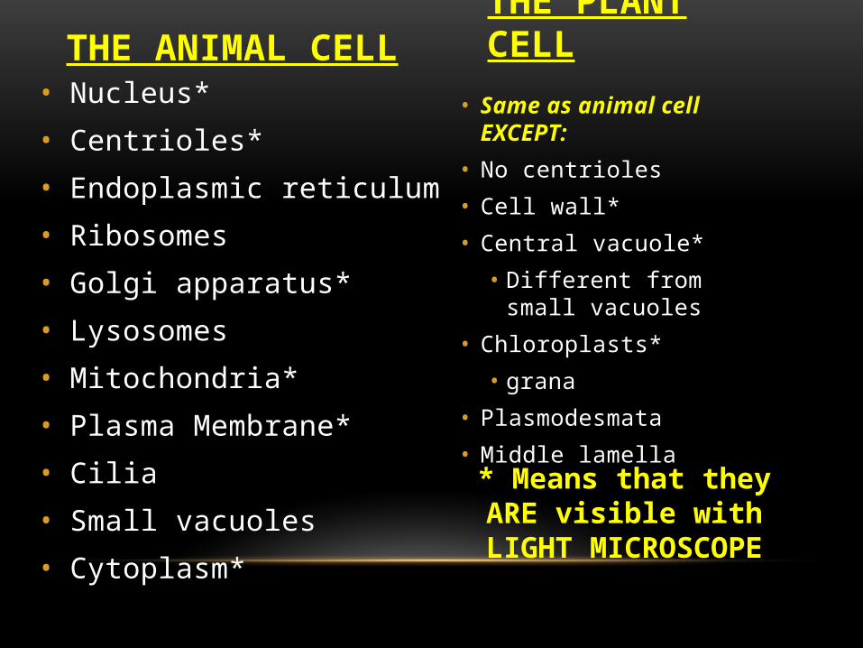
THE ANIMAL CELL• Nucleus*
• Centrioles*
• Endoplasmic reticulum
• Ribosomes
• Golgi apparatus*
• Lysosomes
• Mitochondria*
• Plasma Membrane*
• Cilia
• Small vacuoles
• Cytoplasm*
THE PLANT CELL
• Same as animal cell EXCEPT:
• No centrioles
• Cell wall*
• Central vacuole*
• Different from small vacuoles
• Chloroplasts*
• grana
• Plasmodesmata
• Middle lamella* Means that they ARE
visible with LIGHT MICROSCOPE

• Mesophyll cells
• Spongy
• palisade
• Bundle sheath cells
• Xylem/phloem cells
• Sieve tube elements
• Tracheids
• Root cells
• cortex
• Ciliated epithelial cells
• Muscle cells
• Blood cells
• Nerve cells
TYPES OF CELLS
Animal Cells
Plant Cells

BASIC ORGANELLES• Cell Surface membrane
• At high magnifications (100 000x +) you can see THREE layers (trilaminar appearance)
• Hydrohphilic phosphate outer layer (dark line)
• Hydrophobic fatty acid tail inner layer (light line)
• Hydrophilic phosphate inner layer (dark line)
• 7 nm wide (small!!!!) (0.007 um)
• MICROVILLI finger-like extensions of cell surface membrane
• Typical of animal epithelial cells covering surfaces
• Increase surface area of cell membrane
• Good for absorption in gut and reabsorption in kidney
• Cytoplasm
• Between nucleus and cell surface membrane
• Aqueous, watery material
• Varies from fluid to jelly-like
• Many organelles suspended in here


• Largest organelle
• DOUBLE MEMBRANE
• Surrounded by double membrane called the NUCLEAR ENVELOPE
• Outer membrane of the envelope is continuous with the ENDOPLASMIC RETICULUM
• Nuclear envelope contains many NUCLEAR PORES
• ALLOWS FOR MATERIALS TO BE EXCHANGED BETWEEN NUCLEUS AND CYTOPLASM
• Ex. mRNA and ribosomes leave nucleus
• Ex. Hormones and nutrients enter nucleus
NUCLEUS


INSIDE THE NUCLEUS• Chromatin
• Loosely coiled chromosomes spread out in nucleus of non-dividing cell
• Chromosomes
• Contain DNA organised into functional units called genes
• Genes control the activities of the cell and inheritance
• Gene section of DNA that codes for a specific protein
• Nucleolus
• Dense structure in nucleus that manufactures ribosomes
• Usually darkly stained inside of nucleus in micrographs

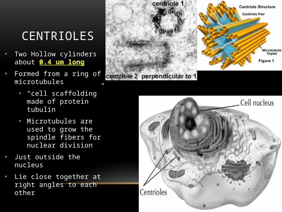
CENTRIOLES• Two Hollow cylinders about 0.4 um
long
• Formed from a ring of microtubules
• “cell scaffolding” made of protein tubulin
• Microtubules are used to grow the spindle fibers for nuclear division
• Just outside the nucleus
• Lie close together at right angles to each other
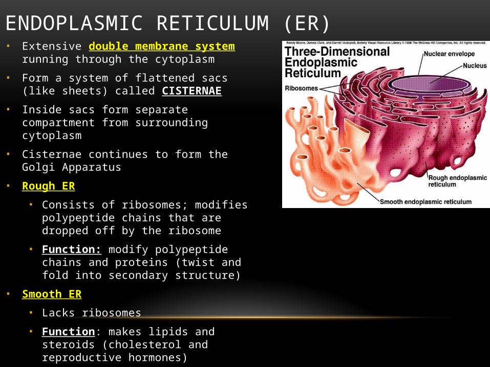
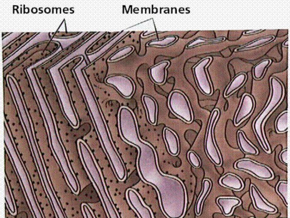
ENDOPLASMIC RETICULUM (ER)• Extensive double membrane system running through
the cytoplasm
• Form a system of flattened sacs (like sheets) called CISTERNAE
• Inside sacs form separate compartment from surrounding cytoplasm
• Cisternae continues to form the Golgi Apparatus
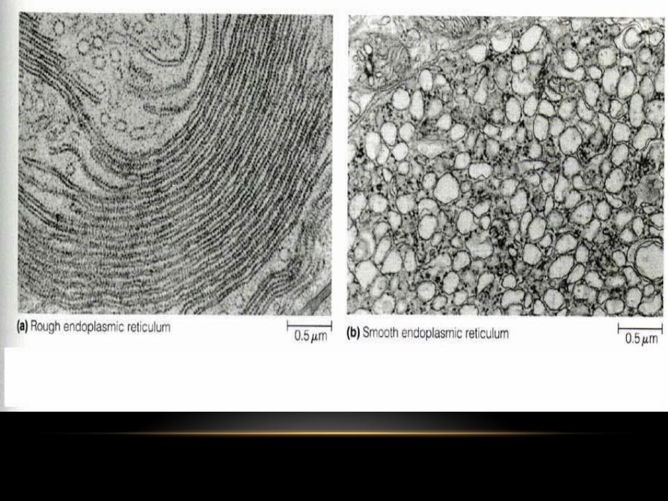
• Rough ER
• Consists of ribosomes; modifies polypeptide chains that are dropped off by the ribosome
• Function: modify polypeptide chains and proteins (twist and fold into secondary structure)
• Smooth ER
• Lacks ribosomes
• Function: makes lipids and steroids (cholesterol and reproductive hormones)
ROUGH ER
• Involved in protein making (synthesis)• So what are we going to see on it?
• ribosomes• Once a protein is made, it leaves the ribosome and
goes into the Rough ER• The rough ER then modifies the protein• All proteins that are exported by the cell are made on
the RER• Membrane proteins are made on the RER too


SMOOTH ER
• NO ribosomes on it
• Looks smooth
• Contains collections of ENZYMES that have specialized tasks• What do enzymes do?
• Tasks include:• Synthesis of membrane lipids
• Detoxification of drugs
• Liver cells• Big in detox therefore….what do u think liver cells have a lot of?


RIBOSOMES• Ribosomal RNA wrapped around protein
• Very small (22 nm in diameter)
• Consists of 2 parts:
• Large subunit
• Small subunit
• Found:
• In Cytoplasm
• On Rough ER
• In nucleus
• Function: hold mRNA in place while tRNA brings over specific amino acids; makes a polypeptide chain
• Site of protein synthesis
• Prokaryotes 70S ribosomes
• Eukaryotes 80S ribosomes (mitochondrial/chloroplasts ribosomes are 70S)


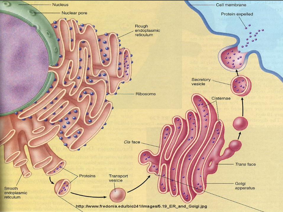
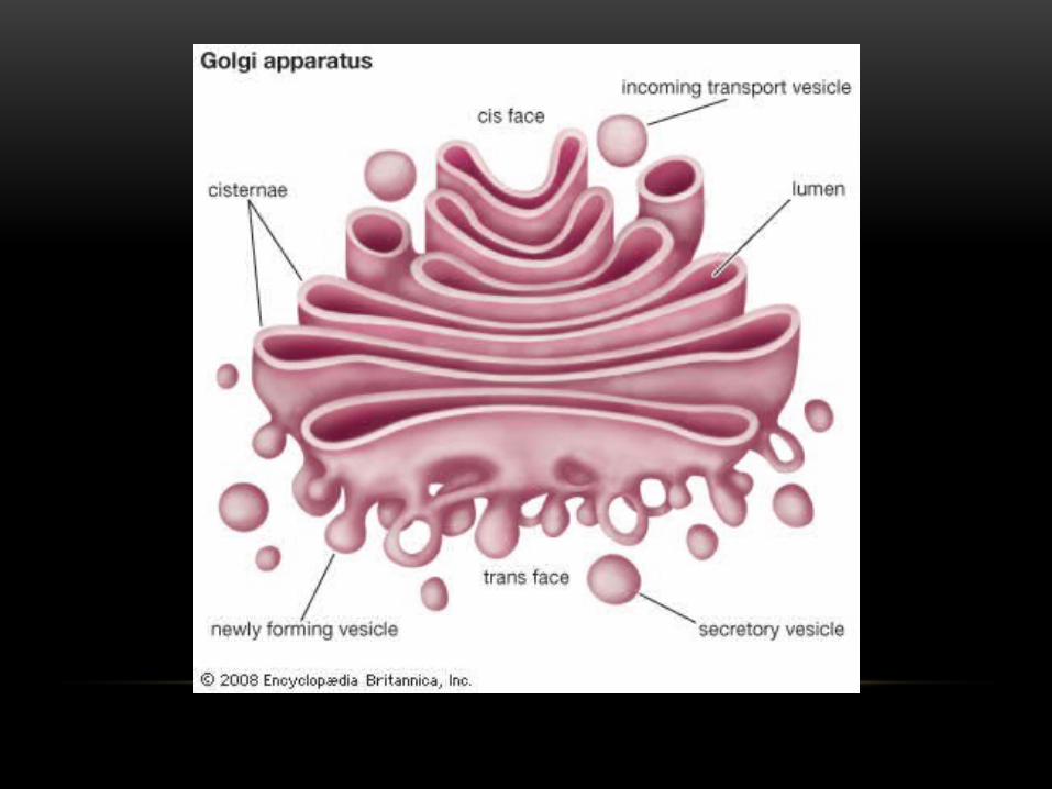
GOLGI APPARATUS (GA)• Stack of flattened sacs (cisternae)
• Constantly being formed at one end from vesicles which bud off from the ER and broken down on the other end to form golgi vesicles that carry the products made in the GA
• FUNCTION: collects, processes and sorts molecules (particularly proteins from the rough ER) ready for transport in Golgi vesicles either to other parts of the cell or out of the cell (secretion)
• They are the CUSTOMIZATION SHOP
• ALSO functions in the creation/production of LYSOSOMES

• Golgi vesicles become lysosomes
• Conversion of sugars into components of the cell wall
• Form secretory vesicles that can release contents via exocytosis
• Addition of sugars to proteins to make Glycoproteins
• Function in cell membrane
• Removal of methoionine (1st amino acid) to create a functioning protein
MORE INFO ON THE GOLGI COMPLEX/BODY….
Examples of Protein Processing Other Important Functions


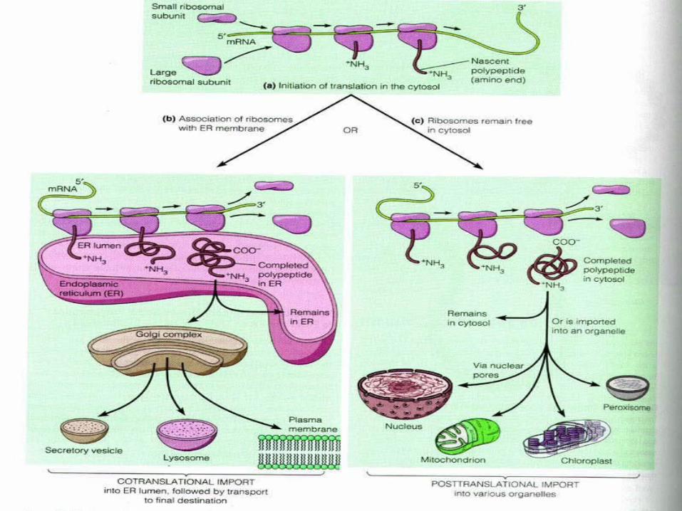
ENDOMEMBRANE SYSTEM & PROTEIN SYNTHESIS
• DNA in nucleus gives message to mRNA
• mRNA leave thru nuclear pore into cytoplasm
• Ribsome “catches” mRNA
• tRNA come over and start adding amino acids together making polypeptide chain
• Polypeptide chain either functions immediately or goes onto next step
• Ribosome deposits polypeptide chain into lumen of the RER
• Polypeptide chain is modified (2* and 3* structure)
• Functioning protein either stays and works in RER or…
• Vesicle buds off RER and transports it to Golgi Apparatus
• Protein is further modified in GA and leaves in a vesicle (either secretory or peroxisome or membrane)
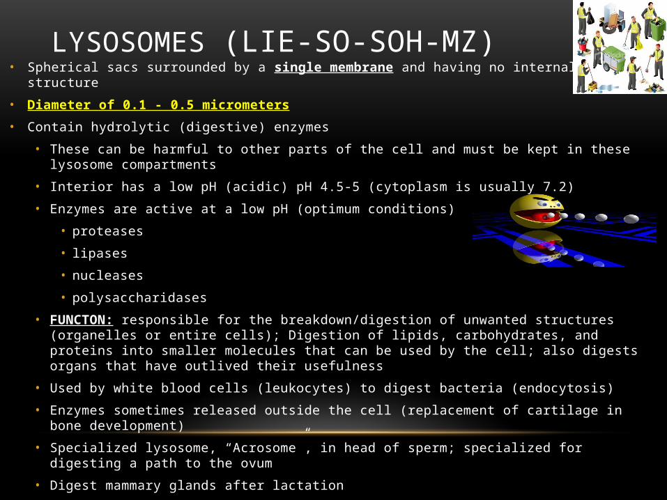
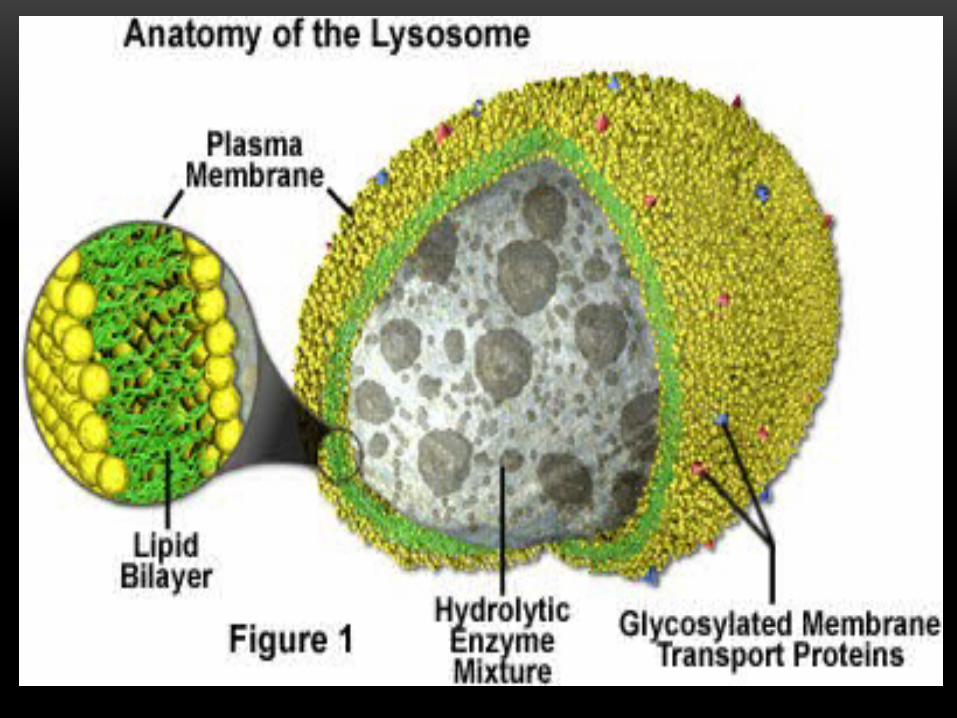
LYSOSOMES (LIE-SO-SOH-MZ)• Spherical sacs surrounded by a single membrane and having no internal structure
• Diameter of 0.1 - 0.5 micrometers
• Contain hydrolytic (digestive) enzymes
• These can be harmful to other parts of the cell and must be kept in these lysosome compartments
• Interior has a low pH (acidic) pH 4.5-5 (cytoplasm is usually 7.2)
• Enzymes are active at a low pH (optimum conditions)
• proteases
• lipases
• nucleases
• polysaccharidases
• FUNCTON: responsible for the breakdown/digestion of unwanted structures (organelles or entire cells); Digestion of lipids, carbohydrates, and proteins into smaller molecules that can be used by the cell; also digests organs that have outlived their usefulness
• Used by white blood cells (leukocytes) to digest bacteria (endocytosis)
• Enzymes sometimes released outside the cell (replacement of cartilage in bone development)
• Specialized lysosome, “Acrosome”, in head of sperm; specialized for digesting a path to the ovum
• Digest mammary glands after lactation

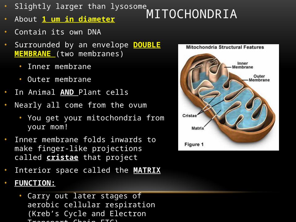
MITOCHONDRIA• Slightly larger than lysosome
• About 1 um in diameter
• Contain its own DNA
• Surrounded by an envelope DOUBLE MEMBRANE (two membranes)
• Inner membrane
• Outer membrane
• In Animal AND Plant cells
• Nearly all come from the ovum
• You get your mitochondria from your mom!
• Inner membrane folds inwards to make finger-like projections called cristae that project
• Interior space called the MATRIX
• FUNCTION:
• Carry out later stages of aerobic cellular respiration (Kreb’s Cycle and Electron Transport Chain…ETC)
• MAKES ATP
• Lipid synthesis

• Much more selective barrier
• Controls precisely what ions and molecules enter the MATRIX
• Protein Porin
• Forms wide aqueous channel in outer membrane
• Easy access for small, water soluble molecules to enter from surrounding cytoplasm into the intermembrane space
MITOCHONDRIA
Outer Membrane
Inner membrane


MITOCHONDRIA=LOTS OF ATP• Site of cell respiration
• ENZYMES important
• Cell Respiration (REQUIRES
OXYGEN=AEROBIC)• 3 stages of Cell respiration
• GLYCOLYSIS
• Makes a lil’ bit of ATP
• Location: CYTOPLASM
• KREBS CYCLE
• Makes a lil’ bit of ATP
• Location: MATRIX of MITOCHONDRIA
• ELECTRON TRANSPORT CHAIN
• Makes A LOT of ATP!!!
• Location: INNER MEMBRANE of MITOCHONDRIA

NO OXYGEN= ANAEROBIC• No oxygen available (or not enough) cell switches to FERMENTATION
• Glycolysis over and over and over
• Happens in CYTOPLASM
• Makes a Little Bit of ATP
• Lactic Acid Fermentation
• Animals
• Muscle cells
• Only can use for a few seconds
• Alcohol Fermentation
• Bacteria and Fungi
• Ex. yeast


ATP HYDROLYSIS
• ATP is a small, water soluble molecule
• Once made in the mitochondria, it can leave and spread to wherever it is needed in the cell
• Energy is released by breaking down ATP ADP + P

ULTRASTRUCTURE OF PLANT CELL
• No centrioles
• Very rarely plant cells will have cilia
• Only plant cells
• Thick cell wall made of cellulose
• Large central vacuole
• chloroplasts

CHLOROPLASTS
• Plant and some Bacteria cells only ( NOT in animal cells)
• Capture energy from the sunlight and convert it into chemical energy…what is this process called?• PHOTOSYNTHESIS
• Like solar power for plants• 2 membranes (Double membrane)
• Inside: large stacks of other membranes that contain chlorophyll


CHLOROPLAST (FOUND IN CELLS IN LEAVES)
• Concentrated in the cells of the mesophyll (inner layer of tissue) in leaf
• Stomata
• Tiny pores on surface of leaf• Allows carbon dioxide and
oxygen in and out of the leaf• Veins
• Carry water and nutrients from roots to leaves
• Deliver organic molecules produced in leaves to other parts of the plant

CHLOROPLAST
• Cellular organelle where photosynthesis takes place• Double membrane
• Outer membrane
• Stroma (fluid filled space)
• Inner membrane
• Thylakoids • Thylakoid membrane contains
CHLOROPHYLL
• Granum
• Intermembrane space
• Contain chemical compound called Chlorophyll• This molecule gives chloroplast its
green color

STRUCTURE OF CHLOROPLAST• Structures organize the many reactions that
take place in photosynthesis
• Stroma
• Thick fluid enclosed by the inner membrane
• Thylakoids
• Disc-like sacs suspended in the stroma
• Has membrane that surrounds inner thylakoid space
• Grana (sing. Granum)
• Stacks of thylakoids

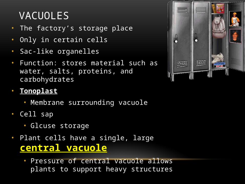
VACUOLES• The factory’s storage place
• Only in certain cells
• Sac-like organelles
• Function: stores material such as water, salts, proteins, and carbohydrates
• Tonoplast
• Membrane surrounding vacuole
• Cell sap
• Glcuse storage
• Plant cells have a single, large central vacuole• Pressure of central vacuole allows plants to support heavy
structures

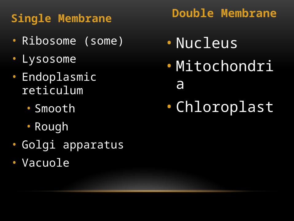
Single Membrane Double Membrane
• Ribosome (some)
• Lysosome
• Endoplasmic reticulum
• Smooth
• Rough
• Golgi apparatus
• Vacuole
• Nucleus• Mitochondria• Chloroplast

Part 3

MICROTUBULES• Long, rigid, hollow tubes
• Small (25 nm in diameter)• Part of cytoskeleton (with actin and
intermediate filaments)
• Protein TUBULIN
• Alpha tubulin
• Beta tublulin
• Alpha + Beta = DIMERS (double molecules)
• PROTOFILAMENTS dimers joined end to end (type of polymerization)
• 13 protofilaments line up next to each other in a ring with hollow center forming microtubule

FUNCTION OF MICROTUBULES
• Support
• Cytoskeleton
• Move secretory vesicles inside and outside the cell
• Nuclear division

MTOC
• MTOCs
• Assemble tubulin to form microtubules in special cell locations
• Microtubule organizing center
• Microtubules formed and broken down here
• Ex. Centrioles (found at base of cilia and flagella)

BASAL BODY

CILIA AND FLAGELLA
• Plural: cilium and flagellum
• Cilia: hundreds of extension of the cell membrane that move like the oars of a boat
• Flagella: one or two long extensions off the cell that move in a whip like fashion
• Enable cells to swim rapidly through liquid

CILIA & FLAGELLA• Long, thin extensions that move in wavelike manner
• Flagella
• Very few extensions
• Long and whip like
• Cilia
• Short extensions (3-4 um long)
• Many present
• Hair like
• Covered with an extension of the plasma membrane
• Contains microtubules (with tubulin protein) that extend throughout its length
• Microtubules arise from structure called the BASAL BODY in cytoplasm
• Identical in structure to centrioles


MOVEMENT OF CILIA AND FLAGELLA
• Caused by microtubules
• Coordinated movement
• Each adjacent cilia is slightly off from its neighbor
• Ripple effect (long grass in the wind)
• Purpose:
• Move substances around the cell (if cell is attached)
• Move the cell itself if it is unattached


ENDOSYMBIONT THEORY
• 1960s
• Mitochondria and chloroplast contain:
• Ribosomes (70S)
• S is unit of measurement of ribosome size
• Measures how fast they sediment in a centrifuge
• Small, circular DNA
• Reproduce
• Make energy
• Mit. & Chl. Were once bacteria that came to live symbiotically in larger cells

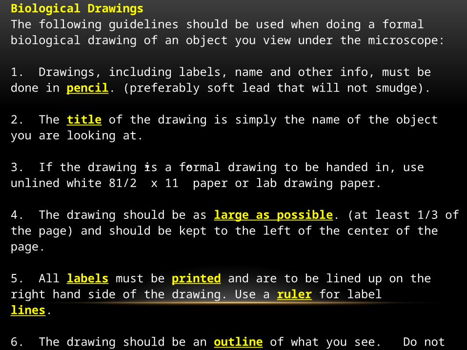
Biological DrawingsThe following guidelines should be used when doing a formal biological drawing of an object you view under the microscope:
1. Drawings, including labels, name and other info, must be done in pencil. (preferably soft lead that will not smudge).
2. The title of the drawing is simply the name of the object you are looking at.
3. If the drawing is a formal drawing to be handed in, use unlined white 81/2” x 11” paper or lab drawing paper.
4. The drawing should be as large as possible. (at least 1/3 of the page) and should be kept to the left of the center of the page.
5. All labels must be printed and are to be lined up on the right hand side of the drawing. Use a ruler for label lines. 6. The drawing should be an outline of what you see. Do not include additional structures just because you think you should see them.
7. Do not shade or sketch. All lines should be solid and complete.
8. Magnification of your drawing MUST be included

Sample


PRACTICE• Anne viewed an amoeba under the high
power 40x objective lens on her microscope and drew the following picture of the amoeba
• She needs to calculate the magnification of her drawing.
• Here is other relevant info:
• Answer the following:a) Convert the diameter of field of view on low power to um.b) Calculate the total magnification under low power and high power.c) Calculate the diameter of the field of view under high power.d) Calculate the size of the object under high power.e) Measure the size of the drawing. Convert to um.f) Calculate the magnification of Anne’s drawing.

Organelle
Plant or Animal (or
both)
Eukaryotic or Prokaryotic or
Both
Size in
um
Size in
nm
Single, double, or
no membrane
Function LM? EM?

MICROSCOPE LAB WORK• Part 1
• Calibrate your eye piece graticule at low, medium and high power
• Determine the field of view at low, medium and high power
• Complete the chart below
• Part 2
• Complete a plan diagram of the plant cell on slide 2
• Make sure to follow the guidelines for biological drawings
• Include the total
Large unit of EG
Medium unit of EG
Small Unit of EG
FOV
Low Power
Medium Power
High Power