agenda 11/28 start development –watch video and do slides note - during slides take notes when...
TRANSCRIPT

Agenda 11/28
Start Development –watch video and do slides http://www.youtube.com/watch?v=_22CFCxDUy0
Note - during slides take notes when indicated (material that you didn’t read but you should know/study for quiz)
Homework - • Finish 47.3 notes, concept checks and online
assignment due tomorrow• Quiz Friday - 11,12, 13, 47.3 all fair game (the more you
study, the better off you will be on unit test in 2 weeks)

• Development occurs at many points in the life cycle of an animal
• This includes metamorphosis and gamete production, as well as embryonic development
© 2011 Pearson Education, Inc.

Figure 47.2
EMBRYONIC DEVELOPMENTSperm
Adultfrog
Egg
Metamorphosis
Larvalstages
Zygote
Blastula
Gastrula
Tail-budembryo
FE
RT
ILIZ
AT
ION
CLEAVAGE
GASTRULATION
OR
GA
NO
-
GE
NE
SIS

• Although animals display different body plans, they share many basic mechanisms of development and use a common set of regulatory genes
• Biologists use model organisms to study development, chosen for the ease with which they can be studied in the laboratory
© 2011 Pearson Education, Inc.

Concept 47.1: Fertilization and cleavage initiate embryonic development
• Fertilization is the formation of a diploid zygote from a haploid egg and sperm
© 2011 Pearson Education, Inc.

Fertilization• Molecules and events at the egg surface play a
crucial role in each step of fertilization– Sperm penetrate the protective layer around the
egg
– Receptors on the egg surface bind to molecules on the sperm surface
– Changes at the egg surface prevent polyspermy, the entry of multiple sperm nuclei into the egg
© 2011 Pearson Education, Inc.

Cleavage• Fertilization is followed by cleavage, a period of
rapid cell division without growth• Cleavage partitions the cytoplasm of one large cell
into many smaller cells called blastomeres• The solid ball of cells is called a morula• The blastula is a ball of cells with a fluid-filled
cavity called a blastocoel
© 2011 Pearson Education, Inc.
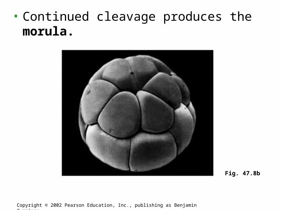
• Continued cleavage produces the morula.
Copyright © 2002 Pearson Education, Inc., publishing as Benjamin Cummings
Fig. 47.8b

– Except for mammals, most animals have both eggs and zygotes with a definite polarity.• Thus, the planes of division follow a specific pattern
relative to the poles of the zygote.• Polarity is defined by the heterogeneous distribution
of substances such as mRNA, proteins, and yolk.– Yolk is most concentrated at the vegetal pole and least
concentrated at the animal pole.
• In some animals, the animal pole defines the anterior end of the animal.
Copyright © 2002 Pearson Education, Inc., publishing as Benjamin Cummings

• A blastocoel forms within the morula blastula
Copyright © 2002 Pearson Education, Inc., publishing as Benjamin Cummings
Fig. 47.8d

Figure 47.6
(a) Fertilized egg (b) Four-cell stage (c) Early blastula (d) Later blastula
50 m
• Animal embryos complete cleavage when the ratio of material in the nucleus relative to the cytoplasm is sufficiently large
© 2011 Pearson Education, Inc.
Regulation of Cleavage

• After cleavage, the rate of cell division slows and the normal cell cycle is restored
• Morphogenesis, the process by which cells occupy their appropriate locations, involves
– Gastrulation, the movement of cells from the blastula surface to the interior of the embryo
– Organogenesis, the formation of organs
© 2011 Pearson Education, Inc.
Concept 47.2: Morphogenesis in animals involves specific changes in
cell shape, position, and survival

Gastrulation• Gastrulation rearranges the cells of a blastula
into a three-layered embryo, called a gastrula
© 2011 Pearson Education, Inc.

• The three layers produced by gastrulation are called embryonic germ layers
– The ectoderm forms the outer layer
– The endoderm lines the digestive tract
– The mesoderm partly fills the space between the endoderm and ectoderm
• Each germ layer contributes to specific structures in the adult animal
© 2011 Pearson Education, Inc.

Go to study area online – Ch. 47
• Watch video of sea urchin development
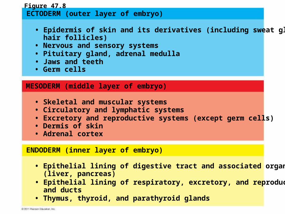
ECTODERM (outer layer of embryo)
MESODERM (middle layer of embryo)
ENDODERM (inner layer of embryo)
• Epidermis of skin and its derivatives (including sweat glands, hair follicles)
• Epithelial lining of digestive tract and associated organs (liver, pancreas)• Epithelial lining of respiratory, excretory, and reproductive tracts and ducts
• Germ cells• Jaws and teeth• Pituitary gland, adrenal medulla• Nervous and sensory systems
• Skeletal and muscular systems• Circulatory and lymphatic systems• Excretory and reproductive systems (except germ cells)• Dermis of skin• Adrenal cortex
• Thymus, thyroid, and parathyroid glands
Figure 47.8

• Gastrulation begins at the vegetal pole of the blastula
• Mesenchyme cells migrate into the blastocoel• The vegetal plate forms from the remaining cells of
the vegetal pole and buckles inward through invagination
© 2011 Pearson Education, Inc.
Gastrulation in Sea Urchins

• The newly formed cavity is called the archenteron
• This opens through the blastopore, which will become the anus
© 2011 Pearson Education, Inc.

Animalpole
Blastocoel
Mesenchymecells
Vegetal plateVegetalpole
Blastocoel
Filopodia
Mesenchymecells
Blastopore
Archenteron
50 m
Ectoderm
Mouth
Mesenchyme(mesoderm formsfuture skeleton)
Blastopore
Blastocoel
Archenteron
Digestive tube (endoderm)
Anus (from blastopore)
Key
Future ectodermFuture mesodermFuture endoderm
Figure 47.9

• Frog gastrulation begins when a group of cells on the dorsal side of the blastula begins to invaginate
• This forms a crease along the region where the gray crescent formed
• The part above the crease is called the dorsal lip of the blastopore
© 2011 Pearson Education, Inc.
Gastrulation in Frogs

• Cells continue to move from the embryo surface into the embryo by involution
• These cells become the endoderm and mesoderm
• Cells on the embryo surface will form the ectoderm
© 2011 Pearson Education, Inc.

Key
Future ectodermFuture mesodermFuture endoderm
SURFACE VIEW CROSS SECTIONAnimal pole
Vegetal poleEarlygastrula
Blastocoel
Dorsal lip ofblasto-pore
BlastoporeDorsal lip ofblastopore
Blastocoelshrinking
Archenteron
Archenteron
Blastocoelremnant
EctodermMesodermEndoderm
Blastopore
Yolk plugBlastopore
Lategastrula
3
2
1
Figure 47.10

Organogenesis
• During organogenesis, various regions of the germ layers develop into rudimentary organs
• Early in vertebrate organogenesis, the notochord forms from mesoderm, and the neural plate forms from ectoderm
© 2011 Pearson Education, Inc.

Figure 47.13
Neural folds
1 mm
Neural fold
Neural plate
Notochord
Ectoderm
Mesoderm
Endoderm
Archenteron
(a) Neural plate formation
(b) Neural tube formation
(c) Somites
Neural fold
Neural plate
Neural crest cells
Outer layerof ectoderm
Neural crest cells
Neural tube
Eye Somites Tail bud
SEM
Neural tube
Notochord
Coelom
Neuralcrestcells
Somite
Archenteron(digestivecavity)
1 mm

• The neural plate soon curves inward, forming the neural tube
• The neural tube will become the central nervous system (brain and spinal cord)
NOW – WATCH FROG DEVELOPMENT VIDEO FROM WEBSITE – ALSO FETAL ULTRASOUNDS
© 2011 Pearson Education, Inc.
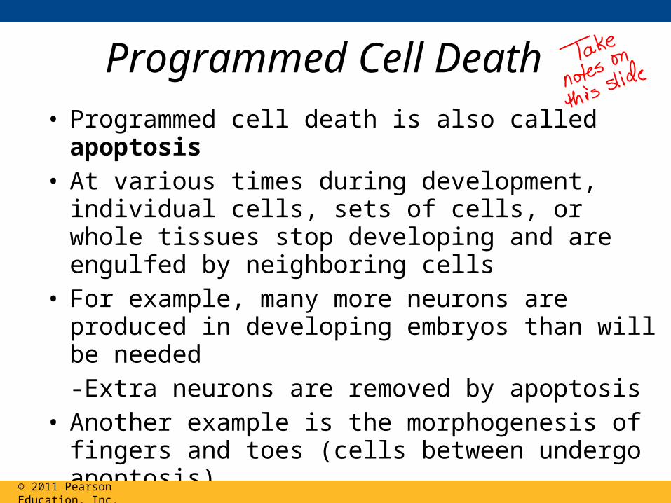
• Programmed cell death is also called apoptosis• At various times during development, individual
cells, sets of cells, or whole tissues stop developing and are engulfed by neighboring cells
• For example, many more neurons are produced in developing embryos than will be needed-Extra neurons are removed by apoptosis
• Another example is the morphogenesis of fingers and toes (cells between undergo apoptosis)
© 2011 Pearson Education, Inc.
Programmed Cell Death

Agenda 11/29• Continue development (slides from 18.4 – this material used to be in 47.3 in
old edition) through bicoid• Take a study break – review/quiz each other on material below
What to study for the quiz:– Cell signaling notes and diagrams in book– Mitosis/Meiosis lab and diagrams– Development notes (Powerpoint posted online) – won’t be anything on quiz
that don’t get through today• Start 47.3 slides – aim to get through fate mapping
• Homework – • Study for quiz tomorrow- 11,12, 13, 47.3 all fair game (the more you study,
the better off you will be on unit test in 2 weeks)

Concept 18.4: A program of differential gene expression leads to the different cell
types in a multicellular organism
• During embryonic development, a fertilized egg gives rise to many different cell types
• Cell types are organized successively into tissues, organs, organ systems, and the whole organism
• Gene expression orchestrates the developmental programs of animals
© 2011 Pearson Education, Inc.

A Genetic Program for Embryonic Development
• The transformation from zygote to adult results from cell division, cell differentiation, and morphogenesis
© 2011 Pearson Education, Inc.
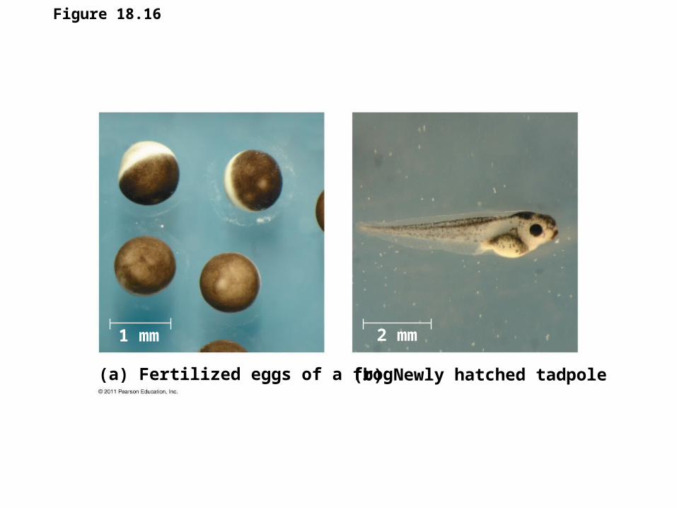
Figure 18.16
(a) Fertilized eggs of a frog (b) Newly hatched tadpole
1 mm 2 mm

• Cell differentiation is the process by which cells become specialized in structure and function
• The physical processes that give an organism its shape constitute morphogenesis
• Differential gene expression results from genes being regulated differently in each cell type
• Materials in the egg can set up gene regulation that is carried out as cells divide
© 2011 Pearson Education, Inc.

Cytoplasmic Determinants and Inductive Signals
• An egg’s cytoplasm contains RNA, proteins, and other substances that are distributed unevenly in the unfertilized egg
• Cytoplasmic determinants are maternal substances in the egg that influence early development
• As the zygote divides by mitosis, cells contain different cytoplasmic determinants, which lead to different gene expression
© 2011 Pearson Education, Inc.
TAKE NOTES
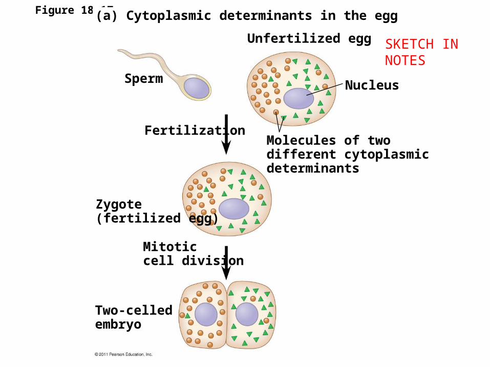
Figure 18.17a(a) Cytoplasmic determinants in the egg
Unfertilized egg
Sperm
Fertilization
Zygote(fertilized egg)
Mitoticcell division
Two-celledembryo
Nucleus
Molecules of twodifferent cytoplasmicdeterminants
SKETCH IN NOTES

• The other important source of developmental information is the environment around the cell, especially signals from nearby embryonic cells
• In the process called induction, signal molecules from embryonic cells cause transcriptional changes in nearby target cells
• Thus, interactions between cells induce differentiation of specialized cell types
© 2011 Pearson Education, Inc.
TAKE NOTES

Figure 18.17b (b) Induction by nearby cells
Early embryo(32 cells)
NUCLEUS
Signaltransductionpathway
Signalreceptor
Signalingmolecule(inducer)
SKETCH IN NOTES

© 2011 Pearson Education, Inc.
Animation: Cell Signaling
Right-click slide / select “Play”
NICE REVIEW OF CH. 11 CELL SIGNALING

Sequential Regulation of Gene Expression During Cellular
Differentiation• Determination commits a cell to its final fate• Determination precedes differentiation• Cell differentiation is marked by the production of
tissue-specific proteins
© 2011 Pearson Education, Inc.
TAKE NOTES

Pattern Formation: Setting Up the Body Plan
• Pattern formation is the development of a spatial organization of tissues and organs
• In animals, pattern formation begins with the establishment of the major axes
• Positional information, the molecular cues that control pattern formation, tells a cell its location relative to the body axes and to neighboring cells
© 2011 Pearson Education, Inc.
KNOW THESE VOCAB WORDS –THEY SHOULD BE IN YOUR CORNELL NOTES

Axis Establishment• Maternal effect genes encode for cytoplasmic
determinants that initially establish the axes of the body of Drosophila
• These maternal effect genes are also called egg-polarity genes because they control orientation of the egg and consequently the fly
© 2011 Pearson Education, Inc.

© 2011 Pearson Education, Inc.
Animation: Development of Head-Tail Axis in Fruit Flies
Right-click slide / select “Play”

• One maternal effect gene, the bicoid gene, affects the front half of the body
• An embryo whose mother has no functional bicoid gene lacks the front half of its body and has duplicate posterior structures at both ends
Bicoid: A Morphogen Determining Head Structures
© 2011 Pearson Education, Inc.

Figure 18.21
Head Tail
Tail Tail
Wild-type larva
Mutant larva (bicoid)
250 m
T1 T2 T3A1 A2 A3 A4 A5 A6
A7A8
A8A7A6A7
A8

• This phenotype suggests that the product of the mother’s bicoid gene is concentrated at the future anterior end
• This hypothesis is an example of the morphogen gradient hypothesis, in which gradients of substances called morphogens establish an embryo’s axes and other features
© 2011 Pearson Education, Inc.
TAKE NOTES – KNOW BICOID EXAMPLE

Figure 18.22
Bicoid mRNA in matureunfertilized egg
Bicoid mRNA in matureunfertilized egg
Fertilization,translation ofbicoid mRNA
Anterior end100 m
Bicoid protein inearly embryo
Bicoid protein inearly embryo
RESULTS
• The bicoid research is important for three reasons– It identified a specific protein required for some
early steps in pattern formation
– It increased understanding of the mother’s role in embryo development
– It demonstrated a key developmental principle that a gradient of molecules can determine polarity and position in the embryo
© 2011 Pearson Education, Inc.
PAUSE HERE FOR STUDY BREAK

Concept 47.3: Cytoplasmic determinants and inductive signals contribute to cell fate specification
• Determination is the term used to describe the process by which a cell or group of cells becomes committed to a particular fate
• Differentiation refers to the resulting specialization in structure and function
© 2011 Pearson Education, Inc.
Fate Mapping• Fate maps are diagrams showing organs and
other structures that arise from each region of an embryo
• Classic studies using frogs indicated that cell lineage in germ layers is traceable to blastula cells
© 2011 Pearson Education, Inc.

Epidermis EpidermisCentralnervoussystemNotochord
Mesoderm
Endoderm
Blastula Neural tube stage(transverse section)
(a) Fate map of a frog embryo
64-cell embryos
Blastomeresinjected with dye
Larvae
(b) Cell lineage analysis in a tunicate
Figure 47.17

• Later studies of C. elegans used the ablation (destruction) of single cells to determine the structures that normally arise from each cell
• The researchers were able to determine the lineage of each of the 959 somatic cells in the worm
© 2011 Pearson Education, Inc.

First cell divisionZygote
HatchingTim
e a
fter
fer
tiliz
ati
on
(h
ou
rs)
Intestine
Intestine
MouthEggs Vulva
Anus
1.2 mmANTERIOR POSTERIOR
Nervoussystem,outer skin,muscula-ture
Muscula-ture, gonads
Outer skin,nervous system
Germ line(futuregametes)
Musculature
10
0Figure 47.18

• Germ cells are the specialized cells that give rise to sperm or eggs
• Complexes of RNA and protein are involved in the specification of germ cell fate
• In C. elegans, such complexes are called P granules, persist throughout development, and can be detected in germ cells of the adult worm
• P granules act as cytoplasmic determinants, fixing germ cell fate at the earliest stage of development
© 2011 Pearson Education, Inc.
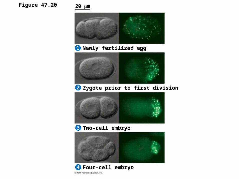
Figure 47.20
Newly fertilized egg
Zygote prior to first division
Two-cell embryo
Four-cell embryo
20 m
2
1
3
4

Figure 47.19
100 m

Axis Formation• A body plan with bilateral symmetry is found
across a range of animals• This body plan exhibits asymmetry across the
dorsal-ventral and anterior-posterior axes• The right-left axis is largely symmetrical
© 2011 Pearson Education, Inc.

• The anterior-posterior axis of the frog embryo is determined during oogenesis
• The animal-vegetal asymmetry indicates where the anterior-posterior axis forms (animal is embryo, vegetal is yolk)
• The dorsal-ventral axis is not determined until fertilization
© 2011 Pearson Education, Inc.
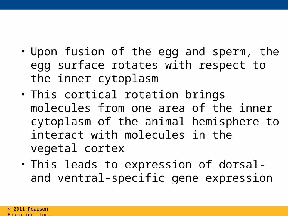
• Upon fusion of the egg and sperm, the egg surface rotates with respect to the inner cytoplasm
• This cortical rotation brings molecules from one area of the inner cytoplasm of the animal hemisphere to interact with molecules in the vegetal cortex
• This leads to expression of dorsal- and ventral-specific gene expression
© 2011 Pearson Education, Inc.

Dorsal
Right
Anterior Posterior
VentralLeft
(a) The three axes of the fully developed embryo
(b) Establishing the axes
Animalhemisphere
Vegetalhemisphere
Animal pole
Vegetal pole
Point ofspermnucleusentry
Gray crescent
Pigmentedcortex
Futuredorsalside
First cleavage
Figure 47.21

• In chicks, gravity is involved in establishing the anterior-posterior axis
• Later, pH differences between the two sides of the blastoderm establish the dorsal-ventral axis
• In mammals, experiments suggest that orientation of the egg and sperm nuclei before fusion may help establish embryonic axes
© 2011 Pearson Education, Inc.

Restricting Developmental Potential
• Hans Spemann performed experiments to determine a cell’s developmental potential (range of structures to which it can give rise)
• Embryonic fates are affected by distribution of determinants and the pattern of cleavage
• The first two blastomeres of the frog embryo are totipotent (can develop into all the possible cell types)
© 2011 Pearson Education, Inc.

Control egg(dorsal view)
1a 1b
Graycrescent
Controlgroup
Experimentalgroup
Experimental egg(side view)
Graycrescent
EXPERIMENT
Thread
Figure 47.22-1

Control egg(dorsal view)
2
1a 1b
Graycrescent
Controlgroup
Experimentalgroup
Experimental egg(side view)
Graycrescent
Thread
Normal NormalBelly piece
EXPERIMENT
RESULTS
Figure 47.22-2

• In mammals, embryonic cells remain totipotent until the 8-cell stage, much longer than other organisms
• Progressive restriction of developmental potential is a general feature of development in all animals
• In general tissue-specific fates of cells are fixed by the late gastrula stage
© 2011 Pearson Education, Inc.

Cell Fate Determination and Pattern Formation by Inductive Signals
• As embryonic cells acquire distinct fates, they influence each other’s fates by induction
© 2011 Pearson Education, Inc.

The “Organizer” of Spemann and Mangold
• Spemann and Mangold transplanted tissues between early gastrulas and found that the transplanted dorsal lip triggered a second gastrulation in the host
• The dorsal lip functions as an organizer of the embryo body plan, inducing changes in surrounding tissues to form notochord, neural tube, and so on
© 2011 Pearson Education, Inc.

Figure 47.23
Dorsal lip ofblastopore
Pigmentedgastrula
(donor embryo)Nonpigmented
gastrula(recipient embryo)
Primary embryo
Secondary (induced) embryo
Primary structures:Neural tubeNotochord
Secondary structures:Notochord (pigmented cells)Neural tube(mostly nonpigmented cells)
EXPERIMENT RESULTS

Formation of the Vertebrate Limb • Inductive signals play a major role in pattern
formation, development of spatial organization• The molecular cues that control pattern formation
are called positional information• This information tells a cell where it is with respect
to the body axes• It determines how the cell and its descendents
respond to future molecular signals
© 2011 Pearson Education, Inc.

• The wings and legs of chicks, like all vertebrate limbs, begin as bumps of tissue called limb buds
© 2011 Pearson Education, Inc.

Figure 47.24
Limb buds
50 m
AnteriorLimb bud
AER
ZPA
Posterior
Apicalectodermalridge (AER)
(a) Organizer regions (b) Wing of chick embryo
Digits
Anterior
Proximal
Dorsal
Posterior
Ventral
Distal
2
34

• The embryonic cells in a limb bud respond to positional information indicating location along three axes
– Proximal-distal axis
– Anterior-posterior axis
– Dorsal-ventral axis
© 2011 Pearson Education, Inc.

• One limb bud–regulating region is the apical ectodermal ridge (AER)
• The AER is thickened ectoderm at the bud’s tip• The second region is the zone of polarizing
activity (ZPA)• The ZPA is mesodermal tissue under the
ectoderm where the posterior side of the bud is attached to the body
© 2011 Pearson Education, Inc.

• Tissue transplantation experiments support the hypothesis that the ZPA produces an inductive signal that conveys positional information indicating “posterior”
© 2011 Pearson Education, Inc.

Figure 47.25
Donorlimbbud
Hostlimbbud
ZPA
Anterior
Posterior
New ZPA
4
4
3
3
2 2
EXPERIMENT
RESULTS

• Sonic hedgehog is an inductive signal for anterior/posterior limb development – gradient from ZPA
• Hox genes also play roles during limb pattern formation
© 2011 Pearson Education, Inc.© 2011 Pearson Education, Inc.

Cilia and Cell Fate• Ciliary function is essential for proper specification of cell
fate in the human embryo• Motile cilia play roles in left-right specification (always
sweep fluid to the left, creating slight asymmetry of morphogens on left-right axis)
• Monocilia (nonmotile cilia) are on every cell and act as antenna to receive signal proteins (such as Sonic hedgehog) – play roles in normal kidney development
© 2011 Pearson Education, Inc.

Figure 47.26
Lungs
Heart
Liver
Spleen
Stomach
Large intestine
Normal locationof internal organs
Location insitus inversus
Caused by defect in motile cilia in a particular part of the embryo