age, gene/environment susceptibility – … · age, gene/environment susceptibility – reykjavik...
TRANSCRIPT

AGE, GENE/ENVIRONMENT SUSCEPTIBILITY – REYKJAVIK STUDY:
Multidisciplinary Applied Phenomics
IN PRESS, AMERICAN JOURNAL OF EPIDEMIOLOGY
DECEMBER 18, 2006
Abstract Word Count: 181
Manuscript Word Count: 3521
Medical Subject Headings (keywords): Aging, Population Genetics, Phenotype, Epidemiology,
Cognition, Cardiovascular Disease, Osteoporosis, Body composition
Writing Committee: Tamara B. Harris, M.D., M.S.1, Lenore J. Launer, Ph.D., M.Sc.1, Gudny
Eiriksdottir, M.Sc.2, Olafur Kjartansson, M.D.3, Palmi V. Jonsson ,M.D. 3,4, Gunnar Sigurdsson,
M.D., Ph.D. 3,4, Gudmundur Thorgeirsson, M.D., Ph.D. 3,4, Thor Aspelund, Ph.D.2, Melissa E.
Garcia, M.P.H. 1, Mary Frances Cotch, Ph.D. 5, Howard J. Hoffman, M.A. 6, Vilmundur
Gudnason, M.D., Ph.D.2,4 for the Age, Gene/Environment Susceptibility -Reykjavik Study
Investigators*
1. Laboratory of Epidemiology, Demography, and Biometry, Intramural Research Program,
National Institute on Aging, Bethesda, MD, USA
2. Icelandic Heart Association, Kopavogur, Iceland
3. Landspitali University Hospital, Reykjavik, Iceland
4. Faculty of Medicine, University of Iceland, Reykjavik, Iceland
5. National Eye Institute, Bethesda, MD, USA
Collaborators: Uggi Agnarsson, M.D. 2,3, Rafn Benediktsson, M.D., Ph.D. 3,4, Halldora
Bjornsdottir, M.D. 3, Bjorn Einarsson, M.D. 3, Jon H. Eliasson M.D. 3, Adalsteinn Gudmundsson,

2
M.D. 3, Thorvaldur Ingvarsson, M.D., Ph.D. (FSA University Hospital – Regional Hospital),
Fridbert Jonasson, M.D., Ph.D. 3,4, Birna Jonsdottir, M.D.2, Maria Jonsdottir, Ph.D. 3, Helgi
Jonsson, M.D., PhD. 3,4, Johannes Kari Kristinsson, M.D., Ph.D. (Sjónlag), Smari Kristinsson,
M.Sc. (Raförninn), Stefan Kristjansson, M.D. (FORNIX), Elin Olafsdottir, M.Sc. M.D. 2, Hannes
Petersen, M.D., Ph.D. 3,4, Nikulas Sigfusson, M.D., Dr. Med. 2, Kristin Siggeirsdottir, O.T.,
M.Sc. 2, Thordur Sigmundsson, M.D. 3, Albert P. Sigurdsson M.D. 3, Sigurdur Sigurdsson, M.Sc.
2, Laufey Steingrimsdottir, Ph.D. (Public Health Institute of Iceland), Sigurlaug
Sveinbjornsdottir, M.D. 3 Consultants: Andrew E. Arai, M.D. (National Heart, Lung, and
Blood, Institute, USA), Michiel Bots, M.D., Ph.D. (Utrecht University, Netherlands), Robert
Detrano, M.D., Ph.D. (University of California, Los Angeles, USA), John Hardy, Ph.D.
(National Institute on Aging, USA), Ron Klein (University of Wisconsin, USA), Barbara Klein
(University of Wisconsin, USA), Thomas F. Lang, Ph.D. (University of California, San
Francisco, USA), Oscar Lopez, M.D. (University of Pittsburgh, USA), Rudy Meijer, M.S.
(Utrecht University, Netherlands), David Owens, M.D. (University of Washington, USA),
Jonathan Plehn, M.D. (George Washington University School of Medicine, USA), Ilmari
Pyykko, M.D., Ph.D. (University of Tampere, Finland), Mark A. van Buchem, M.D., Ph.D.
(University Medical School, Netherlands), Alex Zijdenbos, Ph.D. (Neuralyse, Canada).
Advisory Committee: Vladimir Hachinski, M.D., Ph.D. (University of Western Ontario,
Canada), Nick Bryan, M.D., Ph.D. (University of Pennsylvania, USA), Steven Cummings, M.D.,
M.P.H. (California Pacific Medical Center Research Institute, USA), Karl Tryggvason, M.D.,
Ph.D. (Karolinska Institute, Sweden), Robert Ferrell, Ph.D. (University of Pittsburgh, USA).
Corresponding author: Tamara B. Harris, M.D., M.S., Laboratory of Epidemiology,
Demography and Biometry, Intramural Research Program, National Institute on Aging.

3
Acknowledgements: This study has been funded by NIH contract N01-AG-12100, the NIA
Intramural Research Program, Hjartavernd (the Icelandic Heart Association), and the Althingi
(the Icelandic Parliament). Components of the study were also supported by the NEI, NIDCD,
and NHLBI.

4
ABSTRACT
Anticipating the sequencing of the human genome and description of the human
proteome, the Age, Gene/Environment Susceptibility-Reykjavik Study (AGES-Reykjavik) was
initiated in 2002. AGES-Reykjavik was designed to examine risk factors, including genetic
susceptibility and gene/environment interaction, in relation to disease and disability in old age.
The study is multidisciplinary, providing detailed phenotypes related to the cardiovascular,
neurocognitive (including sensory), and musculoskeletal systems, and to body composition and
metabolic regulation. Relevant quantitative traits, subclinical indicators of disease, and medical
diagnoses are identified using biomarkers, imaging, and other physiologic indicators. The
AGES-Reykjavik sample is drawn from an established population-based cohort, the Reykjavik
Study. This cohort of men and women born between 1907 and 1935 has been followed in
Iceland since 1967 by the Icelandic Heart Association. The AGES-Reykjavik cohort, with
cardiovascular risk factor assessments earlier in life and detailed late life phenotypes of
quantitative traits, will create a comprehensive study of aging nested in a relatively genetically
homogeneous older population. This approach should facilitate identification of genetic factors
that contribute to healthy aging as well as the chronic conditions common in old age.

5
Aging is a complex process that reflects a person’s social and biologic history. Aging
may be accompanied by multiple pathologic conditions that increase disease, reduce cognitive
and physical function, and impair quality of life. To understand better the determinants of aging,
identify potential therapeutic interventions, and design effective prevention programs, a
multidisciplinary approach to study well-defined older populations is needed. This approach
also lends itself well to the study of genetics since the effects of genes often extend well beyond
the single organ system to which a gene was thought to contribute. The rationale for establishing
comprehensively evaluated phenotypes across organ systems was described by Freimer and
Sabatti in what they term the “The Human Phenome Project.” (1). The Age, Gene/Environment
Susceptibility-Reykjavik Study (AGES-Reykjavik) was conceived and designed to provide an
approach to study, among other risk factors, the genetic contribution to conditions of old age.
This paper describes the rationale and design of AGES-Reykjavik, the measurements included in
the study, and provides select descriptive data on the first 2,300 participants.
MATERIALS AND METHODS
Study rationale
AGES-Reykjavik is based on three general hypotheses: first, that genetic variation
contributes to disease occurring in old age; second, that selected diseases common in old age
share genetic, behavioral, and environmental risk factors; and third, that better classification of
phenotypes based on multiple streams of data, including midlife history and subclinical disease,
will further the exploration of how these risk factors are associated with complex traits and
diseases manifest late in life.
AGES-Reykjavik is an epidemiologic study focusing on four biologic systems: vascular,

6
neurocognitive (including sensory), musculoskeletal, and body composition/metabolism. These
four systems were chosen because similar risk factors contribute to physiological changes and
disease in these systems. For instance, inflammation is associated with atherosclerosis (2, 3)
diabetes (4), obesity (5), smoking-related illnesses (6), dementia (7), osteoporosis (8), and
macular degeneration (9).
AGES-Reykjavik stems from the Reykjavik Study, a cohort established in 1967 to
prospectively study cardiovascular disease in Iceland. Combining midlife data from the
Reykjavik Study and old age data from the AGES-Reykjavik allows a life course approach to
better characterize phenotypes. This combination of data can be used to identify patterns of risk
factors and evaluate whether these patterns have remained stable or changed with age. For
instance, previous studies demonstrate convincingly that risk factors such as blood pressure,
weight, and cholesterol measured in late life are influenced by prevalent old age morbidities and
no longer reflect the exposures that initiated these pathologies (10, 11). Furthermore, the midlife
data is unbiased with regard to health history and is far better than retrospective recall.
Apart from improved phenotypic description, the availability of the mid-life data allows
for a complete assessment of nonresponse, particularly how death and refusals might contribute
to bias. This assessment will be enhanced by additional information from hospital records, a
national mortality index with authentication of all death certificates, a Minimum Data Set for
Nursing Home (MDS-NH) and home-care patients (MDS-HC), and archival information from
birth records all available for linkage with the cohort.
To define quantitative traits, subclinical and clinical disease, AGES-Reykjavik includes
extensive state-of-the-art imaging techniques, biochemical measurements, and diagnostic
evaluations. These measures should provide insights into preclinical disease states, identify
patterns of concomitant traits, and increase our ability to understand prognostic indicators

7
underlying pathophysiologic changes. Imaging techniques yield standardized information on
morphometry of organs and tissues in vivo. Use of imaging in epidemiologic studies has been an
effective way to understand subclinical disease particularly in the fields of osteoporosis (12),
atherosclerosis (13), brain structure (14), and body composition (15). Since the imaging
protocols used in AGES-Reykjavik are similar to protocols in other studies (16, 17), we can
compare data directly with these studies. This multi-measurement strategy of phenotypic
definition offers important advantages, and has been successfully employed elsewhere (18).
Some characteristics of Iceland and the Icelandic population should enhance the power to
examine genetic and gene-environment interactions that modulate expression of genes in old age.
The Icelandic population is relatively genetically homogeneous (19), which reduces the problem
of population stratification. Thus, a greater proportion of people at the phenotypic extremes may
share the same genetic susceptibility. Genealogic databases in Iceland allow identification of
relationships in the cohort. The relative isolation and hardship due to deadly infectious
epidemics, few major roads, and foreign rule, coupled with volcanic soil and cold climate, lead
to restricted diet and increased physical activity, until recently. Nonetheless, Iceland has had
high literacy rates and, across the last century, relatively low neonatal mortality. Lastly, Iceland
is freer of air and water pollution than many other countries because most electrical energy is
generated by a geothermal process (20), minimizing several environmental factors affecting
health.
Study design: the Reykjavik Study and AGES-Reykjavik protocols
The Reykjavik Study (RS) originally was comprised of a random sample of 30,795 men
and women born in 1907-1935 and living in Reykjavik in 1967 (21-30). The RS sample was
divided into six groups (groups B, C, A, D, E, and F) by birth year and birth date within month

8
(Table 1). Each group was invited to participate in specific stages. The B group was designated
for longitudinal follow-up and was examined in all stages. The F group was designated a control
group and not included in examinations until 1991. Men and women were examined in separate
years for more efficient clinic operation. Table 1 shows the number from each group sampled at
each stage, with the number examined in each stage in the last column labeled “Respondents”.
Since a standard examination was performed in each stage (Tables 2 and 3 for measures),
longitudinal and cross-sectional data could be used to study secular and individual changes over
the 30-year follow-up period. The stage VI examination (1991-1996) focused on persons aged
70 and older from the F and B groups. It included the core exam components, plus measures of
cognitive and physical function, social support, and other topics particularly relevant to aging.
Surveillance for vital events and cardiovascular disease events has been continual in the cohort
since 1967. Some of the major published research findings from the RS are summarized in
Table 4.
AGES-Reykjavik examinations began in 2002. At that time, there were 11,549
previously examined RS cohort members still alive. From these individuals, we randomly
assigned recruitment order within the six RS groups. First we sampled from the A, B, and C
groups, since these individuals had the largest amount of past examination data. We then
sampled from the rest of the formerly examined participants (D and E groups). We did not
sample within gender to preserve the fact that the RS had been initiated with a random sample of
the population of Reykjavik in these birth cohorts. At the end of AGES-Reykjavik examinations
in February, 2006, 5,764 survivors of the RS cohort had been examined (42 percent male). The
AGES-Reykjavik examination is a single wave of examination, completed in three clinic visits,
with a participant’s full examination completed within a four to six week time window.
Phenotypic data in AGES-Reykjavik are collected using standardized protocols (Table 3).

9
The first clinic visit includes a blood draw, blood pressure, electrocardiography, anthropometry,
and measures of different domains of physical and cognitive function. The questionnaire, based
on the original RS questions, includes health history, life-style practices, a medication survey,
and a food history including early life diet and social aspects of daily life (Table 2). Serum,
plasma, salivary swabs, and urine are obtained for metabolic, hormonal, and inflammatory
markers. White blood cells are obtained, processed, and stored. Chemical measurements are
carried out in the laboratory of the Icelandic Heart Association with independent external
standards. Cells have been saved for transformation for more than half the cohort.
The second exam day includes imaging protocols using magnetic resonance imaging
(MRI), computerized tomography (CT), and ultrasound instrumentation (Table 3). The third
exam includes vision screening, assessment of intraocular pressure, digital retinal photographs
through dilated pupils, a hearing test, a dementia assessment, if indicated, and the exit interview
with a physician or nurse. The clinic, laboratory, and imaging suite are all housed in the same
building. For those unable or unwilling to come to the clinic, a home examination has been
available but was used sparingly.
Dementia case ascertainment is done in a 3-step process. The Mini-Mental State
Examination (31) and the Digit Symbol Substitution Test (32) are administered to all
participants. Individuals screen-positive based on a combination of these tests are administered a
second, more diagnostic test battery, and a subset of these are selected for a neurologic exam.
Proxies for this latter group are interviewed about medical history, and social, cognitive and
daily functioning relevant to the diagnosis. A consensus diagnosis based on international
guidelines is made by a panel that includes a geriatrician, neurologist, neuropsychologist, and
neuroradiologist. We also screen for depression at visit one with follow-up testing for screen-
positives with the M.I.N.I., which gives more detailed diagnostic information about psychiatric

10
morbidity (33).
The image acquisition and reading protocols were designed in conjunction with expert
consultants. Image acquisition is performed by a team of radiographers who have been trained
and certified in each of the protocols. This group, augmented by trained lay readers, also
analyzes all images except the retinal photographs, which are read by an independent reading
center. Scans are first reviewed by a radiologist for major clinical abnormalities. Image analysis
is generally semi-automated. All information, including images are de-identified prior to transfer
into the permanent study database.
Phenotypic data will be combined with supplemental data on clinical outcomes. Sources
of supplemental data include registries of vital status, cardiovascular disease and procedures,
fractures; hospital records with International Classification of Diseases (ICD) codes; the MDS-
NH (34), and the MDS-HC (35, 36). Registries are based on medical record data using pre-
determined algorithmic criteria.
Standardized quality control protocols have been established for the clinical and
laboratory measures, the image acquisition, and image analysis. For all image modalities, a five
to 10 percent random sample is re-read by consulting experts. In addition, a standard set of scans
for each core measure is re-read over the year by the image analysis team to monitor drift in the
readings. For the laboratory, all analyses are controlled with a set of daily internal quality control
samples and quality assurance samples are measured monthly in accordance with the
Scandinavian External Quality Assessment (EQA) organizers. Imaging machines are also
monitored with daily, weekly, and monthly measures.
Genotyping will be carried out both at the Icelandic Heart Association and at other
laboratories. With high throughput genotyping becoming more available, collaborations with
other studies with similar phenotypic data are planned, for initial gene discovery and for

11
replication.
AGES-Reykjavik was approved by the National Bioethics Committee in Iceland that acts
as the Institutional Review Board for the Icelandic Heart Association (approval number: VSN-
00-063), and by the National Institute on Aging Intramural Institutional Review Board. A multi-
stage consent is obtained in AGES-Reykjavik to cover participation, use of specimens and DNA,
and access to administrative records. All requests to merge AGES-Reykjavik data with
administrative, genealogic, hospital, or nationally maintained databases are reviewed by the
Icelandic Data Protection Committee. Release of data for analysis is governed by rules created
by these bodies to protect the privacy of Icelandic participants.
Starting in 2007, all surviving AGES-Reykjavik participants will be recruited to a second
examination. This examination is restricted to components that are central to testing hypotheses
related to the four study areas and will show change over time. The planned measurements are
shown in Tables 2 and 3.
Statistical methods
Selected cardiovascular risk factors are compared in all RS participants eligible for
AGES-Reykjavik, in the first 1,310 men and 1,933 women invited to AGES-Reykjavik, and in
the first 976 men and 1,324 women enrolled. Not described are the additional 3,464 participants
enrolled in AGES-Reykjavik. Eligible are compared to invited and non-responding invited are
compared to enrolled. Comparisons are made for the following: total cholesterol, triglycerides
(log-transformed and then back transformed), fasting glucose, systolic blood pressure, and body
mass index (weight in kilogram divided by height in meters squared) (22). In AGES-Reykjavik,
lipids and glucose were analyzed using a Hitachi 912 (Roche Diagnostics, Switzerland, 1999)
with comparable quality assessment standards as used in the RS.

12
Using SAS Proc Genmod (37), all age-adjusted regression models were created
separately for men and women (Tables 5 and 6). Midlife data was adjusted to age=50 and
AGES-Reykjavik data to age=76. Age-adjusted linear regression was used to compare groups on
continuously distributed data; logistic regression models were used for smoking.
Among the first 2300 enrolled participants, we compared measures of cardiovascular risk
factors from midlife with their current measurements (Table 7). Repeated measures generalized
estimation models were used, with age at entry and time between visits as covariates.
To illustrate the power of obtaining detailed measures on several biologic systems, we
identified a key measurement from each of the four focus areas of the study and examined their
joint prevalence in the first 2,300 of the total 5,764 persons enrolled in the cohort. We examined
trabecular bone mass, performance on two cognitive tests, fasting insulin, and arterial
calcification (Table 8). Trabecular bone mass was measured from the quantitative CT scans of
the femoral neck and spine (38). For insulin, cognition, and trabecular bone density, scores
below gender-specific medians were considered low scores (Table 8). Higher arterial
calcification, imaged with helical CT and calculated as an Agatston score (39), was defined as
having calcification in four of the five sites examined, including the ascending and descending
aorta, the combined coronary arteries, and in the thoracic and abdominal aorta. For individuals
missing data on one site, if all other sites analyzed had calcium present, they were considered at
high risk. For this illustrative example, we selected cut-points that would provide overlap
between traits; if other cut-points had been defined, the overlap proportions would have changed.
RESULTS
Total eligible RS cohort versus randomly selected AGES-Reykjavik invitees

13
There were 11,549 participants from the RS alive as of March 2002, including 4,800 men
(41.6 percent of those alive). From this group, a random sample of 1,310 men was invited to the
AGES-Reykjavik clinic through February 2004. We first compared mean midlife values of
cardiovascular risk factors for the 4,800 living, eligible men to the 1,310 invited to the AGES-
Reykjavik examination (Table 5). Those invited had higher total cholesterol, lower triglycerides,
higher systolic blood pressure, and lower BMI in midlife than the average midlife values for the
pool of men alive. A similar analysis for women also showed differences between women who
participated in the Reykjavik Study and those invited to participate in AGES-Reykjavik, but the
factors that differed were not the same as in men. Of the 6,749 living, eligible women, a random
sample of 1,933 women was invited to attend the AGES-Reykjavik exam. Compared with all
the living RS women, the 1,933 invited had significantly lower triglycerides, fasting blood
glucose, lower BMI, and included a smaller percentage of smokers (Table 6).
Responders versus non-responders through February 2004
Among the 1,310 men invited, 976 (response rate of 75 percent) agreed to participate in
the study. Compared to those who refused, participants had significantly lower midlife
triglycerides, fasting blood glucose, and systolic blood pressure (Table 5). The percent of men
who smoked in midlife was similar in the two groups as was midlife total cholesterol and body
mass index (BMI). Of the 1,933 women invited, 1,324 women participated in the examination
(response rate of 68 percent). Women who participated in AGES-Reykjavik had significantly
lower midlife glucose and systolic blood pressure, and were less likely to have been a smoker
than non-responders (Table 6). BMI, total cholesterol, and triglycerides did not differ between
these groups. In both men and women, nonresponse was greater among persons with a
previously poor cardiovascular risk profile, particularly for systolic blood pressure and blood
14
glucose.
Midlife versus late-life characteristics of first 2,300 participants recruited to the AGES-
Reykjavik Study
Among the first 2,300 participants, all measures differed significantly between the mid-
life and late-life measures with the exception of triglyceride levels in men (Table 7).
Interestingly, other than BMI, midlife and older age measurements were only moderately
correlated, with the lowest correlations for systolic blood pressure and fasting glucose. BMI,
glucose, and systolic blood pressure all increased into old age, as did triglyceride levels in
women; only total cholesterol decreased.
Joint prevalence of health measures
In this older population, overlap between measures representing the four focus areas of
the study (trabecular bone mass, cognitive test performance, fasting insulin, and arterial
calcification) was more common than the occurrence of a single characteristic (Figure 1) -- each
alone was less than three percent, except for arterial calcification which as nine percent. Forty
percent of the participants had three of the four defined characteristics, with the most common
combination being lower trabecular bone, more arterial calcification, and lower cognitive score
(18 percent), while the least common combination involved lower trabecular bone, poorer
cognition, and higher insulin (one percent). Variation among these characteristics can be used to
study successful aging, with few diseases, or to study the extreme of frailty, often accompanied
by multiple health conditions.
DISCUSSION

15
A major goal of AGES-Reykjavik is intensive quantitative trait identification, within and
across biologic systems, for studying the genetic contribution to diseases of old age. Because of
the in-depth characterization within and between multiple physiologic systems, this study should
also create a valuable resource for a comprehensive study of aging.
Many system-specific studies of the contribution of genetics to complex disorders have
been undertaken. To our knowledge, this is one of the few studies designed a priori to
comprehensively phenotype a cohort for multiple diseases, where the target conditions were
selected based on the potential of genetic factors that contribute either to the discrete disease
state or to quantitative traits that might underlie these conditions. This should allow for broader
exploration of contributing genes and should be particularly valuable for analysis with whole
genome SNP markers. The range of phenotypic characterization of the cohort, from clinically
recognized conditions defined by criteria-based diagnoses to novel intermediate endophenotypes
based on non-invasive technologies integrated with genetic, biochemical, physiologic, and
performance-based measures of health and function, should provide a rich basis for newly-
proposed analytic approaches, such as reverse phenotyping (40).
As the world’s population ages, a major challenge is to unravel the pathways to disease
and disability in older persons. Iceland shares the same major chronic diseases as in other
industrialized countries with similar rates of cognitive and physical impairment. Focusing on
this population will allow innovative approaches to the study of how people reach old age and
what factors allow older persons to enjoy a healthy old age. Practically, studies such as this,
which require extensive long-term data, can only be achieved by leveraging longitudinal studies
onto existing cohorts that have already accrued data, thereby facilitating a life course approach to
understanding the trajectories of disease and disability. Studies like this complement the “organ-
specific” studies of health in old age and provide an opportunity for extension of the findings in a

16
context that can identify homologies between and among conditions that may better show factors
that impact on multiple conditions. From this perspective, measurements in the study were
selected based on well-designed population studies contemporary with AGES-Reykjavik and
collaborations with investigators outside of the study will continue to be sought to augment these
measurements.
Studies like AGES-Reykjavik that take advantage of existing data resources, can also
address methodologic problems. The question of selective survival or selective participation
often arises in studies of older populations, although it has been argued that the relationships of
risk factors within the survivors is unaffected by the bias. Because data from earlier life exists in
from the original study, it will be possible to model the effect that both survival and
nonparticipation might have on the direction and strength of associations observed between risk
factors and outcomes. This might be particularly important for estimating risks in older women,
who tend to live longer but to be frailer and therefore have lower participation rates in studies.
Selective participation of healthier older persons in this cohort is reflected in at least two ways.
The response rate for older women is lower than for older men as older women are frailer and
more likely to be institutionalized. Second, the midlife profile of the non-responders shows
higher blood pressure and higher glucose, both major contributors to health in old age. Again,
nesting the study within the Reykjavik Study, these potential biases are known (unlike most
studies of aging where sampling of older persons is carried out de novo) and we hope to use the
earlier data to model sensitivity of our results to these factors.
The design of the AGES-Reykjavik Study represents an integrative approach to
methodologic problems that may affect studies of genetics and studies of aging. As with many
of the ongoing major cohort studies, it is hoped that this study will serve as the basis for ancillary
studies that utilize the biologic specimens and the image database for studies consistent with the

17
original consent obtained from the participants.
REFERENCES
1. Freimer N, Sabatti C. The human phenome project. Nat Genet 2003;34:15-21. 2 Ross R. Atherosclerosis--an inflammatory disease. N Engl J Med 1999;340:115-26. 3. Ershler WB, Keller ET. Age-associated increased interleukin-6 gene expression, late-life
diseases, and frailty. Annu Rev Med 2000;51:245-70. 4. Festa A, D'Agostino R Jr, Tracy RP, et al. Insulin Resistance Atherosclerosis Study.
Elevated levels of acute-phase proteins and plasminogen activator inhibitor-1 predict the development of type 2 diabetes: the insulin resistance atherosclerosis study. Diabetes 2002;51:1131-7.
5. Juge-Aubry CE, Henrichot E, Meier CA. Adipose tissue: a regulator of inflammation. Best
Pract Res Clin Endocrinol Metab 2005;19:547-66. 6. Hogg JC. Pathophysiology of airflow limitation in chronic obstructive pulmonary disease.
Lancet 2004;364:709-21. 7. McGeer PL, McGeer EG. Inflammatory pathogenesis in Alzheimer's disease: biological
mechanisms and cognitive sequeli. Neurobiol Aging 2001;22:799-809. 8. Nanes MS. Tumor necrosis factor-alpha: molecular and cellular mechanisms in skeletal
pathology. Gene 2003;321:1-15. 9. Klein RJ, Zeiss C, Chew EY, et al. Complement factor H polymorphism in age-related
macular degeneration. Science 2005;308:385-9. 10 Launer LJ, Masaki K, Petrovitch H, et al. The association between midlife blood pressure
levels and late-life cognitive function: The Honolulu-Asia Aging Study. JAMA 1995;274:1846-1851.
11. Harris TB, Ballard-Barbasch R, Madans J, et al. Overweight, weight loss, and risk of
coronary heart disease in older women. The NHANES I Epidemiologic Follow-up Study. Am J Epidemiol 1993;137:1318-27.
12. Black DM, Greenspan SL, Ensrud KE, et al. The effects of parathyroid hormone and
alendronate alone or in combination in postmenopausal osteoporosis. N Engl J Med 2003;349:1207-15.
13. Wagenknecht LE, Langefeld CD, Carr JJ, et al. Race-specific relationships between
coronary and carotid artery calcification and carotid intimal medial thickness. Stroke 2004;35:e97-9.
18
14. Kuller LH, Shemanski L, Manolio T, et al. Relationship between ApoE, MRI findings, and
cognitive function in the Cardiovascular Health Study. Stroke 1998;29:388-98. 15 Goodpaster BH, Carlson CL, Visser M, et al. Attenuation of skeletal muscle and strength in
the elderly: The Health ABC Study. J Appl Physiol 2001;90:2157-65. 16. Bild DE, Bluemke DA, Burke GL, et al. Multi-ethnic study of atherosclerosis: objectives
and design. Am J Epidemiol 2002;156:871-81. 17. Breteler MM, van den Ouweland FA, Grobbee DE, et al. A community-based study of
dementia: the Rotterdam Elderly Study. Neuroepidemiology 1992;11:23-8. 18. Dick DM, Jones K, Saccone N, et al. Endophenotypes Successfully Lead to Gene
Identification: Results from the Collaborative Study on the Genetics of Alcoholism. Behav Genet 2005; 1-15.
19. Helgason A, Nicholson G, Stefansson K, et al. A reassessment of genetic diversity in
Icelanders: strong evidence from multiple loci for relative homogeneity caused by genetic drift. Ann Hum Genet 2003;67:281-97.
20. Jonsson PV. Letters from Reykjavik. Annals of Intern Med. 1998;128:941-945. 21. Sigurdsson E, Thorgeirsson G, Sigvaldason H, et al. Unrecognized myocardial infarction:
epidemiology, clinical characteristics, and the prognostic role of angina pectoris. The Reykjavik Study. Ann Intern Med 1995;122:96-102.
22. Jónsdóttir LS, Sigfusson N, SigvaldasonH, et al. Incidence and prevalence of recognized
and unrecognized myocardial infarction in women. The Reykjavik Study. Eur Heart J 1998;19:1011-1018.
23. Andresdottir MB, Sigurdsson G, Sigvaldason H, et al. Fifteen percent of myocardial
infarctions and coronary revascularizations explained by family history unrelated to conventional risk factors. The Reykjavik Cohort Study. Eur Heart J 2002;23:1655-63.
24. Saevarsdottir S, Oskarsson OO, Aspelund T, et al. Mannan binding lectin as an adjunct to
risk assessment for myocardial infarction in individuals with enhanced risk. J Exp Med 2005;201:117-25.
25. Tulinius H, Sigfússon N, Sigvaldason H, et al. Risk factors for malignant diseases: A
cohort study on a population of 22,946 Icelanders. Cancer Epidemiol Biomarkers Prev 1997;6:863-873.
26. Jonsson S, Thorsteinsdottir U, Gudbjartsson DF, et al. Familial risk of lung carcinoma in
the Icelandic population. JAMA 2004;292:3026-9. 27. Vilbergsson S, Sigurdsson G, Sigvaldason H, et al. Prevalence and incidence of NIDDM in
Iceland: evidence for stable incidence among males and females 1967-1991--the Reykjavik

19
Study. Diabet Med 1997;14:491-8. 28. Gunnarsdottir I, Birgisdottir BE, Thorsdottir I, et al. Size at birth and coronary artery
disease in a population with high birth weight. Am J Clin Nutr 2002;76:1290-4. 29. Danesh J, Wheeler JG, Hirschfield GM, et al. C-reactive protein and other circulating
markers of inflammation in the prediction of coronary heart disease. N Engl J Med 2004;350:1387-97.
30. Andresdottir MB, Sigfusson N, Sigvaldason H, et al. Erythrocyte sedimentation rate, an
independent predictor of coronary heart disease in men and women: The Reykjavik Study. Am J Epidemiol 2003;158:844-51.
31. Folstein MF, Folstein SE, McHugh PR. Mini-Mental Status, a practical method for grading
the cognitive state of patients for the clinician. J Psychiatr Res 1975;12:189-198. 32. Wechsler D. Wechsler Adult Intelligence Scale – Revised. New York: The psychological
Corporation, 1981. 33. Sheehan DV, Lecrubier Y, Sheehan KH, et al. The Mini-International Neuropsychiatric
Interview (M.I.N.I.): the development and validation of a structured diagnostic psychiatric interview for DSM-IV and ICD-10. J Clin Psychiatry 1998;59 Suppl 20:22-33;quiz 34-57.
34. Johannesdottir GB, Jonsson PV. Nursing Home Pre-admission Assessment in Reykjavík
1992. Arctic Med Res 1994;53:512-514. 35. Morris JN, Hawes C, Fries BE, et al. Designing the national resident assessment instrument
for nursing homes. Gerontologist 1990;30:293-307. 36. Morris JN, Fries BE, Steel K, et al. Comprehensive clinical assessment in community
setting: applicability of the MDS-HC. J Am Geriatr Soc 1997;45:1017-24. 37. SAS Institute Inc. SAS/STAT 9.1 User’s Guide. Cary, NC: SAS Institute Inc., 2004. 38. Siggurdsson G, Aspelund T, Chang MR, Jonsdottir B, Sigurdson S, Eiriksdottir G,
Gudmundsson A, Harris TB, Gudnason V, Lang TF. Increasing gender difference in bone strength in old age: The Age, Gene/Environment Susceptibility-Reykjavik Study (AGES-Reykjavik). Bone (in press).
39. Budoff MJ, Georgiou D, Brody A, et al. Ultrafast computed tomography as a diagnostic
modality in the detection of coronary artery disease: a multicenter study. Circulation 1996;93:898-904.
40. Schultze TG, McMahon FJ. Defining the phenotype in human genetic studies: Forward
genetics and reverse phenotyping. Hum Hered 2004;58:131-138.

Table 1. Examinations for participants in the Reykjavik Study (1967-1996) and AGES-Reykjavik (2002-2004)*.
Reykjavik Study Number of Participants
Subcohort B C A D E F Stage of Total Sample
Reykjavik Dates of Men 2,955 2,743 2,755 2,282 2,106 2,081 Study Examination Women 3,101 2,990 2,936 2,429 2,191 2,224 Respondents
I 1967-1968 Men 2,203 2,203 1968-1969 Women 2,371 2,371
II 1970-1971 Men 2,072 1,985 4,057 1971-1972 Women 2,049 2,134 4,183
III 1974-1976 Men 1,916 1,785 1,859 5,560 1977-1979 Women 1,014 955 1,931 3,900
IV 1979-1981 Men 1,801 1,443 3,244 1981-1984 Women 1,968 1,619 3,587
V 1985-1987 Men 1,477 1,115 2,592 1987-1991 Women 1,765 1,266 3,028
VI 1991-1994 Men 664 169 833 1994-1996 Women 943 267 1,210 AGES-Reykjavik Number of Participants
AGES- 2002-2004 Men 344 320 305 2 5 0 976 Reykjavik 2002-2004 Women 467 414 426 7 10 0 1,324
* This table shows the cohort recruitment and examination schedule for the Reykjavik Study (RS) and the Age, Gene/Environment Susceptibility- Reykjavik Study (AGES-Reykjavik) through February, 2004. The RS cohort was randomized into six groups or

21
subcohorts (B, C, A, D, E, and F) based on birth dates. The RS examinations were done in six stages, listed on the left, during which different sub-cohorts groups were invited. The B group was designated for longitudinal follow-up and examined at each stage. Men and women were examined separately at each stage to optimize examination clinic logistics. At the bottom, the row labeled ‘AGES-Reykjavik’ represents the number of persons from each of the RS subcohorts who were recruited among the first 2,300 participants to enter the AGES-Reykjavik Study. When AGES-Reykjavik began, 4,800 men and 6.749 women from the RS were alive (as of March, 2002).
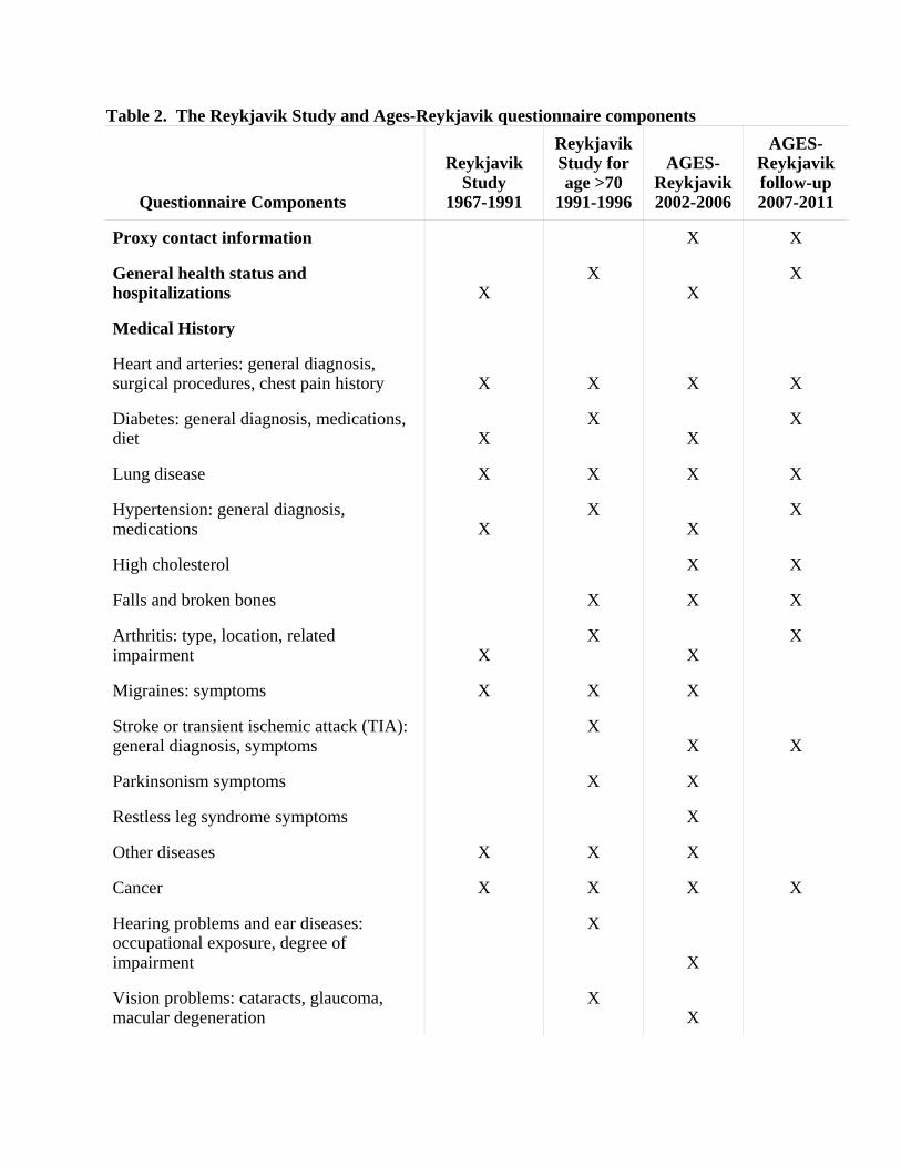
Table 2. The Reykjavik Study and Ages-Reykjavik questionnaire components
Questionnaire Components
Reykjavik Study
1967-1991
Reykjavik Study for age >70
1991-1996
AGES-Reykjavik 2002-2006
AGES-Reykjavik follow-up 2007-2011
Proxy contact information X X
General health status and hospitalizations X
X X
X
Medical History
Heart and arteries: general diagnosis, surgical procedures, chest pain history X
X X
X
Diabetes: general diagnosis, medications, diet X
X X
X
Lung disease X X X X
Hypertension: general diagnosis, medications X
X X
X
High cholesterol X X
Falls and broken bones X X X
Arthritis: type, location, related impairment X
X X
X
Migraines: symptoms X X X
Stroke or transient ischemic attack (TIA): general diagnosis, symptoms
X X
X
Parkinsonism symptoms X X
Restless leg syndrome symptoms X
Other diseases X X X
Cancer X X X X
Hearing problems and ear diseases: occupational exposure, degree of impairment
X
X
Vision problems: cataracts, glaucoma, macular degeneration
X X

23
Dentition: periodontal disease, dentures X
Prostate disease (MEN) X X
Reproductive history (WOMEN): pregnancies, menopause, medications
X X
Weight history X X
Sleeping habits X X
Urinary Incontinence X X
Anxiety X X
Geriatric Depression Scale X X X
Depression history and medications X X X
Subjective memory problems X X X
Social activity and contacts X X X
Coping and perceived stress X
Cognitively stimulating leisure activities X X
Functional limitations: stairs, 500 m walk, activities of daily living (ADL), instrumental activities of daily living, use of assistive devices
X X
X
Family medical history X X X
Education and languages X X X
Occupational history X X X
Wealth indicators X X X
Residence location in youth and mid-life. X X X
Diet history: youth, mid-life, current (old-age)
X X
Smoking and tobacco use history X X X
Alcohol X X
Physical activity: winter, summer, youth, mid-life X
X X
X

24
Table 3. The Reykjavik Study and Ages-Reykjavik examination components
Measurements
Reykjavik Study
1967-1991
Reykjavik Study for age >70
1991-1996
AGES-Reykjavik 2002-2006
AGES-Reykjavikfollow-up 2007-2007
VASCULAR SYSTEM
Pulse, blood pressure X X X X
Electrocardiogram: Heart rate, rhythm, ischemia, silent myocardial infarction (exercise test of subgroup in Reykjavik Study)
X X X X
Heart Rate Variability (measured during cognitive and physical function assessment for stress response)
n=1023
Ultrasonography of Carotid: Intimal/medial thickness, plaque count, carotid distensibility
X
Computerized tomography of vascular calcium: Coronary calcium, calcium volumes for aortic arch, and descending aorta
X X
Digitized retinal photograph: Arterial damage, drusen, retinal exudates
X
Echocardiography: Left ventricular thickness, wall motion, valve structure/function
n=900
Arterial tonometry: Pulse wave velocity n=900 X
Cardiac MRI with gadolinium enhancement: MRI defined MI, cardiac output, wall motion
n=1100
Lipids (laboratory): Total, HDL, LDL cholesterol, triglycerides
X X X X
Renal Function (laboratory): creatinine X X X X
NEUROCOGNITIVE
Neuropsychological testing: Memory, speed of processing, working memory
X X X

25
Mood: Depression symptoms, anxiety X X X
History of depression: depression diagnosis X X
MRI of the brain: Atrophy/ventricular size, infarct size and location, white matter lesion load and location, voxel-based morphometry
X X
Dementia evaluation: Dementia diagnosis and subtype adjudication by clinical consensus
X X
Visual acuity and functional vision X
Audiometry evaluation X
MUSCULOSKELETAL
Computerized tomography of L1/L2 (1mm slices): Integral and trabecular bone quality, structural properties
X X
Computerized tomography of hip (1 mm slices): Integral, cortical, and trabecular bone quality of total and regional femur, structural properties
X X
Hand photographs for osteoarthritis assessment: phalangeal abnormalities
X
Goniometry of knee X
OBESITY/SARCOPENIA AND METABOLISM
Anthropometric measurements: Height, weight, waist circumference
X
X X X
Bioelectrical impedance: Total body fat and non-fat lean
X X
Isometric dynamometry: Quadriceps strength, hand grip strength
X X

26
Computerized tomography of L4/L5: Sagittal diameter, waist and thigh circumference, visceral, subcutaneous, intermuscular, intramuscular fat areas, total and selected muscle areas
X X
Computerized tomography of thigh: Subcutaneous, intermuscular, intramuscular fat areas, total and selected muscle areas
X X
INTEGRATIVE FUNCTION
Health questionnaire: Behavioral risk factors, Social support/network, Medical history – see detailed questionnaire information in Table 2
X X X X
Motor and proprioceptive function: Balance platform, performance measures(TUG, 6meter walk)
X X Perfor-mance
measures
EuroQol EQ-5D questionnaire of health outcomes
X X
Inflammation (laboratory): C-reactive protein, sedimentation rate
X X X X
Stress Response (laboratory): Evening and morning salivary cortisol
X
Glucose Regulation (laboratory): fasting insulin, fasting glucose, hemoglobin A1C
X X X X
Pulmonary Function: spirometry X X 3000
Medications Inventory: prescriptions, over the counter
X X X
IMAGE ARCHIVE: MRI, CT, Ultrasound, Retinal photographs
X X
BIORESPOSITORY: serum, plasma, urine, and cells
X X X X
Abbreviations: MI= myocardial infarction, MRI=magnetic resonance imaging, CT=computerized tomography, HDL=high-density lipoprotein, LDL=low-density lipoprotein,

27
TUG=Timed Up and Go Test,

28
Table 4. Selected Findings from the Reykjavik Study Reference Summary of finding 21,22 Unrecognized Myocardial Infarction (MI) Risk factors and prognosis were similar for recognized and unrecognized MI. Risk of recurrent MI following an unrecognized MI was similar in men and women. Unrecognized MI is as common in women as in men 22 Family history Family history of MI from questionnaire is an independent risk factor for MI that cannot be
explained by the conventional risk factors 24, 29, 30 Inflammation Erythrocyte sedimentation rate is an independent risk factor for MI. C-reactive protein is an independent risk factor for MI but does not add markedly to the
conventional risk factors in prediction of MI. Mannose-binding lectin is predictive of MI in high-risk persons, such as diabetics or those with
raised cholesterol. 25, 26 Smoking and cancer Smoking was the most commonly associated risk factor for the development of neoplasms
among the cardiovascular risk factors. Family history of lung cancer was shown to be an independent risk factor for lung cancer, even
accounting for smoking.

29
Table 5. Midlife values (adjusted to age 50) of selected disease risk factors in eligible, invited and the first 2300 AGES-Reykjavik Study participants: Men.
MEN Eligible from Reykjavik Study cohort members
N=4,800
Invited for AGES-Reykjavik N=1,310
Non-responders to AGES-Reykjavik
N=334
AGES-Reykjavik enrollees N=976
Selected Risk Factors Mean 95% CI Mean 95% CI Mean 95% CI Mean 95% CI
TC (mmo1/L) 6.32 6.29,6.35 6.39 * 6.33,6.45 6.34 6.22,6.46 6.4 6.34,6.47
TG (mmo1/L) 1.15 1.13,1.17 1.11 * 1.08,1.13 1.16 1.11,1.23 1.08 † 1.05,1.11
Fasting Glucose (mmol/L) 4.48 4.46,4.50 4.47 4.44,4.50 4.52 4.46,4.58 4.45 † 4.41,4.48
SBP (mmHg) 136.4 135.8,137.0 137.6 * 136.6,138.6 142.5 140.2,144.9 135.6 ‡ 134.5,136.7
BMI (kg/m2) 25.7 25.6,25.8 25.5 * 25.3,25.7 25.7 25.3,26.0 25.4 25.2,25.6
Smokers (%) 50.2 48.7,51.7 52.1 49.3, 54.8 55.1 49.6, 60.6 51 47.8, 54.2
Abbreviations: CI=Confidence Interval, TC=Total Cholesterol, TG=Triglycerides, SBP=Systolic Blood Pressure, BMI=Body Mass Index * p<0.05 between midlife data of invited and eligible Reykjavik Study cohort members †p<0.05 between midlife data of non-responders and AGES-Reykjavik Study enrollees ‡ p<0.01 between midlife data of non-responders and AGES-Reykjavik Study enrollees

30
Table 6. Midlife values (adjusted to age 50) of selected disease risk factors in eligible, invited and the first 2300 AGES-Reykjavik Study participants: Women.
WOMEN Eligible from Reykjavik Study cohort members
N=6,749
Invited for AGES-Reykjavik N=1,933
Non-responders to AGES-Reykjavik
N=609
AGES-Reykjavik enrollees N=1,324
Selected Risk Factors Mean 95% CI Mean 95% CI Mean 95% CI Mean 95% CI
TC (mmo1/L) 6.32 6.28,6.35 6.28 6.23,6.33 6.36 6.25,6.46 6.26 6.20,6.32
TG (mmo1/L) 0.91 0.90,0.93 0.88 * 0.87,0.90 0.89 0.86,0.93 0.88 0.86,0.90
Fasting Glucose (mmol/L) 4.29 4.27,4.31 4.25 * 4.23,4.28 4.29 4.22,4.36 4.23 ‡ 4.21,4.27
SBP (mmHg) 128.1 127.6,128.7 128.5 127.6,129.4 133.1 131.2,135.0 126.7 § 125.7,127.8
BMI (kg/m2) 24.9 24.8,25.0 24.7 † 24.5,24.8 24.7 24.3,25.1 24.6 24.4,24.9
Smokers (%) 36.3 35.0,37.7 32.3 * 30.1,34.5 36.3 32.1, 40.8 30.8 ‡ 28.4,33.5
Abbreviations: CI=Confidence Interval, TC=Total Cholesterol, TG=Triglycerides, SBP=Systolic Blood Pressure, BMI=Body Mass Index * p<0.01 between midlife data of eligible Reykjavik Study cohort members and those invited † p<0.05 between midlife data of eligible Reykjavik Study cohort members and those invited ‡ p<0.05 between midlife data of non-responders and AGES-Reykjavik Study enrollees § p<0.0001 between midlife data of non-responders and AGES-Reykjavik Study enrollees

31
Table 7. Comparison of midlife Reykjavik Study and late life AGES-Reykjavik measurements of selected cardiovascular risk factors.
Gender Variable
Reykjavik Study age- adjusted values
AGES-Reykjavik
age- adjusted values
Pearson Correlation
between values
10-year change
26-year change
p-value for
correlation
MEN Total cholesterol (mmol/L) 6.40 5.27 0.26 -0.41 -1.06 <0.01
N=976 Triglyceride (mmol/L)* 1.08 1.07 0.44 -0.01 -0.02 0.24
Serum glucose (mmol/L)† 5.52 5.97 0.24 0.17 0.43 <0.01
Systolic blood pressure (mmHg) 135.6 141.9 0.20 2.4 6.2 <0.01
Body mass index (kg/m2) 25.4 26.7 0.66 0.4 1.16 <0.01
WOMEN Total cholesterol (mmol/L) 6.26 6.11 0.27 -0.08 -0.21 <0.01
N=1,324 Triglyceride (mmol/L)* 0.88 1.15 0.46 0.10 0.26 <0.01
Serum glucose (mmol/L)† 5.32 5.70 0.30 0.17 0.43 <0.01
Systolic blood pressure (mmHg) 126.7 141.4 0.31 5.5 14.4 <0.01
Body mass index (kg/m2) 24.6 27.1 0.69 0.9 2.4 <0.01 * Analysis on log transformed values. 10 year change back transformed † The Reykjavik Study (RS) value is blood sugar. Conversion to serum glucose was 1.47+0.91x(RS blood sugar).

32
Table 8. Cut-points used to examine overlap in the four focus areas for AGES-Reykjavik participants. Men
(N=976) Women
(N=1,324)
Median or % 25th and 75th
percentiles Median or % 25th and 75th
percentiles
Trabecular bone mineral density mg/cm3
Lumbar spine 0.09 0.07,0.11 0.07 0.05,0.09
Femoral neck 0.03 0.01,0.06 0.01 -0.01,0.04
Glucose metabolism:
Serum Insulin mU/L 8.52 5.67,12.72 7.85 5.31,11.20 Cognition: Mini-Mental State Exam 27 25, 29 28 26, 29
Digit Symbol Substitution Test 28 21, 36 29 21, 36 Calcification of arteries (% with any calcification)* Coronary arteries 96.40% 81.60%
Ascending aorta 98.70% 98.60%
Descending aorta 84.50% 84.80%
Abdominal aorta L1/L2 96.30% 96.50%
Abdominal aorta L4/L5 91.80% 89.50%
In 4 of 5 aortic areas and coronary arteries (%) 91.10% 85.10% * Agatston scores and calcification measurements in the abdominal aorta at vertebral levels L1/L2 and L4/L5 with values greater than

33
zero indicate that some degree of calcification is present. The percentages in this table reflect the percent of the cohort with any calcification present at the noted location.

34
Figure 1. Independence and overlap of prevalent phenotypes in the AGES-Reykjavik Study, 2002-2004.
High insulin
Arterial calcification
in all areas
High Arterial
1
9 2
8
9
1
7
18
14
2
Poor cognition
Low trabecular bone
13 1
2
2
10

35
Figure 1 Legend: Phenotypes from the Age, Gene/Environment Susceptibility Study (AGES-Reykjavik) are represented by the overlapping circles in this figure, representing the traits of poor cognition, arterial calcification in all areas, high insulin, and low trabecular bone. The phenotypes are further defined in Methods section. Numbers within the circles represent the percent of the cohort with each of these traits. Numbers in areas of overlap indicate the percent of the cohort that has more than one trait. Two percent of the cohort had none of the phenotypes and only 13 percent share all the traits. The number inside the small circle within the ‘poor cognition’ phenotype represents the percent of the cohort which had both poor cognition and low trabecular bone. Similarly, the number inside the small circle within the ‘high insulin’ phenotype represents the portion of the cohort that has both high insulin and arterial calcification in all areas.