african journal of pharmacy and pharmacology · curve of acid pyrogallic in concentrations from...
TRANSCRIPT

Volume 9 Number 8, 29 February, 2015
ISSN 1996-0816
African Journal of Pharmacy and Pharmacology

ABOUT AJPP The African Journal of Pharmacy and Pharmacology (AJPP) is published weekly (one volume per year) by Academic Journals.
African Journal of Pharmacy and Pharmacology (AJPP) is an open access journal that provides rapid publication (weekly) of articles in all areas of Pharmaceutical Science such as Pharmaceutical Microbiology, Pharmaceutical Raw Material Science, Formulations, Molecular modeling, Health sector Reforms, Drug Delivery, Pharmacokinetics and Pharmacodynamics, Pharmacognosy, Social and Administrative Pharmacy, Pharmaceutics and Pharmaceutical Microbiology, Herbal Medicines research, Pharmaceutical Raw Materials development/utilization, Novel drug delivery systems, Polymer/Cosmetic Science, Food/Drug Interaction, Herbal drugs evaluation, Physical Pharmaceutics, Medication management, Cosmetic Science, pharmaceuticals, pharmacology, pharmaceutical research etc. The Journal welcomes the submission of manuscripts that meet the general criteria of significance and scientific excellence. Papers will be published shortly after acceptance. All articles published in AJPP are peer-reviewed.
Submission of Manuscript Submit manuscripts as e-mail attachment to the Editorial Office at: [email protected]. A manuscript number will be mailed to the corresponding author shortly after submission.
The African Journal of Pharmacy and Pharmacology will only accept manuscripts submitted as e-mail attachments. Please read the Instructions for Authors before submitting your manuscript. The manuscript files should be given the last name of the first author.

Editors Sharmilah Pamela Seetulsingh- Goorah Dr.B.RAVISHANKAR Associate Professor, Director and Professor of Experimental Medicine Department of Health Sciences SDM Centre for Ayurveda and Allied Sciences, Faculty of Science, SDM College of Ayurveda Campus, University of Mauritius, Kuthpady, Udupi- 574118 Reduit, Karnataka (INDIA) Mauritius
Dr. Manal Moustafa Zaki Himanshu Gupta Department of Veterinary Hygiene and Management University of Colorado- Anschutz Medical Campus, Faculty of Veterinary Medicine, Cairo University Department of Pharmaceutical Sciences, School of Giza, 11221 Egypt Pharmacy Aurora, CO 80045, USA
Prof. George G. Nomikos Scientific Medical Director
Dr. Shreesh Kumar Ojha Clinical Science Molecular Cardiovascular Research Program Neuroscience College of Medicine TAKEDA GLOBAL RESEARCH & DEVELOPMENT Arizona Health Sciences Center CENTER, INC. 675 North Field Drive Lake Forest, IL University of Arizona 60045 Tucson 85719, Arizona, USA USA
Prof. Mahmoud Mohamed El-Mas
Dr.Victor Valenti Engracia Department of Pharmacology, Department of Speech-Language and Hearing Therapy Faculty of Philosophy and Sciences, UNESP Marilia-SP, Brazil.l Prof. Sutiak Vaclav Dr. Caroline Wagner Rovníková 7, 040 20 Košice, Universidade Federal do Pampa The Slovak Republic, Avenida Pedro Anunciação, s/n The Central Europe, Vila Batista, Caçapava do Sul, RS - Brazil European Union Slovak Republic Slovakia

Editorial Board Prof. Fen Jicai Dr. Sirajunnisa Razack School of life science, Xinjiang University, Department of Chemical Engineering, Annamalai China. University,
Annamalai Nagar, Tamilnadu, Dr. Ana Laura Nicoletti Carvalho India. Av. Dr. Arnaldo, 455, São Paulo, SP. Brazil. Prof. Ehab S. EL Desoky
Professor of pharmacology, Faculty of Medicine Dr. Ming-hui Zhao Assiut University, Assiut, Professor of Medicine Egypt. Director of Renal Division, Department of Medicine Peking University First Hospital Dr. Yakisich, J. Sebastian Beijing 100034 Assistant Professor, Department of Clinical Neuroscience PR. China. R54
Karolinska University Hospital, Huddinge Prof. Ji Junjun 141 86 Stockholm , Guangdong Cardiovascular Institute, Guangdong General Sweden. Hospital, Guangdong Academy of Medical Sciences, China. Prof. Dr. Andrei N. Tchernitchin
Head, Laboratory of Experimental Endocrinology and Prof. Yan Zhang Environmental Pathology LEEPA Faculty of Engineering and Applied Science, University of Chile Medical School, Memorial University of Newfoundland, Chile. Canada.
Dr. Sirajunnisa Razack Dr. Naoufel Madani Department of Chemical Engineering, Medical Intensive Care Unit Annamalai University, Annamalai Nagar, Tamilnadu, University hospital Ibn Sina, Univesity Mohamed V India. Souissi, Rabat, Morocco. Dr. Yasar Tatar
Marmara Unıversıty, Dr. Dong Hui Turkey.
Department of Gynaecology and Obstetrics, the 1st hospital, NanFang University, Dr Nafisa Hassan Ali China. Assistant Professor, Dow institude of medical technology
Dow University of Health Sciences,Chand bbi Road, Karachi, Prof. Ma Hui Pakistan. School of Medicine, Lanzhou University, China. Dr. Krishnan Namboori P. K.
Computational Chemistry Group, Computational Prof. Gu HuiJun Engineering and Networking, School of Medicine, Taizhou university, Amrita Vishwa Vidyapeetham, Amritanagar, Coimbatore- China. 641 112
India.
Dr. Chan Kim Wei Research Officer Prof. Osman Ghani Laboratory of Molecular Biomedicine, University of Sargodha, Institute of Bioscience, Universiti Putra, Pakistan. Malaysia.
Dr. Liu Xiaoji Dr. Fen Cun School of Medicine, Shihezi University, Professor, Department of Pharmacology, Xinjiang China. University, China.

International Journal of Medicine and Medical Sciences
African Journal of Pharmacy and Pharmacology
Table of Contents: Volume 9 Number 8 29 February, 2015
ARTICLES
Research Articles Subchronic toxicity evaluation of the hydroethanolic extract from Endopleura uchi (Huber) Cuatrec in Wistar rats 223 Beatriz M. Sá Clarissa S. Lima, Uriel Davi A. Silva, Helison O. Carvalho, Caio P. Fernandes, Rafael L. Resque, Tania T. de Oliveira and José Carlos T. Carvalho Evaluation of anti-inflammatory activity of the bark of Eysenhardtia polystachya in experimental animal models 230 Rosa Martha Perez Gutierrez
Review: The potential of chalcones as a source of drugs 237 Rosa Martha Perez Gutierrez, Alethia Muñiz-Ramirez and Jahel Valdes Sauceda
Clinical duration of rocuronium becomes shorter at night: A chronopharmacological study 258 Menekse Ozcelik, Ali Abbas Yılmaz, Sirali Oba, Sanem Turhan and Oya Ozatamer Effect of grapefruit juice and sibutramine on body weight loss in obese rats 265 Hadir Farouk, Sawsan S. Mahmoud, Bahia A. El-Sayeh, and Ola A. Sharaf

Vol. 9(8), pp. 223-229, 29 February, 2015 DOI: 10.5897/AJPP2014. 4220 Article Number: 6F0AF5751297 ISSN 1996-0816 Copyright © 2015 Author(s) retain the copyright of this article http://www.academicjournals.org/AJPP
African Journal of Pharmacy and Pharmacology
Full Length Research Paper
Subchronic toxicity evaluation of the hydroethanolic extract from Endopleura uchi (Huber) Cuatrec in
Wistar rats
Beatriz M. Sá1,2, Clarissa S. Lima1,2, Uriel Davi A. Silva1, Helison O. Carvalho1, Caio P. Fernandes1, Rafael L. Resque1, Tania T. de Oliveira3 and José Carlos T. Carvalho1,2*
1Laboratório de Pesquisa em Fármacos,Departamento de Ciências Biológicas e da Saúde, Universidade Federal do Amapá, Amapá, Brasil.
2Programa de Pós-Graduação em Biodiversidade Tropical, Universidade Federal do Amapá, Amapá, Brasil. 3Laboratório de Biofarmacos, Departamento de Bioquimica e Biologia Molecar, Universidade Federal de Viçosa,
Minas Gerais, Brasil.
Received 30 October 2014; Accepted 3 February 2015
Endopleura uchi (Huber) Cuatrec. (Humiriaceae) is a species from Brazilian Amazon rainforest, popularly used against menstrual disorders and uterine inflammation. This study aimed to evaluate the subchronic toxicity of hydroethanolic extract from E. uchi (EHEEu) in Wistar rats based on biochemical and hematological parameters. Rats were treated with daily doses of EHEEu (500 mg/kg - gavage), and then hematological and biochemical parameters were observed. The results shows that the treatment performed produced no signs of toxicity or death, as well as no changes in weight gain or daily intake of water and food. Biochemical and hematological parameters were not modified by EHEEu administration, with the exception of erythrocyte index of rats (males) in the treated group, however, it was not assigned clinical relevance once it remained within the reference range for the species. Thus, subchronic administration of EHEEu produced no toxic effects in Wistar rats. Key words: Endopleura uchi, subchronic treatment, hematology, biochemistry.
INTRODUCTION The traditional use of medicinal plants based on popular knowledge, along with the belief that being natural does not cause adverse reactions, made only a few medicinal plants were evaluated through pre-clinical and clinical
studies to prove its effectiveness and security (Turolla and Nascimento, 2006). However, over time it was realized that certain plants have potentially dangerous substances and therefore should be used with care,
*Corresponding author. E-mail: [email protected]. Author(s) agree that this article remain permanently open access under the terms of the Creative Commons Attribution License 4.0 International License

224 Afr. J. Pharm. Pharmacol. respecting their toxicological risks (Veiga-Junior and Pinto, 2005).Endopleura uchi (Huber) Cuatrec. (Humiriaceae) is a botanical species typical from the Brazilian Amazon rain-forest This plant is found in not too flooded ground forest, which is widely spread in the Amazon Basin.It is popularly known in the region as uchi, uxi, axuá, uchi-pucu, uxi-amarelo, uxi-liso e uxi-ordinário (Cuatrecasas, 1961; Schultes, 1979). The stem bark of E. uchi is used as tea for menstrual disorders and uterine inflammation (Revilla, 2001). Other indications are also known and cited as antimicrobial activity (Politi, 2009), high anti-oxidant activity and absence of cytotoxicity (Politi et al., 2011). These results justify the increasingly growing interest in the study of this plant.
According to Politi (2009), the bark of E. uchi consists mainly of three types of secondary metabolites: tannins, coumarins and saponins. Another phytochemical study from crude ethanol extract of bark led to the isolation of bergenin isocoumarins and 8, 10 dimetoxibergenina, pentacyclic triterpenoids, maslinic acid and its methyl ester maslininate (Luna et al., 2000).
The bergenin extracted from the bark of E. uchi have been identified as mainly responsible against the biological activities such as anti-inflammatory (Nunomura et al., 2009), anti-microbial (Silva et al., 2009), neuroprotective (Takahashi et al., 2003) and antinociceptive (Oliveira et al., 2011). Until this moment, toxicological studies do not support the safe use of E. uchi. However, Politi et al. (2010) evaluated the acute oral toxicity in male rats with an extract of powdered bark of E. uchi. Thus, the present study aimed to evaluate the subchronic toxicity of crude hydroethanolic extract of the stem bark of E. uchi in Wistar rats of both sexes. MATERIALS AND METHODS Collection and authentication The stem barks of E. uchi were collected in April, 2005 in the city of Ananideua, Pará State, Brazil. The voucher specimen of the plant (number 180611) was deposited in the Herbarium of the Brazilian Agricultural Research Corporation - EMBRAPA, Belém, Pará, Brazil. Extract preparation The stem bark of E. uchi were dried in an air circulating oven at 40°C for 72 h and after drying, it was triturated in a knives mill, obtaining the powder of the plant (464 g). Subsequently, this material was macerated in a 75% hydroalcoholic solution in a ratio of 1:5 at room temperature for 7 days, under stirring. Then, the mash was filtered and concentrated on a rotaevaporator (Quimis Model Q 218.2) at 45°C until complete evaporation of the solvent. The concentrated filtrate was subjected to lyophilization providing 10.65% yield.
Analysis of the content of total polyphenols and total tannins The method of Brazilian Pharmacopeia, 5th edition (2010) described for Rhatany species (Ratanhiae radix) was adapted and subjected to linear regression curve prepared with pyrogallic acid, all analyses were performed in triplicate. The polyphenols contents were calculated from the equation of the line obtained by standard curve of acid pyrogallic in concentrations from 0.01 to 0.05 mg/ml submitted to reaction with phosphomolybdic tungstic acid in alkaline medium following the method described by Carvalho et al. (2013). Animals Rats (Rattus norvegicus albinus), Wistar strain (males and females), weighing around 170 to 215 g from the vivarium of the Center for Reproductive Biology, Federal University of Juiz de Fora were used. These went through a period of adjustment, kept under controlled lighting conditions (cycle 12 h light/dark), temperature (23 ± 2°C) and received water and food ad libitum. The experimental protocol was approved by the Ethics Committee of the Federal University of Amapá (Case No. 001A/2012). Treatment of animals and evaluation of biochemical and hematological parameters Twenty rats corresponding to two groups n = 10/group (5 males and 5 females) were randomized into five subgroups and treated for 22 consecutive days orally with EHEEu at a dose of 500 mg/kg (treated group) and distilled water (control group). Parameters indicative of toxicity and blood-biochemical analyzes were performed according to the method described by Silva et al. (2005), with some adaptations. Parameters such as aspartate aminotransferase (AST), alanine aminotransferase (ALT), total cholesterol, HDL cholesterol, triglycerides, alkaline phosphatase (ALP), albumin, glucose, creatinine, total and differential blood count were determined using the multiparametric equipment for biochemical determination (Alizé) from Biomérieux and automatic hematologic cells analyzer HumaCount Plus. Statistical analysis The results obtained in different analyzes were expressed as average ± standard error of the average (average ± SEA) for each experimental group. For biochemical and hematological analysis, we used Mann - Whitney, and to compare the data of weight gain, water and food intake the student "t" test (unpaired) was used. Test was performed using GraphPad Prism software® (version 5.03). Results with p < 0.05 were considered statistically significant. RESULTS The lyophilized extract showed total polyphenols equal to 7.01% corresponding to an average of 0.021 ± 0.008 mg/ml (n = 3). The percentage of total tannins was 1.5% with 0.0045 ± 0.0011 mg/ml (n = 3) representing 21.4% of all polyphenols in the extract (Figure 1). The oral admini-stration for 22 days with EHEEu at a dose of 500 mg/kg in rats, males and females, did not affect the weight gain

Beatriz et al. 225
Concentartion (mg/ml)
Figure 1. Standard curve of pyrogallic acid for spectrophotometrically (λ = 760 nm), concentrations from 0.01 to 0.05 mg/mL subjected to reaction with phosphomolybdictungstic acid in alkaline medium with read-after 2 minutes of reaction. Linear regression of the results obtained r2 = 0.9987 correlation coefficient with the equation of the line y = 10.450x 0.0118.
Table 1. Effect of the treatment (po) of EHEEu (500 mg/kg) and distilled water (control) for 22 consecutive days, on the biochemical parameters of Wistar rats (males and females).
Parameter Female Male
Control EHEEu Control EHEEu AST (U/L) 241.6 ± 50.0 245.0 ± 39.9 193.2 ± 27.6 228.0 ± 37.1 ALT (U/L) 83.6 ± 22.9 79.0 ± 17.8 53.0 ± 4.8 54.5 ± 5.8 Total cholesterol (mg/dl) 66.5 ± 1.8 73.8 ± 4.6 61.6 ± 3.3 65.6 ± 4.2 HDL cholesterol (mg/dl) 31.6 ± 1.2 30.7 ± 1.4 31.2 ± 1.1 30.4 ± 0.8 Triglycerides (mg/dl) 82.0 ± 4.5 76.8 ± 6.3 55.5± 2.3 74.4 ± 1.6 ALP (U/L) 23.6 ± 2.4 26.2 ± 6.2 90.8 ± 8.9 90.0 ± 17.9 Albumin (g/dl) 3.5 ± 0,0 3.6 ± 0,1 3.7 ± 0.0 3.6 ± 0.0 Glucose (mg/dl) 111.7 ± 20.9 123.3 ±17.3 154.5 ± 6.75 146.4 ± 13.5 Creatinine (mg/dl) 0.5 ± 0.0 0.6 ± 0.0 0.4 ± 0.0 0.5 ± 0.0
The values represent mean ± S.E.M. (n = 5/group). *p < 0.05 compared to the control group (Mann-Whitney test). AST: Aspartate aminotransferase; ALT: Alanine aminotransferase; FA: Alkaline phosphatase.
gain and daily food and water intake of these animals, since significant changes were not observed compared to the control group (Figures 2, 3 and 4). The level of serum AST, both in males and females (control and treated) and ALT in males (control and treated) were above the reference values (Table 1). The treatment of animals with EHEEu did not alter the levels of triglycerides and total cholesterol, however, the levels of HDL cholesterol in males and females (control and treated group) animals remained below the reference values (Table 1). Oral treatment of animals with EHEEu did not interfere in hematologic rates, except for the values of mean
corpuscular hemoglobin concentration (MCHC) of animals (males), which showed a significant increase compared to the control group. The differential count of lymphocytes, monocytes, neutrophils and eosinophils showed similar values, with no statistically significant differences between the control group and treated with EHEEu (Table 2). DISCUSSION Polyphenols vary from simple molecules to complex

226 Afr. J. Pharm. Pharmacol.
1 5 10 15 20150
200
250
300Control (males)EHEEu 500 mg/kg (males)Control (females)EHEEu 500 mg/kg (females)
Time (day)
Bod
y we
ight
(g)
Figure 2. Effects of the administration (po) of EHEEu (500 mg/kg) and distilled water (control) on the body weight of Wistar rats (males and females), by 22 consecutive days. The values represent mean ± SEM. (n = 5/group). *p < 0.05 compared to the control group.
Wat
er (m
l)
Figure 3. Effects of the administration (po) of EHEEu (500 mg/kg) and distilled water (control) on the daily water consumption of Wistar rats (males and females), for 22 consecutive days. The values represent mean ± S.E.M. (n = 5/group). *p < 0.05 compared to the control group.
molecules with a high degree of polymerization and are classified into several classes of secondary metabolites such as flavonoids, phenolic acids, simple phenols, coumarins, tannins, lignins, and tocopherols among others and are easily oxidizable by metals light, heat and enzymatic processes and are associated the antioxidant actions (Shahidi and Naczk 1995; Simões et al., 2004). In this study, the EHEEu showed concentration significant for tannins.
One of the indicators of adverse effects of drugs and chemicals is the change in animal body weight (Tofovic
and Jackson, 1999; Raza et al., 2002; Teo et al., 2002). Systemic toxicity can also be diagnosed by decreased water intake, diet, behavioral changes such as apathy and prostration, and by the appearance of rough hair coat (Melo, 2001). In this study, it was observed that the gain in body weight in rats (males) from control and EHEEu treated groups was higher than those rats (females) from both groups. However, it was found that oral administration for 22 days with EHEEu in rats, males and females did not affect the weight gain and daily food and water intake of these animals (Figures 2, 3 and 4).

Beatriz et al. 227
0
5
10
15
20
25Control (males)EHEEu 500 mg/kg (males)Control (females)EHEEu 500 mg/kg (females)
Food
(g)
Figure 4. Effects of the administration (po) of EHEEu (500 mg/kg) and distilled water (control) on the daily feed intake of Wistar rats (males and females), for 22 consecutive days. The values represent mean ± S.E.M. (n = 5/group). *p < 0.05 compared to the control group.
Table 2. Effect of the treatment (po) of EHEEu (500 mg/kg) and distilled water (control) for 22 consecutive days, on hematological parameters of Wistar rats (males and females).
Parameter Females Males
Control EHEEu Control EHEEu RBC (x106/mm3) 7.9 ± 0.1 7.9 ± 0.3 9.2 ± 0.0 9.1 ± 0.2 HGB (g/dl) 14.8 ± 0.1 15.1 ± 0.5 15.7 ± 0.1 15.9 ± 0.2 HCT (%) 41.3 ± 0.4 40.8 ± 1.7 48.0 ± 0.3 47.0 ± 1.7 MCV (fl) 52.2 ± 0.6 51.5 ± 0.6 52.0 ± 0.3 51.2 ± 0.5 MCH (pg) 18.6 ± 0.1 19.1 ± 0.4 17.0 ± 0.1 17.4 ± 0.2 MCCH (g/dl) 35.7 ± 0.3 37.1 ± 0.5 32.7 ± 0.1 34.7 ± 0.4* WBC (×103/mm3) 3.7 ± 0.6 3.4 ± 0.4 3.6 ± 0.6 3.2 ± 0.7 Lymphocytes (%) 56.2 ± 3.8 52.2 ± 3.6 63.8 ± 2.1 61.8 ± 1.3
Monocytes (%) 2.4 ± 0.6 3.0 ± 0.3 1.6 ± 0.4 2.0 ± 0.5
Segmented (%) 40.0 ± 3.5 43.0 ± 3.6 32.6 ± 2.2 34.6 ± 1.2
Eosinophils (%) 1.4 ± 0.2 1.8 ± 0.3 2.0 ± 0.8 1.6 ± 0.5
Platelets (×103/mm3) 1069.0 ± 35.5 996.25 ± 13.9 1026.3 ± 159.0 1007± 63.1
The values represent mean ± S.E.M. (n = 5/group). *p < 0,05 compared to the control group (Mann-Whitney test). RBC: Red Blood Cells; HGB: Hemoglobin; HCT: Hematocrit; MVC: Mean corpuscular volume; MCH: Mean corpuscular hemoglobin; MCCH: Mean corpuscular hemoglobin concentration.
Although the food intake of the treated group was lower than in the control group (Figure 4), the fact is not associated with EHEEu toxic effects because there was no interference with the body mass gain of these animals during treatment. During the experimental period, the animals of different groups showed no clinical signs of toxicity and no death was registered. The decrease in glomerular filtration, in general, leads to increased plasma creatinine concentrations. In rats, plasma levels of creatinine change can be a reliable indicator for the presence of renal injury, because its serum level is not
influenced by diet, age or sex (Alves, 2007). The administration EHEEu does not change serum creatinine levels in animals, which indicates that renal function was not affected (Table 1).
According to Alves (2007), some enzymes can be used as indicators of hepatic injury, such as ALT, AST and ALP. ALT is found mainly in the cytoplasm of hepato-cytes, while 80% of AST is present in mitochondria. In light hepatocellular damage the serum is predominantly cytoplasmic, while in severe injuries the mitochondrial enzyme is released (Motta, 2003). The ALP is present

228 Afr. J. Pharm. Pharmacol. mainly at bone tissue, at the hepatobiliary system and the gastrointestinal mucosa, it is indicative of cholestasis, which may lead to increased serum levels up to 10 times (Scheffer and Gonzalez, 2003). In the present study, these enzymes were not altered after administration of EHEEu, however the level of serum AST, both in males and females (control and treated) and ALT in males (control and treated) were above the reference values (Table 1) (Clifford and Giknis, 2008).
The importance of lipid levels is fundamental because elevated levels of total cholesterol are closely related to risk of ischemic coronary disease (Araújo et al., 2011). Treatment of animals with EHEEu did not alter the levels of triglycerides and total cholesterol, probably indicating that there was no change in the lipid metabolism of these animals, however, the levels of HDL cholesterol in males and females (control and treated group) animals remained below the reference values (Table 1) (Dantas et al., 2006).
Despite the existence of own mechanisms to control the physiological parameters values, it is known that in certain groups of animals, such as rats and mice, these parameters can vary, mainly related to gender, strain, genotype and may be influenced by age, diet, handling, environment, among other factors (Pinheiro et al., 2003). Although some biochemical parameters in animals treated with EHEEu are below or above the reference values, cannot be assigned clinical importance because the same results were found in the control group, with no statistically significant difference between them (Table 1). However, it is necessary for the histopathological study of the liver and kidneys of these animals.
According to Silva et al. (2012), hematological parameters are important for the toxicity study, due to the hematopoietic system that is highly sensitive to the toxic agents’ activities, such as those with cytotoxic or mutagenic potential. These toxicants may result in many changes such as qualitative or quantitative, transient or permanent and may also limit the use of drugs. In oral treatment of animals with EHEEu, in hematologic rates, only the values of mean corpuscular hemoglobin concentration (MCHC) of animals (males) was changed, which showed a significant increase compared to the control group (Table 2). According to Alves (2007), analysis of red blood cell (RBC) indices are important indicators in determining the morphological type of anemia. The increased erythrocyte MCHC index of male animals had no clinical relevance because they have values within the range recommended by the study of Charles River (Giknis and Clifford, 2008). Conclusion With the obtained results, we can suggest that oral
treatment with EHEEu at a dose of 500 mg/kg for 22 days produced no signs of systemic toxicity in Wistar rats males and females when compared to the control group. So the EHEEu showed high degree of safety at dose and period in which the animals were exposed. AKNOWLEDGEMENT The authors acknowledge CNPq (National Research Council, Project Number 402332 / 2013-0) and CAPES (AUXPE Project Number 3292/2013). Conflict of interest The authors declare that there are no conflicts of interest. REFERENCES Alves NM (2007). Estudo farmacognóstico e da toxicidade experimental
(aguda e subaguda) do guatambu (Aspidosperma subincanum Mart.). Dissertação de Mestrado, Universidade de Brasília, Brasil.
Araújo TOP, Goes LEJ, Rocha AG, Costa MP, Santos LHS, Bertato AC, Gileno MC (2011). Benefícios da cafeína sobre os níveis séricos de colesterol e triglicerídeos. Rev. Uniara 14(1):118-126.
Brazilian Pharmacopeia (2010). Métodos aplicados a medicamentos e físicos e químicos. Farmacopeia Brasileira, Brasília (DF), 5th edition 2:59-93.
Carvalho HO, Medeiros BJL, Sá BM, Araújo JTC, Kawakami MYM, Favacho HAS, Carvalho JCT (2013). Study of dissolution profiles and desintegration of capsules containing the dried hydroethanolic extract of Calophyllum brasiliense. Braz. J. Pharmacogn. 23(1):194-199.
Clifford CB, Giknis MLA (2008). Clinical Laboratory Parameter for Crl: WI(Han). Charles River Laboratories.
Cuatrecasas JA (1961). A taxonomic revision of Humiriaceae. Contributions from the United States National Herbarium. Bull. US Nat. Mus. 35(2):25-214.
Dantas JA, Ambiel CR, Cuman RKN, Baroni S, Bersani-Amado CA (2006). Valores de referência de alguns parâmetros fisiológicos de ratos do Biotério Central da Universidade Estadual de Maringá, Estado do Paraná. Acta Sci. Health Sci. 28(2):165-170.
Luna JS, Silva TM, Bento ES, Sant´ana AEG (2000). Isolamento e Identificação estrutural dos constituintes químicos de Endopleura uchi (Humiriaceae). In: Reunião Anual da Sociedade Brasileira de Química, Minas Gerais, Brasil.
Mello FB (2001). Estudo dos efeitos de Lantana camara (Verbenaceae) sobre a fertilidade e reprodução de ratos. Dissertação de Mestrado, Universidade Federal do Rio Grande do Sul, Brasil.
Motta VT (2003). Bioquímica Clínica para o Laboratório: princípios e interpretações. Ed. Robe, São Paulo, Brasil.
Nunomura RCS, Oliveira VG, DA Silva SL, Nunomura SM (2009). Characterization of bergenin in Endopleura uchi bark and its anti-inflammatory activity. J. Braz. Chem. Soc. 20(6):1060-1064.
Oliveira CM, Nonato FR, Lima FO, Couto RD, David JP, David JM, Soares MBP, Villarreal CF (2011). Antinociceptive properties of bergenin. J. Nat. Prod. 74(10):2062-2068.
Pinheiro DCSN, Favali CBF, Filho AAS, Silva ACM, Filgueiras TM, Lima MGS (2003). Parâmetros hematológicos de camundongos e ratos do biotério central da Universidade Federal do Ceará. Bol. Inf. COBEA (3):6-9.
Politi FAS, Mello JCP, Migliato KF, Nepomuceno ALA, Moreira, RRD, Pietro RCLR (2011). Antimicrobial, Cytotoxic and Antioxidant

Activities and Determination of the Total Tannin Content of Bark Extracts Endopleura uchi. Int. J. Mol. Sci. 12:2757-2768.
Politi FAS, Moreira RRD, Salgado HRN, Pietro RCLR (2010). Testes preliminares de motilidade intestinal e toxicidade oral aguda com extrato de cascas pulverizadas de Endopleura uchi (Huber) Cuatrec. (Humiriaceae) em camundongos. Rev Pan-Amaz Saude 1(1):187-189.
Politi FAZ (2009). Estudos farmacognósticos e avaliação de atividades biológicas de extratos obtidos das cascas pulverizadas de Endopleura uchi (Huber) Cuatrec. (Humiriaceae). Dissertação de Mestrado, Universidade Estadual Paulista Júlio de Mesquita Filho, Brasil.
Raza M, AL-Shabanah OA, EL-Hadiyah TM, AL-Majed AA (2002). Effect of prolonged vigabatrin treatment on hematological and biochemical parameters in plasma, liver and kidney of Swiss albino mice. Sci. Pharmac. 70:135-145.
Revilla J (2001). Plantas da Amazônia: oportunidades econômicas e sustentáveis. Ed. Sebrae/Inpa, Manaus, Brasil.
Scheffer MC (2002). Fisiologia pós-colheita de espécies medicinais, condimentares e aromáticas. In: Wachowicz, CM, Carvalho, RIN. Fisiologia vegetal: Produção e pós-colheita. Curitiba: Champagnat, Pp.383-404.
Scheffer JF, González FHD (2003). Enzimologia clínica em medicina veterinária. Available at: http://www6.ufrgs.br/favet/lacvet/restrito/pdf/rev_jfss.pdf [Accessed April 2012.
Schultes RE(1979). De plantis toxicariis e mundo novo tropicale commentationes XXI. Interesting native uses of the Humiriaceae in the northwest Amazon. J. Ethnopharmacol. 1(1):89-94.
Shahidi F, Naczk M (1995). Food Phenolics: Sources,Chemistry,Effects and Applications. Technomic Publishing Company Inc., Lancaster (Pennsylvania). Pp. 281-319.
Silva EJR, Aguiar FJS, Gonçalves ES, Souza IMV, Dimech GS, Fraga MCCA, Coelho MCOC, Wanderley AG (2005). Avaliação do tratamento subcrônico com o estrato hidroalcólico de Calendula officinalis L. sobre os parâmetros bioquímicos e hematológicos em ratas Wistar. Ver. Bras. Farmacogn. 15(2):88-93.
Beatriz et al. 229 Silva SL, Oliveira VG, Yano T, Nunomura RCS (2009). Antimicrobial
activity of bergenin from Endopleura uchi (Huber) Cuatrec. Acta Amaz. 39(1):187-191.
Silva SN, Abreu IC, Silva GFC, Ribeiro RM, Lopes AS, Cartagenes MSS, Freire SMF, Borges ACR, Borges MOR (2012). The toxicity evaluation of Syzygium cumini leaves in rodents. Rev. Bras. Farmacogn. 22(1):102-108.
Simões CMO, Schenkel EP, Gosmann, G, Mello JCP, Mentz LA, Petrovick PR (Org.) (2004). Farmacognosia: da planta ao medicamento. 5 ed. Porto Alegre/ Florianópolis: Editora UFRGS/Editora UFSC.
Takahashi H, Kosaka M, Watanabe Y, Nakade K, Fukuyama Y (2003). Synthesis and neuroprotective activity of bergenin derivatives with antioxidant activity. Bioorg. Med. Chem. 11(1):1781-1788.
Teo S, Stirling D, Thomas S, Hoberman A, Kiorpes A, Khetani V (2002). A 90 day oral gavage toxicity study of D- methylphenidate and D, L-methylphenidate in Sprague Dawley rats. Toxicology 179:183-196.
Tofovic SP, Jackson EK (1999). Effects of long-term caffeine consumption on renal function in spontaneously hypertensive heart failure prone rats. J. Cardiovasc. Pharmacol. 33(3):360-366.
Turolla MSR, Nascimento ES (2006). Informações toxicológicas de alguns fitoterápicos utilizados no Brasil. Rev. Bras Ciên. Farm. 42(2):289-306.
Veiga-Júnior VF, Pinto AC (2005). Plantas medicinais: cura segura? Quim. Nov 28(3):519-528.

Vol. 9(8), pp. 230-236, 29 February, 2015 DOI: 10.5897/AJPP2015. 4266 Article Number: 34EFD5E51346 ISSN 1996-0816 Copyright © 2015 Author(s) retain the copyright of this article http://www.academicjournals.org/AJPP
African Journal of Pharmacy and Pharmacology
Full Length Research Paper
Evaluation of anti-inflammatory activity of the bark of Eysenhardtia polystachya in experimental animal
models
Rosa Martha Perez Gutierrez
Laboratory of Research on Natural Products, School of Chemical Engineering and Extractive Industries del IPN Unidad Profesional Adolfo Lopez Mateos, Zacatenco, del IPN. Av. Instituto Politécnico Nacional S/N. CP.07508, México.
Received 1 January, 2015; Accepted 16 February, 2015
The bark of Eysenhardtia polystachya, has been used in Mexican folk medicine for treatment of anti-inflammatory diseases. This study aimed to investigate the anti-inflammatory activity, on various animal models using carrageenan-induced oedema, cotton pellets-induced granuloma, induction of acute inflammation by histamine, croton oil induced ear oedema, activity of myeloperoxidase, adjuvant-induced arthritis, quantification of tumor necrosis factors alpha (TNFα), interleukin-1 beta (IL-1β), prostaglandin E2 (PGE2) and leukotriene B4 (LTB4) in arthritic rat. Methanol extract (PAM) was also performed by assessing the activities of lipoxygenase and xanthine-oxidase. Our data indicate that PAM exhibited significant anti-inflammatory activity in all the trials of paw and ear oedema induced exhibiting also anti-arthritic activity. PAM could also markedly inhibit production of proinflammatory cytokines, especially TNFα, IL-1β, PGE2 and LTB4. These effects resulted in an attenuation of the inflammatory cytokines and ultimately suppression of the oedema. The extract also inhibited lipoxygenase and xanthine-oxidase. It was seen that PAM is effective on chronic inflammation and acute inflammation. Key words: Eysenhardtia polystachya, anti-inflammatory, cytokines, inflammation.
INTRODUCTION The tree Eysenhardtia polystachya, (Ortega) Sarg, belonging to the Leguminosae family, is known as “palo azul” and has widely been used for the treatment of nephrolithiasis, as a blood depurative, diuretic and anti-rheumatic and bladder disorders developed with diabetes (Perez et al., 1998). Phytochemical studies indicate that E. polystachya contains polyphenols (Burns et al., 1984). In another study, isoflavans displayed moderate
cytotoxic activity against KB cell lines (Alvarez et al., 1999). The methanolic extract of branches displayed hypoglycaemic activity (Alvarez and Delgado, 1999a). In another report, methanolic bark extract was further separated by column chromatography, yielding four known substances: (-)-epicatechin, (+)-afzelechin, eriodictyol, (+)-quercetin-3-O-p-D-galactopyranoside, all of which showed scavenging properties against free
E-mail: [email protected] Author(s) agree that this article remain permanently open access under the terms of the Creative Commons Attribution License 4.0 International License

radical 1,1-diphenyl-2-picrylhydrazyl (DPPH) (Narvaez-Mastache et al., 2008). In previous studies, the hypoglycemic, antioxidant potential and advanced glycation endproducts (AGEs) inhibition capacity of the methanol-water extract from the bark of E. polystachya in vitro assays, and also using diabetes-induced oxidative damage in the liver, kidney and pancreas was evaluated (Gutierrez and Baez, 2014). This study was undertaken to evaluate the anti-inflammatory potential of plant extracts using various animal models. MATERIALS AND METHODS Plant Bark of E. polystacya was collected in Ixmiquilpan, Hidalgo, México in June 2011. Voucher specimens (4532) were deposited in the Herbario de la ENEP-Iztacala UNAM for further reference. Animals Male Wistar rats weighing 150 to 200 g were obtained from the Laboratory Animal Services Center, Escuela Nacional de Ciencias Biologicas-IPN, Mexico. All animals were acclimated for 1 week under 12 h light and 12 h dark cycle at room temperature of 22±1°C. Chow diet and water were provided ad libitum. Experiments reported in this study were carried out following the guidelines stated in Principles of Laboratory Animal Care (National Institute of Health Publication (NIH) 85-23, revised 1985 and the Mexican Official Normativity (NOM-062-Z00-1999)). All animals’ procedures were performed in accordance with the recommendations for the care and use of laboratory animals (756/lab/ENCB). Preparation of plant extracts A total of 1000 g of the bark of E. polystachya were dried and powdered in a mechanical grinder. The ground material was extracted with 5 L of hexane (PAH), chloroform (PAC) and methanol (PAM) consecutively using a soxhlet apparatus. These extracts were filtered and concentrated by a rotary vacuum evaporator and kept in a vacuum desiccator for complete solvent removal. Carrageenan-induced rat paw oedema The rats were divided into 5 groups (n=6). Acute inflammation was produced by the subplantar administration of 0.1 ml of 1% carrageenan in normal saline in the right paw of the rats. The different groups were treated with PAH, PAC and PAM (50, 100, and 200 mg/kg, p.o.), indomethacin (10 mg/kg, p.o.) and control vehicle were administered orally. The paw volume was measured at 0 and 3 h after carrageenan injection using plethysmometer. The animals were pretreated with the extracts 1 h before the administration of carrageenan. The extracts and the standard used for this study were prepared in the same manner as mentioned earlier. The ratio of the anti-inflammatory effect of the extracts was calculated by the following equation: anti-inflammatory activity (%) = (1- D/C) × 100, where D represents the percentage difference in paw volume after extract was administered to the rats, and C represents the percentage difference of volume in the control groups (Washiyama et al., 2009).
Gutierrez 231 Cotton pellets-induced granuloma The rats were divided into four groups (n=6). After shaving, the rats were anaesthetized and 10 mg of sterile cotton pellets were inserted, one in each axilla. The extracts (50, 100, 200 mg/kg, p.o.) and indomethacin (10 mg/kg, p.o.) and control vehicle were administered orally for 7 consecutive days from the day of cotton pellet implantation. The animals were anaesthetized on the eighth day and cotton pellets were removed surgically and made free from extraneous tissues. The pellets were incubated at 37°C for 24 h and dried at 60°C to constant weight. Increment in the dry weight of the pellets was taken as measure of granuloma formation (Gupta et al., 2003). Induction of acute inflammation in rat hind paws by histamine The anti-inflammatory activity of the extract was measured with phlogistic agents (namely, histamine, 5-HT) which act as mediator of inflammation. Acute inflammation in the hind paws was induced by the subcutaneous injection of 0.05 ml of the prepared solutions of histamine (1%) into the right hind paws of the rats. The left hind paws without injection were used as control. The volumes (ml) of both hind paws of rat were measured using a plethysmometer (Plethysmometer 7150, UGO Basile, Italy) at 1 h before induction and 0.5, 1, 2, 3, 4, 6 h after induction of the inflammation. The increased volumes (paw edema) of the right hind paws of rats were calculated by the following equation: the increased rate (%) = (B-A)/A × 100, where A and B represent the paw volumes before induction of inflammation and at different time points after the induction, respectively. Extracts (100, 50, 25, 12.5 mg/kg) or vehicle were intraperitoneally administrated 10 min prior to histamine injection. The reference drug, indomethacin (10 mg/kg), was orally administered 1 h prior to histamine injection (Jing-Rong et al., 2008). Croton oil induced ear edema in mouse The edema was induced by application of 20 µl of croton oil (200 µg) diluted in a solution of acetone/water (7:3) to the inner surface of the mouse's left ear. The right ear received only the vehicle (20 µl). Immediately after injecting the phlogistic agent, in the groups of treated animals we applied 20 µl of the total extract (1.25, 2.5, 5.0, 7.5 mg) to the left ear. In the control group, 20 µl of the vehicle was applied to the left ear. After 6 h, the animals were killed, and the ears were sectioned in discs 6.0 mm in diameter and weighed (mg) in an analytical balance. The percentage of inhibition of edema was determined (Van Arman, 1974). Activity of myeloperoxidase (MPO) The MPO activity was evaluated in the supernatant of homogenates of the ear sections (controls and those treated with crude extract, 5.0 mg; or indomethacin, 1.0 mg), according to the technique described by Bradley el al. (1982). The ear tissue was placed in 50 mM potassium phosphate buffer, pH 6.0, containing 0.5% hexadeciltrimethylammonium bromide (Sigma, 1 ml/50 mg of tissue) in a Potter homogenizer. The homogenate was vortex-mixed and centrifuged for 5.0 min at 2500 rpm. Ten microliters of the supernatant thus obtained was added to a 96- well microplate, in triplicate, followed by addition of 200 µl of a buffer solution containing O-dianisidine dihydrochloride (Sigma, 16.7 mg), bidistilled water (90 ml), potassium phosphate buffer (10 ml) and 1% H2O2 (50 µl). The enzyme activity was determined by measuring the absorbancy (460 nm).

232 Afr. J. Pharm. Pharmacol.
Table 1. Effect of the PAM extract on carrageenan induced paw oedema and on cotton-pellet induced granuloma in rats.
Treatment Dose mg/kg Paw edema (Percentage inhibition) at different time intervals
1 2 3 4 Carrageenan control 0 0 0 0 0 PAM 50 12.44 ± 2.22b 19.41 ± 6.12b 23.07 ± 6.43b 34.61 ± 7.38b PAM 100 28.31 ± 4.52a 36.26 ± 8.58a 48.45 ± 4.67a 61.53 ± 8.10a PAM 200 37.62 ± 5.31a 48.73± 7.52a 60.32 ± 7.28a 73.26 ± 7.31a Indomethacin 10 52.23 ± 6.23b 57.62 ± 3.49b 73.21 ± 5.52a 78.43 ± 6.28a STD 10 21.47 ± 7.21a 39.46 ± 4.72a 55.43 ± 6.78a 68.12 ± 4.21a
Values represent mean ± SEM (n=8). One way ANOVA followed by Dunnet’s multiple comparison test. ap<0.05, bp<0.01, statistically significant relative to control. Diclofenac sodium (STD).
Adjuvant-induced developing arthritis in rat Adjuvant arthritis was induced in rat by the subplantar injection of 0.02 ml freshly prepared suspension (5.0 mg/ml) of steam killed Mycobacterium tuberculosis (Difco, USA) prepared in liquid paraffin in the left hind foot pad. The volume of the injected (primary inflanmmatory response) (Arrigoni-Martelli and Bramm, 1975) and uninjected (secondary immune mediated response) paws were quantitated on day 13 after the adjuvant injection. The volume of the injected as well as uninjected paws was measured by a volume differential meter model 7101, Ugo Basile, Italy and the effect determined on day 13. Quantification of tumor necrosis factors alpha (TNFα), interleukin-1 beta (IL-1β), prostaglandin E2 (PGE2) and leukotriene B4 (LTB4) in serum samples of arthritic rat Samples of the serum obtained on day 14 from different groups of animals were prepared for the analysis of cytokine mediators. TNFα, IL-1β, PGE2 and LTB4 were estimated using commercially available kits based on sandwich and competitive enzyme-linked immunosorbent assay (ELlSA) technique (R&D Systems, MN, USA) according to the manufacturer's instructions. All cytokine concentrations were carried out by means of colorimetric measurement at 450 nm on an ELISA plate reader (Multiskan, Thermo Electron Corporation, MA, USA) by interpolation from a standard curve. Enzyme inhibition assay Xanthine oxidase (XO) inhibition assay The xanthine oxidase inhibition activity was assayed on a spectrophometer according to a method previously described by Owen and Timothy (1999). The assay mixture consisted of 150 µl of phosphate buffer (0.066 M; pH 7.5), 50 µl of extract solution (1 mg/ml in phosphate buffer), and 50 µl of enzyme solution (0.28 U/ml). After pre-ineubation at room temperature (25°C) for 3 min, the reaction was initiated by addition of 250 µl of substrate solution (Xanthine, 0.15 M in the same buffer). A blank without enzyme solution was also prepared. The reaction was monitored for 3 min at 295 nm and velocity (Vo) was recorded. Phosphate buffer was used as negative control (activity of the enzyme without extract solution). AlIopurinol was used as positive control. The percentage of xanthine oxidase inhibition was calculated using the following equation: Inhibition (%) = [(Vocontrol Vosample) × 100]/Vocontrol
Vocontrol is the activity of enzyme without macerate/fraction and Vosample is the nzyme activity in the presence of extract or allopurinol Lipoxygenase (LOX) inhibition assay The lipoxygenase inhibiting activity was assayed spectrophotometrically as described by Lyckander and Malterud (1992). Briefly 100 µl of the enzyme solution (at the final concentration of 200 U/ml) was prepared in boric acid buffer (0.2 M; pH 9) and mixed with 25 µl of extract solution (1 mg/ml in boric acid buffer) and then incubated at room temperature for 3 min. Reaction was subsequently initiated by the addition of substrate solution (Iinoleic acid, 250 µM), and the velocity was recorded for 2 min at 234 nm. Negative control was prepared and contained 1% methanol solution without extract solution. Quercetin was used as positive control. The percentage of lipoxygenase inhibition was calculated according to the following equation: Inhibition (%) = [(Vocontrol Vosimple) × 100]/Vocontrol Vocontrol is the activity of enzyme in absence of extract solution and Vosimple is the activity of the enzyme in the presence of extract or quercetin. Statistical analysis All data are expressed as mean ± standard error of mean (SEM). The level of statistical significance was determined by analysis of variance followed by Duncan’s new multiple range test. RESULTS The hexane, chloroform and methanol extracts of the bark of E. polystachya was evaluated for anti-inflammatory activity in experimental animal models. The methanol extract (PAM) exhibited significant (p<0.05) anti-inflammatory activity in all assays, instead hexane and chloroform extracts showed no anti-inflammatory activity (the results are not presented here).
As shown in Table 1, the methanol extract showed maximum inhibition of 73.26% at the dose of 200 mg/kg after 4 h of drug treatment in carrageenan induced paw oedema, whereas the standard drug indomethacin and

Gutierrez 233
Table 2. Effect of the PAM extract on cotton-pellet induced granuloma in rats. Treatment Dose (mg/kg) Weight of cotton pellet Percentage of inhibition Cotton-Pellet control 0 48.4 ± 2.8 - PAM 50 37.1 ± 6.0 23.3 PAM 100 30.5 ± 24.7 37.0b PAM 200 22.7 ± 23.9 53.19a Indomethacin 10 21.3 ± 4.7 56.0a
Values represent mean ± SEM (n=8). One way ANOVA followed by Dunnet’s multiple comparison test. ap<0.05, bp<0.01, statistically significant relative to control.
Table 3. Inhibition of histamine-induced paw edema of rats by treatment of methanol Extract (PAM).
Doses mg/kg
Time (h) after histamine injection 0 1 2 3 4
Control 0 36.8 ± 7.9 27.5 ±6 .9 22.2 ± 7.6 17.5 ± 5.6 13.7 ± 4.4 12.5 0 27.2 ± 3.5 (26)a 18.1 ± 2.8 (34)b 16.1 ± 4.5 (27)a 14.2 ± 1.9 (19)a 5.4 ± 1.32 (60)b 25 0 25.0 ± 6.1 (32)a 17.1 ± 2.7 (38)b 15.2 ± 3.8 (31)b 10.3 ± 4.7 (41)a 4.8 ± 0.78 (64)a 50 0 21.3 ± 2.9 (42)b 16.4 ±2.9 (40)a 13.4 ± 2.9 (39)a 8.1 ± 3.7 (53)a 4.3 ± 1.23 (68)a 100 0 19.3 ± 4.7 (47)b 15.3 ± 5.2 (44)a 12.1 ± 4.6 (45)b 5.8 ± 2.4 (66)a 3.9 ± 0.76 (71)a In 10 0 14.2 ±1.8 (61)a 14.6 ± 3.6 (46)a 11.8 ± 6.2 (47)a 5.0 ± 1.0 (71)b 3.1 ± 0.98 (77)a
PAM and the vehicle were i.p. injected 10 min prior to the induction. Reference drug was orally administered 1 h in advance. Values are the mean ± SEM (n=8). ap<0.05, bp<0.01. Increase of paw volume (%). Indomethacin (In).
Table 4. Effect of the PAM extract on edema of the ear and the activity of MPO.
Group (mg) Ear edema (mg) Group (mg) MPO activity (UMPO/mg) Basal 7.3 ± 1.4 Basal 0.001 ± 0.0004 CO 14.6 ± 4.3 CO 0.98 ± 0.05 CO + 1.25 13.3 ± 4.2 CO + 5.0 0.41 ± 0.07ab CO + 2.5 12.4 ± 3.7 CO + Indo 0.26 ± 0.05ab CO + 5.0 11.1 ±3.8a - - CO + 7.5 10.0 ± 1.4a - - CO + Indo 8.1 ± 2.6ab - -
Indomethacin (Indo), 1 mg which was administered topically, was used as the reference anti-inflammatory (positive control). Ears which received only an application of the vehicle (basal). Each column represents the mean weight of the ears ± SEM, 6 h after application of the croton oil (CO) and the MPO-mediated activity± SEM, ap<0.01, compared to the control group (CO), bp<0.05, compared to the Indo group (ANOVA, Tukey's test).
diclofenac sodium showed 78.43 and 68.12% of inhibition, respectively.
In the chronic model (cotton pellets-induced granuloma) PAM at dose of 200 mg/kg and standard drug (indomethacin) showed decreased formation of granuloma tissue of 53.19 and 56.0%, respectively (Table 2).
In histamine-induced rat paw edema, measurements were conducted at 0.5, 1, 2, 3, 4 and 6 h after subcutaneous injection of histamine. As shown in Table 3, the hind paw edema of rats rapidly decreased from 1 h onward after the injection. The left hind paws that were used as control showed no increase of paw.
Application of croton oil to the left ear of the mice induced a very evident inflammatory response by 6 h. The weight of the ear doubled as compared to the right ear (basal, with no croton oil applied). PAM failed to inhibit the intensity of edema at a dose of 1.25 mg; however, at doses of 2.5, 5.0, and 7.5 mg, this extract significantly (p<0.01) reduced the intensity of edema (Table 4).
MPO is an enzyme present in the intracellular granules of neutrophils, and can be used as a marker for the influx of polymorphonuclear leukocytes into inflamed tissues. Application of croton oil induced an increase in MPO activity on the order of 20-fold, at 6 h after application of

234 Afr. J. Pharm. Pharmacol.
Table 5. Effect of the PAM extract on antiarthritic activity in Micobecterium tuberculosis induced arthritis in rat.
Doses (mg/kg) Oedema (mean ± mm) Control 1.03 ± 0.02 1 0.90 ± 0.05 2 0.74 ± 0.08ab 4 0.60 ± 0.06ab Prednisolone 5 0.52 ± 0.04a
ap<0.01, compared to the control group , bp<0.05, compared to the Prednisolone group (ANOVA, Tukey's test).
Table 6. Effect of the PAM extract on TNFα, IL-1β, PGE2 and LTB4 in arthritis induced rat.
Doses (mg/kg) TNFα (pg/ml) IL-1β (pg/ml) PGE2(pg/ml) LTB4 (pg/ml) Control 4500 1980 701 470
1 3100a 1488a 531b 402 2 2200a 975b 419a 358 4 2000b 769b 263a 268a
Prednisolone 5 1007b 526b 143b 161b
Table 7. Inhibitory activities PAM against lipoxygenase (LOX) and xanthine oxidase (XO). Treatment LOX XO PAM 46.1 ± 3.29 67.17 ± 4.19 Allopurinol ND 75.32 ± 5.84 Quercetin 54.21 ± 6.43 71.50 ± 5.16
Values are the mean ± SEM (n=6). ap<0.01, bp<0.05, Student's t-test. Showing significant inhibition of TNFα, IL-1β, PGE2 parameters at higher dose levels of 2 and 4 mg/kg. Expression of LTB4 was also inhibited but was not very significant statistically compared with prednisolone.
the stimulus. PAM extract (5.0 mg) and indomethacin (1.0 mg) significantly inhibited the activity of the enzyme. These results are as shown in Table 3. PAM showed statistically significant dose related inhibition of oedema in the injected paw with a maximum effect at dose levels of 2 and 4 mg/kg orally (Table 5). The PAM administered groups also did not show significant swelling in the uninjected paw (secondary response) of the experimental animals when compared with the vehicle control group.
Antiarthritic activity of PAM in M. tuberculosis induced inflammatory arthritis in mice (injected paw) is as shown in Table 6. At dose of 4 mg/kg (p.o.), PAM significantly decreased TNFα, IL-1β, PGE2 and LTB4 levels.
The activity of the extracts from E. polystachya against XO and LOX, were evaluated and the results are summarized as shown in Table 7. The data indicated that PAM possesses strong activity in XO and LOX enzyme inhibition compared to that produced by allopurinol and quercetin.
DISCUSSION In this investigation, PAM has been demonstrated to have anti-inflammatory effect at the dosages of 50, 100 and 200 mg/kg on the carrageenan-induced paw edema in rats. Carrageenan induced oedema is commonly used as an experimental animal model for acute inflammation and the time course of edema development in carrageenan induced paw edema in rats is generally represented by a biphasic curve (Gepdiremen et al., 2004), of which the first phase occurs within 1 h of injection and it is mediated by the release of histamine and 5-HT induced hind paw edema. Prostaglandins play a major role in the development of the second phase which is measured at 3 h time, which indicates that the methanolic extract exhibits its anti-inflammatory action by means of either inhibiting the synthesis, release or action of inflammatory mediators, namely, histamine, serotonin and prostaglandins might be involved in inflammation in the

later phase.
In the current study, the anti-inflammatory effect of PAM was further evaluated in the histamine-induced paw edema in rats. Histamine is a potent mediator to act in acute inflammation; it is produced in the early phase of acute inflammation to increase vascular permeability. Since the action of histamine is transient, the inflam-mation decreases very quickly after induction. Hence, the early inflammatory response appears to be mediated mainly by histamine. In our study, PAM showed strong and dose-dependent inhibition on the paw edema in the early phase of the inflammation, implying that PAM exert the anti-inflammatory effect by acting on the early phase of the inflammation.
Chronic inflammation is a reaction arising when the acute response is insufficient to eliminate proinflamma-tory agents. Chronic inflammation includes a proliferation of fibroblasts and the infiltration of neutrophils and exudation (Dunne, 1990). Chronic inflammation occurs by means of the development of proliferative cells. These cells can be either spread or in granuloma form. PAM showed significant anti-inflammatory activity in cotton-pellet induced granuloma and thus found to be effective in chronic inflammatory conditions, which reflected its effi-cacy in inhibiting the increase in the number of fibroblasts and synthesis of collagen and mucopoly-saccharides during granuloma tissue formation (Recio et al., 1995).
PAM showed significant (p<0.05) activity on edema of the ear induced by local application of croton oil which involves the activation of phospholipase A2 and, conse-quently, biosynthesis of prostaglandins and leucotriens (Rotelli et al., 2003).
PAM possess significantly dose-related antiarthritic activity in M. tuberculosis-induced adjuvant arthritis test in rat, which is considered the close to simulating human rheumatoid arthritis. The appearance of secondary lesions (uninjected paw swelling) is the manifestation of cell mediated immunity (T cell response) and the suppression of this response by PAM suggests it to have immunosuppressive activity (Luster et al., 1982).
PAM significantly inhibited the expression of TNF-α, LTB4 and IL-1β 1evels which are potent triggers involved in leukocyte migration (Crofford el al., 1997) in arthritic animal and this was also evident in the inhibition of cell migration and the exudate volume by decreasing the influx of leucocytes. TNF-α, a key proinflammatory mediator of this activated immune network, connects other ancillary cells in the process of activation with a detrimental outcome (Aggarwal and Gutterman, 1992). PGE2 is another polypeptide mediator, activates lymphocytes and induces several components of the host's acute-phase response to infection and injury (Schrader and Thompson, 1994).
LOX and XO representing the key enzymes in the biosynthesis of leukotrienes that have been postulated to play an important role in the pathophysiology of several inflammatory and allergic diseases. Leukotrienes function as initiators of inflammation and their inhibition is considered
Gutierrez 235 to be partly responsible for the anti-inflammatory activity (Ammon et al., 1992). In the present study, PAM showed good anti-LOX and anti-XO activities. The inhibition percentage by PAM on XO is comparable to that allopurinol, is a therapeutic drug used to treat gout, which also suffers from many side effects such as hypersensitivity syndrome. Thus, there is a need to develop compounds with XO activity which is devoid of the undesirable side effects of allopurinol. Flavonoids and polyphenolic crude extracts have been reported to possess xanthine oxidase and lipoxygenase inhibitory activities (Halliwill, 1996). The high antioxidant capacity of PAM may be due to the presence of flavonoids or polyphenols in the extract (Perez and Baez, 2014).
The results of this study demonstrated that methanol extract of the bark of E. polystachya acts as an anti-inflammatory agent. It also can be a good source of effective crude inhibitors for XO and LOX. The finding presented in this study are encouraging and substanciate the search for newer pharmacophores in palo azul behind the anti-inflammatory effect. Conflict of interest Authors declare that there are no conflicts of interest. REFERENCES Alvarez L, Rios MY, Esquivel C, Chavez MI, Delgado G, Aguilar MI
(1999). Cytotoxic isoflavans from Eysenhardtia polystachya. J. Nat. Prod. 61:767-770.
Alvarez L, Delgado G (1999a). C- and O-Glycosyl-cx-hydroxydihydrochalcones from Eysenhardtia polystachya. Phytochemistry 50:681-687.
Aggarwal BB, Gutterman JU (1992). Tumor necrosis factor. Human Cvtokines. Blackwell Scientific Publications: Boston, MA, USA.
Ammon HPT, Annazodo MI, Safayhi H, Dhawan BN, Scrimal R (1992). Curcumin: a potent inhibitor of leukotriene B4 formation in rat peritoneal polymorphonuclear neutrophils (PMNL). Planta Med. 58:226-229.
Arrigoni-Martelli E, Bramm E (1975). Investigations in the influence of cyclophosphamide gold sodium thiomalate and D-penicillamine on nystatin oedema and adjuvant arthritis. Agents Actions 5:264-267.
Bradley PP, Priebat DA, Chiristensen RD, Rothstein G (1982). Measurement of cutaneous inflammation: estimation of neutrophil content with an enzyme marker. J. Investig. Dermatol. 78:206-209.
Burns DT, Dalgarno BG, Ggargan P, Grimshaw J (1984). An isoflavone and a coumestan from Eysenhardtia polystachya-robert boyle's fluorescent acid-base indicator. Phytochemistry 3:167-169.
Chimenti F, Fioravanti R, Bolasco A, Chimenti P, Secci D, Rossi F, Yáñez M (2009). Chalcones: A valid scaffold for monoamine oxidases inhibitors. J. Med. Chem. 52:2818–2824.
Crofford LJ, Tan B, McCarthy CJ, Hia T (1997). Involvement of nuclear factor Kappa B in the regulation of ciclooxygenase-2 expression by interlukin-1 in rheumatoid synoviocytes. Arthitis Rheum. 40:226-236.
Dunne MW (1990). Pathophysiology: Concepts of Altered Health States with Contributors,' ed. by Porth C. M., Lippincott, Philadelphia. pp. 165-176.
Gepdiremen A, Mshvildadze V, Suleyman H, Elias R (2004). Acute and chronic anti-inflammatory effects of Hedera cochica in rats, J. Ethnopharmacol. 94:191-195.
Gupta M, Mazumdar UK, Sivakumar T, Laxmi MV, Somalingappa SK,

236 Afr. J. Pharm. Pharmacol.
Sambathkumar R, Manikandan L (2003). Evaluation of anti-inflammatory activity of chloroform Extract of Bryonia laciniosa in experimental animal models. Biol. Pharm. Bull. 26:1342-1344.
Jing-Rong W, Hua Z, Zhi-Hong JG, Yuen FWG, Liang L (2008). In vivo anti-inflammnatory and analgesic activities of a purified saponin fraction derived from the root of Ilex pubescens. Biol. Pharm. Bull. 31:643-650.
Halliwill B (1996). Antioxidants in human health and diseases. Ann. Rev. Nutr. 16:33-50.
Luster ML, Dean JH, Boorman GA (1982). A cell mediated S immunity and its application in toxicology. En vivo Health Perspect. 43:31-36.
Lyckander IM, Malterud KE (1992). Lipophilic flavonoids from Orthosiphon spicatus as inhibitors of 15-lipoxygenase. Acta Pharm. Nordica. 4:159-166.
Narvaez-Mastache JM, Novillo F, Delgado G (2008). Antioxidant aryl- prenylcoumarin, flavan-3-ols and flavonoids from Eysenhardtia subcoriacea. Phytochemistry. 69: 451-456.
Owen PL, Timothy J (1999). Xanthine oxidase inhibitory activity of northeastern North American plant remedies used for gout. J. Ethnopharmacol. 64:149-160.
Perez RMG, Vargas R, Perez GS, Zavala S (1998). Antiurolithiatic
activity of Eysenhardtia polystachya aqueous extract on rats- Phytoter. Res. 12:144-145.
Gutierrez RM, Baez EG (2014). Evaluation of hypoglycemic, antioxidant and antiglycating activities of the Eysenhardtia polystachya. Pharmcog. Mag. (Supplement 2)10:S404-S418.
Recio MC, Giner RM, Manez S, Ros JL (1995). Structural requirements for the anti-Inflammatory activity of natural triterpenoids. Planta Med. 61:182-185.
Rotelli AE, Guardia T, Juárez AO, De La Rocha NE, Pelzer LE (2003) Comparative study of flavonoids in experimental models of inflammation. Pharmacol. Res. 48:601-606.
Schrader JW, Thompson AW (1994). The cytokine Hanbook, 2nd edn. Academic Press: London UK.
Van Arman CG (1974). Anti-inflammatory drugs. Clin. Pharm. Ther. 16:900-904
Washiyama M. Sasaki, Y, Hosokawa T, Nagumo S (2009). Anti-inflammatory constituents of Sappan lignum. Biol. Pharm. Bull. 32:941-944.

Vol. 9(8), pp. 237-257, 29 February, 2015 DOI: 10.5897/AJPP2015. 4267 Article Number: EAF80FD51347 ISSN 1996-0816 Copyright © 2015 Author(s) retain the copyright of this article http://www.academicjournals.org/AJPP
African Journal of Pharmacy and Pharmacology
Review
Review: The potential of chalcones as a source of drugs
Rosa Martha Perez Gutierrez*, Alethia Muñiz-Ramirez and Jahel Valdes Sauceda
Laboratorio de Investigacion de Productos Naturales, Escuela Superior de Ingeniería Química e Industrias Extractivas,
IPN. Av. Instituto Politecnico Nacional S/N Unidad Profesional Adolfo Lopez Mateos, Col. Zacatenco, CP 07758, Mexico D.F., Mexico.
Received 3 January, 2014; Accepted 30 January, 2015
Chalcone and dihydrochalcones are intermediates in the biosynthesis of flavonoids and isoflavonoids in plants. These compounds are widely investigated for their anticancer, anti-inflammatory, antimicrobial, antiprotozoal, antifilarial, larvicidal, anticonvulsant, anti-rheumatoid and antioxidant activities and their use as food additives. Chalcones are considered to be an active ingredient in a large number of medicinal herbs. Further chemical investigation of these plants has now resulted in the isolation of chalcone and biologically active derivatives. Chalcone and their derivatives are an attractive molecular scaffold for the search of new biologically active molecules. This review provides a comprehensive analysis of the source plants, chemistry, structure-activity, pharmacological reports of chalcone and derivatives isolated and identified from plants. In recent years a considerable number of investigations conducted on the biological activities of these compounds suggested a wide range of clinical applications. Key words: Chalcones, dihydrochalcones, derivatives, bioactives, flavonoids, phytochemistry.
INTRODUCTION Chalcones structure differs considerably from the other members of the flavonoid family. Approximately 201 aglycone structures with varied patterns of hydroxylation, and in some cases, methylation and prenylation, are known. Although many chalcones occur as glycosides, the majority are found as free aglycones. Chalcones are isomerized to flavanones in plants by the enzyme chalcone isomerase, but are readily isomerized in vitro in
the presence of acid (Seigler, 2002). The biological effect of chalcones was found to be dependent on the presence, the number and position of functional groups such as methoxy, glycosides, hydroxyl, halogens, etc. in both A and B rings (Dhar, 2003).
Chalcones are abundant in edible plants fruits, vegetables, spices, tea and have also been shown to display pharmacologicall varied effects (Chimenti et al.,
*Corresponding author. E-mail: [email protected]. Author(s) agree that this article remain permanently open access under the terms of the Creative Commons Attribution License 4.0 International License

238 Afr. J. Pharm. Pharmacol. 2009). They present a broad spectrum of biological acti-vities such as anticancer, anti-inflammatory, antimalarial, antifungal, antilipidemic, antiprotozoal (antileishmanial and antitrypanosomal), antibacterial, antifilarial, larvicidal, antioxidant, anticonvulsant antimicrobial and antiviral (Rahman, 2011). There has been a tremendous interest in these compounds (Appendix 1) as evidenced by the voluminous work. Therefore, we aimed to compile an up to date and comprehensive review of chalcones that covers their traditional and folk medicine uses, phytochemistry and pharmacology. BIOLOGICAL ACTIVITY Scientific investigations of the medicinal properties of chalcones dates back to the 1980s. A summary of the findings of these studies performed is presented below. Antiinflammatory activity Recent reports indicate the importance of chalcones as anti-inflammatory agents involved in the inhibition of cell migration and the inhibition of TNF-α production in mouse model. Chalcone derivatives have been extensively reported to inhibit NO synthesis, iNOS and cycloxygenase 2 protein expression in lipopolysaccharide (LPS) stimulated cells. The structure-activity analysis demonstrated that chalcones with substituents that reduce the electronic density in the B ring, such as chlorine atoms or nitro groups, show better biological activity and selectivity in the inhibition of nitrite production, and position 2 in B ring seems to be more important (Wu et al., 2011). Six chalcones were isolated from Angelica keiskei 2',4',4-trihydroxy-3'-[2-hydroxy-7-methyl-3-methylene-6-octaenyl]chalcone] (1), 2',4',4-trihydroxy-3'-geranylchalcone (2), 2',4',4-trihydroxy-3'-[6-hydroxy-3,7-dimethyl-2,7-octadienyl]chalcone (3), 2',4-dihydroxy-4'-methoxy-3'-[2-hydroperoxy-3-methyl-3-butenyl] chalcone (4), 2',4-dihydroxy-4'-methoxy-3'-geranylchalcone (5), and 2',4-dihydroxy-4'-methoxy-3'-[3-methyl-3-butenyl]chalcone (6). Among them, compounds 1 to 3 showed potent inhibitory activity of IL-6 production in TNF-α-stimulated MG-63 cell, while compounds 4 to 6 did not. The inhibitory activity of IL-6 production in TNF-α-stimulated MG-63 cell is likely to be affected by the presence of C-4' hydroxyl group in the chalcone moiety (Shin et al., 2011).
The chalcone derivatives isolated from the fruits of Malotus philippinensis called mallotophilippens C (7), D (8) and E (9) xanthohumol (10), and dihydroxanthohumol (11) inhibited the production of NO induced by LPS and IFN-y in murine macrophage-like cell line, RAW 264.7. Furthermore, mallotophilippens inhibited inducible iNOS,
COX-2, IL-6 and IL-113 mRNA gene expression (Nowakowska, 2007). Daikonya and co-workers hypothesized that the main inhibitory mechanism of these compounds may be the inactivation of the nuclear factor KB (NF-KB) (Daikonya et al., 2004). Antimicrobial effect Licochalcone A (12) is a retrochalcone isolated from the roots and rhizomes of Glycyrrhiza inflata. It is active against a wide range of Gram positive organisms but not against Gram negative bacteria and eukaryotes. Licochalcone A structure-activity relationship study showed that, of the two phenolic hydroxyl (OH) groups attached to rings A and B of licochalcone A, the OH on ring A was more important for antibacterial activity. The prenyl side chain on ring B contributed to lipophilicity, and could be replaced by groups with comparable lipophilíc character, like n-hexyl, without loss of antibacterial activity. Licochalcone A has been used as a lead compound for the design of more potent antibacterial agents based on the chalcone template (Liu et al., 2008).
Drewes and van Vuuren (2008) isolated from flowers of Helichrysum gymnocomum the chalcones 4',6', 8',trihydroxychalcone (14) and 2-hydroxy-4',6'-dibenzyloxychalcone (13) which had minimum inhibitory concentration (MIC) value below 64 µg against of pathogens including Staphylococcus aureus and the S. aureus methicillin and gentamycin resistant strain. The existence of the benzyloxy group, as well as the presence of the unsubstituted B-ring in chalcone play a role in influencing the antimicrobial activity. Other studies show that Artocarpus nobilis (Moraceaes) yielded 2',4',4-trihydroxy-3'-geranylchalcone (15), 2',4',4-trihydroxy-3'-[6-hydroxy-3,7-dimethyl-'2(E),7-oetadienyl] chalcone (16), 2',4',4-trihydroxy-3'-['2-hydroxy-7-methyl-3-methylene-6-oetaenyl]chalcone (17), 2',3,4,4'-tetrahydroxy-3'-geranylchalcone (18) and 2'3,4,4'-tetrahydroxy-3'-[6-hydroxy-3,7-dimethyl-2(E),7-octadienyl] chalcone (19). All the compounds showed fungicidal activity at 5 µg/spot against Cladosporium cladosporioides. Furthermore, four chalcones, were isolated from an ethanol extract of the leaves of Maclura tinctoria (L.) Gaud. Compounds 2',4',4,2"-tetrahydroxy-3'-[3"-methylbutyl-3"'enyl]chalcone] (20), isovachalcone (21), bakuchalcone (22), and bavachromanol. Isovabachalcone was active against Candida albicans (IC50 of 3 µg.ml-1) and Cryptococcus neoformans (IC50 of 7 µg ml-1) (Jayasinghe et al., 2004). Other studies show that the methanolic extract of Zuccagnia punctata consisting of 2',4'-dihydroxy-3'-methoxychalcone (23) and 2',4'-dihydroxy chalcone (24) displayed very good acti-vities (MIC = 6.25 and 3.12 µg ml-1) against Phomopsis

longicolla Hobbs CE117, and (MIC = 6.25 µg ml-1) against Colletotrichum truncatum CE175 (Svetaz et al., 2004). 2',4'-Dihidroxy-3',5'-dimethyl-6' methoxychalcone (25) (Belofsky et al., 2004) isolated from Dalea versicolor exhibited individually and in synergy with known antibiotics (berberin, erythromycin and tetracycline) the activity towards the human pathogen S. aureus and the opportunistic pathogen B. cereus. This compound in the presence of berberine effected a dramatic 30-fold increase in activity against B. cereus. Antiosteoporosis effect Dimeric dihydrochalcone cycloaltilisin 6 (26) and AC-5-1 (27) were isolated of the bud covers of Artocarpus altilis. All the compounds shown to be potent inhibitors of cathepsin K (is a cysteine protease that has been implicated in osteoporosis). Cycloaltilisin 6 was found to be the most potent inhibitor with an IC50 of 98 nM followed by AC-5-1 with an IC50 of 170 nM and cycloaltilisin 7 (28) with an IC50 of 840 nM (Patil et al., 2002). Antioxidant effect The methanol extract of Maclura tinctoria stem bark led to the isolation of four chalcone glycosides 4'-O-β-D-(2"-p-coumaroyl)glucopyranosyl-4,2',3'-trihydroxychalcone (29), 4'-O-β-D-(2"-p-coumaroyl)-6"-acetylglucopyranosyl-4,2',3'-trihydroxychalcone (30), 3'-(3-methyl-2-butenyl)-4'O-β-D-(glucopyranosyl-4,2'-dihydroxy chalcone (31) and 4'-O-β-D-(2"-acetyl-6"-cinnamoyl)glucopyranosyl-4,2',3'-trihydroxychalcone (32). The results showed that 3'-(3-methyl-2-butenyl)-4'O-β-D-(glucopyranosyl-4,2'-dihydroxychalcone was the most active chalcone in antioxidant assays (Cioffi et al., 2003). The fruit and seeds of Cedrelopis grevei (Ptaeroxylaceae) yielded uvangoletin (33), flavokawin B (34), 5,7-dimethylpinocembrin (35), 2'-methoxyhelikrausichalcone (36), and the prenylated chalcones, cedrediprenone (37) and cedreprenone (38) (Koorbanally et al., 2003). The antioxidant effect of some dihydrochalcones has been reported in apple fruits (Malus domestica). Phloridzin (39), seboldin (40) and trilobatin (41) were isolated from the leaf of M. domestica. Phloridzin had a high activity in the oxygen radical antioxidant capacity (ORAC) assay, it have ability to prevent oxidative-dependent formation of AGEs the phenylephrine-induced contraction of isolated rat mesenteric arteries. Sieboldin clearly demonstrated antioxidant activity and prevented vasoeonstrietion and inhibited AGEs formation (De Bernonville et al., 2010).
Gutierrez et al. 239 Eight dihydrochalcones were isolated from the roots of Anneslea fragrans var. lanceolata, davidigenin-2'-O-(6"-O-4'''-hydroxybenzoyl)-β-glucoside (42), davidigenin-2'-O-(2"-O-4'"-hydroxybenzoyL)-β-glucoside (43), davidigen-2'-O-(3"-O-4'"-hydroxybenzoyl)-β-glucoside (44), davidigenin-2'-O-(6"-O-syringoyl)-β-glucopyranoside (45), 1-O-3,4-dimethoxy-5-hydroxyphenyl-6-O-(3,5-di-O-methylgalloyl)-β-gluco- pyranoside (46) davidioside (47), 4'-O-methyldavidioside (48) and davidigenin (49). Compounds 46 to 49 showed weak DPPH radical scavenging activity, whereas the other chalcones did not display any DPPH radical scavenging activity. The 2,6-dimethoxy groups of the syringoyl moiety may further stabilize the phenoxyl radicals enhancing the radical scavenging ability of compounds 45 and 46 (Huang et al., 2012).
Syzygium jambos ALston, afforded three compounds phloretin 4'-O-methyl ether (2',6'-dihydroxy-4'-methoxydihydrochalcone) (50), myrigalone G (51) and myrigalone B (52), which showed antioxidant activity higher than that of α-tocopherol by spectrophotometry method (Jayasinghe et al., 2004). Aspalathin (53) and nothofagin (54) were isolated from Rooibos (Aspalathus linearis). The most potent radical scavengers were aspalathin (IC50 = 3.33 µM) and EGCG (IC50 = 3.46 µM), followed by nothofagin (IC50 = 4.04 µM), [90]. Antiplasmodial effect Worldwide, 300-500 million people are infected with malaria each year. Most cases occur in sub-Saharan Africa, with approximately 2 million people dying there each year. Unfortunately, the emergence of malarial parasite strains resistant to chloroquine has eroded this drug´s efficacy. Extensive programs are underway to screen natural products and synthetic derivatives for new agents to treat chloroquine-resistant malaria. The n-hexane extract of leaves of Piper hostmannianum var. berbicense (Miq.) (Piperaeeae) exhibited interesting activity against Plasmodium falciparum (IC50 = 8 µg ml-1) (Portet et al., 2007). An activity bioassay-guided fractio-nation led to the isolation of dihydrochalcones hostmanin A (55), hostmanin B (56), hostmanin C (57) hostmanin D (58) and 2',6'-dihydroxy-4'-methoxydihydrochalcone (59), as well as linderatone (60), adunctin E (61) and (-)-methyllinderatin (62). All chalcones were actives in vitro against Plasmodium falciparum, whereas linderatone and (-)-methyllinderatin were considered to be potentially interesting. Anticancer activities Since apoptosis is one of the most potent defenses

240 Afr. J. Pharm. Pharmacol. against cancer development, efforts have been made to develop a chemoprevention and therapeutic strategies that selectively trigger apoptosis in malignant cancer cells. Particularly interesting are the properties of chalcones in the induction of apoptosis and their ability to change mitochondrial membrane potential (Sabzevari et al., 2004). In cancer, it has been reported that chalcones interfere in several points of the signal transduction pathways related to cellular proliferation, angiogenesis, metastasis, apoptosis and the reversal of multidrug resis-tance. The largenumber of research articles and patents related to chalcones is already an indication of their importance as a lead class of compounds. Chalcones with fewer hydroxyl groups on rings A and B were more effective in this regards, as compared to chalcones containing more hydroxyl groups. This difference was attributed to the acidity of the phenolic hydroxyl groups. One of the most widely cited mechanisms by which chalcones exert their cytotoxic activity is that of the interference with the mitotic phase of the cell cycle. A large number of methoxylated chalcones with antimitotic activity against HeLa cells was discovered. Other studies show that the capacity of 2’-hydroxychalcones with different methoxy subtitutions on ring B to inhibit cellular proliferation, induce apoptosis and correlate it with the chemical reactive indexes in HepG2 hepatocellular carcinoma cells (Echeverria et al., 2009).
Later, Bertl et al. (2004) studied the potential antiangiogenic effects of xanthohumol (63) and isoxanthohumol (64), chalcones isolated from Humulus lupulus (hopse). In in vitro conditions they observed a reduction of newly formed capillary growth by xanthohumol at a concentration range of 0.5 to 10 µM (lC50 value of 2.2 µM). The inhibitory effect of isoxanthohumol was weaker. Furthermore, xanthohumol effectively blocksed tumour angiogenesis and tumour growth in vivo and interferes with several steps in the angiogenic process. Xanthohumol also reduced vascular endothelial growth factor (VEGF) secretion, decreased cell invasion and metalloprotease production in acute and chronic myelogenous leukemia cell lines (DellEva et al., 2007). Moreover, licochalcone E (65), a retrochalcone isolated from the roots of Glycyrrhiza inflata, was found to be an inducer of apoptosis in endothelial cells by modulating NFKB and members of the Bcl-2 family (Mojzis et al., 2008).
Similarly, 2',4'-dihydroxy-6'-methoxy-3',5'-dimethylchalcone (66), extracted from the dried flower Cleistocalyx operculatus, blocked antiangiogenesis in vitro as well as in vivo. In in vitro conditions it reversibly inhibited VEGF receptor tyrosine kinase phosphorylation. It also inhibited MAPI< and AKT activation of VEGF receptor signal transduction. Systemic administration of this chalcone resulted in the inhibition of subcutaneous tumour growth of human hepatocarcinoma Bel7402 and
lung cancer GLC-82 xenografts and a decrease in the tumour vessel density (Zhu et al., 2005).
TRAIL is a naturally occurring anticancer agent appearing in soluble form or expressed in immune cells. TRAIL mediates in vitro and in vivo apoptosis in cancer cells. Cytotoxic effects of chalcones and dihydrochalcone 2',6'-dihydroxy-4'-methoxychalcone (67), 2',6'-dihydroxy-4'-methoxydihydro chalcone (68) 2' 6' -dihydroxy-4,4' –dimethoxy dihydrochalcone (69) and phloretin (70) markedly augment TRAIL mediated apoptosis in LNCaP cells. Sensitization of prostate cancer cells to TRIAL-mediated apoptosis by chalcones and dihydrochalcones suggest the potential role of these compounds in anticancer immune defense in which endogenous TRAIL takes part. The TRAIL-mediated cytotoxic and apoptotic pathways may be a target of the chemopreventive agents in prostate cancer cells and the overcoming TRAIL-resistance by chalcones and dihydrochalcones may be one of the mechanisms responsible for their cancer preventive effects (Szliszka et al., 2010). The phytochemical study of chloroform extract of Calythropsis aurea (Myrtaceae) yielded two chalcones calythropsin (71) and dihydrocalythropsin (72). Calythropsin showed no detectable activity in vitro tubulin polymerization assay, however it showed weak cytotoxic activity against L1210 cells with IC50 of 7 µM (Beutler et al., 1993).
In another study, the chalcone derricin (73) and lonchocarpin (74) were isolated from hexanic extract from the roots of Lonchocarpus sericeus (Fabaceae). Both chalcones possessed cytotoxicity against CEM Leukaemia cell line, inhibiting cell growth with IC50 lower than 20 µg/ml. Lonchocarpin was cytotoxic against tumoral cells, but had no effect on sea urchin egg development at tested concentrations. In fact, lonchocarpin was also the least active substance against leukaemia cells presenting a maximal inhibition of 77% in higher tested concentration, while derricin almost completely stopped cell growth (Cunha et al., 2003).
Dihydrochalcones 10',6'-diacetoxy-4,4'-dimethoxydihydrochalcone (75), 4,2',6'-trihydroxy-4'-methoxy dihydrochalcone (76), 2',6'-dihydroxy-4'-methoxydihydrochalcone (77) and chalcone 2',4'-diacetoxy chalcone (78) isolated from the leaves of Carthamus arborescens showed cytotoxic activity on cell lines P-388, A-549 and HT-29. Of these chalcones 10',6'-diacetoxy-4,4'-dimethoxy- dihydrochalcone was the most potent against human cell line tested (Barrero et al., 1997). Litseaone A (79) and B (80) were isolated of the stem bark of Litsea rubescens and Litsea pedunculata. Both compounds exhibited moderate cytotoxic activities with IC50 values of 23.0 and 21.5 µg ml-1 against liver carcinoma (HepG-2) cell line. Chalcones displayed potent cytotoxic activities with IC50 values lower than 14.0 µg ml-1 against myeloid leukaemia (HL-60) and epidermoid carcinoma (A431) cell lines were more active than DDP.

Litseaone A exhibited cytotoxic activity against myeloid leukaemia (HL-60) with IC50 value 2.1-fold more sensitive to DDP. These chalcones were found to contain the rare epoxy or ethylidenedioxy group. This is the first report on the presence of chalcone in the Litsea plant genus (Li et al., 2011). Syzygium samarangense (Bloom) (Myrtaceae), known commonly as wax jambu, is an evergreen tree with origins in Asia. Three C-methylated chalcones, 2',4'-dihydroxy-3',5'-dimethyl-6'-methoxychalcone (81), stercurensin (82), and cardamonin (83) were isolated (Resurrección-Magno et al., 2005).
In another study, the dihydrochalcone 2',4'- dihydroxy-4,6'-dimethoxydihydrochalcone (84) was isolated from the ethyl ether extract of lryanthera juruensis Warb (Myristicaceae) and it was found to be a major cytotoxic metabolite when tested against a panel of cancer cell lines (122). Panduratin A (85) is a cyclohexanyl chalcone found in Boesenbergia rotunda induced apoptosis on A375 cancer cells, which was mediated by prolonged ER stress at least in part via the PERK/eIF2α/ATF4/CHOP pathway revealeing that mitochondrion is the primary acting site of Panduratin A on A375 cancer cells (Lai et al., 2015). Flavokawain B (34), a kava chalcone, showed a strong in vitro activity against osteosarcoma cell lines. This compound inhibited cell proliferation, induced apoptosis and cell cycle arrest. Furthermore, in contrast to conventional chemotherapeutic drugs, showed less toxicity in normal bone marrow cells (Tao et al., 2013). Cardomonin (83) inhibited prostate cancer cell proliferation and decreased the expression of NFkB1. Moreover, analysis by flow cytometry showed that this compound induced DNA fragmentation, suggesting an effect on apoptosis induction in the PC-3 cell line (Pascoal et al., 2014). Antiviral effect Licochalcone G (84), licochalcone A (12), echinantin (86), 5-prenylbutein (87), licochalcone D (88), isoliquiritigenin (89), licoagrochalcone A (90), and kanzonol C (91) were isolated from the acetone extract of the Glycyrrhiza inflata. All the isolated compounds shown activity against NAs from influenza viruses. The non-prenylated chalcones echinantin and isoliquiritigenin (IC50 5.80 ± 0.30 and 8.41 ± 0.39 µg ml-1, respectively) exhibited higher activity than the prenylated compounds 5-prenylbutein, the C-5 hydroxy derivative of licoagrochalcone A (IC50 25.87 ± 2.03 µg ml-1) (Go et al., 2005). Xanthohumol (10), chalcone, isolated from Humulus lupulus is a selective inhibitor of HIV-1. The EC50's of xanthohumol on inhibiting HIV-1 p24 antigen and RT production were 1:28 and 1:35 µg ml-1, respectively. Xanthohumol also showed activity against BVDV, HSV-2, and HSV-1, as well as additionally
Gutierrez et al. 241 against cytomegalovirus (CMV) (Buckwold et al., 2004).
Licochalcones A (12) and B (92) as well as 3,3',4,4'-tetrahydroxy-2-methoxy chalcone (93) suppressed the TPA-induced HIV promoter, whereas they did not cause any apparent reduction in the Luc activity in pCMVLuc transfected cells. These chalcones had a negative effect on HIV transcription, possibly because they bind to some specific protein factors. Additionally, cardamonin exhibited an appreciable anti-HIV-I PR activity (75.1% inhibition) with an IC50 value of 31 µg ml-1 (Xu et al., 2000). Glycycoumarin, glycyrin, glycyrol and liquiritigenin isolated from Glycyrrhiza uralensis, as well as isoliquiritigenin, licochalcone A and glabridin, develop antivirals activity against hepatitis C virus (HCV) infection (Adianti et al., 2014). Tyrosinase inhibitor effect Chalcone (94), 4-hydroxychalcone (95), 4'-hydroxychalcone (96), 2'-hydroxychalcone (97), 2',4'-dihydroxychalcone (98), 2',4-dihydroxychalcone (99), 2',4',4-trihydroxychalcone (100) and 2',4',3,4-tetrahydroxychalcone (101) were tested as inhibitors of tyrosinase mono- and diphenolase activities, showing that the most important factor in their efficacy is the location of the hydroxyl groups on both aromatic rings, with a significant preference to a 4-substituted B ring, rather than a substituted A ring. Neither the number of hydroxyls nor the presence of a catechol moiety on ring B correlated with the increasing tyrosinase inhibition potency. Surprisingly, the addition of a second OH to 4-HC at position 2' (ring A) negated tyrosinase inhibition activity, as observed in 2',4-dihydroxychalcone which was practically inactive (Seo et al., 2003). CONCLUSION The pharmacological studies conducted on chalcones indicate the immense potential of these compounds in the treatment of conditions such as osteoporosis, cancer, influenza viruses, as inhibitor of the HIV-1, antimicrobial, tyrosinase inhibitor, plasmodial etc. Not surprisingly, chalcones also exhibits antioxidant and anti-inflammatory effects as oxidative injury underlies many of these diseases. However, the diverse pharmacological activities of the chalcones have only been assayed in in vitro tests using laboratory animals, and the results obtained may not necessarily be applicable situation in humans. While there are gaps in the studies conducted so far, which need to be bridged in order to exploit the full medicinal potential of chalcones, it is still very clear that there are compounds which are already widely used and also have an extraordinary potential for the future.

242 Afr. J. Pharm. Pharmacol. ABBREVIATIONS: AGEs, Advanced glycation end-prcducts; A-549, human non-small cell lung cancer; AKT, protein kinase B; COX-2, cycloxygenase 2; DPPH, 1,1-diphenyl-2-picrylhydrazyl; IFN-y; interferon gamma; IL-6, interleukin 6; IL-13, interleukin 13; iNOS, NO synthetase; HepG-2, liver carcinoma; HT-29, human colon cancer; LPS, lipopolysaccharide; MAPI, multiple activation key; NF-κB, nuclear factor kappa-light-chain-enhancer of activated B cells; NO, nitrous oxide; ORAC, oxygen radical absorbance capacity; P-388, murine leukemia; TNF-α, tumor necrosis factor-α; VEGF, vascular endothelial growth factor. Conflict of interest Authors declare that there are no conflicts of interest. REFERENCES Adianti M, Aoki C, Komoto M, Deng L, Shoji I, Wahyuni TS, Lusida MI,
Soetjipto, Fuchino H, Kawahara N, Hotta H (2014). Anti-hepatitis C virus compounds obtained from Glycyrrhiza uralensis and other Glycyrrhiza species. Microbiol Immunol. 58:180-7.
Barrero AF, Herrador MM, Arteaga F (1997). Cytotoxic activity of flavonoids from Carthamu arborescens, Ononis natrix ssp. Ramosissima and Centaurea malacitan. Fitoterapia 68:281-283.
Belofsky G, Percivill D, Lewis K, Tegos GP, Ekart J (2004). Phenolic metabolites of Dalea versicolor that enhance antibiotic activity against model pathogenic. J. Nat. Prod. 67:481-484.
Bertl E, Becker H, Eicher T, Herhaus C, Kapadia G, Bartsch H, Gerhauser C (2004). lnhibition of endothelial cell functions by novel potential cancer chemopreventive agents. Biochem. Biophys. Res. Commun. 325:287-95.
Beutler JA, Cardelllna JH, Gray NG, Tanya R, Prather TR (1993).Two new cytotoxic chalcones from Calythropsis aurea. J. Nat. Prod. 56:1718-1722.
Buckwold VE, Wilson RJ, Nalca A, Beer BB, Voss TG, Turpin JA, Buckheit RW (2004). Antiviral activity of hop constituents against a series of DNA and RNA viruses. Antiviral Res. 61:57-62.
Chimenti F, Fioravanti R, Bolasco A,Chimenti P, Secci D, Rossi F, Yáñez M (2009). Chalcones: A valid scaffold for monoamine oxidases inhibitors. J. Med. Chem. 52:2818–2824.
Cioffi C, Escobar LM, Braca A, Tommasi N (2003). Antioxidant chalcone glycosides and flavanones from Maclura (Chlorophora) tinctoria. J. Nat. Prod. 66:1061-1064.
Cunha GMH, Fontenele JB, Helio V, Junlor N, Sousar FCM, Sílveira ER, Nogueira NAP (2003). Cytotoxic activity of chalcones isolated from Lonchocarpus sericeus (Pocr.) Kunth. Phytoter Res. 17: 155-159.
Daikonya A, Katsuki S, Kitanaka S (2004). Antiallergic agents from natural sources. Inhibition of nitric oxide production by novel chalcone derivatives from Mallotus philippinensis (Euphorbiaceae). Chem. Pharm. Bull. 52:1326-1329.
De Bernonville TD, Guyot S, Paulin J. Gaucher M, Loufrani L, Henrion D, Debre S, Guilet D, Richomme P, Dat JF, Brisset M (2010). Dihydrochalcones: Implication in resistance to oxidative stress and bioactivities against advanced glycation end-products and vasoconstriction. Phytochemistry 71: 443-452.
DellEva R, Ambrosini C, Vannini N, Piaggio G, Albini A, Ferrari N (2007). AKT/NF-kappaB inhibitor xanthohumol targets cell growth and angiogenesis in hematologic malignancies. Cancer 110: 2007-2011.
Dhar DN (2003). The Chemistry of Chalcones and Related
Compounds. John Wiley, New York. P 1981. Drewes SE, van Vuuren SF (2008). Antimicrobial acylphloroglucinols
and dibenzyloxy flavonoids from flowers of Helichrysum gymnocomum. Phytochemistry 69:1745-1749.
Echeverria C, Santibañez JF, Donoso-Tauda O, Escobar CA, Ramirez-Tagle R (2009). Structural antitumoral activity relationships of synthetic chalcones. Int. J. Mol. Sci. 10:221–231.
Go ML, Wu X, Liu XL (2005). Chalcones: An update on cytotoxic and chemoprotective properties. Curr. Med. Chem. 12:483-499.
Jayasinghe L, Bais B, Padmini C, Hara N. Fujimoto Y (2004). Geranyl chalcone derivative with antifungal and radical scavenging properties from the leaves of Artocarpus nobilis. Phytochemistry 65:1287-1290.
Huang H, Ko H, Jin Y, Yang S, Shih Y, Chen I (2012). Dihydrochalcone glucosides and antioxidant activity from the roots of Anneslea fragrans var. lanceolata. Phytochemistry 78: 120-125.
Koorbanally NA, Randrianarivelojosia M, Mulholland DA, van Ufford LQ, van den Berg JJA (2003). Chalcones from the seed of Cedrelopsis grevei (Ptaeroxylaceae). Phytochemistry 62:1225-1229.
Lai SL, Wong PF, Lim TK, Lin Q, Mustafa MR (2015). Cytotoxic mechanisms of Panduratin A on A375 melanoma cells: A quantitative and temporal proteomics analysis. Proteomics
Li L, Zhao X, Luo Y, Jing-Feng Z, Yang X, Zhang H (2011). Novel cytotoxic chalcones from Litsea rubescens and Litsea pedunculata. Bioorg. Med. Chem. Lett. 21:7431-7433.
Liu X, Xu YJ, G0 ML (2008). Functionalized chalcones with basic functionalities have antibacterial activity against drug sensitive Staphylococcus aureus. European J. Med. Chem. 43:1681-1687.
Mojzis J, Varinska L, Mojzisova G, Kostova I, Mirossay L (2008). Antiangiogenic effects of flavonoids and chalcones. Pharmacol. Res. 57:259-265.
Nowakowska Z (2007). A review of anti-infective and anti-inflammatory chalcones. Eur. J. Med. Chem. 42:125-137.
Pascoal AC, Ehrenfried CA, Lopez BG, de Araujo TM, Pascoal VD, Gilioli R, Anhe GF, Ruiz AL, Carvalho JE, Stefanello ME, Salvador MJ (2014). Antiproliferative activity and induction of apoptosis in PC-3 cells by the chalcone cardamonin from Campomanesia adamantium (Myrtaceae) in a bioactivity-guided study. Molecules 19:1843-55.
Patil AD, Freyer AJ, Kíllmer L (2002). Offen, P; Taylor, P.B; Votta, B.J; Johnson, R.K; A new dimer dihydrochalcone and a new prenylated flavone from the bud covers of Artocarpus altilis: Potent inhibitors of cathepsin K. J. Nat. Prod. 65:624-627.
Portet B, Fabre N, Rourny V,Gornitzka H, Bourdy G, Chevalley S, Sauvain M, Valentin A, Moulis C (2007). Activity-guided isolation of antiplasmodial dihydrochalcones and flavanones from Piper hostmannianum var. berbicense. Phytochemistry 68:1312-1320.
Rahman MA (2011). Chalcone: A Valuable Insight into the recent advances and potential pharmacological activities. Chem. Sci. 29:1-16.
Resurrección-Magno MHC, Villasenor IM, Harada N (2005). Antihyperglycaemic flavonoids from Syzygium samaran- gense (Blume) Merr.& Perry. Phytother. Res. 19:246-253.
Sabzevari O, Galati G, Moridani M, Siraki A, Obrien P (2004). Molecular cytotoxic mechanisms of anticancer hydrochalcones. Chem Biol Interact. 148:57–67.
Seigler D (2002). Plant secondary metabolism. Kluwer academic publishers. Snd Printing. pp. 173-175.
Seo SY, Sharma VK, Sharma N (2003). Mushroom tyrosinase: recent prospects. J. Agric. Food Chem. 51:2837-2853.
Shin JE, Choi EJ, Jin Q, Jin H, Woo E (2011). Chalcones isolated from Angelica keiskei and their inhibition of IL- 6 production in TNF-α- stimulated MG-63 Cell. Arch. Pharm. Res. 34:437-442.
Svetaz L, Tapia A, Lopez SN, Furlan RLE, Petenatti E, Rschmeda-Hirschmann G, Zacchino SA (2004). Antifungal chalcones and new caffeic acid esters from Zuccagnia punctata acting against soybean infecting fungi. J. Agric. Food Chem. 52:3297-3300.
Szliszka E, Czuba ZP, Mazur B, Paradysz A, Krol W (2010). Chalcones and dihydrochalcones augment TRAIL-mediated

apoptosis in prostate cancer Cells. Molecules 15:5336-5353. Tao J, Carol L, Lauren SK, Ramez EY Xiaolin ZG, Hoang BH (2013).
Flavokawain B, a kava chalcone, inhibits growth of human osteosarcoma cells through G2/M cell cycle arrest and apoptosis Mol. Cancer 12:55.
Wu J, Li J, Cai Y, Pan Y, Ye F, Zhang Y, Zhao Y, Yang SLX, Liang G (2011). Evaluation and discovery of novel synthetic chalcone deri-vatives as anti- inflammatory agents. J. Med. Chem. 54:8110-8123.
Xu HX, Wan M, Dong H, But P (2000). Ihibitory activity of flavonoids and tannins against HIV-1 protease. Biol. Pharm. Bull. 23:1072-1076.
Gutierrez et al. 243 Zhu XF, Xie BF, Zhou JM, Feng GK, Liu ZC, Wei XY, Zhang FX, Liu
MF (2005). Blockade of vascular endothelial growth factor receptor signal pathway and antitumor activity of ON-III (2',4'- dihydroxy-6'-methoxy-3',5'-dimethylchalcone), a component from Chinese herbal medicine. Mol. Pharmacol. 67:1444-1450.

244 Afr. J. Pharm. Pharmacol.
Appendix 1. Compound names and their structures.
S/NO Name of compound Molecular structure
1 2',4',4-trihydroxy-3'-[2-hydroxy-7-methyl-3-methylene-6-octaenyl]chalcone]
OOH
HO
OH
OH
2 2',4',4-trihydroxy-3'-geranylchalcone
OOH
HO OH
3 2',4',4-trihydroxy-3'-[6-hydroxy-3,7-dimethyl-2,7-octadienyl]chalcone
OOH
HO OH
OH
4
2',4-dihydroxy-4'-methoxy-3'-[2-hydroperoxy-3-methyl-3-butenyl] chalcone
OOH
MeO OH
OOH
5 2',4-dihydroxy-4'-methoxy-3'-geranylchalcone
OOH
MeO OH
6 2',4-dihydroxy-4'-methoxy-3'-[3-methyl-3-butenyl]chalcone
OOH
MeO OH
7 Mallotophilippens C
O
HO
O
OH
OH

Gutierrez et al. 245
8 Mallotophilippens D
O
HO
O
OH
OHOH
9 Mallotophilippens E
O
HO
O
OH
OHOH
10 Xanthohumol
O
HO
OH
OH
OMe
OH
11 Dihydroxanthohumol
O
HO
OH
OH
OMe
OH
12 Licochalcone A
O
OHHO
MeO
13 2-Hydroxy-4',6'-dibenzyloxychalcone
O
OH
O
O
14 benzyloxy group
O
OH
OH
HO

246 Afr. J. Pharm. Pharmacol.
15 2',4',4-trihydroxy-3'-geranylchalcone
OOH
OHHO
16 2',4',4-trihydroxy-3'-[6-hydroxy-3,7-dimethyl-'2(E),7-oetadienyl] chalcone
OOH
OHHOOH
17 2',4',4-trihydroxy-3'-['2-hydroxy-7-methyl-3-methylene-6-oetaenyl)chalcone
OOH
OHHOOH
18 2',3,4,4'-tetrahi-droxy-3'-geranylchalcone
OOH
OHHO
OH
19 2'3,4,4'-tetrahydroxy-3'-[6-hydroxy-3,7-dimethyl-2(E),7-octadienyl) chalcone
OOH
OHHO
OH
OH
20 2',4',4,2"-tetrahydroxy-3'-[3"-ethyl]chalcone
O
HO
OH
OHOH
21 Isovachalcone
OOH
OHO
HO
22 Bakuchalcone
OOH
OHO
OH
HO

Gutierrez et al. 247
23 2',4'-dihydroxy-3'-methoxychalcone
OOH
HO
MeO
24 2',4'-dihydroxy chalcone
OOH
HO
25 2',4'-Dihidroxy-3',5'-dimethyl-6' methoxychalcone
O
OMeHO
OH
26 Dimeric dihydrochalcone cycloaltilisin 6
O
HO
OH
OH
HO
O
OH
OH
OH
HO
27 AC-5-1
O
HO
OH
OH
HO
28 Cycloaltilisin 7
O
OH
O
O
OH

248 Afr. J. Pharm. Pharmacol.
29 4'-O-β-D-(2"-p-coumaroyl)glucopyranosyl-4,2',3'-trihydroxychalcone
OOH
OO
HO
HO
HO
O
HO
OH
O
OH
30 4'-O-β-D-(2"-p-coumaroyl)-6"-acetylglucopyranosyl-4,2',3'-trihydroxychalcone
OOH
OO
O
HO
HO
O
HO
OH
O
OH
O
31 3'-(3-methyl-2-butenyl)-4'O-β-D-(glucopyranosyl-4,2'-dihydroxy chalcone
OOH
OO
HO
HO
HO
OH
OH
32 4'-O-β-D-(2"-acetyl-6"-cinnamoyl)glucopyranosyl-4,2',3'-trihydroxychalcone
OOH
OO
O
HO
HO
O
HO
OH
O
O
33 Uvangoletin
OOMe
MeO O
34 Flavokawin B
OOMe
OHO
OH

Gutierrez et al. 249
35 5,7-dimethylpinocembrin
OOH
HO OMe
36 2'-methoxyhelikrausichalcone
OOH
MeO OMe
37 Cedrediprenone
OOH
HO O
OH
38 cedreprenone
OOMe
OHO
39 Phloridzin
O
OHO OH
O
OHOH
HO
HO
OH
40 Seboldin
OOH
O OH
O
OH
HO
HO
OH OH
OH
41 Trilobatin
OOH
O OH
O
OH
HO
HO
OH
OH
42 Davidigenin-2'-O-(6"-O-4'''-Hydroxybenzoyl)-¡l-glucoside
OO
HO OH
O
HO
OH
O
O
HO
MeO
HO
MeO

250 Afr. J. Pharm. Pharmacol.
43 Davidigenin-2'-O-(2"-O-4'"-hydroxybenzoyL)-β-glucoside
OO
HO OH
O
HO
HO
O
O
OH
OH
44 Davidigen-2'-O-(3"-O-4'"-hydroxybenzoyl)-β-glucoside
OO
HO OH
O
HO
O
OH
O
HO
OH
45 Davidigenin-2'-O-(6"-O-syringoyl)-β-glucopyranoside
OOH
HO OH
46 1-O-3,4-dimethoxy-5-hydroxyphenyl-6-O-(3,5-di-O-methylgalloyl)-β-glucopy ranoside
O
OMe
OH
OMe
O
OH
OH
HO
O
O
MeO
HO OMe
47 Davidioside
OOH
HO OH
48 4'-O-methyldavidioside
OO
MeO OH
OHO
HO
HO
OH

Gutierrez et al. 251
49 Davidigenin
OO
HO OH
O
HO
HO
OH
O
OMeO
HO
MeO
50 Phloretin 4'-O-methyl ether (2',6'-dihydroxy-4'-methoxydihydrochalcone)
OOH
MeO OH
51 Myrigalone G
OOH
MeO OH
52 Aspalathin
OOH
HO OH OH
OHO
O
OH
HO
HO
53 Nothofagin
OOH
HO OH OH
O
O
OH
HO
HO
54 Dihydrochalcones hostmanin A
O
O
OH
MeO
55 Hostmanin B
O
O
OH
MeO

252 Afr. J. Pharm. Pharmacol.
56 Hostmanin C
OOH
MeO O
HHOH
57 Hostmanin D
OOH
HO OMe
O O
58 2',6'-dihydroxy-4'-methoxydihydrochalcone
O
OH
OH
MeO
59 linderatone
O
O
OH
HO
60 Adunctin E
OOH
MeO O
HHOH
61 (-)-methyllinderatin
OOH
MeO OH
H
H
62 Xanthohumol
OOMe
OHHO OH

Gutierrez et al. 253
63 Isoxanthohumol
O
HO
OH
OMe
64 licochalcone E
O
OHHO
MeO
65 2',4'-dihydroxy-6'-methoxy-3',5'-dimethylchalcone
O
HO
OH
OMe
66 2',6'-dihydroxy-4'-methoxychalcone
O
OH
OH
MeO
67 2',6'-dihydroxy-4'-methoxydihydro chalcone
O
OH
OH
MeO
68 2' 6' -dihydroxy-4,4' –dimethoxy dihydrochalcone
O
OH
OH
MeO OMe
69 Phloretin
O
OH
OH
HO OH
70 Calythropsin
HO
O
OH
HO OMe
71 Dihydrocalythropsin
HO
O
OH
HO OMe
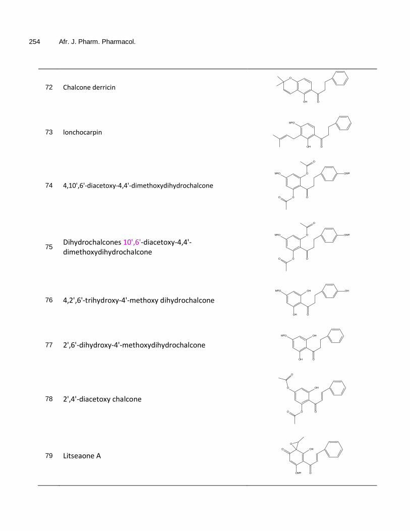
254 Afr. J. Pharm. Pharmacol.
72 Chalcone derricin
OOH
O
73 lonchocarpin
OOH
MeO
74 4,10',6'-diacetoxy-4,4'-dimethoxydihydrochalcone
O
O
O
MeO OMe
O
O
75 Dihydrochalcones 10',6'-diacetoxy-4,4'-dimethoxydihydrochalcone
O
O
O
MeO OMe
O
O
76 4,2',6'-trihydroxy-4'-methoxy dihydrochalcone
O
OH
OH
MeO OH
77 2',6'-dihydroxy-4'-methoxydihydrochalcone
O
OH
OH
MeO
78 2',4'-diacetoxy chalcone
O
OH
O
O
O
O
79 Litseaone A
OOMe
O
O
OH

Gutierrez et al. 255
80 Litseaone B
OOMe
MeO O
O
81 Stercurensin
OOH
HO OMe
82 cardamonin
OOH
HO OMe
83 2',4'- dihydroxy-4,6'-dimethoxydihydrochalcone
OOH
HO OMe OMe
84 Licochalcone G
OOH
HO
MeO
OH
85 licochalcone A
O
HO
MeO
OH
86 Echinantin
O
HO
MeO
OH
87 5-prenylbutein
O
HO OH
OH
OH

256 Afr. J. Pharm. Pharmacol.
88 licochalcone D
O
HO
MeO
OH
OH
89 Isoliquiritigenin
O
HO OH
OH
90 licoagrochalcone A
O
HO OH
OH
91 kanzonol C
O
HO OH
OH
92 Licochalcones A
O
HO
MeO
OH
OH
93 Licochalcones B
O
HO
MeO
OH
OHHO
94 Chalcone
O
95 4-Hydroxychalcone
O
OH
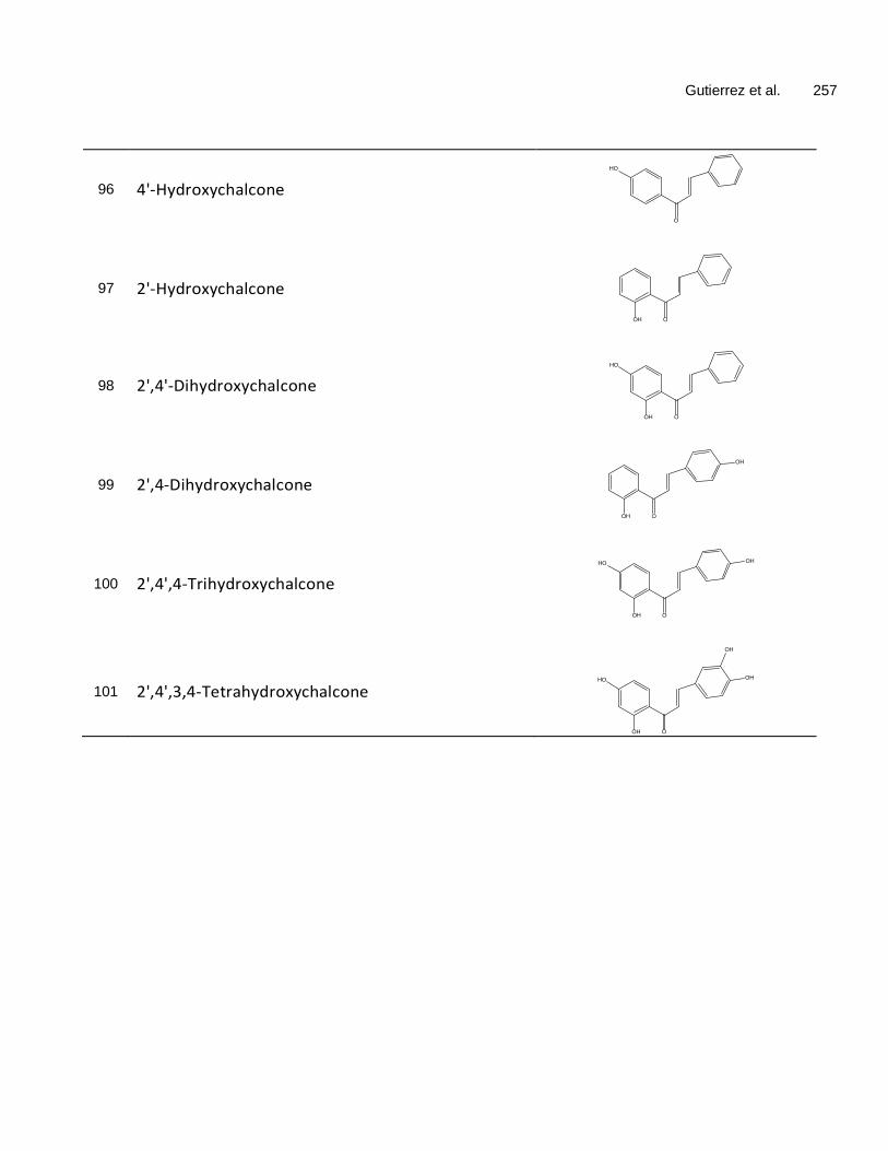
Gutierrez et al. 257
96 4'-Hydroxychalcone
O
HO
97 2'-Hydroxychalcone
OOH
98 2',4'-Dihydroxychalcone
OOH
HO
99 2',4-Dihydroxychalcone
OOH
OH
100 2',4',4-Trihydroxychalcone
OOH
OHHO
101 2',4',3,4-Tetrahydroxychalcone
OOH
OHHO
OH

Vol. 9(8), pp. 258-264, 29 February, 2015 DOI: 10.5897/AJPP2014. 4223 Article Number: 9CAAF6B51348 ISSN 1996-0816 Copyright © 2015 Author(s) retain the copyright of this article http://www.academicjournals.org/AJPP
African Journal of Pharmacy and Pharmacology
Full Length Research Paper
Clinical duration of rocuronium becomes shorter at night: A chronopharmacological study
Menekse Ozcelik*, Ali Abbas Yılmaz, Sirali Oba, Sanem Turhan and Oya Ozatamer
Department of Anesthesiology and ICM, Ankara University School of Medicine, Ankara, Turkey
Received 5 November, 2014; Accepted 3 February, 2015
The effects of circadian rhythms on the duration of neuromuscular blocking agents are under investigated. Therefore, the aim of this prospective, observational study was to investigate whether the dosing-time of rocuronium would cause significant differences in the neuromuscular block duration during the four different period of a day in a diurnal based study design. Forty patients were allocated into one of the following four groups according to the surgery timing during a day. Patients undergoing surgery between 07:00 am and 01:00 pm were included in Group M (Group morning, n = 10), between 01:00 pm and 07:00 pm in Group AN (Group afternoon, n = 10), between 07:00 pm and 01:00 am in Group E (Group evening, n = 10), and between 01:00 am and 07:00 am in Group N (Group night, n = 10). All patients received rocuronium in a dose of 0.6 mg/kg. The time to T1 10% and clinical duration were significantly shorter in Group N [24.4 ± 8.0, 28.5 ± 9.0 min, mean ± standard deviation (SD)] when compared with Group AN (32.1 ± 8.6, 36.7 ± 9.1 min, mean ± SD) and Group E (32.5 ± 9.9, 37.0 ± 11.4 min, mean ± SD) (p < 0.05). T1 10% time and clinical duration of rocuronium were significantly shorter when used at nighttime compared to afternoon and evening part of the day. The increased hepatic blood flow and metabolism of rocuronium may be the responsible factors related to this shortening at night. Key words: Rocuronium, chronobiology, circadian rhythm, neuromuscular non-depolarizing agents, anesthesia, intravenous.
INTRODUCTION The biologic rhythms are physiological circumstances that provide the adaptation of an organism to the external environment in the absence of environmental time determinants (Chassard and Bruguerolle, 2004). The chronobiology investigates these rhythms and their mechanisms. The most well recognized biologic rhythm is the circadian rhythm that is characterized by a biologic cycle of approximately 24 h (Chassard et al., 2005). There are a great number of studies related to drug and biologic rhythm interactions in clinical medicine
(Chassard et al., 2007). However, probably due to two main reasons mentioned, the influence of chronobio-logy on the clinical practice of anesthesiology remains to be determined. The first reason is the combined use of several drugs in the course of anesthesia and therefore, the fear of underestimating or overestimating the interactions between biologic rhythms and anesthetic drugs. In addition to this, because of the concept considering that the circadian rhythms have been overcome with the homeostasis, most of the anesthe-
*Corresponding author. E-mail: [email protected] Author(s) agree that this article remain permanently open access under the terms of the Creative Commons Attribution License 4.0 International License

siologists have not been interested in chronobiology in their clinical practice (Cheeseman et al., 2007).In the field of anesthesia, the drug dose-response relationship represents an extremely important issue. A large number of clinical and laboratory studies have been designed to establish the factors affecting this dose-response relationship from past to present. Rocuronium is the most rapid onset and the most commonly used non-depolarizing muscle blocker currently available. Factors such as age (Baykara et al., 2002), weight (Leykin et al., 2004), temperature of the patient (Beaufort et al., 1995), as well as the anesthetic agent used for anesthesia maintenance (Maidatsi et al., 2004) affect the duration of action of rocuronium. Whether the chronobiology is another variable affecting the duration of action of rocuronium is controversial. Only one study was under-taken to evaluate the influence of time of day on the clinical duration of rocuronium in the available literature (Cheeseman et al., 2007). However, its design did not include all the time course of a day. The night part of the day denoting the resting period of human individuals had not been considered in the study design. Therefore, we have undertaken a study to in-vestigate whether the dosing-time of rocuronium would cause significant differences in the neuromuscular block duration during the whole day in a diurnal based study design. MATERIALS AND METHODS After local ethics committee approval, the patients undergoing elective or emergency abdominal surgery with an expected duration less than four hours between January, 2006 and July, 2006 were enrolled into the study which was conducted according to the Declaration of Helsinki and Good clinical research practice (GCRP) guidelines for pharmacodynamic studies of neuromuscular blocking agents (Fuchs-Buder et al., 2007). The inclusion criteria were 18 to 59 years of age, American Society of Anesthesiologist (ASA) status 1 or 2, actual body weight in the range of 20% of the ideal body weight, normal renal and hepatic function, no history of cardiac and neuromuscular diseases. The exclusion criteria were expected difficult airway, known allergy to one of the study drugs, history of drug usage likely to affect the neuromuscular function. The information about the general anesthesia, monitorization techniques, and the study protocol were given to all subjects and then the informed consent were requested from the patients undergoing elective and emergency surgery the day before and one hour before surgery, respectively.
The patients were allocated into one of the following four groups according to the surgery timing during a day. The patients undergoing elective surgeries between 07:00 am and 01:00 pm were included in Group M (Group morning, n = 10) and between 01:00 pm and 07:00 pm were included in Group AN (Group afternoon, n = 10). The patients undergoing emergent surgeries between 07:00 pm and 01:00 am were included in Group E (Group evening, n = 10) and between 01:00 am and 07:00 am were included in Group N (Group night, n = 10).
After arrival to the operating room, a 20-gauge intravenous (iv) catheter was inserted on the dorsum of the hand or antecubital fossa. Intravenous midazolam (0.4 mg/kg) premedication was given to all patients following routine ASA monitorization including electrocardiogram with continuous ST-T analysis, non-invasive blood pressure, body temperature, and pulse oximetry (Viridia CMS
Ozcelik et al. 259 M1166A, Hewlett Packard, Germany). In addition, Bispectral Index (A-2000 XP, Aspect Medical Systems, Inc, USA), weighted sum of several electroencephalographic sub parameters was installed to monitor depth of anesthesia. Neuromuscular function was assessed quantitatively with an acceleromyograph (TOF-Watch ® SX, Organon, Dublin, Ireland) connected to a laptop computer installed with appropriate software via an interface. All patients were warmed with a blanket laid under the patients. Anesthetic management Total intravenous anesthesia (TIVA) with target controlled infusion (TCI) technique using propofol and remifentanil was utilized in anesthesia induction and maintenance of all patients. The demographic data such as age, actual body weight, height and sex were set on the TCI infusion pump (Orchestra ® Base Primea, Fresenius Kabi, Germany), which enables the drug concentration in the plasma (Cp) and at the effect site (Ce) such as brain, to be continuously controlled. The pharmacokinetic model described by Schnider et al. (1998) uses age as a co-variate to improve the accuracy of the model. Because this model has a smaller-volume central compartment and equilibrates more quickly with the effect site, our patients received TCI propofol driven by the Schnider model with effect-site control. Meanwhile, the infusion rate of TCI remifentanil was controlled by Minto's pharmacokinetic model incorporated into software that was previously used specifically for remifentanil (Minto et al., 1997). Syringes containing 1% propofol (10 mg/ml) and remifentanil (50 μg/ml) were simultaneously loaded onto the device and connected to the patient's intravenous catheter using a three-way stopcock.
The anesthesia induction was provided by remifentanil infusion with an effect-site concentration at 7.5 ng/ml following propofol infusion with an effect-site concentration at 2.5 µg/ml. Once the BIS value was lower than 80, the mask ventilation with 100% oxygen was started.
After reaching the target concentrations of anesthetic drugs and before administration of rocuronium, a 50 Hz tetanic stimulation for 5 s was administered followed after 1 min by immobilization of the patient’s fingers. Then the ulnar nerve at the wrist was stimulated in a train-of-four (TOF) mode every 15 s for 3 min. When the calibration process by CAL 2 mode was completed and then stable baseline of the response to TOF was reached, 0.6 mg/kg rocuronium iv was administered in 5 s. The time between rocuronium injection and 95% depression of first twitch (T1) response was noted as “onset time”. As soon as onset time was defined, all patients were intubated. All patients were mechanically ventilated with a tidal volume of 7 ml/kg and frequency of 12/min. In order to achieve the goal of maintenance of desired end-tidal carbon dioxide level (between 35 and 45 mmHg), firstly tidal volume was increased by 20% and secondly frequency of mandatory respirations was increased to 16/min.
During the maintenance period of anesthesia the remifentanil effect-site concentration was adapted by step of 2.5 ng/ml according to the hemodynamic parameters in every 5 min. In case of bradycardia and hypotension, atropine 0.5 mg and ephedrine 10 mg iv were injected, respectively. Meanwhile, propofol effect-site concentration was adjusted to intend intraoperative BIS value of 40 to 60. If BIS value was lower than 40, propofol effect-site concentration had been lowered by 0.5 µg/ml. On the contrary, propofol effect-site concentration had been increased by 0.5 µg/ml if BIS value was higher than 60. The anesthetic management was schematized in Figure 1. During the anesthesia maintenance, the time elapsing between TOF count 0 and first twitch reappearance was noted as “deep neuromuscular block duration”. Moreover, the time elapsing between administration of rocuronium and recovery to 10 and 25% of first twitch height (T1 10%, T1 25%) were recorded as “the time to T1 10% and clinical duration”, respectively. When

260 Afr. J. Pharm. Pharmacol.
Figure 1. Anesthetic management algorithm.
Table 1. Demographic data (n=patient number).
Group Group M (n=10) Group AN (n=10) Group E (n=10) Group N (n=10) p Age (year) 42±8 38±12 32±13 30±11 NS Weight (kg) 69±10 66±9 69±10 64±8 NS Height (cm) 173±8 168±6 179±9 169±6 NS Actual body weight (kg) 70±9 65±7 64±10 63±7 NS ASA Classification (I/II) (n) 9/1 9/1 8/2 8/2 NS
All values are shown as mean±SD, NS, statistically not significant. Group Morning (Group M): patients undergoing surgery between 07:00 am and 01:00 pm; Group Afternoon (Group AN): patients undergoing surgery between 01:00 pm and 07:00 pm; Group Evening (Grup E): patients undergoing surgery between 07:00 pm and 01.00 am; Group Night (Group N): patients undergoing surgery between 01.00 am and 07.00 am.
“clinical duration” was achieved, if necessary, additional rocuronium in a dose of 0.15 mg/kg iv was administered and the time between the injection of rocuronium and the second recovery to 25% of first twitch height was recorded as “second clinical duration”. If an additional rocuronium was not necessary due to the end of surgery, 30 μg/kg neostigmine and 10 μg/kg atropine were administered for the recovery of neuromuscular blockade. Along the recovery period, the time from 25 to 75% of first twitch height recovery was recorded as “recovery index” and the time between T1 25% and TOF ratio of 90% was also recorded as “complete recovery interval”. If the TOF ratio was above 80 and the BIS level was above 70, the patients were then extubated.
All patients were followed in terms of their peripheral temperatures and BIS levels and all these parameters were recorded. In addition to the duration of anesthesia and surgery, the total and hourly propofol, remifentanil and rocuronium consumptions were also noted. Statistical analysis The statistical analyses were made by using SPSS 12.0 program
(Statistical Program for Social Sciences, Chicago, IL, USA) for Windows with a personal computer. The numeric data were analyzed by the t-test and presented as mean ± SD. Chi-square test was used for the evaluation of categorical variables; correlation analysis was used to determine the relationship between the variables. In comparison of 3 or more groups, Kruskall-Wallis, a non-parametric test was used, in comparison of 2 groups Mann-Whitney U test was used. No power analysis was done. For statistical comparisons, p <0.05 were considered as significant. RESULTS Forty patients were enrolled into the study. None of them were excluded due to any reason. No significant difference was found between groups in terms of patient demographics provided in Table 1. Intraoperative hemodynamic parameters were similar between groups and no hemodynamic adverse effect was found. The onset time of rocuronium did not differ between 4 groups with a mean (SD) of 2.65 (1.0) min. Similarly, there was

Ozcelik et al. 261
Table 2. Neuromuscular blockade parameters (n=patient number).
Neuromuscular parameters (min) Group M (n=10)
Group AN (n=10)
Group E (n=10)
Group N (n=10)
Onset time 2.8 ± 1.1 2.4 ± 0.9 2.4 ± 1.0 3.0 ± 1.1 Deep neuromuscular blockade duration 19.0 ± 4.0 25.8 ± 9.3 24.6 ± 7.4 19.2 ± 8.5 The time to T1 10% 27.2 ± 6.6 32.1 ± 8.6 32.5 ± 9.9 24.4 ± 8.0* Clinical duration 31.0 ± 7.7 36.7 ± 9.1 37.0 ± 11.4 28.5 ± 9.0* Second clinical duration 21.4 ± 9.0 17.5 ± 4.4 16.8 ± 7.3 19.0 ± 5.0 Recovery index 5.5 ± 2.8 5.6 ± 2.8 7.6 ± 4.1 5.5 ± 3.2 Complete recovery interval 9.6 ± 2.8 10.0 ± 3.8 12.6 ± 4.9 10.8 ± 4.0
All values are shown as mean ± SD. Group Morning (Group M): patients undergoing surgery between 07:00 am and 01:00 pm; Group Afternoon (Group AN): patients undergoing surgery between 01:00 pm and 07:00 pm; Group Evening (Grup E): patients undergoing surgery between 07:00 pm and 01.00 am; Group Night (Group N): patients undergoing surgery between 01.00 am and 07.00 am. * p < 0.05 compared with Group AN and E.
Table 3. Intraoperative drug consumptions (n=patient number).
Parameter (mg) Group M (n=10)
Group AN (n=10)
Group E (n=10)
Group N (n=10)
Total rocuronium consumption 84 ± 33 66 ± 22 73 ± 32 68 ± 25 Hourly rocuronium consumption 45 ± 12 41 ± 11 43 ± 14 45 ± 9 Total propofol consumption 708 ± 263 686 ± 429 671 ± 264 498 ± 216 Hourly propofol consumption 375 ± 78 388 ± 81 391 ± 101 329 ± 57 Total remifentanil consumption 2390 ± 957 2763 ± 1703 2782 ± 1268 2228 ± 775 Hourly remifentanil consumption 1265 ± 359 1521 ± 409 1592 ± 416 1526 ± 424
All values are shown as mean±SD. Group Morning (Group M): patients undergoing surgery between 07:00 am and 01:00 pm; Group Afternoon (Group AN): patients undergoing surgery between 01:00 pm and 07:00 pm; Group Evening (Grup E): patients undergoing surgery between 07:00 pm and 01.00 am; Group Night (Group N): patients undergoing surgery between 01.00 am and 07.00 am.*p < 0.05 compared with Group AN and E.
no difference related to the deep neuromuscular blockade duration of rocuronium among groups (Table 2). In contrast, there was a clear relationship between the administration time of rocuronium and the estimates of time to T1 10% and clinical duration of rocuronium. The time to T1 10% was significantly shorter in Group N (24.4 ± 8.0 min, mean ± SD) when compared with Group AN (32.1 ± 8.6 min, mean ± SD) and Group E (32.5 ± 9.9 min, mean ± SD) (p < 0.05). The shortening in T1 10% observed in Group N was 24 and 24.9% compared with Group AN and Group E, respectively.
There was no difference between Group M, Group AN and Group E in terms of time to T1 10%. The time to T1 10% did not differ between Group M and Group N. When the clinical duration of rocuronium was compared between 4 groups, it was found that clinical duration was shorter in Group N (28.5 ± 9.0 min, mean ± SD) when compared with Group AN (36.7 ± 9.1 min, mean ± SD) and Group E (37.0 ± 11.4 min, mean ± SD) (p < 0.05). The longest and the shortest clinical durations were noted in Group AN with a 61.4 min and in Group N with a 20.5 min, respectively. The shortening in clinical duration
observed in Group N was 22.3 and 22.9% compared with Group AN and Group E, respectively. There was no difference between Group M, Group AN and Group E in terms of clinical duration. There was also no difference in clinical duration between Group M and Group N. These findings were listed in Table 2.
Although the second clinical duration was shorter in Group N compared with Group M, this was not statistically significant. The recovery index and complete recovery interval did not differ at any time throughout the day between groups (Table 2). The BIS levels and peripheral temperature measurements were similar between the groups (Figures 2 and 3). There was no statistically significant difference between four groups in
terms of intraoperative total and hourly propofol, remifentanil and rocuronium consumptions (Table 3). DISCUSSION This is the first human study investigating the effects of the rocuronium administration time on the neuromuscular

262 Afr. J. Pharm. Pharmacol.
Figure 2. Intraoperative BIS values.
Figure 3. Intraoperative peripheral temperature measurements.
Time
BIS
val
ue
110.0 90.0 70.0 50.0 30.0
Tem
pera
ture
Time
34 33.8 33.6 33.4 33.2 33 32.8 32.6 32.4 32.2

blockade characteristics during the 24 h. The primary finding of our study was that, T1 10% time and clinical duration of rocuronium were significantly shorter at night compared to afternoon and evening part of the day under total intravenous anesthesia with propofol and remifentanil. The onset time of rocuronium is approximately 1.5 to 1.7 min considering its pharmacodynamic properties (Naguib and Lien, 2009). However, the onset time was found longer in our study (mean, 2.65 min).
Na and coworkers demonstrated that the prior administration of remifentanil during TCI propofol and remifentanil anesthesia prolonged the onset time of rocuronium due to decreased cardiac output in their well-designed study (Na et al., 2012). This may also explain our prolonged onset time due to our comparable sequence of anesthesia induction. Nevertheless, there was no difference in terms of onset time or deep neuromuscular blockade of rocuronium between the 4 study groups.
There was a clear relationship between the dosing time of rocuronium and the time to T1 10%, and clinical duration. The time to T1 10% and clinical duration were shorter at night compared with afternoon and evening application. These findings were not consistent with the study data previously published.
Cheeseman et al. (2007) demonstrated that the minimum duration of action of rocuronium dosing of 0.6 mg/kg was between 14:00 and 17:00 corresponding to the timing of our Group AN. There was an important difference in study design between these two studies. Unlike from our study, there was no patient enrollment in the night period of a day due to some anticipated difficulties. Therefore, the authors could not conclude on the clinical duration of rocuronium during the resting period of participants.
The clinical duration of a neuromuscular blocker indicates that the duration of neuromuscular blocker action becomes terminated, as it has been mostly metabolized in human body. In practice, if the clinical duration of a neuromuscular blocker has been achieved, anesthesiologist should give either an additional neuromuscular blocker or a reversal agent of neuromuscular blocker according to the circumstances (that is, ongoing or ending surgery). Therefore, quantifying these parameters is very important to decide what should be done in the course of a surgical anesthesia.
The mechanism underlying this chrono-pharmacological aspect of rocuronium may be attributed to its metabolism pathway. A large portion of circulating rocuronium is taken by the liver, metabolized and then excreted into the bile. Lemmer and Nold (1991) found significant circadian differences in estimated hepatic blood flow (EHBF) measured by injection of indocyanine green during the different part of the day in healty male volunteers. In this study, the peak EHBF was reached at
Ozcelik et al. 263 04:00 am. They also showed no difference in the distribution volume. Likewise, in another study investigating the oxaliplatin pharmacokinetics and chronopharmacological aspects, the hepatic blood flow was found maximum between 02:00 am and 08:00 am, and minimum at 02:00 pm within a day (Levi et al., 2000). Therefore, we may speculate that the clinical duration of rocuronium appears to be shorter with bedtime compared with AN or E administration.
There was an absence of statistical significance in terms of second clinical duration between four groups. The additional rocuronium dosage was 0.15 mg/kg. Therefore, this finding can be explained by application error in smaller drug doses rather than the hepatic blood flow differences. There were no difference regarding recovery index and complete recovery interval between groups. There are a number of limitations in this study. Probably, the most important one is the formation of four different groups instead of two study groups. This would be more appropriate for investigations on circadian rhythms. The number of patients were not determined using a power analysis statistically. In addition to this, this study was not sufficient to establish the real mechanism related to diurnal variation of clinical duration of rocuronium.
Future clinical trials assessing the blood rocuronium levels or rocuronium and receptor interactions are needed to clarify this issue. Additionally, the effect of the diurnal changes associated with elective or emergency was another potential confounding factor in the study design. This effect was underestimated in this study. Conclusion This study demonstrated that the clinical duration of rocuronium became shorter at night compared with the noon and evening part of the day under propofol and remifentanil anesthesia. Although the effect of this shortening on clinical practice looks like limited, it could be more significant as enough data about the diurnal changes related with rocuronium are provided in the future. ACKNOWLEDGEMENTS The authors acknowledge the many insightful suggestions of Hakan Zengil, M.D, Prof, lecturer of Gazi University Faculty of Medicine Department of Pharmacology and the president of Chronobiology Association in Turkey, to this work. The authors also thank Basak Meco, M.D, DESA, for the assistance in the preparation of the manuscript. Conflict of interest There is no conflict of interest as regard this study.

264 Afr. J. Pharm. Pharmacol. REFERENCES Baykara N, Woelfel S, Fine GF, Solak M, Toker K, Brandom BW (2002).
Predicting recovery from deep neuromuscular block by rocuronium in children and adults. J. Clin. Anesth. 14:214-217.
Beaufort AM, Wierda JM, Belopavlovic M, Nederveen PJ, Kleef UW, Agoston S (1995). The influence of hypothermia (surface cooling) on the time-course of action and on the pharmacokinetics of rocuronium in humans. Eur. J. Anaesthesiol. 11: S95-106.
Chassard D, Allaouchiche B, Boselli E (2005). Time is everything: the pendulum swings on. Anesthesiology 103:454-456.
Chassard D, Bruguerolle B (2004). Chronobiology and Anesthesia. Anesthesiology 100:413-427.
Chassard D, Duflo F, de Queiroz Siqueira M, Allaouchiche B, Boselli E (2007). Chronobiology and anaesthesia. Curr. Opin. Anaesthesiol. 20:186-190.
Cheeseman JF, Merry AF, Pawley M, de Souza RL, Warman GR (2007). The effect of time of day on the duration of neuromuscular blockade elicited by rocuronium. Anaesthesia 62:1114-1120.
Fuchs-Buder T, Claudius C, Skovgaard T, Eriksson I, Mirakhur RK, Viby-Mogensen J (2007). Good clinical research practice in pharmacodynamic studies of neuromuscular blocking agents II:the Stockholm revision. Acta. Anaesthesiol. Scand. 51:789-808.
Lemmer B, Nold G (1991). Circadian changes in estimated hepatic blood flow in healty subjects. Br. J. Clin. Pharm. 32:627-629.
Levi F, Metzger G, Massari C, Milano G (2000). Oxaliplatin pharmacokinetics and chronopharmacological aspects. Clin. Pharmacokinet. 38:1-21.
Leykin Y, Pellis T, Lucca M, Lomangino G, Marzano B, Gullo A (2004).
The pharmacodynamics effects of rocuronium when dosed according to real body weight or ideal body weight in morbidly obese patients. Anesth. Analg. 99:1086-1089.
Maidatsi PG, Zaralidou AT, Gorgias NK, Amaniti EN, Karakoulas KA, Minto CF, Schnider TW, Shafer SL (1997). Pharmacokinetics and pharmacodynamics of remifentanil. II. Model application. Anesthesiology 86:24-33.
Na HS, Hwang JW, Park SH, Oh AY, Park HP, Jeon YT, Do SH (2012). Drug-administration sequence of target-controlled propofol and remifentanil influences the onset of rocuronium. A double-blind, randomized trial. Acta. Anaesthesiol. Scand. 56:558-564.
Naguib M, Lien CA (2009). Pharmacology of muscle relaxants and their antagonists. In: Miller’s Anesthesia, ed: Miller R. pp. 859-911.
Schnider TW, Minto CF, Gambus PL, Andresen C, Goodale DB, Shafer SL, Youngs EJ (1998). The influence of method of administration and covariates on the pharmaco- kinetics of propofol in adult volunteers. Anesthesiology 88:1170-1182.

Vol. 9(8), pp. 265-273, 29 February, 2015 DOI: 10.5897/AJPP2014. 4175 Article Number: 81888CE51349 ISSN 1996-0816 Copyright © 2015 Author(s) retain the copyright of this article http://www.academicjournals.org/AJPP
African Journal of Pharmacy and Pharmacology
Full Length Research Paper
Effect of grapefruit juice and sibutramine on body weight loss in obese rats
Hadir Farouk1*, Sawsan S. Mahmoud1, Bahia A. El-Sayeh2, and Ola A. Sharaf1
1Pharmacology Department, National Research Center, Egypt.
2Pharmacology Department, Faculty of Pharmacy, Cairo University, Egypt.
Received 14 September, 2014; Accepted 16 February, 2015
Grapefruit (Citrus Paradise, family Rutaceae) is a citrus fruit that is low in calories and rich in dietary fibers. Sibutramine (Sibutramine hydrochloride monohydrate) is an anti-obesity drug that enhances satiety. The effect of grapefruit juice and sibutramine on body weight and neurotransmitters controlling appetite was investigated in obese rats. Rats were assigned to two dietary groups for 3 weeks; control group (n=6) was fed commercial standard pellets diet and obese group (n=24) was fed cafeteria diet (hypercaloric diet consisting of highly palatable food). The effect of sibutramine and grapefruit juice was studied on obese rats. Statistical difference and interactions were evaluated through one-way analysis of variance test (one-way ANOVA) followed by Dunnett’s test was used for means of different groups. For all statistical tests done, a 0.05 level of probability was used as the criterion for significance. Grapefruit juice produced its weight reduction effect after 1 week of administration and lasted till the end of the experiment and did not affect brain neurotransmitters. Sibutramine produced its weight reduction effect after 1 week of administration and lasted for only 2 weeks and produced an increase in brain noradrenaline while grapefruit juice produced its effect from the first week till the end of the study. It can be concluded that grapefruit juice is better than sibutramine since its effect lasted till the end of the experiment and also did not affect brain noradrenaline. Key words: Obesity, sibutramine, grapefruit juice, cafeteria diet.
INTRODUCTION Obesity represents one of the most serious global health issues that have increased to the extent that it could be considered pandemic (Abolfotouh et al., 2008).
Obesity has reached epidemic proportions globally (WHO, 2003). A recent report from the World Health Organization estimated that approximately 500 million individuals are obese while 1.5 billion are overweight worldwide (WHO, 2011). Obesity occurs through a
longstanding imbalance between energy intake and energy expenditure, influenced by a complex biologic system that regulates appetite (Wilding, 2011).
During the past decade, overweight and obesity joined underweight, malnutrition, and infectious diseases as major health problems threatening the developing world (Hossain et al., 2007). At least 2.6 million people each year die as a result of being overweight or obese
*Corresponding author. E-mail: [email protected]. Author(s) agree that this article remain permanently open access under the terms of the Creative Commons Attribution License 4.0 International License

266 Afr. J. Pharm. Pharmacol. (WHO, 2010).
It seems clear that an energy-dense, high fat diet, palatable foods with high caloric density and increasingly sedentary lifestyles are particularly conducive to the development of obesity (Pi-Sunyer, 2002). Obesity is a risk factor for many chronic conditions including diabetes and cardiovascular diseases (Wang et al., 2014).
Serotonin, particularly via the 5HT2c receptor, has also been implicated in satiety signaling. The hypothalamus is densely innervated by serotonin-containing fibres origin-nating in the mid-brain raphe nucleus. Serotonin is the target of a number of obesity drugs such as the re-uptake inhibitors sibutramine (Jackson et al., 1997).
In the hypothalamus, the paraventricular nucleus (PVN) (the area associated with control of food intake) (Jackson et al., 1997) is innervated by norepinephrine (NE) fibers and is a site at which infusion of exogenous NE elicits eating at low doses. Two subtypes of α-adrenergic recep-tors within the PVN exert antagonistic actions on eating in the rat: activation of PVN α2-adrenoceptors increases eating, α2-adrenoceptor agonists such as clonidine have been shown to increase food intake in rats (Jackson et al., 1997). On the other hand activation of PVN α1-adrenoceptors suppresses eating (Wellman, 2000).
Grapefruit (Citrus Paradise, family Rutaceae) (Scora et al., 1982) is a citrus fruit that is low in calories and rich in dietary fibers (Stump et al., 2006). It is a commercial crop of great importance, mainly for the juice industry, and also as a source of essential oils and pectin (Hodgson, 1967). It had been shown that grapefruit juice decreased body weight (Fujioka et al., 2006).
Sibutramine is an anti-obesity drug that enhances satiety (suppressing the appetite) (Kim et al., 2003). It acts centrally as an inhibitor of both norepinephrine and serotonin reuptake. Due to inhibition of noradrenaline reuptake, sibutramine is expected to increase systolic and diastolic blood pressure as well as pulse rate with the result that the drug therapy being discontinued in about 5% of patients (Yanovski and Yanovski, 2002).
The aim of this work was to study the effect of grapefruit juice and sibutramine on obesity induced by cafeteria diet in rats MATERIALS AND METHODS Animals Thirty female Sprague-Dawley albino rats weighing 120 to 140 g purchased from the animal house colony of the National Research Center (Dokki, Giza, Egypt) were kept in the animal house under hygienic conditions at room temperature with reversed 12 h light, dark cycle. The animals were fed on commercial standard pellets and given water ad libitum for an adaptation period of 1 week. Experiments were performed according to the national regulations of animal welfare and institutional animal committee (IAEC). Animal grouping Rats were then divided into two groups. The first group (6 rats) was
fed on commercial standard pellets throughout the experiment and served as control group. The second group (24 rats) was fed high calorie highly palatable “cafeteria diet” consisting of chocolate and cookies (Hamilton and Doods, 2002) and served as obese group in addition to standard pellets for 3 weeks for the induction of obesity.
After 3 weeks, the obese group was divided into 3 subgroups: Group I received cafeteria diet for 3 weeks and served as positive control. Group II received grapefruit juice (4 ml, 3 times daily, by gavage) (Fujioka et al., 2006; Paget and Barnes, 1964) together with feeding cafeteria diet for 3 weeks. Group III received sibutramine (3 mg/kg/day, by gavage) (Brown et al., 2001) together with feeding cafeteria diet and standard pellets for 3 weeks.
Body was recorded weekly. Percent change of body weight gain was calculated as:
Final weight – Initial weight Change of body weight (%) = × 100 Initial weight At the end of the experimental period, blood samples were collected, rats were killed by decapitation and brain tissues were isolated.
Blood samples (3 ml) were collected from retro-orbital plexus in dry centrifuge tubes and left to clot at room temperature. Samples were centrifuged at 1500 rpm for 10 min, the clear supernatant was separated and used for determination of serum glucose, triglycerides, glutamic oxaloacetic transaminase (GOT), glutamic pyruvic transaminase (GPT), creatinine and urea. Preparation of brain samples The brain was immediately excised on ice and weighed then homogenized in 75% high performance liquid chromatography (HPLC) methanol (1/10 weight/volume) using a homogenizer (Jake and Kunkle IKA labortechnick, Ultra-turrax T25, Germany) surrounded with an ice jacket and the homogenates were centrifuged in a cooling centrifuge (Sigma and laborzentrifugen, 2K15, Germany) for 15 min at 5000 rpm. The supernatant was used for determination of monoamines’ concentration by HPLC according to the method described by Pagel et al. (2000). Statistical analysis Data were expressed as mean ± standard error (SE). Statistical differences and interactions were evaluated using one-way analysis of variance test (One-way ANOVA) followed by Dunnett’s test. For all statistical tests done, a 0.05 level of probability was used as the criterion of significance. RESULTS Effect of feeding cafeteria diet for 3 weeks on body weight gain of rats Feeding cafeteria diet for 3 weeks significantly increased body weight gain of rats, as compared to normal control values (50.11 ± 2.35 and 21.36 ± 1.92 g at P<0.05, respectively) (Figure 1).

Farouk et al. 267
Cha
nge
of b
ody
wei
ght g
ain
(%)
Figure 1. Effect of feeding cafeteria diet for 3 weeks on body weight gain of rats. *Significantly different from normal control at P<0.05.
Effect of grapefruit juice and sibutramine on food consumption Grapefruit juice significantly decreased food consumption after the second and the third weeks (323.17 ± 18.53 and 317.39 ± 29.04 Kcal at P<0.05, respectively). Sibutramine significantly decreased food consumption after the second week only (354.25 ± 13.49 Kcal at P<0.05) (Figure 2). Effect of grapefruit juice and sibutramine on body weight Grapefruit juice significantly decreased body weight gain after weeks 1, 2 and 3 (7.1 ±0.26, -3.58 ± 0.31 and 4.42 ± 0.45 g at P<0.05 respectively). Sibutramine significantly decreased body weight gain after week 1 and week 2 (-2 ± 0.25 and 6.2 ±0.65 g at P<0.05 respectively) (Figure 3). Effect of grapefruit juice and sibutramine on serum glucose and triglycerides Cafeteria diet fed rats (positive control) did not show any significant effect on serum glucose compared to normal control. All treated groups did not show any significant effect on serum glucose as compared to normal control. Cafeteria diet fed rats (positive control) showed a significant increase in serum triglycerides as compared to normal control (312.44 ± 15.24 mg/dl) at P<0.05.
Grapefruit juice did not show any significant effect on serum triglycerides as compared to normal control but it showed significant decrease in serum triglycerides as compared to positive control (141.7 ± 8 mg/dl at P<0.05). Sibutramine showed significant increase in serum tri-glycerides compared to normal control and did not show any significant effect as compared to positive control (259.12 ± 22.7 mg/dl) (Figure 4). Effect of grapefruit juice and sibutramine on serum GPT and GOT Cafeteria diet fed rats (positive control) showed a significant increase in serum GPT compared to normal control (49.75 ± 2.4 U/ml) at P<0.05. All treated groups showed significant decrease in serum GPT compared to positive control and did not show any significant difference compared to normal control. All treated groups did not show any significant difference in serum GOT compared to control groups (Figure 5). Effect of grapefruit juice and sibutramine on serum creatinine and urea Cafeteria diet fed rats showed significant increase in serum creatinine compared to normal control (0.721 ± 0.022 mg/dl at P<0.05). All treated groups did not show any significant effect compared to positive control. Cafeteria diet fed rats showed significant decrease in

268 Afr. J. Pharm. Pharmacol.
Week 1 0
0 Week 2 Week 3 Figure 2. Effect of grapefruit juice and sibutramine on food consumption. *Significantly different from positive control at P<0.05.
Cha
nge
of b
ody
wei
ght g
ain
(%)
Figure 3. Effect of grapefruit and sibutramine on body weight gain in weeks 1, 2 and 3. *Significantly different from positive control at P<0.05.

Farouk et al. 269
Con
cent
ratio
n (m
g/dl
)
Serum glucose after 3 weeks
Serum triglycerides after 3 weeks
Figure 4. Effect of grapefruit and sibutramine on serum glucose and triglycerides after 3 weeks.
Con
cent
ratio
n (U
/ml)
Serum GOT after 3 weeks Serum GPT after 3 weeks
Figure 5. Effect of grapefruit and sibutramine on serum GOT and GPT after 3 weeks.
serum urea compared to normal control (23.56 ± 1.53 mg/dl at P<0.05). Grapefruit juice did not show any significant effect on serum urea compared to the control groups. Sibutramine showed significant increase in serum urea compared to positive control but did not show any significant effect compared to normal control (Figure 6).
Effect of grapefruit juice and sibutramine on brain serotonin and noradrenaline Cafeteria diet did not show any significant effect on brain serotonin compared to normal control. All treated groups did not show any significant effect on serotonin compared to control groups. Sibutramine showed significant

270 Afr. J. Pharm. Pharmacol.
Con
cent
ratio
n (m
g/dl
)
Serum creatinine after 3 weeks Serum urea after 3 weeks
Figure 6. Effect of grapefruit and sibutramine on serum creatinine and urea after 3 weeks. aSignificantly different from normal control at P<0.05. bSignificantly different from positive control at P<0.05.
Con
cent
ratio
n (µ
g/g)
Brain noradrenaline after 3 weeks
Brain serotonin after 3 weeks
Figure 7. Effect of grapefruit and sibutramine on brain noradrenaline and serotonin after 3 weeks. *Significantly different from positive control at P<0.05.
increase in brain noradrenaline concentration compared to control groups (1.583 ± 0.03 μg/g at P<0.05) (Figure 7). DISCUSSION Cafeteria diet fed rats represent a useful model for human obesity studies because the cafeteria diet is a palatable hypercaloric and hyperlipidic diet that induces voluntary hyperphagia and fast body weight gain
(Rodríguez et al., 2001). Exposure to a palatable diet had long-term effects on feeding patterns. Rats became overweight because they initially ate more frequently and ultimately ate more of foods with higher energy density (Martire et al., 2013).
In the present study, cafeteria (CAF) diet was used to induce obesity in rats (Hamilton and Doods, 2002). Such diet increases body weight and adipose mass in rats even after a short period of use (Rodríguez et al., 2004).
In the present study, grapefruit juice significantly decreased body weight gain after weeks 1, 2 and 3 and

after 3 weeks of administration (at the end of the treatment) compared to positive control group. The mechanism of this reduced weight may be due to delayed gastric emptying secondary to grapefruit and its relative acidity (Fujioka et al., 2006). It has been noted in other studies that a decrease in pH of gastric contents can delay gastric emptying which causes gastric distension which contribute to satiety (Chaw et al., 2001) and so decrease in body weight gain.
The decrease in body weight gain may also be attributed to nootkatone, a constituent of grapefruit, which is naturally occurring adenosine monophosphate-activated protein kinase (AMPK) activator. AMPK is a serine/threonine kinase that is implicated in the control of energy metabolism and is considered to be a molecular target for the suppression of obesity (Murase et al., 2010). It consists of three proteins (α, β, and γ subunits) that together make a functional enzyme. Each of these three subunits takes on a specific role in both the stability and activity of AMPK (Stapleton et al., 1996). The net effect of AMPK activation is stimulation of hepatic fatty acid oxidation and ketogenesis, inhibition of cholesterol synthesis, lipogenesis, and triglyceride synthesis, stimu-lation of skeletal muscle fatty acid oxidation and muscle glucose uptake, and modulation of insulin secretion by pancreatic beta-cells (Winder and Hardie, 1999).
The energy-sensing capability of AMPK can be attributed to its ability to detect and react to fluctuations in the AMP:ATP ratio that take place during rest and exercise (muscle stimulation) (Winder and Hardie, 1996). During exercise, AMPK activity increases while the muscle cell experiences metabolic stress, and brought about by an extreme cellular demand for ATP. Upon activation, AMPK increases cellular energy levels by inhibiting anabolic energy consuming pathways (such as fatty acid synthesis and protein synthesis) and stimulating energy producing catabolic pathways (such as fatty acid oxidation and glucose transport) (Thomson et al., 2007).
Nootkatone significantly reduced high-fat and high-sucrose diet-induced body weight gain. These findings indicate that nootkatone is beneficial toward preventing obesity due to enhanced energy metabolism through AMPK activation in skeletal muscle and liver (Murase et al., 2010). The present study revealed that sibutramine (3 mg/kg) produced significant reduction of body weight gain after the first and the second weeks only, as compared to obese control values. No significant change of body weight gain was observed after the third week of drug administration, as compared to obese control values.
It has been reported that sibutramine produces significant reduction of body weight when used in conjunction with caloric restriction all over the period of its administration (Florentin et al., 2007). In the present study CAF diet did not produce a significant increase in the level of serum glucose compared to normal control, but it increased the serum triglycerides level (Kuhlmann et al., 2005). These results may be attributed to
Farouk et al. 271 hyperinsulinemia and hyperlipidemia (Akiyamaab et al., 1996).
Studies have shown that hyperinsulinima is a major cause of obesity because insulin causes the body to store more fats (Reaven et al., 2002). Hyperinsulinemia accelerates hepatic triglyceride (TG) production which leads to elevated plasma TG concentrations (Akiyamaab et al., 1996) and this may explain why TG showed high serum level in cafeteria diet fed rats. Data of the present study showed that grapefruit juice produced a significant reduction in serum triglycerides level compared to positive control group. This is in harmony with a previous study that showed that hesperidin and naringin, bioactive compounds of citrus fruits (grapefruit), are powerful plasma lipid lowering flavonones (Shela et al., 2007). Also a previous work showed that diet supplemented with fresh grapefruit positively influences serum lipid levels of all fractions, especially serum triglycerides. The addition of fresh grapefruit to generally accepted diets could be beneficial for hyperlipidemic, especially hypertriglyceridemic (Gorinstein et al., 2006). While compared to normal control there was no significant difference which suggests that grapefruit may normalize the serum triglycerides level of obese rats as grapefruit is rich in flavonoids. Several studies have shown that flavonoids possess lipolytic activity via inhibition of cAMP-phosphodiesterase and maintain lipolysis-inducing cAMP levels. Lipolysis is a catabolic process leading to the breakdown of TG stored in fat cells (adipocytes) and the release of free fatty acids (FFA) and glycerol. And so grapefruit prevents obesity and helps to decrease body weight and body fat (Dallas et al., 2008).
Results of the current study revealed that CAF-diet-fed rats produced a significant increase in serum GPT level while non significant effect on serum GOT was shown as compared to normal control group. This may be explained by previous studies that showed that in young healthy subjects, a fast food-based diet (CAF diet) dra-matically increased serum GPT (Marchesini et al., 2009), as well as that, a study done on human showed the effect of obesity was particularly important in the case of GPT than GOT (Robinson and Whitehead, 1989). Also, sev-eral studies showed that hyperlipidemia causes fatty liver disease which increases serum GPT (Kim et al., 2008).
In the present study, grapefruit juice showed an improvement in the level of serum GPT compared to positive control group. This study further shows that CAF diet produced a significant increase in serum creatinine and reduction in serum urea level compared to normal control group. These findings can be explained by the increase in nitrogen retention in CAF diet fed rats. The marked N2 retention was due to a decrease in amino acid catabolism and in urea production by the liver (Sadhu, 2010). Therefore daily N2 retention was greatly enhanced in these animals which is in agreement with the known protein-sparing effect of fat (excess dietary fats spare or retain the body protein) (Estornell et al., 1994).

272 Afr. J. Pharm. Pharmacol.
The decrease in urea production may be due to the following facts: (i) the rate of synthesis of urea from precursors by isolated hepatocytes from cafeteria-diet-fed rats was lower than in controls, (ii) in cafeteria-diet-fed rats the activities of all the enzymes of the urea cycle are decreased, the major percentage decreases are those of carbamoylphosphate synthetase and of argininosuccinate synthetase, the enzymes involved in the regulation of the overall rate of the cycle, when rats are switched to normal chow diet, the enzyme activities return to normal values; (iii) ihe uptake of amino acids by liver of cafeteria-diet-fed rats is lower than in controls. These results contrast with those obtained previously by using other models of obesity in rat (that is genetic or hypothalamic), in which N excretion was increased (Barber et al., 1985).
Data of the present study showed that all treated groups showed a significant increase in serum creatinine level compared to normal control, but when compared with positive control they did not show any significant effect. This means that all treatments cannot normalize serum creatinine level in the presence of CAF diet. Sibutramine produced a significant increase in brain noradrenaline level compared to positive control. This is due to the reuptake inhibition effect of sibutramine on brain noradrenaline (Brown et al., 2001).
The present data showed that sibutramine did not produce a significant effect on brain serotonin level after 3 weeks of administration compared to control groups. This may be due to the low dose of sibutramine used in the present study (3 mg/kg) since previous study showed that low doses of sibutramine had no effect on serotonin concentration and only high doses (5 mg/kg) increased serotonin level (Balcioglu and Wurtaman, 2000). This may explain why the effect of sibutramine on body weight did not last till the end of the experiment since there is a synergistic interaction between serotonin and noradrenaline (Jackson et al., 1997). Conclusion Conclusively, grapefruit juice decreased the body weight gain from the first week till the end of the study and also decreased food consumption without affecting brain noradrenaline level. Sibutramine decreased the body weight gain and food consumption only in week 2 (that is, it did not last till the end of the treatment) but it increased the brain noradrenaline level which may lead to an increase in blood pressure.
So from the present study, it was concluded that grapefruit juice is better than sibutramine since its effect lasted till the end of the experiment and also did not affect brain noradrenaline. Conflict of interest The authors declare no conflict of interest.
REFERENCES Abolfotouh MA, Soliman LA, Mansour E, Faraghaly M, El-Dawaiaty AA
(2008). Central obesity among adults in Egypt: Prevalence and associated morbidity. East. Mediterr. Health J. 14.
Akiyamaab T, Tachibanaab I, Shiroharaab H, Watanabeab N, Otsuki M (1996). High-fat hypercaloric diet induces obesity, glucose intolerance and hyperlipidemia in normal adult male Wistar rat. Diabetes Res. Clin. Pract. 31(1):27-35.
Balcioglu A, Wurtman RJ (2000). Sibutramine, a serotonin uptake inhibitor, increases dopamine concentrations in rat striatal and hypothalamic extracellular fluid. Neuropharmacology 39(12):2352-2359.
Barber T, Vina JR, Vina J, Cabo J (1985). Decreased urea synthesis in cafeteria-diet-induced obesity in the rat. Biochem. J. 230:675-681.
Brown M, Bing C, King P, Pickavance L, Heal D, Wilding J (2001). Sibutramine reduces feeding, body fat and improves insulin resistance in dietary-obese male wistar rats independently of hypothalamic neuropeptide Y. Br. J. Pharmacol. 132:1898-1904.
Chaw CS, Yazaki E, Evans DF (2001). The effect of pH on the gastric emptying of liquids measured by electrical impedance tomography and pH-sensitive radiotelemetry capsule. Int. J. Pharm. 227:167–175.
Dallas C, Gerbi A, Tenca G, Juchaux F, Bernard F (2008). Lipolytic effect of a polyphenolic citrus dry extract of red orange, grapefruit, orange (SINETROL) in human body fat adipocytes. Mechanism of action by inhibition of cAMP-phosphodiesterase (PDE). Phytomedicine 15(10):783-792.
Estornell E, Barber T, Cabo J (1994). Improved nitrogen metabolism in rats fed on lipid-rich liquid diets. Br. J. Nutr. 71:361-373.
Florentin M, Liberopulos EN, Elisaf MS (2007). Sibutramine-associated adverse effects: a practical guide for its safe use. Obes. Rev. 9(4):378-387.
Fujioka K, Greenway F, Sheard J, Ying Y (2006). The effects of grapefruit on weight and insulin resistance: relationship to the metabolic syndrome. J. Med. Food 9(1):49-54.
Gorinstein S, Caspi A, Libman I, Lerner H T, Huang D, Leontowicz H, Leontowicz M, Tashma Z, Katrich E, Feng S, Trakhtenberg S (2006). Red Grapefruit Positively Influences Serum Triglyceride Level in Patients Suffering from Coronary Atherosclerosis: Studies in Vitro and in Humans. J. Agric. Food Chem.54 (5):1887-1892.
Hamilton BS, Doods HN (2002). Chronic application of MTII in a rat model of obesity results in sustained weight loss. Obes. Res. 10(3):182-187.
Hodgson RW (1967). Horticultural varieties of Citrus. In: Reuter W, Webber HJ, Batchelor LD (eds.), The Citrus Industry. History, World Distribution, Botany and Varieties, Vol. I. University of California Press. pp. 431-591.
Hossain P, Kawar B, El Nahas M (2007). Obesity and Diabetes in the Developing World— A Growing Challenge. N. Engl. J. Med. 356(3):213-215.
Jackson HC, Bearham MC, Hutchins LJ, Mazurkiewicz SE, Needham AM, Heal DJ (1997). Investigation of the mechanisms underlying the hypophagic effects of the 5-HT and noradrenaline reuptake inhibitor, sibutramine, in the rat. Br. J. Pharmacol. 121(8):1613-1618.
Kim SH, Lee YM, Jee SH, Nam CM (2003). Effect of Sibutramine on Weight Loss and Blood Pressure: A Meta-analysis of Controlled Trials. Obes. Res. 11:1116–1123.
Kim WR, Flamm SL, Di Bisceglie AM, Bodenheimer HC (2008). Serum activity of alanine aminotransferase (ALT) as an indicator of health and disease. Hepatology 47(4)1363-1370.
Kuhlmann J, Neumann-Haefelin C, Belz U, Kramer W, Juretschke HP, Herling AW (2005). Correlation between insulin resistance and intramyocellular lipid levels in rats. Magn. Res. Med. 53(6):1275–1282.
Marchesini G, Centis E, Nuccitelli C (2009). The effect of life time changes in the treatment of NAFLD. Poster presentation at Liver and metabolic syndrome. Falk Liver Conference (Part II), Hannover, Germany.
Martire SI, Holmes N, Westbrook RF, Morris MJ (2013). Altered feeding patterns in rats exposed to a palatable cafeteria diet: increased snacking and its implications for development of obesity. PloS one 8(4):e60407.

Murase T, Misawa K, Haramizu S, Minegishi Y, Hase T (2010).
Nootkatone, a characteristic constituent of grapefruit, stimulates energy metabolism and prevents diet-induced obesity by activating AMPK. Am. J. Physiol. Endocrinol. Metab. 299(2):E266-E275.
Pagel P, Blome J, Wolf HU (2000). High-performance liquid chromatographic separation and measurements of various biogenic compounds possibly involved in the pathomechanism of Parkinson’s disease. J. Chromatography B. 746:297-304.
Paget GE, Barnes JM (1964). Evaluation of Drug Activities. In: Lawrence DR, Bacharach AL (Eds.), Pharmacometrics Vol. 1. Academic Press.
Pi-Sunyer FX (2002). The Obesity Epidemic: Pathophysiology and Consequences of Obesity. Obesity Res. 10(suppl.2):S97-S104.
Reaven GMD, Strom TK, Fox B (2002). Syndrome X: Overcoming the Silent Killer that Can Give You a Heart Attack. Published by Simon and Schuster, New York.
Robinson D, Whitehead TP (1989). Effect of body mass and other factors on serum liver enzyme levels in men attending for well population screening. Ann. Clin. Biochem. 26:393-400.
Rodríguez E, Monjo M, Rodríguez-Cuenca S, Pujol E, Amengual B, Roca P, Palou A (2001). Sexual dimorphism in the adrenergic control of rat brown adipose tissue response to overfeeding. Pflugers Arch. Eur. J. Phys. 442(3):396-403.
Rodríguez E, Ribot J, Rodríguez AM, Palou A (2004). PPAR- 2 Expression in Response to Cafeteria Diet: Gender- and Depot-Specific Effects. Obes. Res. 12:1455–1463.
Sadhu GV (2010). Process for the preparation of Caralluma extract and a formulation prepared thereof. US Patent App. 12/696,518.
Scora RW, Kumamot J, Soost RK, Nauer EM (1982). Contribution to the origin of the grapefruit Citrus paradisi (Rutaceae). Syst. Bot. 7(2):170-177.
Shela G, Hanna L, Maria L, Ryszard K, Mikolaj G, Zenon J, Yong-Seo P, Soon-Teck J, Seong-Gook K, Simon T (2007). Effect of hesperidin and naringin on the plasma lipid profile and plasma antioxidant activity in rats fed a cholesterol-containing diet. J. Sci. Food Agric. 87(7):1257-1262.
Farouk et al. 273 Stapleton D, Mitchelhill KI, Gao G, Widmer J, Michell BJ, The T, House
CM, Fernandez CS, Cox T, Witters LA, Kemp BE (1996). Mammalian AMP-activated protein kinase subfamily. J. Biol. Chem. 271(2):611-614.
Stump AL, Mayo T, Blum A (2006). Management of grapefruit-drug interactions. Am. Fam. Physician 74(4):605-608.
Thomson DM, Porter BB, Tall JH, Kim HJ, Barrow JR, Winder WW (2007). Skeletal muscle and heart LKB1 deficiency causes decreased voluntary running and reduced muscle mitochondrial marker enzyme expression in mice. Am. J. Physiol. Endocrinol. Metab. 292(1):E196-202.
Yanovski SZ, Yanovski A (2002). Drug therapy: obesity. N. Engl. J. Med. 346(8):591-602.
Wang JB, Patterson RE, Ang A, Emond J A, Shetty N, Arab L (2014). Timing of energy intake during the day is associated with the risk of obesity in adults. J. Hum. Nutr. Diet 27(s2):255-262.
Wellman PJ (2000). Norepinephrine and the control of food intake. Nutrition 16(10):837-842.
Wilding JPH (2011). Pathophysiology and aetiology of obesity. Medicine 39(1):6-10.
Winder WW, Hardie DG (1999). AMP-activated protein kinase, a metabolic master switch: possible roles in type 2 diabetes. Am. J. Physiol. 277(1 Pt. 1):E1-10.
Winder WW, Hardie DG (1996). Inactivation of acetyl-CoA carboxylase and activation of AMP-activated protein kinase in muscle during exercise. Am. J. Physiol. 270(2 Pt. 1):E299-304.
World Health Organization (WHO), (2003). Obesity and overweight. Global strategy on diet, physical activity and health.
World Health Organization (WHO), (2010). 10 facts on obesity. Available at: http://www.who.int/features/factfiles/obesity/facts/en/index1.html.
World Health Organization (WHO), (2011). Obesity and overweight: fact sheet No 311. Available at: http://www.who.int/mediacentre/factsheets/fs311/en/.

African Journal of Pharmacy and
Pharmacology
Related Journals Published by Academic Journals
■ Journal of Medicinal Plant Research■ African Journal of Pharmacy and Pharmacology■ Journal of Dentistry and Oral Hygiene■ International Journal of Nursing and Midwifery■ Journal of Parasitology and Vector Biology■ Journal of Pharmacognosy and Phytotherapy■ Journal of Toxicology and Environmental Health
Sciences