affinity purification of protein complexes in c. elegans (ip) with an affinity purified polyclonal...
TRANSCRIPT

CHAPTER 11
Affinity Purification of ProteinComplexes in C. elegans
Esther Zanin*, Julien Dumonty, Reto Gassmann*,Iain Cheesemanx, Paul Maddoxz, Shirin Bahmanyar*,Ana Carvalho*, Sherry Niessen{, John R. Yates III{,Karen Oegema* and Arshad Desai**Ludwig Institute for Cancer Research and Department of Cellular & Molecular Medicine, University ofCalifornia San Diego, La Jolla, California, USA
yInstitut Curie-UMR144, Paris, France
zInstitute forResearch in Immunology andCancer, Department of Pathology andCell Biology, University ofMontreal, Montreal, Quebec, Canada
xWhitehead Institute for Biomedical Research, MIT, Cambridge, MA, USA
{The Skaggs Institute for Chemical Biology and Department of Chemical Physiology, The Center forPhysiological Proteomics, The Scripps Research Institute, La Jolla, California, USA
AbstractI. IntroductionII. Generating Tools for Biochemistry
A. Generating a Polyclonal AntibodyB. Assessing Antibody Specificity by Immunoblotting after RNAiC. Tandem Affinity Purification TagsD. Introduction of Transgenes for Expression of Tagged ProteinsE. Validating the Functionality of the Transgene-Encoded Tagged Protein
III. The Optimal Starting Material for Protein PurificationA. Growing Worms in Large-Scale Liquid CultureB. Unsynchronized Liquid Starter CultureC. Monitoring Worm CulturesD. Harvesting Worms from Liquid CulturesE. Isolation of Embryos and Synchronization as Starved L1 LarvaeF. Seeding Synchronized Cultures Using Starved L1 LarvaeG. Freezing Adult Worms for ImmunoprecipitationH. Freezing Embryos for Immunoprecipitation
METHODS IN CELL BIOLOGY, VOL 106Copyright 2011, Elsevier Inc. All rights reserved. 289
0091-679X/10 $35.00DOI 10.1016/B978-0-12-544172-8.00011-6
I. Enriching for Specific Age EmbryosJ. Enriching for Meiosis I Arrested Embryos
IV. Isolation of Nuclei from EmbryosV. Preparing Worm and Embryonic ExtractVI. Single-step Immunoprecipitation
A. Covalent Coupling of Antibodies to Protein A BeadsB. ImmunoprecipitationC. Sample Buffer Elution: For Silver Staining & ImmunoblottingD. Glycine Elution: For Mass SpectrometryE. Urea Elution: For Mass Spectrometry
VII. Tandem Affinity Purification Using a LAP TagA. TEV CleavageB. S Protein AgaroseC. Sample Buffer Elution: For Silver Staining & ImmunoblottingD. Urea Elution: For Mass Spectrometry
VIII. Mass Spectrometry & Prioritization for Follow-up ExperimentsIX. SummaryX. Solutions and Media
A. Worm ReagentsB. Large-Scale Liquid CultureC. Isolation of Nuclei from EmbryosD. Single-step ImmunoprecipitationE. Tandem Affinity Purification using a LAP Tag
XI. EquipmentAcknowledgmentsReferences
Abstract
C. elegans is a powerful metazoanmodel system to address fundamental questionsin cell and developmental biology. Research in C. elegans has traditionally focusedon genetic, physiological, and cell biological approaches. However, C. elegans isalso a facile system for biochemistry: worms are easy to grow in large quantities, thefunctionality of tagged fusion proteins can be assessed using mutants or RNAi, andthe relevance of putative interaction partners can be rapidly tested in vivo.Combining biochemistry with function-based genetic and RNA interference screenscan rapidly accelerate the delineation of protein networks and pathways in diversecontexts. In this chapter, we focus on two strategies to identify protein–proteininteractions: single-step immunoprecipitation and tandem affinity purification.We describe methods for growth of worms in large-scale liquid culture, preparationof worm and embryo extracts, immunoprecipitation, and tandem affinity purifica-tion. In addition, we describe methods to test specificity of antibodies, strategiesfor optimizing starting material, and approaches to distinguish specific from non-specific interactions.
290 Esther Zanin et al.
ABBREVIATIONSAPC, Anaphase-promoting complex; CBP, Calmodulin-binding peptide; ChIP,Chromatin Immunoprecipitation; DMP, Dimethylpimelidimate; dsRNA, Doublestranded RNA; GST, Glutathione S-transferase; h, Hour; IP, Immunoprecipitation;LAP, Localization and Affinity Purification; MosSCI, Mos1 mediated Single Copytransgene Insertion; nAChR, Nicotinic acetylcholine receptor; RNAi, RNA inter-ference; RT, Room temperature; TAP Tandem Affinity Purification; TEV, Tobaccoetch virus.
I. Introduction
Caenorhabditis elegans is widely recognized as a powerful model system for celland developmental biology. The landmark work that described the cell lineage fromembryo to adult provided the foundation to study cell biology in the context ofdevelopment in C. elegans (Sulston et al., 1983). Research in C. elegans hastraditionally emphasized genetic and physiological approaches to elucidate genefunction. Classical epistasis analysis groups genes isolated by mutagenesis screensinto distinct pathways (Huang and Sternberg, 2006). In the past decade, genome-wide RNAi screens have greatly accelerated the annotation of gene functions(Fernandez et al., 2005; Kamath et al., 2003; Piano et al., 2000, 2002;Sonnichsen et al., 2005). Until recently, biochemical studies have lagged behind,primarily due to the historical trajectory of C. elegans research. However,C. elegans is a facile system for biochemical approaches as it is straightforwardto grow worms in large quantities, assess the functionality of tagged fusion proteinsusing mutants or RNAi, and test the relevance of any identified interacting proteinrapidly through in silico analysis and in vivo methods (Audhya and Desai, 2008;Moresco et al., 2010).In this chapter, we focus onmethods inC. elegans for isolating protein complexes
and identifying new interacting proteins using mass spectrometry. In addition, wedescribe cloning vectors that are useful for protein tagging and methods to assessantibody specificity. To identify new interaction proteins we employ two majorstrategies. In the first strategy, the target protein is purified using single-stepimmunoprecipitation (IP) with an affinity purified polyclonal antibody and theentire immunoprecipitate is subjected to mass spectrometric analysis.Immunoprecipitation of the endogenous protein has several advantages: proteinexpression is controlled by the endogenous promoter and protein function is notaltered by addition of a tag. The drawback of this approach is that a large number ofproteins are detected using current highly sensitive mass spectrometry methods.This poses a challenge for discriminating between relevant and non-specific inter-actions and therefore the significance of co-purified proteins needs to be carefullyevaluated in follow-up work. A potential additional drawback is that binding of theprimary antibodymay block association with a subset of interacting components. Inthe second strategy, we use tandem affinity purification (TAP) to isolate high
11. Affinity Purification of Protein Complexes in C. elegans 291
affinity complexes (Rigaut et al., 1999). Two sequential affinity purification stepssignificantly reduce background and lead to clean isolation of protein complexes.Two potential drawbacks of this approach are that the tag has the potential to alterprotein function and that the stringency of the two-step purification procedure maycause loss of low affinity interacting proteins. As either strategy has drawbacks,whenever possible we conduct both in parallel. Such a dual approach was criticalin defining the protein network that constitutes the core microtubule-binding siteof the chromosome during cell division (Cheeseman et al., 2004; Desai et al.,2003).Below we discuss first the tools necessary for biochemical analysis of a protein of
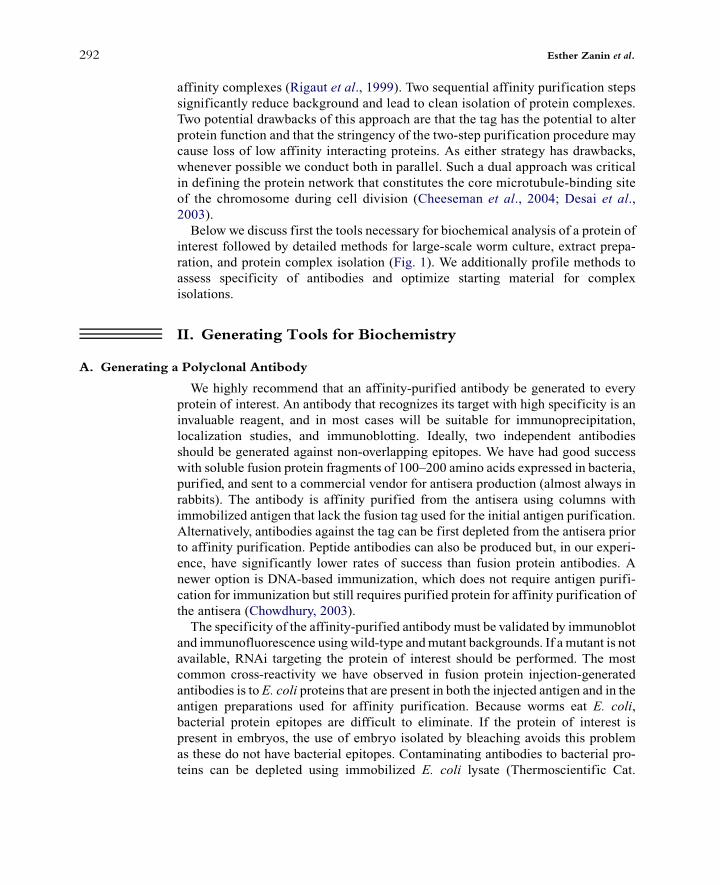
interest followed by detailed methods for large-scale worm culture, extract prepa-ration, and protein complex isolation (Fig. 1). We additionally profile methods toassess specificity of antibodies and optimize starting material for complexisolations.
II. Generating Tools for Biochemistry
A. Generating a Polyclonal Antibody
We highly recommend that an affinity-purified antibody be generated to everyprotein of interest. An antibody that recognizes its target with high specificity is aninvaluable reagent, and in most cases will be suitable for immunoprecipitation,localization studies, and immunoblotting. Ideally, two independent antibodiesshould be generated against non-overlapping epitopes. We have had good successwith soluble fusion protein fragments of 100–200 amino acids expressed in bacteria,purified, and sent to a commercial vendor for antisera production (almost always inrabbits). The antibody is affinity purified from the antisera using columns withimmobilized antigen that lack the fusion tag used for the initial antigen purification.Alternatively, antibodies against the tag can be first depleted from the antisera priorto affinity purification. Peptide antibodies can also be produced but, in our experi-ence, have significantly lower rates of success than fusion protein antibodies. Anewer option is DNA-based immunization, which does not require antigen purifi-cation for immunization but still requires purified protein for affinity purification ofthe antisera (Chowdhury, 2003).The specificity of the affinity-purified antibody must be validated by immunoblot
and immunofluorescence usingwild-type andmutant backgrounds. If a mutant is notavailable, RNAi targeting the protein of interest should be performed. The mostcommon cross-reactivity we have observed in fusion protein injection-generatedantibodies is to E. coli proteins that are present in both the injected antigen and in theantigen preparations used for affinity purification. Because worms eat E. coli,bacterial protein epitopes are difficult to eliminate. If the protein of interest ispresent in embryos, the use of embryo isolated by bleaching avoids this problemas these do not have bacterial epitopes. Contaminating antibodies to bacterial pro-teins can be depleted using immobilized E. coli lysate (Thermoscientific Cat.
292 Esther Zanin et al.
#44938) and also blocked by adding an unrelated fusion protein preparation thatharbors similar contaminants. For both immunofluorescence and immunoblotting,we typically use 0.5–1 mg/mL affinity-purified antibody in the primary antibodyincubation step. To deplete contaminating anti-bacterial protein antibodies, weincubate 500 mL of 10 mg/mL affinity purified antibody (diluted in AbDil: PBS
[(Fig._1)TD$FIG]
Fig. 1 Experimental outline for protein complex identification inC. elegans.Wild-type or LAP-tagged
strain is grown on NGM plates until larvae are starved. With starved larvae an unsynchronized starter
culture is inoculated. Embryos are isolated by bleaching and hatched in the absence of food to obtain
starved L1 larvae. Starved L1 larvae are used to set up six synchronized liquid cultures. After several
rounds of synchronized liquid culture, when sufficient amounts of worms/embryos are obtained, the
extract is prepared and protein complexes are purified by immunoprecipitation and analyzed by mass
spectrometry. Approximate time for each experiment is indicated. (See color plate.)
11. Affinity Purification of Protein Complexes in C. elegans 293
+ 2% BSA + 0.1% Triton X-100 + 0.1% sodium azide) with 100 mL of E. coliprotein agarose for 1–2 h at room temperature. The supernatant is then mixed with50 mg/mL final concentration of an unrelated fusion protein purified from bacteria(harboring the same tag as the antigen) and then used for immunoblotting. Thisprocedure eliminates anti-bacterial protein antibody cross-reactivity even in highlysensitive chemiluminescent detection.
B. Assessing Antibody Specificity by Immunoblotting after RNAi
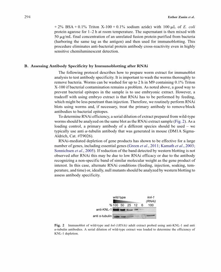
The following protocol describes how to prepare worm extract for immunoblotanalysis to test antibody specificity. It is important to wash the worms thoroughly toremove bacteria. Worms can be washed for up to 2 h in M9 containing 0.1% TritonX-100 if bacterial contamination remains a problem. As noted above, a good way toprevent bacterial epitopes in the sample is to use embryonic extract. However, atradeoff with using embryo extract is that RNAi has to be performed by feeding,which might be less penetrant than injection. Therefore, we routinely perform RNAiblots using worms and, if necessary, treat the primary antibody to remove/blockantibodies to bacterial epitopes.To determine RNAi efficiency, a serial dilution of extract prepared fromwild-type
worms should be analyzed on the same blot as the RNAi extract sample (Fig. 2). As aloading control, a primary antibody of a different species should be used – wetypically use anti a-tubulin antibody that was generated in mouse (DM1A Sigma-Aldrich, Cat. #T9026).RNAi-mediated depletion of gene products has shown to be effective for a large
number of genes, including essential genes (Green et al., 2011; Kamath et al., 2003;Sonnichsen et al., 2005). If reduction of the band detected by western blotting is notobserved after RNAi this may be due to low RNAi efficacy or due to the antibodyrecognizing a non-specific band of similar molecular weight as the gene product ofinterest. In this case, alternate RNAi conditions (feeding, injection, soaking, tem-perature, and time) or, ideally, null mutants should be analyzed by western blotting toassess antibody specificity.
[(Fig._2)TD$FIG]
Fig. 2 Immunoblot of wild-type and knl-1(RNAi) adult extract probed using anti-KNL-1 and anti
a-tubulin antibodies. A serial dilution of wild-type extract was loaded to determine the efficiency of
KNL-1 depletion.
294 Esther Zanin et al.
Before startingPipet 15 mL water into two screw-cap tubes, mark the liquid level, and remove the
water.Put distilled water in sonicating water bath (Bransonic Ultrasonic Cleaner 3510)
and turn heating to 80 !C or to maximum. If the sonicating water bath does not haveheating capability, boiling water should be added prior to use.
1. Transfer 30 RNAi and 30 control worms into 0.5 mL M9 in marked tubes. Wetypically use injection as a method for RNAi as it has the best penetrance for earlyembryonic depletions, but soaking or feeding can also be used.
2. Add 1 mL M9 and pellet at 200g in a microcentrifuge. Carefully remove super-natant leaving the worms undisturbed.
3. Repeat step 2 two times.4. After last wash, remove all liquid down to 15 mLmark and add 15 mL 2" Sample
Buffer.5. Place in sonicating water bath at 80 !C. Sonicate on maximum setting for
15–20 min.6. Microcentrifuge at 200g and check that worms have dissolved (you should not
see a pellet). If a significant pellet remains boil the samples at 95 !C in a heatblock with intermittent vortexing.
7. Lightly centrifuge, mix by flicking, and load#10 mL/lane for immunoblots (aimfor one worm per microliter of final sample).
C. Tandem Affinity Purification Tags
Tandem Affinity Purification allows the isolation of protein complexes in highpurity. A composite tag is fused to the protein of interest containing two differentpurification tags separated by a protease cleavage site (Fig. 3). The original TAP tagused a domain of protein A that binds to IgG and a calmodulin-binding peptide(CBP) separated by a highly specific tobacco etch virus (TEV) protease site(Rigaut et al., 1999). In C. elegans, a version of this approach that we have usedwith significant success is the localization and affinity purification (LAP) tag(Cheeseman and Desai, 2005). The LAP tag is a modification of the TAP tag thatcan be used to both affinity purify the fusion protein and study its in vivo localizationdynamics. The LAP tag contains GFP (or mCherry) and S peptide (that bindsS protein with high affinity) separated by a TEV cleavage site (Fig. 3A). TheLAP-tag fusion protein is first purified using anti-GFP-coupled beads, releasedfrom the beads by TEV protease cleavage and further purified in a second step overS protein agarose. The LAP tag has been successfully used in several studies toisolate new protein complexes (Audhya et al., 2005; Cheeseman et al., 2004;Dammermann et al., 2009; Gassmann et al., 2008). When using this tag for analysisof protein complexes containing RNA, it should be kept in mind that the binding of Speptide to S protein reconstitutes an active ribonuclease.Several LAP-tag containing vectors are available: pIC26 allows fusing the target
protein at its N-terminus to the LAP tag using a SpeI restriction site (Fig. 3B–D)
11. Affinity Purification of Protein Complexes in C. elegans 295
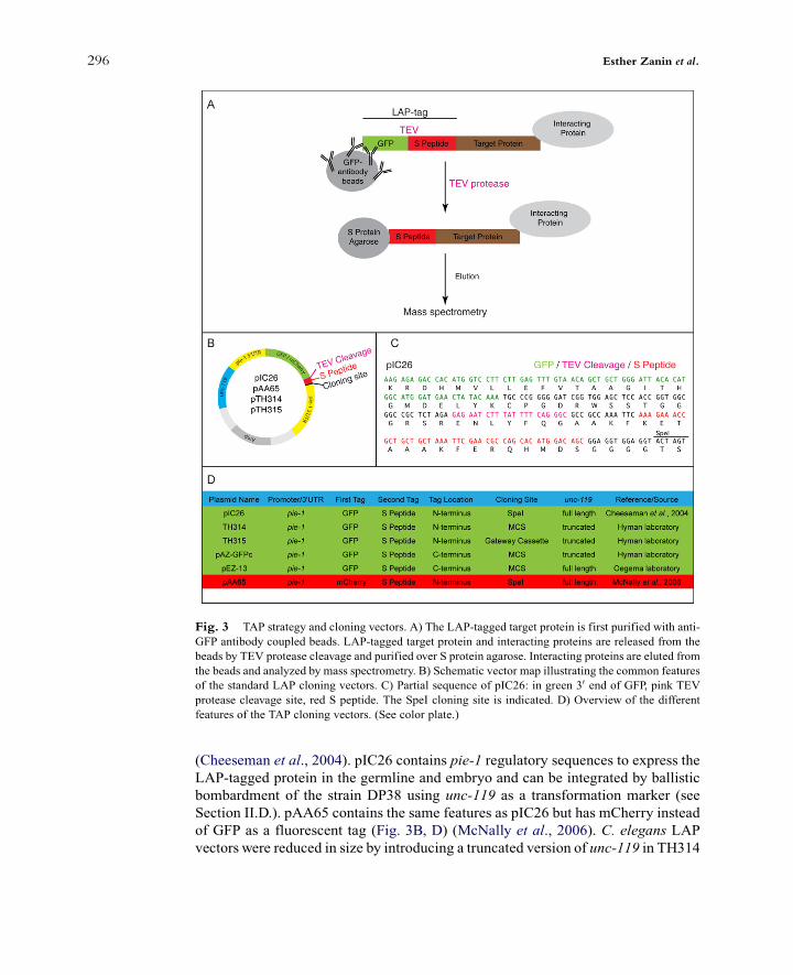
(Cheeseman et al., 2004). pIC26 contains pie-1 regulatory sequences to express theLAP-tagged protein in the germline and embryo and can be integrated by ballisticbombardment of the strain DP38 using unc-119 as a transformation marker (seeSection II.D.). pAA65 contains the same features as pIC26 but has mCherry insteadof GFP as a fluorescent tag (Fig. 3B, D) (McNally et al., 2006). C. elegans LAPvectors were reduced in size by introducing a truncated version of unc-119 in TH314
[(Fig._3)TD$FIG]
Fig. 3 TAP strategy and cloning vectors. A) The LAP-tagged target protein is first purified with anti-
GFP antibody coupled beads. LAP-tagged target protein and interacting proteins are released from the
beads by TEV protease cleavage and purified over S protein agarose. Interacting proteins are eluted from
the beads and analyzed by mass spectrometry. B) Schematic vector map illustrating the common features
of the standard LAP cloning vectors. C) Partial sequence of pIC26: in green 30 end of GFP, pink TEV
protease cleavage site, red S peptide. The SpeI cloning site is indicated. D) Overview of the different
features of the TAP cloning vectors. (See color plate.)
296 Esther Zanin et al.

and TH315 (Fig. 3D). However, incomplete rescue of the Unc phenotype makes theidentification of transformants more challenging. The target gene can be inserted inthese vectors at the 30 end of GFP by either Gateway cloning (TH315) or conven-tional cloning (TH314). To fuse the protein of interest at the C-terminus to the LAPtag either pAZ-GFPc (truncated unc-119) or pEZ-13 (full length unc-119) can beused (Fig. 3D). The LAP tag can easily be transferred into other vectors using thecassette present in pIC26.Instead of fusing both tags to one protein, it is also possible to fuse them to different
members of the same protein complex. Such a ‘‘split’’ TAP tag was used to isolatenew binding partners of the integral membrane nicotinic acetylcholine receptor(nAChR) (Gottschalk et al., 2005). Additional epitopes have been implemented fortandem affinity purifications (Polanowska et al., 2004; Schaffer et al., 2010).
D. Introduction of Transgenes for Expression of Tagged Proteins
For somatic expression of transgenes, heritable and repetitive extrachromosomalarrays are often sufficient; for example, Gottschalk et al. used an array to expressTAP-tagged nAChR subunits (Gottschalk et al., 2005). Injecting DNA in theC. elegans germline will generate extrachromosomal arrays (Mello and Fire,1995;Mello et al., 1991). However, a transgene in an array is typically overexpressedin somatic cells and rapidly silenced in germ cells (Kelly and Fire, 1998; Seydouxand Strome, 1999). The variable degree of heritability of the arrays can also make itdifficult to obtain sufficient material from large-scale cultures.An alternative to arrays is ballistic bombardment where small transgene-coated
gold particles are introduced into the worm tissue at high speed (Praitis et al., 2001).Bombardments are performed in the DP38 strain that carries a mutation in the unc-119 gene. The DP38 strain is unable to move and does not transition to the dauerstage. A copy of the unc-119(+) gene is introduced in the same vector as thetransgene and transformants are identified by wild-type movement and dauer for-mation. Ballistic bombardment yields low-copy number integrations at random sitesin the genome. Bombarded transgenes may not be expressed at the endogenous levelnor at all relevant developmental stages. Another drawback of generating transgeniclines by ballistic bombardment is that the integration sites are different for eachtransgene making it difficult to comparewild-type and engineeredmutants. Detailedprocedure for ballistic bombardment is described in Green et al. (2008) and on theSeydoux laboratory website (http://www.bs.jhmi.edu/MBG/SeydouxLab/vectors/index.html).A recent technique, MosSCI (Mos1 mediated Single Copy transgene Insertion)
circumvents the problems associated with arrays and bombarded lines by directingthe transgene at a fixed locus in the genome (Frokjaer-Jensen et al., 2008).Transformants are identified using the same strategy as for ballistic bombardment:the injected strain contains an unc-119(ed3)mutation that is rescued by introducingthe wild-type unc-119(+) gene on the vector harboring the transgene. Description of
11. Affinity Purification of Protein Complexes in C. elegans 297
the vectors and a detailed protocol for MosSCI can be found on the webpage of theJorgensen laboratory that developed this method (http://sites.google.com/site/jor-gensenmossci/Home).
E. Validating the Functionality of the Transgene-Encoded Tagged Protein
Before using the tagged protein for biochemical studies it is important to validateits functionality. The most straightforward test is whether the fusion protein canrescue a mutant phenotype. If a mutant is not available, functional tests can beconducted using RNAi. This requires that the endogenous messenger RNA is spe-cifically targeted by a dsRNA. Re-encoding transgenes or using a different 30UTRonthe transgene are two strategies that have been used for this purpose (Audhya et al.,2005; Dammermann et al., 2008). A detailed description of how we re-encodetransgenes was recently presented (Green et al., 2008).
III. The Optimal Starting Material for Protein Purification
For proteins that are widely expressed, adult worms are the most straightforwardstartingmaterial to use since they are easily grown in large-scale using liquid culture.If the protein of interest is enriched in embryos, embryonic extracts can be made bybleaching adult hermaphrodites to isolate embryos. By carefully choosing the incu-bation temperature it is also possible to enrich the embryonic extract either for young(<50 cells) or old embryos (>200 cells) (see Section III.I.). If the target protein ispresent only in specific cell types, one can try to enrich for these cell types by usingconditional loss of function mutations. For example, by using a temperature-sensi-tive mutant that does not form a germline one can enrich for somatic cells (Beananand Strome, 1992). We describe in Section III.I. a strategy that uses a temperature-sensitive meiotic arrest mutant to enrich for embryos in meiosis I. While flowcytometry sorting methods have been developed to isolate specific cell types forexpression and small RNA analysis, the amounts enriched are not yet sufficient forbiochemical methods (Cinar et al., 2005; Colosimo et al., 2004; Fernandez et al.,2010; Zhang et al., 2002). Future miniaturization of protein isolation methods islikely to enable proteomic characterization of individual cell types sorted by flowcytometry.
A. Growing Worms in Large-Scale Liquid Culture
Worms are relatively straightforward to grow in biochemical quantities usingliquid cultures. Growing worms on egg plates is an alternative approach and adetailed protocol for their use is described by Mains & McGhee (Hope, 1999).The following protocols describe growing worms in large-scale liquid culture toobtain sufficient amount of either adult worms or embryos to perform
298 Esther Zanin et al.
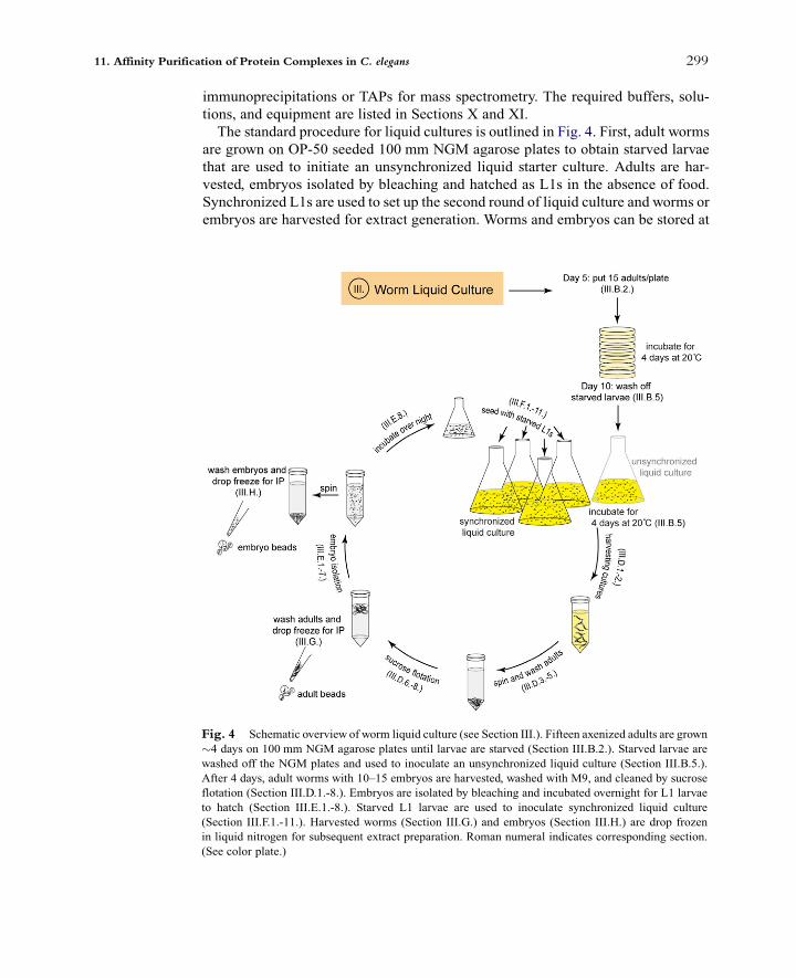
immunoprecipitations or TAPs for mass spectrometry. The required buffers, solu-tions, and equipment are listed in Sections X and XI.The standard procedure for liquid cultures is outlined in Fig. 4. First, adult worms
are grown on OP-50 seeded 100 mm NGM agarose plates to obtain starved larvaethat are used to initiate an unsynchronized liquid starter culture. Adults are har-vested, embryos isolated by bleaching and hatched as L1s in the absence of food.Synchronized L1s are used to set up the second round of liquid culture and worms orembryos are harvested for extract generation. Worms and embryos can be stored at
[(Fig._4)TD$FIG]
Fig. 4 Schematic overview of worm liquid culture (see Section III.). Fifteen axenized adults are grown
#4 days on 100 mm NGM agarose plates until larvae are starved (Section III.B.2.). Starved larvae are
washed off the NGM plates and used to inoculate an unsynchronized liquid culture (Section III.B.5.).
After 4 days, adult worms with 10–15 embryos are harvested, washed with M9, and cleaned by sucrose
flotation (Section III.D.1.-8.). Embryos are isolated by bleaching and incubated overnight for L1 larvae
to hatch (Section III.E.1.-8.). Starved L1 larvae are used to inoculate synchronized liquid culture
(Section III.F.1.-11.). Harvested worms (Section III.G.) and embryos (Section III.H.) are drop frozen
in liquid nitrogen for subsequent extract preparation. Roman numeral indicates corresponding section.
(See color plate.)
11. Affinity Purification of Protein Complexes in C. elegans 299

$80 !C until sufficient amounts for immunoprecipitation are obtained. 500 mLliquid worm culture yields about 7 g of worms and 0.7 g of embryos.Growth of worms in liquid culture should be carefully and continuously moni-
tored. It is important to determine under a dissecting microscope the age of theworms, whether they have sufficient amounts of food, and if contamination ispresent. Cultures must be handled under sterile conditions, preferably in a dedicatedlaminar flow hood to avoid contamination. Growth temperature can be adjustedbetween 16 !C and 25 !C and it is recommended to use a cooling shaker that reliablyholds temperature.
B. Unsynchronized Liquid Starter Culture
Since contamination is a serious problem in liquid culture, we recommend startingwith freshly axenized worms. These are then amplified on 100 mm NGM agaroseplates by putting 15 cleaned worms onto each plate (Fig. 4). After incubating theplates for 4–5 days at 20 !C the just starved larvae are used to start the unsynchro-nized liquid culture.
1. Day 1: Pipet 5 mL 2MNaOH and 5 mL of bleach at edge of bacteria on a 60 mmNGM agarose plate. Transfer 10 adult worms into NaOH/bleach drop. Wait untilL1 larvae hatch and transfer L1 larvae onto new plates and grow until they areadults.
2. Day 5: Seed 100 mm NGM agarose plates with 15 cleaned adults. You will need7–8 plates per 500 mL of liquid culture. Incubate plates at 20 !C for 4–5 daysuntil worms are just starved.
3. Day 8: Start a 50 mL overnight of OP-50-1 in LB + 50 mg/mL streptomycin.OP-50-1 is a streptomycin resistant E. coli strain.
4. Day 9: Start 1.5 L culture of OP-50-1 in LB + 50 mg/mL streptomycin. You willneed 1.5 L of bacterial culture as food for 500 mL of C. elegans culture.
5. Day 10:
% Harvest bacterial culture at 4200 rpm for 15 min in sterile 1 L centrifugebottles. Pour off LB. Bacterial food can be stored in 50 mL conicals at 4 !Cfor several weeks.
% Make 500 mL Complete S Basal.% Resuspend bacterial pellet in 20 mL Complete S Basal and transfer into asterile 2.8 L wide bottom Fernbach flask with 500 mL Complete S Basal.
% Rinse 7–8 plates of just starved larvae with 10 mL of sterile M9. To collect asmany worms as possible from plates repeat wash with 5 mL of M9. Checkplates to make sure most worms were washed off. Collect in a 50 mL conical.
% Pellet worms at 600g in centrifuge for 3 min with slow deceleration. Removesupernatant with a sterile pipet and discard. Resuspend worms in 5 mL of freshM9 and add to flask containing the Complete S Basal with bacteria. Shake theinoculated culture at 20 !C at 200–230 rpm. Adjust growth time by varyingtemperature between 16 !C and 25 !C.
300 Esther Zanin et al.
6. Days 11–13: Growth of the culture should be carefully followed (see SectionIII C.) and worms harvested when majority are adults. The culture will takeabout 3–3.5 days depending on age of worms on starter plates and the desiredstate of the final culture.
Note: The bacterial food for liquid culture can be obtained from fermentor facil-ities. However, we have had contamination as well as growth problems with exter-nally supplied food and, consequently, prefer growing up our own bacterial cultures.
C. Monitoring Worm Cultures
Worm liquid cultures should bemonitored under a dissectingmicroscope once a day.This allows assessment of the developmental stage and health of the worms, ensuresthat worms have enough food, and confirms that there is no significant contamination.
1. In a laminar flow hood transfer 1 mL of the culturewith a sterile pipet into 1.5 mLmicrocentrifuge tubes.
2. Spin in microcentrifuge 600g for 3 min.3. Carefully pipet #20 mL worm slurry using a cut-off tip onto a glass slide. Place
cover slip on top and look at the worms under dissecting microscope.
D. Harvesting Worms from Liquid Cultures
Worms are harvested by settling under gravity and cleaned by flotation on asucrose cushion, which separates healthy worms from bacteria and debris (Fig. 4).The sucrose floated worms are washed and used to isolate embryos.
1. When the majority of worms in the culture have 10–15 embryos, transfer thecultures into 500 mL graduated cylinders. Settle worms in ice water bath for 1 h.
2. Aspirate off media using a sterile 5 mL pipet and transfer slurry (brown film atbottom) to two 50 mL conicals per L culture.
3. Pellet in centrifuge at 600g for 3 min with slow deceleration.4. Aspirate off supernatant, collect worms into 50 mL conical, and add cold M9.5. Pellet in centrifuge at 600g for 3 min and aspirate off supernatant.6. Resuspend pelleted worms by adding cold M9 to the 25 mL mark. To this add
25 mL cold 60% (w/v) sucrose.Mix and centrifuge immediately at 1500g for 5 min.7. After the spin, adult worms will form a layer at the top of the tube. Remove adults
down to the 35 mL mark with 5 mL pipet and transfer to a new conical.8. Wash worms by adding cold M9 to 50 mL mark. Pellet the worms at 600g for
3 min and carefully remove supernatant.
Note: It is important towork rapidly during the sucrose flotation step. Do not leavethe worms for too long in sucrose and wash them out of sucrose rapidly aftercollection from the top of the cushion.
11. Affinity Purification of Protein Complexes in C. elegans 301
E. Isolation of Embryos and Synchronization as Starved L1 Larvae
The washed worms are bleached to isolate embryos (Fig. 4). Good bleachingefficiency depends on small volumes (maximum 5 mL worm pellet per 50 mLconical) and freshness of bleach. Bleaching should be followed by eye under adissecting microscope. Bleaching sterilizes the culture; after bleaching, sterile tech-nique becomes critical again so that no contaminants are introduced into the materialthat will be used to inoculate the synchronized liquid culture.
1. To worm pellet add 25 mL 0.1 M NaCl and mix by pipeting up and down twice.Settle worms on ice for 5 min.
2. Aspirate off supernatant including worms that have not settled to the bottom andadd 0.1 M NaCl up to a volume of 30 mL.
3. Mix 5 mL 5 N NaOH with 10 mL bleach in conical. Immediately add NaOH/bleach mix to 30 mL worm suspension.
4. Vortex at maximum speed for 5 s and stand tube at RT for 2 min. Repeat fourtimes for a total bleaching time of 7–9 min. Follow bleaching by examiningsamples on a glass slide under a dissecting microscope. Stop bleaching whenonly embryos remain. The color of the bleach mixture will change to a burntorange as worms are dissolved.
5. Immediately centrifuge at 800g for 1 min at 4 !C. Aspirate off supernatant.6. Add sterile water to a total volume of 50 mL, mix by inverting the tube, and
centrifuge at 800g for 2 min.7. Repeat step 6. Washed embryos from synchronized liquid culture can be frozen
for immunoprecipitation as described in Section III.H.8. Add 35 mL M9 and transfer to a 50 mL flask. Rinse the conical with an extra
10 mL of sterile M9 and add to flask. Shake at 20 !C until embryos hatch and arestarved L1 larvae (#18–20 h).
F. Seeding Synchronized Cultures Using Starved L1 Larvae
Starved L1 larvae from 500 mL starter flask can be expanded to inoculate up to sixsynchronized liquid cultures.
1. 2 Days before: Start 300 mL culture of OP-50-1 in LB + 50 mg/mL streptomycin.2. Day before: Start six 1.5 L cultures of OP-50-1 in LB + 50 mg/mL streptomycin.3. Harvest bacterial cultures at 4200 rpm for 15 min in sterile 1 L centrifuge
bottles. Pour off LB.4. Make 3 L Complete S Basal and distribute into six 2.8 L Fernbach flasks.5. Resuspend bacterial pellet of each 1.5 L culture in 20 mL Complete S Basal and
transfer into sterile 2.8 L Fernbach flask with 500 mL Complete S Basal.6. When flasks with bacterial food are ready, start processing starved L1s. In the
hood, transfer L1s to a 50 mL conical. Chill on ice for 5–10 min.7. Spin at 600g for 3 min. Carefully remove supernatant. Bring up to 50 mL with
sterile cold M9.
302 Esther Zanin et al.

8. Repeat step 7.9. Add sterile M9 such that total volume is#5 mL, transfer L1s to 15 mL conical,
and pellet by spinning at 600g for 2 min. Immediately after the spin, use a pen tomark the volume of the pellet on the side of the tube. Estimate the volume ofpellet by adding known volumes of water to a separate conical.
10. Resuspend L1s to total volume of 12 mL using sterile M9 and look at a sampleunder dissection microscope; estimate percentage of L1s relative to deadembryos/worm parts/clumps.
11. Seed each flask with equivalent of 50 mL pure L1 pellet (e.g., if pellet volume is0.6 mL and % L1 in the resuspension is 70% then seed with 1.4 mL of theresuspended pellet). Avoid overseeding or cultures will starve.
12. Put flasks at 20 !C at 230 rpm. Grow for 48 h while monitoring cultures underdissecting microscope.
G. Freezing Adult Worms for Immunoprecipitation
Once the liquid cultures are ready collect and wash worms as described in SectionIII.D.Wash worms by adding 50 mL cold 1" Lysis Buffer (with protease inhibitors).Remove Lysis Buffer until only a small amount remains. Freeze adult worms bydispensing from a pipette drop by drop in liquid nitrogen, which will form smallbeads (Fig. 4). Store at $80 !C.
H. Freezing Embryos for Immunoprecipitation
Bleach adult worms as described in Section III.E. and continue after step 7. Washembryo pellet with 50 mL cold 1" Lysis Buffer (with protease inhibitors). Freezeembryos by dispensing from a pipette drop by drop in liquid nitrogen. Store embryobeads at $80 !C.
I. Enriching for Specific Age Embryos
While precise synchronization of embryos is not possible, it is straightforward toenrich for old or young embryo populations by varying growth conditions andcarefully monitoring worms in the culture under a dissecting microscope. Generatesynchronized starved L1 larvae (see Section III.E.) and inoculate worm cultures (seeSection III.F.). To obtain worms that just started embryo production, incubate the flaskfor #64 h at 17 !C. Using these growth conditions worms typically contained up tofive embryos, the majority of which have <50 cells (Fig. 5A). If several flasks ofworm culture are grown simultaneously, it is necessary to monitor each flask sepa-rately, as the time at which embryo production begins may vary between flasks. It isalso critical to avoid contamination, which may adversely affect synchronous growthof the worms. To obtain worms with mostly old embryos (>200 cells), cultures areincubated for #64 h at 19 !C (Fig. 5A). Embryos are frozen as described in Section
11. Affinity Purification of Protein Complexes in C. elegans 303

III.H. To determine the age of the embryos, about#5 mL of packed embryos are fixedin 1 mL of cold methanol for at least 30 min, then incubated with 1 mg/mL Hoechststain in PBS+0.1% Triton X-100 for 10 min. After three washes with PBS+0.1%Triton X-100, embryos are suspended in 30 mL PBS, and 5 mL are mixed on a18"18 mm coverslip with 15 mL of mounting medium. The coverslip is carefullyplaced on a slide, sealed with nail polish, and the number of nuclei per embryo isdetermined using a fluorescence microscope (Fig. 5A).
J. Enriching for Meiosis I Arrested Embryos
Adult worms contain about 15 mitotically dividing embryos and at most twomeiotic embryos because meiosis is completed 30 min after fertilization. To enrichfor meiosis I metaphase, we used a mutation in a subunit of the anaphase-pro-moting complex (APC). We chose a temperature-sensitive allele (g48) of emb-27(Cassada et al., 1981; Golden et al., 2000). At permissive temperature (16 !C)emb-27(g48) worms contain about 15 mitotically dividing embryos, similar towild type. At the restrictive temperature (24–25 !C), emb-27(g48) embryos arrestat metaphase of meiosis I and worms accumulate meiotically-arrested fertilizedembryos. Mutant worms are initially grown at 16 !C as described in Section III.
[(Fig._5)TD$FIG]
Fig. 5 A) Early and late embryo preparation stained with Hoechst. B) Meiosis I arrested and endo-
mitotic chromosomes stained with Hoechst.
304 Esther Zanin et al.
A.-E. It is important to keep the temperature at or below 16 !C to prevent meta-phase arrest. Bleached larvae are used to inoculate a synchronized liquid culture(see Section III.F.) and are shifted to 24 !C when the majority of worms are at theL4 stage. The time point of temperature shifting has to be chosen carefullybecause an early shift causes larval arrest and a late shift will ‘‘contaminate’’the extract with mitotic embryos. Worms are harvested when the majority contains4–8 one-cell meiotic-arrested embryos and worm extract is prepared as describedin Section V. Timing is once again critical since after 4–5 h the embryos overcomethe meiotic arrest and start cycling endo-mitotically (DNA is replicated but celldivision does not occur). The quality of the culture can be assayed by cutting 2–3worms in a drop of L-15 blastomere culture medium (Edgar, 1995) containing1 mg/mL Hoechst 33342, and analyzing the shape of the chromosomes.Metaphase I arrested embryos display six maternal chromosomes that have atypical oval shape (Fig. 5B). Endo-mitotic embryos contain decondensed chromo-somes that have a fibrous-like appearance and tend to detach from the anteriorcortex of the embryo (Fig. 5B).The above two examples are specific to our research interests but related strategies
can be used to enrich for cell types or stages of interest prior to protein complexisolations.
IV. Isolation of Nuclei from Embryos
If the protein of interest is a nuclear protein or if DNA–protein interactions arebeing analyzed as in Chromatin Immunoprecipitation (ChIP) experiments, nucleican be isolated from embryos prior to further analysis. Embryos are isolated bybleaching as described in Section III.E Steps 1–7; however it is important thatembryos are not frozen prior to nuclei isolation. To isolate nuclei, the embryoeggshell is digested using chitinase (Fig. 6A). Dounce homogenization in hypo-tonic buffer liberates intact nuclei. A 100g centrifugation removes large debrisand a subsequent 2000g centrifugation pellets the nuclei. A final centrifugationstep over a sucrose cushion is used to separate the nuclei from membrane con-taminants. Enrichment of nuclei can be easily followed by the emergence of thecore histone bands on Coomassie-stained gels (Fig. 6B). Because the chitinasestep is performed at room temperature, nuclei must be purified from freshlyisolated embryos to avoid protein degradation. 1 mL of embryo pellet typicallyyields #100 mL of nuclei.
1. Harvest embryos by bleaching as described in Section III.E. until step 7.2. Add two embryo pellet volumes of Embryo Buffer and 250 mL of chitinase
stock solution per mL of embryo pellet.3. Rotate at RT for #30 min. Monitor eggshell integrity under dissecting micro-
scope. Embryos without an eggshell will lose their oval shape and fall apart intoclumps of cells.
11. Affinity Purification of Protein Complexes in C. elegans 305
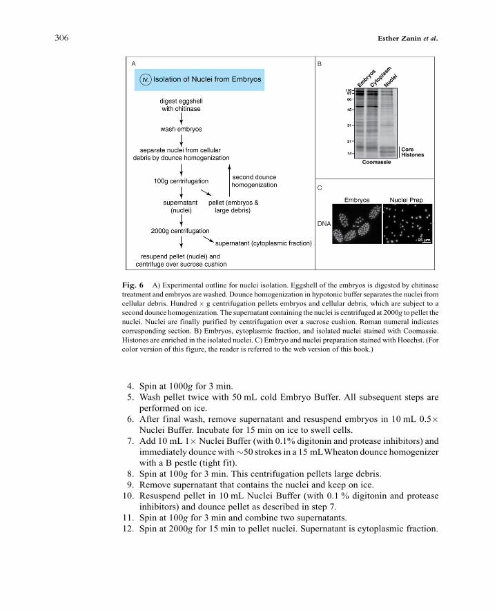
4. Spin at 1000g for 3 min.5. Wash pellet twice with 50 mL cold Embryo Buffer. All subsequent steps are
performed on ice.6. After final wash, remove supernatant and resuspend embryos in 10 mL 0.5"
Nuclei Buffer. Incubate for 15 min on ice to swell cells.7. Add 10 mL 1"Nuclei Buffer (with 0.1% digitonin and protease inhibitors) and
immediately douncewith#50 strokes in a 15 mLWheaton dounce homogenizerwith a B pestle (tight fit).
8. Spin at 100g for 3 min. This centrifugation pellets large debris.9. Remove supernatant that contains the nuclei and keep on ice.10. Resuspend pellet in 10 mL Nuclei Buffer (with 0.1 % digitonin and protease
inhibitors) and dounce pellet as described in step 7.11. Spin at 100g for 3 min and combine two supernatants.12. Spin at 2000g for 15 min to pellet nuclei. Supernatant is cytoplasmic fraction.
[(Fig._6)TD$FIG]
Fig. 6 A) Experimental outline for nuclei isolation. Eggshell of the embryos is digested by chitinase
treatment and embryos arewashed. Dounce homogenization in hypotonic buffer separates the nuclei from
cellular debris. Hundred " g centrifugation pellets embryos and cellular debris, which are subject to a
second dounce homogenization. The supernatant containing the nuclei is centrifuged at 2000g to pellet the
nuclei. Nuclei are finally purified by centrifugation over a sucrose cushion. Roman numeral indicates
corresponding section. B) Embryos, cytoplasmic fraction, and isolated nuclei stained with Coomassie.
Histones are enriched in the isolated nuclei. C) Embryo and nuclei preparation stained with Hoechst. (For
color version of this figure, the reader is referred to the web version of this book.)
306 Esther Zanin et al.

13. Resuspend pellet in 500 mL Nuclei Buffer (with 0.1% digitonin and proteaseinhibitors) and mix with 5 mL 30% (w/v) sucrose cushion in Nuclei Buffer with0.1% digitonin.
14. Centrifuge at 2000g for 15 min. The pellet will be enriched for nuclei.15. Check integrity of the nuclei under a microscope after incubation with 1 mg/mL
Hoechst stain (Fig. 6C).
V. Preparing Worm and Embryonic Extract
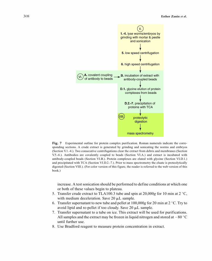
Extract can be prepared using whole worms harvested from liquid culture or fromembryos isolated by bleaching. Lysis of worms and embryos is performed in anisotonic buffer. Subsequent to lysis, the salt concentration can be raised to 300 mMKCl to enhance stringency of protein complex isolation. Worms and embryos arelysed by grinding in liquid nitrogen followed by sonication (Fig. 7). Two consecutivecentrifugations remove membranes and lipids and the supernatant is used for com-plex isolation. The lysis conditions described here are not well-suited for membrane-associated proteins. A protocol for isolating membrane proteins is described inGottschalk et al. (2005).
1. Pre-cool a mortar and pestle by filling with liquid nitrogen for at least 5 min.2. Weigh out frozen adult/embryo beads. For one immunoprecipitation use#1 g of
frozen worm/embryo beads. Grind frozen beads to a fine powder: initially breakdown worm/embryo beads by gentle tapping (try not to lose too many beads asthey have a tendency to jump out of the mortar), then grind. Keep mortar cold byaddingmore liquid nitrogen as necessary andwaiting for it to evaporate. For morethan 40 g of worms, one can also use a warring blender cooled with liquidnitrogen. For embryo extracts, the freeze-grinding step may be skipped, as son-ication is sufficient to release cytoplasmic contents.
3. Add an equal volume of 1.5"Lysis Buffer (with protease inhibitors) to each gramof adult/embryo beads. Keep 20 mL sample for gel analysis.
4. Set up ice-water bath and sonicate with a tip sonicator (e.g., Branson DigitalSonifier). It is critical to prevent heating of the sample during sonication. For aBranson Digital Sonifier with a microtip use the following settings:
% 30% amplitude for 3 min total (15 s on; 45 s off – after each 1 min of sonicationwait #2 min to chill)
% 40% amplitude for 30 s (15 s on; 45 s off) Save 20 mL sample.
We recommend optimizing the sonication protocol by two methods:Use a dissecting microscope to monitor worm/embryo lysis. At least 80–90% ofthe worms/embryos must be lysed.Use Bradford reagent or UV absorbance to directly monitor lysis. As cells lyse,the protein concentration and A260 absorbance (due to nucleic acid release) will
11. Affinity Purification of Protein Complexes in C. elegans 307
increase. A test sonication should be performed to define conditions at which oneor both of these values begin to plateau.
5. Transfer crude extract to TLA100.3 tube and spin at 20,000g for 10 min at 2 !C,with medium deceleration. Save 20 mL sample.
6. Transfer supernatant to new tube and pellet at 100,000g for 20 min at 2 !C. Try toavoid lipid and re-pellet if too cloudy. Save 20 mL sample.
7. Transfer supernatant to a tube on ice. This extract will be used for purifications.All samples and the extract may be frozen in liquid nitrogen and stored at$80 !Cuntil further use.
8. Use Bradford reagent to measure protein concentration in extract.
[(Fig._7)TD$FIG]
Fig. 7 Experimental outline for protein complex purification. Roman numerals indicate the corre-
sponding sections. A crude extract is generated by grinding and sonicating the worms and embryos
(Section V.1.-4.). Two consecutive centrifugations clear the extract from debris and membranes (Section
V.5.-6.). Antibodies are covalently coupled to beads (Section VI.A.) and extract is incubated with
antibody-coupled beads (Section VI.B.). Protein complexes are eluted with glycine (Section VI.D.1.)
and precipitated with TCA (Section VI.D.2.-7.). Prior to mass spectrometry the eluate is proteolytically
digested (Section VIII.). (For color version of this figure, the reader is referred to the web version of this
book.)
308 Esther Zanin et al.

VI. Single-step Immunoprecipitation
Immunoprecipitation using specific antibodies is a powerful method for analyz-ing protein–protein interactions and identifying protein complexes. Single-steppurification using polyclonal antibodies followed bymass spectrometry on the entireeluate commonly indentifies several hundred to a thousand proteins making itchallenging to distinguish between signal and noise. Background can arise fromgeneral non-specific binding to beads, to constant regions of antibody chains, and topartially denatured antibodies. Background can also be specific to individual affin-ity-purified antibodies – consequently a random IgG control cannot be used todiscriminate between true signal and noise. We recommend using as a control apolyclonal rabbit antibody raised against GST (glutathione S-transferase) that isaffinity-purified using the same procedures used for the antibody to the targetprotein. In addition, whenever possible, we recommend parallel immunoprecipita-tions with either two antibodies to the same protein (preferablewith non-overlappingepitopes) or one antibody and one tagged fusion protein. Purification from a mutantstrain is an ideal negative control but is not feasible if the mutation is lethal. If atagged protein is immunoprecipitated, an untagged strain can be used as a negativecontrol.The following two examples illustrate how redundant purification strategies and
suitable negative controls helped pinpoint bona fide complex members. Polyclonalrabbit antibodies to two essential chromosome segregation proteins, KNL-1 andKNL-3, identified in an RNAi screen, were used to purify complexes containingthese proteins (Cheeseman et al., 2004; Desai et al., 2003). Each protein was presentin the other immunoprecipitation, allowing cross-referencing of the two immuno-precipitations to identify 11 proteins in common (Fig. 8) (in total the two
[(Fig._8)TD$FIG]
Fig. 8 Identification of 10-protein kinetochore complex. Immunoprecipitation of KNL-1 and KNL-3
isolatedmore than 130 interacting proteins with 11 proteins common in both immunoprecipitations. MIS-
12 and KBP-1, two common interactors, were LAP tagged and purified isolating 11 common interactors
of which 10were found also inKNL-1 andKNL-3 immunoprecipitations. (For color version of this figure,
the reader is referred to the web version of this book.)
11. Affinity Purification of Protein Complexes in C. elegans 309

immunoprecipitations identified over 130 proteins). MIS-12 and KBP-1, two of thenewly discovered proteins that were in the common set were LAP tagged and tandemaffinity purified revealing a 10-protein complex containing both KNL-1 and KNL-3(Fig. 8). Functional validation confirmed that all of the proteins that co-purifiedwere involved in chromosome segregation.In a second example, novel interaction partners of C. elegans Dicer (DCR-1), a
RNA endoribonuclease that is required in mammalian cells for small RNA gener-ation, were identified by single-step purification (Duchaine et al., 2006). Two single-step DCR-1 immunoprecipitations were performed: one using adult worms with apolyclonal anti-DCR-1 antibody and one using embryos that express HA-taggedDCR-1. Negative control purifications were performed in a dcr-1 mutant strain, anuntagged wild-type strain and with unrelated antibodies. Co-purified proteins wereconsidered high confidence interactors if they were reproducibly identified in atleast two independent purifications and not present in the negative control purifica-tions. Twenty high confidence interactors were analyzed further with biologicalassays. Twelve of the 20 interactors could be linked to DCR-1 activities or itssmall-RNA products.These two examples illustrate the importance of experimental design that
incorporates redundancy at the outset. The need for employing such strategieshas been magnified by the significant technical advances in protein mass spec-trometry – currently, one-step immunoprecipitations with an affinity-purifiedantibody frequently yield between 100 and 1000 distinct proteins in the immu-noprecipitate. We note that existing datasets can provide computational tools forfiltering the output of mass spectrometric analysis. For example, over the yearswe have conducted a large number of protein isolation and mass spectrometryexperiments in C. elegans embryos. The targets for this analysis span a number ofcellular processes. This cumulative dataset helps identify potentially interestingcandidates from a new purification experiment as opposed to frequently observedco-purifying ‘‘sticky’’ proteins. New label free quantitative mass spectrometrymethods can also be employed to help discriminate true signal from noise(Hubner et al., 2010).We note that, prior to mass spectrometry, it is important to assess the solubility
of the protein target and the efficiency of the immunoprecipitation using immu-noblotting. For this purpose, samples of the crude extract, low-speed supernatant,high-speed supernatant, the supernatant after incubation with antibody beads, andthe immunoprecipitate are important to prepare and store, keeping track of thevolumes at each step. Such an effort can quickly identify potential confoundingproblems (e.g., low solubility or poor immunoprecipitation efficiency) and guideapproaches to overcome them.Below, we describe how to covalently couple the affinity-purified antibody to
protein A beads, use the antibody-coupled beads for immunoprecipitation, and elutebound proteins from the beads for analysis by mass spectrometry, silver staining andimmunoblotting (Fig. 7).
310 Esther Zanin et al.
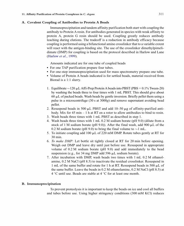
A. Covalent Coupling of Antibodies to Protein A Beads
Immunoprecipitation and tandem affinity purification both start with coupling theantibody to Protein A resin. For antibodies generated in species with weak affinity toprotein A, protein G resin should be used. Coupling greatly reduces antibodyleaching during elutions. The tradeoff is a reduction in antibody efficacy becausecoupling is performed using a bifunctional amine crosslinker that to a variable extentwill react with the antigen-binding site. The use of the crosslinker dimethylpimeli-dimate (DMP) for coupling is based on the protocol described in Harlow and Lane(Harlow et al., 1999).
Amounts indicated are for one tube of coupled beads% For one TAP purification prepare four tubes.% For one-step immunoprecipitation used for mass spectrometry prepare one tube.% Volume of Protein A beads indicated is for settled beads, material received fromBiorad is a 1:1 slurry.
1. Equilibrate#120 mLAffi-PrepProteinAbeads into PBST (PBS + 0.1%Tween-20)by washing the beads three to four times with 1 mL PBST. This should give about60 mL of packed beads. Wash beads by gentle inversion. Briefly pellet them using apulse in a microcentrifuge (30 s at 3000g) and remove supernatant avoiding beadpellet.
2. Resuspend beads in 500 mL PBST and add 10–50 mg of affinity-purified anti-body. Mix for 45 min – 1 h at RT on a rotor to allow antibodies to bind to resin.
3. Wash beads three times with 1 mL PBST as described in step 1.4. Wash beads three times with 1 mL 0.2 M sodium borate (pH 9.0) (dilute from a
stock of 1 M sodium borate (pH 9.0)). After the final wash, add 900 mL of the0.2 M sodium borate (pH 9.0) to bring the final volume to #1 mL.
5. To initiate coupling add 100 mL of 220 mM DMP. Rotate tubes gently at RT for30 min.
6. To make DMP: Let bottle sit tightly closed at RT for 20 min before opening.Weigh out DMP and leave dry until just before use. Resuspend in appropriatevolume of 0.2 M sodium borate (pH 9.0) and add immediately to the beadsuspension (e.g., for 34 mg DMP add 596 mL sodium borate).
7. After incubation with DMP, wash beads two times with 1 mL 0.2 M ethanol-amine, 0.2 M NaCl (pH 8.5) to inactivate the residual crosslinker. Resuspend in1 mL of the same buffer and rotate for 1 h at RT. Resuspend beads in 500 mL ofthe same buffer. Leave the beads in 0.2 M ethanolamine, 0.2 M NaCl (pH 8.5) at4 !C until use. Beads are stable at 4 !C for at least one month.
B. Immunoprecipitation
To prevent proteolysis it is important to keep the beads on ice and cool all buffersand tubes before use. Using higher stringency conditions (300 mM KCl) reduces
11. Affinity Purification of Protein Complexes in C. elegans 311

background although there is also an increased likelihood of losing meaningful lowaffinity interactions.
1. Pre-elute 100 mL of antibody-coupled beads three times with 1 mL of100 mM glycine (pH 2.6) to remove antibody that is not covalently coupledto the beads. Do not leave beads for a long time in glycine or the antibody willdenature.
2. Wash beads three times with 1 mL cold Lysis Buffer (with 0.5 mM DTT) toneutralize glycine and prepare the beads for immunoprecipitation.
3. Mix beads with 900 mL extract for at least 1 h at 4 !C on rotating platform.4. Rinse beads two times with 1 mL cold Lysis Buffer (with 0.5 mM DTT and
protease inhibitors).5. On rotator in cold room wash beads two times for 5 min with 1 mL cold Lysis
Buffer (with 0.5 mM DTT and protease inhibitors).6. Wash five times with 1 mL Lysis Buffer (with 0.5 mM DTT) without detergent
(NP-40) or protease inhibitors. Remove as much supernatant as possible.
Note: The presence of detergents can interfere with mass spectrometry. Therefore,it is important to wash the sample thoroughly in detergent-free buffer afterimmunoprecipitation.
C. Sample Buffer Elution: For Silver Staining & Immunoblotting
1. Elute beads by heating in 100 mL of 2" Sample Buffer without DTT for 10 minat 70 !C.
2. Pellet beads, transfer supernatant to a new tube, and add DTT to 100 mM (1/9supernatant volume of 1 M DTT stock; Elution 1).
3. Add 100 mL 2" Sample Buffer with DTT to pelleted beads (Elution 2).4. Boil both elution samples for 5 min and analyze by silver staining or immunoblot.
Both elutions will contain immunoprecipitated proteins although amounts ineach may vary; Elution 2 will have more IgG contamination than Elution 1.
Note: For immunoblots conducted using rabbit primary antibodies, the secondaryantibody will detect any IgG released by the elution from the beads. For silverstaining, load 5–10 mL directly. For immunoblots, load 5–10 mL of a 1/10 dilutionmade in Sample Buffer.
D. Glycine Elution: For Mass Spectrometry
1. After standard immunoprecipitation (see Section VI.B), elute beads three timeswith 150 mL 100 mM glycine (pH 2.6). Pool elutions and neutralize by adding150 m L 2 M Tris (pH 8.5). Neutralize the beads by washing two times with150 mL Lysis Buffer (without detergent) and pool with eluate. Total volume willbe 900 mL. Make sure you remove all the beads.
312 Esther Zanin et al.

2. Add 1/5th volume 100% trichloroacetic acid (TCA; #200 mL).3. Leave samples at 4 !C overnight.4. Spin for 30 min at maximum speed in microcentrifuge. Remove supernatant and
spin again for 1 min. Remove any residual supernatant with gel loading tip,leaving 5–10 mL behind.
5. Wash twice with 500 mL cold acetone. Spin 10 min at maximum speed inmicrocentrifuge.
6. Dry the protein pellet by spinning briefly in a speed vac.7. Freeze in liquid nitrogen and store at $80 !C. The protein pellet is suitable for
direct mass spectrometric analysis.8. After elution with glycine, the beads should be boiled in Sample Buffer and
analyzed by silver staining/immunoblotting to assess elution efficiency.
E. Urea Elution: For Mass Spectrometry
Urea elution can also be used for mass spectrometry. In practice, we find thatglycine elution works better for elution of the antigen from the antibody and fordetection of the purified antigen in mass spectrometric analysis.
1. After standard immunoprecipitation (see Section VI.B), wash beads with Pre-urea Wash Buffer. Remove all residual supernatant.
2. Add 75 mL Urea Elution Buffer and rotate for 30 min at RT.3. Pellet beads and transfer eluate to a new tube. Re-pellet to ensure removal of all
protein A beads.4. Remove 50 mL of elution and drop freeze in liquid nitrogen to send for mass
spectrometry. Add Sample Buffer to the rest to run on a gel.
VII. Tandem Affinity Purification Using a LAP Tag
As a first step, the extract is incubated with anti-GFP antibody-coupledprotein A beads (Fig. 3A). For this purpose, we use in-house rabbit polyclonalanti-GFP antibodies generated by injecting purified GFP in rabbits.Recombinant GFP-binding domains from single-chain antibodies have also beenused successfully for affinity purification of GFP-tagged proteins (Trinkle-Mulcahy et al., 2008). After immunoprecipitation of GFP, the fusion proteinis released by TEV protease cleavage (Fig. 3A). The subsequent purification onS Protein agarose further enriches for complexes containing the fusion protein.Extract prepared from an untagged strain can serve as negative control for theTAP procedure.
A. TEV Cleavage
Perform immunoprecipitation using anti-GFP antibody-coupled beads asdescribed in Section VI.B.
11. Affinity Purification of Protein Complexes in C. elegans 313
Note: At this stage, it is possible to elute boundmaterial using glycine as describedin Section VI.D. and compare the elution of the one-step GFP immunoprecipitationto the two-step LAP.
1. Pool beads into a single tube and fill with Lysis Buffer (with 300 mM KCl, 0.5mM DTT).
2. Add#30 mL of purified TEV protease (1 mg/mL) and rock tubes for 4 h at 4 !C.Add an additional 30 mL of TEV and rock tubes overnight.
3. Pellet beads and transfer supernatant to a new tube. Add 350 mL Lysis Buffer tobeads to remove any residual cleaved protein.
B. S Protein Agarose
1. Wash tube of 85 mL S protein agarose three times with 1 mL Lysis Buffer (with300 mM KCl).
2. Add TEV protease eluted supernatant to S protein agarose and rock for 3 h at4 !C.
3. Pellet beads and wash three times with Lysis Buffer (with 300 mM KCl).4. Wash one time with Lysis Buffer with 100 mM KCl without detergent (NP-40).
C. Sample Buffer Elution: For Silver Staining & Immunoblotting
Perform Sample Buffer Elution as described in Section VI.C.
D. Urea Elution: For Mass Spectrometry
Perform Urea Elution as described in Section VI.E.
VIII. Mass Spectrometry & Prioritization for Follow-upExperiments
Protein mass spectrometry has made remarkable advances in the recent decade.We will not discuss the details of the methodology, which are extensively reviewedelsewhere (Cravatt et al., 2007; Yates et al., 2009). We typically do not separateproteins from immunoprecipitates on gels prior to mass spectrometry. Instead, theentire eluate is proteolytically digested and the peptide mixture is separated bymultidimensional liquid chromatography. The mass/charge ratio of ionized peptidesis determined in the first mass analyzer, and then the peptides are fragmented in thecollision cell and passed through the second mass analyzer to determine amino acidsequence. As mentioned above, current methods can yield up to 1000 proteins inone-step immunoprecipitation performed using an antibody that passes generallyaccepted antibody specificity criteria. This abundance of information makes it
314 Esther Zanin et al.
challenging to sort relevant hits from background and necessitates redundant strat-egies. New technical developments, including labeling-based and label-free meth-ods, may also aid in this effort. In practical terms, the challenge in analyzing proteinmixtures by mass spectrometry lies primarily in developing a good working rela-tionship with a mass spectrometry-focused laboratory or core facility.Prioritization of potential interactors from lists generated bymass spectrometry of
immunoprecipiates/tandem affinity purifications for follow-up experiments is per-haps the greatest challenge faced in biochemical analysis of protein complexes. Inthis regard, it is helpful to think of the initial list of proteins identified as hits from aprimary screen, with a variety of secondary screens being necessary to separate thewheat from the chaff. Redundant strategies and elimination of common contami-nants are important means for filtering such lists. In addition, the extensive genomicresources available in C. elegans that are archived on Wormbase provide an invalu-able tool. Finally, the ability to rapidly test gene function is perhaps the mostimportant, as it motivates in-depth analysis to validate the initially observed putativephysical interaction.
IX. Summary
Although genetic and cell biological analysis continue to be central to elucidatinggene function in C. elegans, identifying protein–protein interactions is increasinglybeing employed to develop comprehensive understanding of cellular pathways. Inthis chapter, we discussed biochemical methods and outlined protocols currentlyused to isolate protein complexes in C. elegans.
X. Solutions and Media
A. Worm Reagents% NGM agarose plates (100 mm and 60 mm)
3 g NaCl25 g agarose2.5 g peptone975 mL ddH2OAutoclave 35 min and place in 55 !C water bath.When cooled sterilely add:1 mL cholesterol (5 mg/mL in EtOH)1 mL 1 M CaCl21 mL 1 M MgSO4
25 mL 1 M KH2PO4 (pH 6.0)
11. Affinity Purification of Protein Complexes in C. elegans 315
% M9
10 g NaCl12 g Na2HPO4
6 g KH2PO4
0.5 g MgSO4&7H2OAdd ddH2O to 2 L and autoclave 35 min.
% 2 M NaOH% Bleach
Fischer Cat. #SS290 (parafilm when not in use to minimize exposure to air andstore at 4 ˚C)
% OP-50
B. Large-Scale Liquid Culture% 60% (w/v) sucrose in M9% 0.1 M NaCl% OP-50-1
streptomycin resistant E. coli strain
% LB (2 x)
100 g of LB1800 mL ddH2OAutoclave 35 min
% LB + 50 mg/mL streptomycin% S Basal (1 L)
5.9 g NaCl50 mL of 1 M KH2PO4 (pH 6.0)Add ddH2O to 1 LSplit into two 500 mL bottles; to each bottle add 0.5 mL cholesterol (5 mg/mLin EtOH; should form a light cloudy precipitate).Autoclave and store at RT.
% Trace Metals Solution
Disodium EDTA 1.86 g (5 mM)FeSO4&7H2O 0.69 g (2.5 mM)MnCl2&4H2O 0.2 g (1 mM)ZnSO4&7H2O 0.29 g (1 mM)CuSO4&5H2O 0.025 g (0.1 mM)
Dissolve in 1 L water; filter sterilize, aliquot into 50 mL conicals, and store in dark.
316 Esther Zanin et al.

% 1 M potassium citrate (pH 6.0)
268.8 g tripotassium citrate26.3 g citric acid monohydrateAdd ddH2O to 900 mL, adjust pH to 6.0 using 10 N KOH and add ddH2O to1 L. Autoclave and store at RT.
% 1 M MgSO4
% 1 M CaCl2% Complete S Basal
Working under sterile conditions in the laminar flow hood add the following to500 mL S Basal:5 mL 1 M potassium citrate (pH 6.0)5 mL Trace Metals Solution1.5 mL 1 M MgSO4
1.5 mL 1 M CaCl2
C. Isolation of Nuclei from Embryos% 30% (w/v) sucrose in Nuclei Buffer% Embryo Buffer
25 mM Hepes KOH (pH 7.6)118 mM NaCl48 mM KCl2 mM CaCl22 mM MgCl2
% Nuclei Buffer
10 mM Tris:HCl (pH 8.0)80 mM KCl2 mM EDTA0.75 mM spermidine0.3 mM spermineSpermine and spermidine are prepared as 1 M stocks in ddH2O and stored at –20 !C. Add immediately before use.Add 1 tablet Complete EDTA-Free Protease Inhibitor Cocktail Tablets (RocheApplied Science, Cat. #1873580) per 10 mL buffer just prior to use.
% 10% (w/v) digitonin
(Sigma, Cat. #37006) stock solution is prepared in ddH2O by boiling andfiltering. Store aliquots at – 20 !C. Briefly boil, chill on ice, and add toNuclei Buffer immediately before use.
11. Affinity Purification of Protein Complexes in C. elegans 317

% Chitinase
Dilute in Embryo Buffer at 2 U/mL and store aliquots at – 20 !C. The efficiencyof chitinase treatment varies and the optimal length of digestion should be re-determined for each new batch.
% Hoechst 33342
D. Single-step Immunoprecipitation
Antibody coupling
% 10–50 mg of affinity-purified antibody% Affi-Prep Protein A beads (Biorad Cat. #156-0006)% PBST (PBS + 0.1% Tween-20)% 1 M sodium borate (pH 9.0)
Dissolve 61.8 g of boric acid in 800 mL of ddH2O and adjust the pH to 9.0 withNaOH pellets. Bring volume to 1 L with ddH2O and filter to sterilize. Storeat RT.
% 0.2 M sodium borate (pH 9.0)
Dilute from a stock of 1 M sodium borate (pH 9.0).
% 220 mM DMP
(Sigma Cat. #D8388; FW259.2; stored in a dessicated box at$20 !C) Let bottlesit tightly closed at RT for 20 min before opening.Weigh out DMP and leave dryuntil just before use. Resuspend in appropriate volume of 0.2 M sodium borate(pH 9.0) and add immediately to the bead suspension (e.g., for 34 mg DMP add596 mL sodium borate).
% 0.2 M ethanolamine, 0.2 M NaCl (pH 8.5)
Dissolve 12.2 g of ethanolamine and 11.7 g of NaCl in ddH2O, adjust the pH to8.5 with HCl. Add ddH2O to 1 L and filter to sterilize. Store at RT.
Immunoprecipitation
% 100 mM glycine (pH 2.6)
Dissolved 7.5 g glycine in 800 mL of ddH2O and adjust pH to 2.6 with HCl. AddddH2O to 1 L. Filter to sterilize and store at 4 !C.
% Lysis Buffer
50 mM HEPES (pH 7.4)1 mM EGTA
318 Esther Zanin et al.

1 mM MgCl2100 mM KCl10% glycerol0.05% NP-40DTT goes off with time, so add it to the buffer just before the experiment.Just prior to use add one tablet Complete EDTA-Free Protease InhibitorCocktail Tablets (Roche Applied Science, #1873580) to 12 mL Lysis Buffer.Use Lysis Buffer with 300 mM KCL as indicated in text.
% 3x Sample Buffer
6% SDS240 mM Tris (pH 6.8)30% Glycerol#0.04% (w/v) Bromophenol blueAdd 50 mL 100% 2-ME to 1 mL just before use.
% 1 M DTT
Dissolve 7.7 g DTT in 50 mL sterile ddH2O. Store 1 mL aliquots at –20 !C.
% Pre-urea Wash Buffer
50 mM Tris (pH 8.5)1 mM EGTA75 mM KClFilter to sterilize and store at RT.
% Urea Elution Buffer
50 mM Tris (pH 8.5)8 M urea (Invitrogen Cat. # 15505-035)Store at RT and make fresh on the day of use.
% 2 M Tris (pH 8.5)
Dissolve 121.1 g Tris base in 800 mL ddH2O and adjust pH to 8.5 with HCl. AddddH2O to 1 L. Sterilize by autoclaving and store at RT.
% 100% TCA (Sigma Aldrich Cat. # T0699100 mL)
E. Tandem Affinity Purification using a LAP Tag% GFP antibody% TEV protease 6His-TEV protease (1 mg/mL stock), purified using nickel-nitrilo-triacetic acid (Ni-NTA) agarose and gel filtration (Kapust et al., 2001; Parks et al.,1995), or available from Invitrogen (Cat. #12575-015)
% S protein agarose (Novagen Cat. #69704)
11. Affinity Purification of Protein Complexes in C. elegans 319

XI. Equipment
% 2.8 L Fernbach flasks% microcentrifuge% 0.5 mL and 1.5 mL microcentrifuge tubes% 1 L centrifuge bottles% 1.5 mL screw-cap tubes% 15 mL and 50 mL conicals% Dissecting microscope% Graduated cylinders% Mortar and pestle% Ultracentrifuge with TLA100.3 rotor (Beckman, #349622)% Sonicator with pulse capacity (e.g., Branson Digital Sonifier)% Wheaton dounce homogenizer with pestle B% Cooling shaker (Kuhner Shaker)% Bransonic Ultrasonic Cleaner 3510% Liquid nitrogen dewar flask
Acknowledgments
We acknowledge support of grants from the NIH to A.D. (GM074215) and K.O. (GM074207), and by
funding from the Ludwig Institute for Cancer Research to A.D. and K.O. This work is supported by the
National Center for Research Resources of the National Institutes of Health by a grant to T.N.D. entitled
‘‘Comprehensive Biology: Exploiting the Yeast Genome’’ (P41 RR11823). E.Z. is supported by a research
fellowship from the DFG (ZA619/1-1).
References
Audhya, A., and Desai, A. (2008). Proteomics in Caenorhabditis elegans. Brief Funct. Genomic
Proteomic 7, 205–210.
Audhya, A., Hyndman, F., McLeod, I. X., Maddox, A. S., Yates III, J. R., Desai, A., and Oegema, K.
(2005). A complex containing the Sm protein CAR-1 and the RNA helicase CGH-1 is required for
embryonic cytokinesis in Caenorhabditis elegans. J. Cell. Biol. 171, 267–279.
Beanan,M. J., and Strome, S. (1992). Characterization of a germ-line proliferationmutation inC. elegans.
Development 116, 755–766.
Cassada, R., Isnenghi, E., Culotti, M., and von Ehrenstein, G. (1981). Genetic analysis of temperature-
sensitive embryogenesis mutants in Caenorhabditis elegans. Dev. Biol. 84, 193–205.
Cheeseman, I. M., and Desai, A. (2005). A combined approach for the localization and tandem affinity
purification of protein complexes from metazoans. Sci. STKE 2005, l1.
Cheeseman, I. M., Niessen, S., Anderson, S., Hyndman, F., Yates III, J. R., Oegema, K., and Desai, A.
(2004). A conserved protein network controls assembly of the outer kinetochore and its ability to sustain
tension. Genes Dev. 18, 2255–2268.
Chowdhury, P. S. (2003). DNA immunization as a means to generate antibodies to proteins.Methods Mol.
Biol. 207, 57–62.
Cinar, H., Keles, S., and Jin, Y. (2005). Expression profiling of GABAergic motor neurons in
Caenorhabditis elegans. Curr. Biol. 15, 340–346.
320 Esther Zanin et al.

Colosimo,M. E., Brown,A.,Mukhopadhyay, S., Gabel, C., Lanjuin, A. E., Samuel, A. D., and Sengupta, P.
(2004). Identification of thermosensory and olfactory neuron-specific genes via expression profiling of
single neuron types. Curr. Biol. 14, 2245–2251.
Cravatt, B. F., Simon, G.M., andYates III, J. R. (2007). The biological impact of mass-spectrometry-based
proteomics. Nature 450, 991–1000.
Dammermann, A., Maddox, P. S., Desai, A., and Oegema, K. (2008). SAS-4 is recruited to a dynamic
structure in newly forming centrioles that is stabilized by the gamma-tubulin-mediated addition of
centriolar microtubules. J. Cell. Biol. 180, 771–785.
Dammermann, A., Pemble, H., Mitchell, B. J., McLeod, I., Yates, J. R., Kintner, C., Desai, A. B., and
Oegema, K. (2009). The hydrolethalus syndrome protein HYLS-1 links core centriole structure to cilia
formation. Genes Dev. 23, 2046–2059.
Desai, A., Rybina, S., Muller-Reichert, T., Shevchenko, A., Hyman, A., and Oegema, K. (2003). KNL-1
directs assembly of the microtubule-binding interface of the kinetochore in C. elegans. Genes Dev. 17,
2421–2435.
Duchaine, T. F., Wohlschlegel, J. A., Kennedy, S., Bei, Y., Conte Jr, D., Pang, K., Brownell, D. R.,
Harding, S., Mitani, S., and Ruvkun, G., et al. (2006). Functional proteomics reveals the
biochemical niche of C. elegans DCR-1 in multiple small-RNA-mediated pathways. Cell 124,
343–354.
Edgar, L. G. (1995). Blastomere culture and analysis. Methods Cell Biol. 48, 303–321.
Fernandez, A. G., Gunsalus, K. C., Huang, J., Chuang, L. S., Ying, N., Liang, H. L., Tang, C., Schetter, A.
J., Zegar, C., and Rual, J. F., et al. (2005). New genes with roles in theC. elegans embryo revealed using
RNAi of ovary-enriched ORFeome clones. Genome Res. 15, 250–259.
Fernandez, A. G., Mis, E. K., Bargmann, B. O., Birnbaum, K. D., and Piano, F. (2010). Automated sorting
of live C. elegans using laFACS. Nat. Methods 7, 417–418.
Frokjaer-Jensen, C., Davis, M.W., Hopkins, C. E., Newman, B. J., Thummel, J. M., Olesen, S. P., Grunnet,
M., and Jorgensen, E. M. (2008). Single-copy insertion of transgenes in Caenorhabditis elegans. Nat.
Genet. 40, 1375–1383.
Gassmann, R., Essex, A., Hu, J. S., Maddox, P. S., Motegi, F., Sugimoto, A., O’Rourke, S. M., Bowerman,
B., McLeod, I., and Yates, J. R., et al. (2008). A new mechanism controlling kinetochore-microtubule
interactions revealed by comparison of two dynein-targeting components: SPDL-1 and the Rod/Zwilch/
Zw10 complex. Genes Dev. 22, 2385–2399.
Golden, A., Sadler, P. L., Wallenfang, M. R., Schumacher, J. M., Hamill, D. R., Bates, G., Bowerman, B.,
Seydoux, G., and Shakes, D. C. (2000). Metaphase to anaphase (mat) transition-defective mutants in
Caenorhabditis elegans. J. Cell. Biol. 151, 1469–1482.
Gottschalk, A., Almedom, R. B., Schedletzky, T., Anderson, S. D., Yates III, J. R., and Schafer, W. R.
(2005). Identification and characterization of novel nicotinic receptor-associated proteins in
Caenorhabditis elegans. EMBO J. 24, 2566–2578.
Green, R. A., Audhya, A., Pozniakovsky, A., Dammermann, A., Pemble, H., Monen, J., Portier, N.,
Hyman, A., Desai, A., and Oegema, K. (2008). Expression and imaging of fluorescent proteins in
the C. elegans gonad and early embryo. Methods Cell Biol. 85, 179–218.
Green, R. A., Kao, H. L., Audhya, A., Arur, S., Mayers, J. R., Fridolfsson, H. N., Schulman, M.,
Schloissnig, S., Niessen, S., and Laband, K., et al. (2011). A high-resolution C. elegans essential gene
network based on phenotypic profiling of a complex tissue. Cell 145, 470–482.
Harlow, E. L., David., Harlow, E., and Lane, D. (1999). Using Antibodies: A Laboratory Manual. Cold
Spring Harbor Laboratory Press, New York.
Hope, I. A. (1999). C. elegans: A Practical Approach. Oxford University Press, Oxford & New York.
Huang, L. S., and Sternberg, P. W. (2006). Genetic dissection of developmental pathways.WormBook 14,
1–19.
Hubner, N. C., Bird, A. W., Cox, J., Splettstoesser, B., Bandilla, P., Poser, I., Hyman, A., and Mann, M.
(2010). Quantitative proteomics combined with BAC TransgeneOmics reveals in vivo protein interac-
tions. J. Cell Biol. 189, 739–754.
11. Affinity Purification of Protein Complexes in C. elegans 321

Kamath, R. S., Fraser, A. G., Dong, Y., Poulin, G., Durbin, R., Gotta,M., Kanapin, A., Le Bot, N.,Moreno,
S., and Sohrmann, M., et al. (2003). Systematic functional analysis of the Caenorhabditis elegans
genome using RNAi. Nature 421, 231–237.
Kapust, R. B., Tozser, J., Fox, J. D., Anderson, D. E., Cherry, S., Copeland, T. D., andWaugh, D. S. (2001).
Tobacco etch virus protease: mechanism of autolysis and rational design of stable mutants with wild-
type catalytic proficiency. Protein Eng. 14, 993–1000.
Kelly, W. G., and Fire, A. (1998). Chromatin silencing and the maintenance of a functional germline in
Caenorhabditis elegans. Development 125, 2451–2456.
McNally, K., Audhya, A., Oegema, K., and McNally, F. J. (2006). Katanin controls mitotic and meiotic
spindle length. J. Cell Biol. 175, 881–891.
Mello, C., and Fire, A. (1995). DNA transformation. Methods Cell Biol. 48, 451–482.
Mello, C. C., Kramer, J. M., Stinchcomb, D., and Ambros, V. (1991). Efficient gene transfer inC. elegans:
extrachromosomal maintenance and integration of transforming sequences. EMBO J. 10, 3959–3970.
Moresco, J. J., Carvalho, P. C., and Yates III, J. R. (2010). Identifying components of protein complexes in
C. elegans using co-immunoprecipitation and mass spectrometry. J. Proteomics 73(11), 2198–2204.
Parks, T. D., Howard, E. D., Wolpert, T. J., Arp, D. J., and Dougherty, W. G. (1995). Expression and
purification of a recombinant tobacco etch virus NIa proteinase: biochemical analyses of the full-length
and a naturally occurring truncated proteinase form. Virology 210, 194–201.
Piano, F., Schetter, A. J., Mangone, M., Stein, L., and Kemphues, K. J. (2000). RNAi analysis of genes
expressed in the ovary of Caenorhabditis elegans. Curr. Biol. 10, 1619–1622.
Piano, F., Schetter, A. J., Morton, D. G., Gunsalus, K. C., Reinke, V., Kim, S. K., and Kemphues, K. J.
(2002). Gene clustering based on RNAi phenotypes of ovary-enriched genes in C. elegans. Curr. Biol.
12, 1959–1964.
Polanowska, J., Martin, J. S., Fisher, R., Scopa, T., Rae, I., and Boulton, S. J. (2004). Tandem immunoaf-
finity purification of protein complexes from Caenorhabditis elegans. Biotechniques 36, 778–780 782.
Praitis, V., Casey, E., Collar, D., and Austin, J. (2001). Creation of low-copy integrated transgenic lines in
Caenorhabditis elegans. Genetics 157, 1217–1226.
Rigaut, G., Shevchenko, A., Rutz, B., Wilm, M., Mann, M., and Seraphin, B. (1999). A generic protein
purification method for protein complex characterization and proteome exploration. Nat. Biotechnol.
17, 1030–1032.
Schaffer, U., Schlosser, A., Muller, K.M., Schafer, A., Katava, N., Baumeister, R., and Schulze, E. (2010).
SnAvi – a new tandem tag for high-affinity protein-complex purification. Nucleic Acids Res. 38, e91.
Seydoux, G., and Strome, S. (1999). Launching the germline in Caenorhabditis elegans: regulation of
gene expression in early germ cells. Development 126, 3275–3283.
Sonnichsen, B., Koski, L. B., Walsh, A., Marschall, P., Neumann, B., Brehm,M., Alleaume, A. M., Artelt,
J., Bettencourt, P., and Cassin, E., et al. (2005). Full-genome RNAi profiling of early embryogenesis in
Caenorhabditis elegans. Nature 434, 462–469.
Sulston, J. E., Schierenberg, E.,White, J. G., and Thomson, J. N. (1983). The embryonic cell lineage of the
nematode Caenorhabditis elegans. Dev. Biol. 100, 64–119.
Trinkle-Mulcahy, L., Boulon, S., Lam, Y. W., Urcia, R., Boisvert, F. M., Vandermoere, F., Morrice, N. A.,
Swift, S., Rothbauer, U., and Leonhardt, H., et al. (2008). Identifying specific protein interaction
partners using quantitative mass spectrometry and bead proteomes. J. Cell. Biol. 183, 223–239.
Yates, J. R., Ruse, C. I., and Nakorchevsky, A. (2009). Proteomics by mass spectrometry: approaches,
advances, and applications. Annu. Rev. Biomed. Eng. 11, 49–79.
Zhang, Y., Ma, C., Delohery, T., Nasipak, B., Foat, B. C., Bounoutas, A., Bussemaker, H. J., Kim, S. K.,
and Chalfie, M. (2002). Identification of genes expressed in C. elegans touch receptor neurons. Nature
418, 331–335.
322 Esther Zanin et al.