aesthetic surgery journal - prosites,...
TRANSCRIPT

http://aes.sagepub.com/Aesthetic Surgery Journal
http://aes.sagepub.com/content/33/1/43The online version of this article can be found at:
DOI: 10.1177/1090820X12469534
2013 33: 43Aesthetic Surgery JournalJoe Gryskiewicz
Dual-Plane Breast Augmentation for Minimal Ptosis Pseudoptosis (the ''In-Between'' Patient)
Published by:
http://www.sagepublications.com
On behalf of:
American Society for Aesthetic Plastic Surgery
can be found at:Aesthetic Surgery JournalAdditional services and information for
http://aes.sagepub.com/cgi/alertsEmail Alerts:
http://aes.sagepub.com/subscriptionsSubscriptions:
http://www.sagepub.com/journalsReprints.navReprints:
http://www.sagepub.com/journalsPermissions.navPermissions:
What is This?
- Dec 31, 2012Version of Record >>
at ASAPS on January 4, 2013aes.sagepub.comDownloaded from

Breast Surgery
Aesthetic Surgery Journal33(1) 43 –65© 2013 The American Society for Aesthetic Plastic Surgery, Inc.Reprints and permission: http://www .sagepub.com/journalsPermissions.navDOI: 10.1177/1090820X12469534www.aestheticsurgeryjournal.com
Breast augmentation is no longer considered a routine procedure.1-7 Selecting the proper approach requires detailed analysis of tissue cover, implant style, shape, posi-tion of the breast, position of the nipple-areola complex (NAC), and ptosis. Glandular ptosis or pseudoptosis refers to the condition in which a percentage of the gland falls below the inframammary fold (IMF). If pseudoptosis is minimal, these patients may be treated with a traditional breast augmentation (BA) procedure. Frank ptosis refers to an NAC located below center on the mound at the IMF or lower; patients with frank ptosis have traditionally been treated with mastopexy. Patients who fall in between these 2 categories—in other words, they have minimal ptosis—represent a unique challenge when they present for BA. In fact, the author’s typical patient with minimal ptosis has consulted 2 to 4 surgeons prior to presentation, and she is often confused by conflicting clinical opinions and surgical recommendations. The patient often recognizes that she has minimal ptosis but is generally adamant against addi-tional breast scars.
Dual-plane breast augmentation (DPBA) can be a use-ful surgical approach for patients with minimal or glandu-lar ptosis because DPBA enhances the tissue draping interplay between the gland and the implant. With this
Dual-Plane Breast Augmentation for Minimal Ptosis Pseudoptosis (the “In-Between” Patient)
Joe Gryskiewicz, MD, FACS
AbstractBackground: Dual-plane breast augmentation (DPBA) can be an effective technique for treating patients with ptotic breasts who fall somewhere in between the traditional requirements for breast augmentation versus a more extensive augmentation-mastopexy.Objectives: The author outlines indications for DPBA, describes the technique, and presents outcomes data from patients receiving gel or saline implants in an effort to clarify the advantages of DPBA compared with breast augmentation with or without mastopexy.Methods: This 8-year retrospective comparative study reviewed the results of saline implants placed with a DPBA technique in a single surgeon's practice during the final 4 years of the FDA moratorium (phase 1) compared with both gel and saline implants placed during the 4 years after the moratorium was lifted (phase 2). Patients were consecutive. The entire patient cohort (n = 1999 for primary BA; 3998 implants) was assessed for outcomes.Results: Of the entire cohort, 24.2% were followed for over 1 year; 23.5% of the 256 DPBA patients were followed for over 1 year. Mean (SD) age was 33.6 (8.7) years. The difference in revision rates between BA versus DPBA was 4.6% (95% confidence interval [CI], 0.7-8.5). Conclusions: The DPBA approach is most likely suited for patients with minimal ptosis who fall into a “gray area” between normal anatomy (treated with a traditional BA) and frank ptosis (which would require mastopexy). Knowledge of this approach will allow surgeons to more effectively treat patients who present with unique “in-between” anatomy not addressed by ordinary BA and avoid more extensive mastopexy scars.
Level of Evidence: 3
Keywordsbreast surgery, gel implants, breast augmentation, mammary ptosis, dual plane, breast implants, augmentation mastopexy
Accepted for publication June 29, 2012.
Dr Gryskiewicz is an Adjunct Professor at the University of Minnesota Academic Health Center, School of Dentistry Cleft Palate/Craniofacial Clinics, Minneapolis, Minnesota.
Corresponding Author:Dr Joe Gryskiewicz, 303 E. Nicollet Blvd # 330, Burnsville, MN 55337-4594, USA. E-mail: [email protected]
Scan this code with your smartphone to see the operative video. Need help? Visit www.aestheticsurgeryjournal.com.
at ASAPS on January 4, 2013aes.sagepub.comDownloaded from

44 Aesthetic Surgery Journal 33(1)
technique, the implant is placed in the partial submuscular pocket customary for BA, but the technique also adds ret-romammary dissection release of the caudal edge of the pectoralis major muscle (PMM) parallel to the ribs. Releasing the retromammary attachments to the PMM liberates the NAC to rise slightly, rather than forcing the NAC even further inferiorly from a high-riding implant as well as allowing better parenchymal draping.
The hypothesis of this article is that some patients can avoid a mastopexy procedure by undergoing DPBA instead. Clinically, it is necessary to establish which patients are best served by augmentation alone, by DPBA, or by augmentation mastopexy. Studies and discussions have delineated the indi-cations, technique, and risks.8-10 Among these, an original article from the United States on primary DPBA has not been published for more than 10 years; the last original study on DPBA was published in 2001 (and later reprinted in a 2006 supplement), well before the US Food and Drug Administra–tion’s (FDA) silicone gel implant moratorium was lifted on November 17, 2006.11 Several modifications and secondary procedures have been described.12-15 Therefore, the plastic surgery literature could benefit from a study comparing newer gel implants with saline implants. To meet these needs, the author conducted a chart review covering 8 years of patient experience comparing saline implants placed with a DPBA technique during the final 4 years of the moratorium (phase 1) to both gel and saline implants placed during the 4 years after the moratorium was lifted (phase 2).
MEthOds
Phase 1 of this study (January 1, 2003, to December 31, 2006) was a retrospective chart review of 1003 cosmetic saline BA patients who presented to the author’s clinic. Phase 2 was a retrospective chart review of 996 gel and saline cosmetic BA patients treated by the author between January 1, 2007, and December 3, 2010. The patients were consecutive. This study was approved by the Institutional Review Board of the University of Minnesota, Minneapolis, Minnesota (Study #1111M07181).
The author performed 1003 cosmetic BA procedures exclusively with saline implants (2006 implants) during phase 1 and 996 (1992 implants) cosmetic BA procedures during phase 2, for a total of 1999 primary BA patients over 8 years (3998 implants). The entire group was assessed for outcomes and revisions for (1) deflation, (2) malposition, (3) size change, (4) change saline to silicone, (5) capsular contracture (CC), (6) explant, (7) hematoma, (8) mastopexy, (9) lipotransfer (visible wrinkling), (10) partial closure (skin separation), (11) infection, (12) explant with breast reduction, and (13) insertion of sec-ondary implant. Outcomes for gel versus saline implants were compared during phase 2. On the basis of the author’s prior surgical outcomes (use of gel from 1983-1992), the patients in phase 2 were told that an equivalent visual result would be obtained with either device, saline or silicone. A standardized checklist was used by all clinic
personnel to ensure that the same preoperative discussion took place with each patient, including the pros and cons of saline versus gel. Patients were allowed to try on and palpate both gel and saline implants. The patient ulti-mately made the final decision on which device she pre-ferred. The choice of implant type is treated as an “explanatory (independent) variable” in this study. Preoperative and post-operative measurements, patient age, date of surgery, and implant size/type/manufacturer were also recorded. All implants placed throughout the study were smooth, round, medium or high profile from a single manufacturer (Allergan, Inc, Irvine, California).
Preoperative Analysis
The management algorithm for the DPBA candidate was used as a decision-making tree (Figure 1). Patients found to have the mid-range criteria on the algorithm, including (1) pseudoptosis, ≤15% of the glandular tissue below the IMF; (2) a center-point of the NAC at or above the IMF; (3) a constricted lower pole (NAC-IMF <5 cm); or (4) a tight IMF, were felt to be reasonable candidates for DPBA. Assessments were made from these measurements to deter-mine which patients would be served best by (1) straight-forward breast augmentation, (2) DPBA, or (3) mastopexy. Specifically, DPBA patients must realize that they may require a mastopexy in the future.
With the patient standing, a large, colored index card was placed in each IMF to meticulously ascertain whether any breast tissue (or the NAC) passed below the horizontal plane of the card (ie, below the IMF). The exact center of the gland was noted, and the difference between the actual NAC position and the center of the gland was meas-ured to determine whether the patient had a low-set NAC compared with center. The ideal DPBA candidate would have an NAC trajectory that points straight anteriorly. In patients whose NAC points downward, the DPBA outcome may be more limited.
Anthropometric measurements were recorded on a clinic chart worksheet. These measures included the dis-tance from the IMF and NAC to the umbilicus, the distance from the NAC to the IMF under stretch, breast width, the distance from the sternal notch to the NAC, breast height, and NAC-to-NAC distance. During this final measurement, any difference in NAC height between sides was evaluated and explained to the patient while she looked in a full-length mirror. The fact that NAC height differences would not be corrected by DPBA was emphasized to the patient. Care was taken to compensate for a size difference when formulating the surgical plan, especially if saline implants were chosen. Any difference in size between breasts was ascertained with the patient in 3 different positions: standing, forward leaning, and supine. Each patient was also asked whether she noticed any size dif-ference when wearing a bra. More ptotic breasts required additional attention to detail in sizing because size differ-ences were potentially more difficult to calculate since the
at ASAPS on January 4, 2013aes.sagepub.comDownloaded from

Gryskiewicz 45
tissue mound is more mobile. Patient photos were then printed and analyzed for size differences. If the size differ-ence was appreciable between each breast but similar to the manufactured sizes of prefilled gel implants, gel implants were placed to achieve adequate volume sym-metry in a patient opting for gel. If saline implants were chosen, volume adjustments were made during surgery to compensate for a discrepancy between sides.
Noncandidates
The decision between a DPBA and a mastopexy procedure is perhaps the hardest one in this “in-between” group of patients. Two questions are key: First, does the patient want the NAC in the center of the breast mound? Second, does she want her areola to be smaller? Generally, only some type of mastopexy will produce these results. The
Figure 1. Decision-making algorithm for dual-plane breast augmentation based on the “SOAP” (subjective-objective-assessment-plan) method for medical charting. BA, breast augmentation; DPBA, dual-plane breast augmentation; IMF, inframammary fold; NAC, nipple-areola complex.
at ASAPS on January 4, 2013aes.sagepub.comDownloaded from

46 Aesthetic Surgery Journal 33(1)
management algorithm for the DPBA candidate was used as a decision-making tree for noncandidates as well (Figure 1). Patients found to be better or worse than the mid-range criteria on the algorithm were excluded and underwent straightforward augmentation or augmentation mastopexy. Patients were directly involved in the decision-making process.
The general weight of the breast had to be assessed because DPBA will not easily mobilize the NAC on a heavier mound as easily as a less weighty gland, nor will DPBA lift a down-pointing NAC from below the IMF (Figure 2). Patients were examined in a supine position during the consultation. While standing at the patient’s feet, the surgeon looked cephalad. If the gland(s) fell far laterally, this indicated a marked amount of excess skin
that would limit a pleasing outcome from the DPBA (Figure 3).These patients are examples of noncandidates for DPBA. The excess skin envelope will not conform suf-ficiently to the implant. Skin reduction would be required. These patients should be considered for a mastopexy, which removes some inferior pole skin.
A tuberous breast deformity is technically not a con-traindication to DPBA per se. That said, patients with a tuberous breast deformity are at increased risk for a post-operative double fold, visible tight crease, low NAC, large areola, and uncorrected asymmetry. The patients shown in Figures 4 and 5 have a type II tuberous breast deformity (proposed by Grolleau et al16), putting them at higher risk for a residual tight crease postoperatively; they are not good candidates for DPBA. However, both patients were opposed to having mastopexy incisions and instead under-went DPBA. They both developed a double fold requiring postoperative molding with a bra (which will be discussed under the section detailing postoperative care), and both demonstrated residual asymmetry with enlarged areolae. Patients with a tuberous breast deformity need to be edu-cated about the likelihood of a suboptimal result if they opt for a DPBA. The surgeon should establish excellent rapport preoperatively with these questionable DPBA candidates.
Implant Sizing
Implant size must be commensurate with assessments of tissue cover, breast diameter, and chest wall width.3 Base diameter of the implant is the most important measure-ment compared with breast diameter. Implant size selec-tion has been an issue of much discussion in the literature.17-20 The author employed a wide variety of methods, but the final implant size was always established preoperatively. Great care was taken to be certain that the final implant size equated to the patients’ dimensions and tissues. In determining the final size selection, the author always placed the highest priority on preoperative
Figure 2. This 54-year-old woman demonstrates 3 contraindications to dual-plane breast augmentation because (1) her nipple-areola complex is 2 cm below the inframammary fold, which is (2) pointing almost straight downward, and (3) her breasts are heavy.
Figure 3. (A) This 34-year-old woman is a noncandidate for dual-plane breast augmentation (DPBA) because her breasts fall far laterally in the supine position. She requires a mastopexy. (B) The desired breast position would be more toward the midline, as demonstrated, for her to be a candidate for DPBA.
at ASAPS on January 4, 2013aes.sagepub.comDownloaded from
Gryskiewicz 47
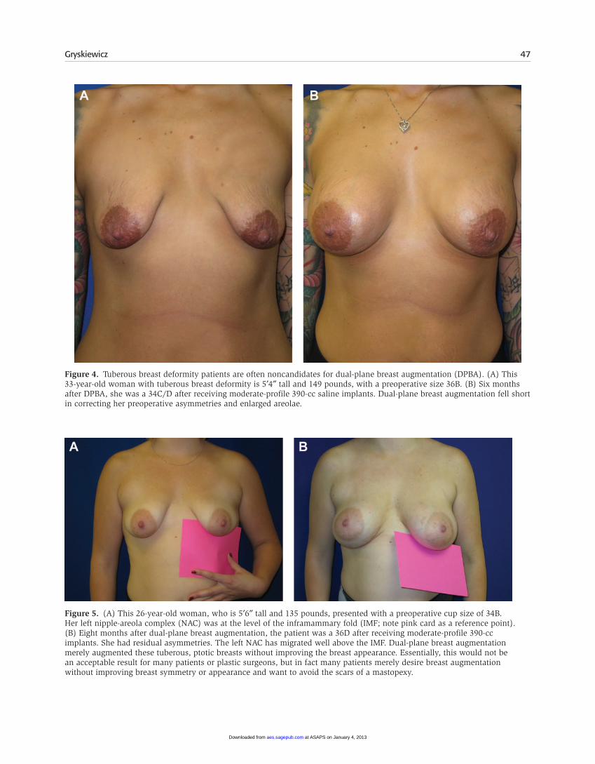
Figure 4. Tuberous breast deformity patients are often noncandidates for dual-plane breast augmentation (DPBA). (A) This 33-year-old woman with tuberous breast deformity is 5′4″ tall and 149 pounds, with a preoperative size 36B. (B) Six months after DPBA, she was a 34C/D after receiving moderate-profile 390-cc saline implants. Dual-plane breast augmentation fell short in correcting her preoperative asymmetries and enlarged areolae.
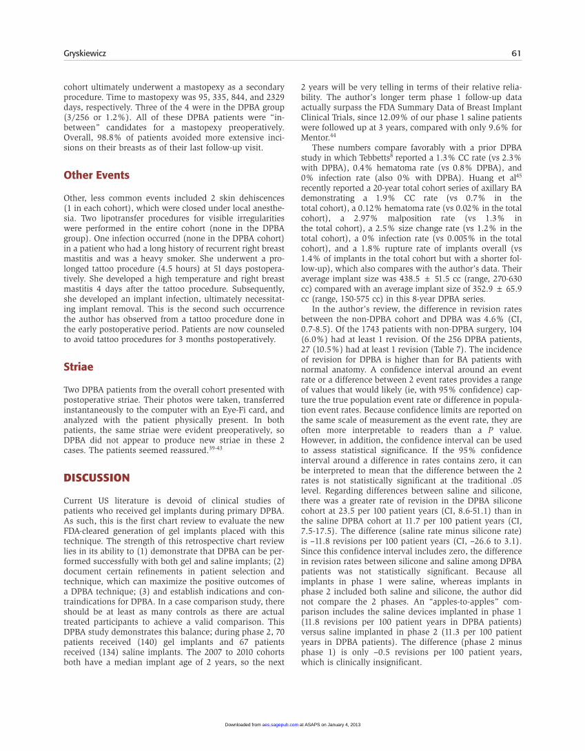
Figure 5. (A) This 26-year-old woman, who is 5′6″ tall and 135 pounds, presented with a preoperative cup size of 34B. Her left nipple-areola complex (NAC) was at the level of the inframammary fold (IMF; note pink card as a reference point). (B) Eight months after dual-plane breast augmentation, the patient was a 36D after receiving moderate-profile 390-cc implants. She had residual asymmetries. The left NAC has migrated well above the IMF. Dual-plane breast augmentation merely augmented these tuberous, ptotic breasts without improving the breast appearance. Essentially, this would not be an acceptable result for many patients or plastic surgeons, but in fact many patients merely desire breast augmentation without improving breast symmetry or appearance and want to avoid the scars of a mastopexy.
at ASAPS on January 4, 2013aes.sagepub.comDownloaded from

48 Aesthetic Surgery Journal 33(1)
measurements and potential tissue cover. Then, the patients’ verbal requests were factored into the analysis.
During consultation, patient photos were obtained, transferred instantaneously to the computer with an Eye-Fi card (Eye-Fi, Mountainview, California), printed, and analyzed. Each patient tried multiple gel sizers with a bra, under a thin T-shirt. The profile of the implant was secondary, whereas the implant diameter vis-à-vis the diameter of the breasts was primary. A gel sizer placed in a bra preoperatively does not have the same surrounding forces placed on it as a surgical pocket, so approximately 30 to 50 cc was added to the final volume to account for flattening of the implant in the partial submuscular pocket.
Once the procedure was scheduled, the patient was given an instruction card admonishing against blood thin-ners and nicotine in the perioperative period. Strict, relia-ble methods of birth control were discussed, and all patients signed a legal release regarding potential preg-nancy. Patients were instructed to wash with 4% chlorhex-idine gluconate soap for a total of 3 preoperative showers at least 1 hour apart the evening before and the morning of surgery, scrubbing the torso to lower the number of skin contaminants.
Dual-Plane Surgical Technique
Preoperatively and with a chaperone present, the patient’s chest was marked with the entire surgical plan and implant sizes. The surgeon reviewed the “ideal” photos brought by the patient during the preoperative consultation as well as the drawings of the surgical plan. These images were hung in the operating room for reference during the procedure.
The surgeon began the procedure with 4 pair of powder-free gloves: first a half-size larger, worn under the normal size, both of which were worn under 2 additional pair that were a full size larger than normal. As the case proceeded, 1 outer pair was removed, such that only an unused pair of gloves touched each implant as it was inserted. Only the surgeon handled the implants. Each patient received 1 dose of intravenous (IV) cefuroxime 1.5 g (GlaxoSmithKline, Research Triangle Park, North Carolina) during the hour prior to surgery because of its exquisite coverage of Staphylococcus epidermidis. The patient was put under general anesthesia. After a sterile paint with tinted ChloraPrep (2% chlorhexidine gluconate + isopropyl alco-hol; CareFusion, Leawood, Kansas) was applied, the patient was covered with sterile draping. Tegaderm (3M Health Care, St Paul, Minnesota) was also applied to each NAC during the last year of this study, to sequester any bacteria-laden nipple discharge that might have been expressed dur-ing the breast procedure (Figure 6).
If a periareolar approach was selected, a zigzag incision was made.21 An endoscopic axillary approach has been described in the literature,8 including recent reports in which a portion of pectoralis was excised.22,23 The author made an inframammary incision 3 cm long for saline or
4 to 6 cm for gel, depending on implant size and the patient’s skin elasticity. Two planes were dissected for a combination of pocket locations (Figure 7). The subpecto-ral dissection was accomplished first. Dissection was car-ried down to the underlying pectoralis fascia. In some cases, the PMM attached slightly above the IMF. If the PMM contracted from the Bovie (Conmed Cautery System 2450; Conmed, Utica, New York), the breast bounced, indicating that dissection was heading in the correct direction. If the rectus muscle contracted, dissection was then angled more superiorly. If the local area around the incision contracted, the surgeon recognized that this was potentially a result of intercostal muscle contraction. To avoid penetrating between the ribs, dissection was cau-tiously redirected superiorly, remaining over the surface of the adjacent rib, because the PMM could have inserted cephalad to the current plane of dissection. Upward trac-tion on the cephalic edge of the incision elevates the PMM off the ribcage. The intercostal muscles do not elevate.
Dissection remained superficial to the periosteum and perichondrium to minimize the patient’s postoperative pain and discomfort. To develop the pocket, electrocautery was applied in coagulation mode for prospective hemostasis. The pectoralis muscle was not released along the medial sternal border. Once the medial pocket was established, the PMM was separated from the pectoralis minor muscle. Dissection advanced from superior-medial to inferior in the lateral pocket to better delineate the fusion area between the 2 pectoralis muscles. The serratus fascia was not elevated. The lateral border of the PMM was not ele-vated unless necessary to accommodate the implant. Care was taken not to overdissect laterally.
Once dissection of the partial subpectoral space was complete, dissection proceeded in the more superficial subglandular plane (retromammary plane). The retro-mammary plane was dissected to the level of the inferior
Figure 6. Tegaderm “nipple-shield” sequesters any bacteria-laden nipple discharge that may be expressed during a breast procedure.
at ASAPS on January 4, 2013aes.sagepub.comDownloaded from

Gryskiewicz 49
edge of the areola (6 o’clock) for minor ptosis or tight folds, but if attachments were loose or the conditions were more severe, dissection was carried to the superior edge of the areola (12 o’clock). Care was taken not to release the muscle too cephalad in the retromammary plane. The breast was moved up and down to determine whether adequate release had been obtained. Space was allowed so that the implant would maximally contact the parenchyma and project anteriorly without muscle restriction.
Once both planes were dissected, the medial inferior edge of the PMM was divided parallel to a rib, 1 cm above the IMF level (Figure 8). The dissection extended across the medial inferior portion of the muscle to the lateral edge of the sternum but did not extend superiorly along the sternal border. Finally, judicious release of the inferior pocket with blunt finger dissection was done on a case-by-case basis by stretching and sparing (not transecting) the IMF ligament and dermal attachments to lower the IMF. Following this type of dissection, the implant had pectoral coverage superiorly, as well as pectoral/fascial coverage laterally, and was exposed to the overlying parenchyma inferiorly down to the inframammary fold, as in Figure 7B.
Once dissection of the pocket was accomplished, air was drawn into the pocket by gently elevating the gland off the ribs with a finger or dissector. The incision was
pinched closed with the opposite hand. In this way, the outline of the pocket perimeter was delineated, obviating the need for a sizer (Figure 9). Following the “air-sizer” maneuver, any pocket irregularities were modified as nec-essary. The pocket was copiously irrigated with Adams pancidal triple antibiotic solution (bacitracin 50 000 units [Xgen Pharmaceuticals, New York, New York]; cefazolin 1 g [Sandoz, Inc, Princeton, New Jersey]; gentamicin 80 mg [APP Pharmaceuticals, Schaumburg, Illinois]), which also provided coverage for methicillin-resistant Staphylococcus aureus (MRSA) and methicillin-resistant S epidermidis (MRSE).24-27
The “no-touch” technique was maintained by removing the outer pair of 4 pair of gloves at this juncture. The implant container was opened by the assistant, and the Adams antibiotic solution was poured over the implant. Sensorcaine 0.25% with epinephrine 1:200 000 (APP Pharmaceuticals) was instilled into the pocket immedi-ately prior to implant placement. The implants were then placed into each dual-plane pocket. It is possible to unin-tentionally insert the implant into the retromammary space, so care was taken to utilize a long enough retractor to elevate below the pectoralis muscle.
A Richardson general surgery retractor (#BT479R; Aesculap, Inc, Center Valley, Pennsylvania) was used for
Figure 7. (A) Two planes are dissected for a combination of pocket locations. Once dissection of the partial subpectoral space is complete, dissection proceeds in the more superficial subglandular plane (retromammary plane) enhancing the tissue-draping interplay between the gland and implant. (B) This figure illustrates the crux of dual-plane breast augmentation. Release of the retromammary plane allows the caudal edge of the pectoralis muscle to migrate superiorly, causing the inferior-anterior edge of the implant to now adequately abut against the posterior surface of the breast parenchyma, supporting the gland and nipple-areola complex up and forward.
at ASAPS on January 4, 2013aes.sagepub.comDownloaded from

50 Aesthetic Surgery Journal 33(1)
gel implant insertion because its 4-cm width accommo-dates a gel implant. Gentle upward traction was placed on the retractor as the gel implant was being inserted. Once a seal had been established at the incision/mouth of the pocket by the gel implant, the retractor was lifted quickly and forcefully to facilitate insertion with negative pres-sure. Often, a suction sound was audible if this maneuver was done correctly. If saline implants were placed, a closed fill system was employed.
Minor adjustments were made as necessary once the implant was placed. Care was taken to ensure that the muscle was pulled down over the implant. If the pocket was reentered for hemostasis or further adjustment, fin-gers or instruments were immersed in the triple antibiotic solution before reentering the pocket. The position of the implant was meticulously assessed from multiple angles in both supine and sitting positions. To stand at the foot of the bed and on each side provided helpful angles for the surgeon, while the anesthesiologist looked from above the drape, focusing on the 12-o’clock position. All patients were placed in a full 90-degree sitting position for the final assessment. Contour irregularities and lack of symmetry became more obvious from a position that simulated the pull of gravity. The “ideal” photos the patient had chosen preoperatively were used as a reference for implant posi-tion (medial vs lateral, inferior vs superior). Finally, the surgeon held a towel tightly across the NAC with the patient in the sitting position (Figure 10). This “evening gown” test was useful to compare upper pole asym-metries.
The incision was closed with a running 2-0 Vicryl suture (Ethicon, Inc, New Brunswick, New Jersey) and the dermis was everted with interrupted and running subcu-ticular 3-0 Monocryl sutures (Ethicon, Inc). Mastisol liquid
Figure 8. The medial inferior edge of the pectoralis major muscle is divided parallel to the ribs, 1 cm above the inframammary fold. The implant will have pectoral coverage superiorly, as well as pectoral fascial coverage laterally, and be exposed to the overlying parenchyma inferiorly down to the inframammary fold. (Inset) An intraoperative judgment is made whether to carry the retromammary dissection superior to the 6-o’clock nipple-areola complex level or even higher, to the 12-o’clock level. IMF, inframammary fold; PMM, pectoralis major muscle.
Figure 9. (A) Once dissection of the pocket is accomplished, air can be drawn into the pocket by gently elevating the gland off of the ribs. (B) The incision is pinched closed, demonstrating the pocket perimeter. Following the “air-sizer” maneuver, any pocket irregularities are modified.
at ASAPS on January 4, 2013aes.sagepub.comDownloaded from

Gryskiewicz 51
adhesive (Ferndale Lab, Ferndale, Michigan) was placed on the incision and allowed to dry, and Steri-Strips (3-M, St Paul, Minnesota) were applied. Patients were dis-charged with only these adhesive skin closure tapes over their incisions. No bra or dressings were applied routinely. Occasionally, a superior pole breast wrap (Clear Point Medical, Champlain, New York) was applied intraopera-tively (or even postoperatively) in selected patients if the implants were found to be riding high. A video of the surgical procedure is available at www.aestheticsurgery-journal.com. You may also use any smartphone to scan the code on the first page of this article to be taken directly to the video on www.YouTube.com.
Postoperative Care
Double FoldSome patients who preoperatively had an NAC at the IMF level developed problems with postoperative double folds secondary to a tight crease. A tight crease may maintain its contour and resist expansion by the underlying implant, causing flattening along its original length. Patients who presented postoperatively with a double fold were instructed to wear a “cutout” bra (Figure 11). To create this bra, the patient brought in a standard, inexpensive underwire bra.
The lower half of the cup, from immediately above the underwire up to the NAC level, was cut away with a scis-sors. Patients were instructed to wear the “cutout” bra 7 days a week, 24 hours a day, for the first postoperative month (except when they were bathing or showering). The “cutout” bra invariably expanded the tight IMF because the wire compressed the new IMF and allowed the original fold to relax more superiorly. With this method, the author has found that most taut tissue will succumb to the implant forces and efface over 1 to 3 months.
Multiple steps from prior publications were followed to provide quicker patient recovery.28-30 In an effort to avoid CC, patients were instructed, as noted above, to begin rais-ing their arms over their heads and to perform manual displacement exercises hours after surgery. Patients were instructed to massage their implants in all directions except downward, to avoid tension on the incision. Regardless of the preferred CC prevention routine, patients were encouraged to continue with the exercises for years, rather than weeks or months.31 If a patient presented with a unilateral firm breast at any time during the postopera-tive period, she was started on a protocol that included exercises, massage, high-dose vitamin E (2000 U/d),32 and nonsteroidal anti-inflammatory medication. A printed pro-tocol was provided to each patient to aid in her compli-ance and understanding of individual responsibility. If a
Figure 10. (A, B, C) A towel is held tightly across the nipple-areola complex while the patient is in the full 90-degree sitting position. This “evening gown” test is useful to compare upper pole asymmetries.
at ASAPS on January 4, 2013aes.sagepub.comDownloaded from

52 Aesthetic Surgery Journal 33(1)
CC was diagnosed, a 90-day treatment protocol of mon-telukast (Singulair; Merck & Co, West Point, Pennsylvania) was initiated with the option to repeat it once for a total duration of 6 months (although this is an off-label usage). Montelukast is not available in generic formulation, may be expensive, and may not be covered by insurance. The out-of-pocket cost to the patient was approximately $400 for a 3-month supply, which may be prohibitive.33-38
Photographic analysis was used to define a positive outcome. All patients had 5 standardized preoperative and postoperative photographs, including frontal, side, and oblique views. The surgeon analyzed the photos preopera-tively with the patient for asymmetries and ptosis. In turn, all postoperative photos were analyzed and compared with the preoperative photos by 2 third parties (the clinic registered nurse [RN] and the aesthetician) and by the surgeon. Postoperative photos were reviewed personally with all patients in the clinic by the RN and surgeon and given directly to each patient upon request.
REsuLts
One hundred eighteen patients (11.8% of phase 1 cohort) underwent DPBA in the first phase of the study and 138 (13.9% of phase 2 cohort) underwent DPBA in the second phase, for a total of 256 overall (12.8%). Clinical results of 5 patients with combinations of ptosis, glandular ptosis, constricted lower pole, and tight IMF can be seen in Figures 12 through 14. Additional patient images are available in an online-only appendix at www.aestheticsurgeryjournal.com.
Duration of Follow-up
Duration of follow-up was calculated overall and by implant type for all patients. About a quarter of patients—24.2% of the entire cohort of 1999 patients (n = 3998 implants) and 23.4% of the 256 DPBA patients—were followed for 1 year or more. Also, 25.9% of the 1554
Figure 11. (A) This 24-year-old woman had a double fold, which can be produced from a tight crease present preoperatively. The tight fold may maintain its contour and resist expansion by the underlying implant, causing flattening along its original length. Arrows point to each end of the tight fold. (B) Patients with a double fold are placed in an underwire “cutout” bra. (C) Seven weeks postoperatively, the patient demonstrates effacement of the double fold.
at ASAPS on January 4, 2013aes.sagepub.comDownloaded from

Gryskiewicz 53
saline implant patients (n = 3108 saline implants) and 18.2% of the 445 silicone gel patients (n = 890 silicone
implants) were followed for 1 year or more, and 28.0% of the saline DPBA patients (n = 186) and 11.4% of the sili-
Figure 12. (A, C, E) This 47-year-old woman presented with ptosis and a very low-set nipple-areola complex. She was 5′4″ and 130 pounds, with a preoperative size of 34B. (B, D, F) One year after bilateral dual-plane breast augmentation (DPBA) with a 450-cc implant on the left and a 450-cc implant on the right. The patient’s postoperative cup size was 34C. Postoperatively, more breast tissue is visible throughout the inferior pole because the DPBA releases the inferior pole tissue, allowing it to rise up like a pendulum.
at ASAPS on January 4, 2013aes.sagepub.comDownloaded from

54 Aesthetic Surgery Journal 33(1)
Figure 13. (A, C, E) This 39-year-old woman presented with pseudoptosis. She was 5′5″ and 135 pounds, with a preoperative cup size of 38B. (B, D, F) Sixteen months after bilateral inframammary dual-plane breast augmentation with a 480-cc implant on the left and a 455-cc implant on the right. The patient’s postoperative size was 38D. Postoperatively, on lateral view, more breast tissue was visible between the inframammary fold (IMF) and the nipple-areola complex. The IMF is lower after augmentation (see the distance from the IMF to anterior elbow crease on the oblique and lateral views).
at ASAPS on January 4, 2013aes.sagepub.comDownloaded from

Gryskiewicz 55
Figure 14. (A, C, E) This 40-year-old woman presented with a tight, irregular, asymmetric inframammary fold (IMF) with a low-set nipple-areola complex (NAC). She was 5′4″ and weighed 140 pounds, with a preoperative cup size of 36B. (B, D, F) Two years after bilateral inframammary dual-plane breast augmentation with a 390-cc implant on the left and a 390-cc implant on the right. The patient’s postoperative size was 38D. The IMF is lower after augmentation (see the distance from the IMF to anterior elbow crease on the oblique and lateral views). Postoperatively on the frontal view, more breast tissue is visible between the inferior visual border and the elevated NAC.
at ASAPS on January 4, 2013aes.sagepub.comDownloaded from

56 Aesthetic Surgery Journal 33(1)
cone gel DPBA patients (n = 70) were followed for 1 year or more. The smaller percentage of follow-up for the gel subset is because the phase 2 four-year study of gel and saline implants closed more recently than the phase 1 eight-year study, which included only saline implants. Most of the 22 patients in the entire cohort and the 3 DPBA patients who did not return for follow-up lived out of state, and these women were followed with phone calls during the first months postoperatively. Of the 1999 patients in the overall cohort, 1003 (50.2%) were in phase 1 and 996 (49.8%) were in phase 2, which is a remarkably consistent number for each phase. Of the entire cohort (N = 1999), DPBA accounted for 11.8% of patients in phase 1 and 13.9% in phase 2. Of the 256 DPBA patients, 118 (46.1%) were in phase 1 and 138 (53.9%) were in phase 2. Once the FDA moratorium was lifted and phase 2 began, it took a while for silicone to be accepted by our patients. Of the 996 patients in phase 2, 551 (55.3%) received saline implants and 445 (44.7%) received sili-cone. The number of DPBA per calendar year varied from 23 to 49. All patients were advised to follow up annually with our office for implant examinations for the rest of their lives.
Demographics
The mean (SD) age of all patients was 33.6 (8.7) years (range, 18-69 years). The median age was 32.9 years. The mean (SD) age of DPBA patients was 36.4 (8.5) years (range, 20-65 years). The median age was 35.4 years.
Implant Characteristics
All patients received 2 implants initially. The mean (SD) implant size in all patients was 350.3 (61.2) cc (range, 120-800 cc). Mean (SD) implant size in DPBA patients was 353.2 (64.7) cc (range, 150-575 cc). All implants placed throughout the study were smooth, round, medium or high profile from a single manufacturer (Allergan, Inc).
Revisions and Untoward Events
There is an implicit assumption in the following results that phase 1 saline and phase 2 saline are combinable, which would make sense because the same manufactur-er’s saline implant models and the surgical equipment and techniques were similar enough over the 2 phases of saline experience. Since the data from all saline implants were assumed to be combinable, the comparison of saline versus silicone was not confined to phase 2. However, phase 1 patients, all of whom received saline implants, had a longer period of observation during which an unto-ward event could occur, whereas patients who received silicone implants had, on average, shorter follow-up time. Therefore, revision rates were normalized by patient years of follow-up. That is, the number of events per patient
year of observation was used to assess relative risk to level the playing field when comparing saline and silicone.
Overall, 1868 patients had no revisions and 131 patients (6.6%) had at least 1 revision. Of the 131 total revisions, 109 patients had 1 revision, 21 had 2 revisions, and 1 had 3 revisions. Of the 256 DPBA patients, 229 had no revi-sions, 27 (10.5%) had at least 1 revision, and 3 had 2 revisions. No DPBA patient had 3 revisional procedures. Of the 1743 patients with non-DPBA surgery, 104 (6.0%) had at least 1 revision. The difference in revision rates between types of surgery was 4.6% (confidence interval [CI], 0.7-8.5). In patients receiving saline implants, the non-DBPA and DBPA revision rates were 6.9% and 11.3%, respectively. In patients receiving silicone implants, these rates were 2.4% and 8.6%, respectively. The pattern is also similar over time. In phase 1, the non-DBPA and DBPA revision rates were 8.9% and 14.4%, respectively. In phase 2, they were 2.9% and 7.2%, respectively.
Saline implants required more revisions in the overall cohort and in the DPBA group than silicone gel implants. Of the 1554 patients with saline implants, 116 (7.5%) had at least 1 revision. Of the 445 patients with silicone implants, 15 (3.4%) had at least 1 revisional procedure. Of the 186 DPBA patients with saline implants, 21 (11.3%) had at least 1 revi-sion. Of the 70 DPBA patients with silicone implants, 6 (8.6%) had at least 1 revision. These data could yield an unfair comparison because the saline cohort had longer fol-low-up times than did the silicone patients and therefore had more time for events to occur. Therefore, summarization by patient year of follow-up helps to adjust for differences in follow-up time. The revision rates for any kind of secondary procedure were 9.4 revisions per 100 patient years (CI, 7.9-11.1) for saline patients versus 7.1 (CI, 4.0-11.7) for silicone. The difference is 2.3 (CI, –2.0 to 6.7). With DPBA patients, there were more events in the silicone cohort at 23.5 per 100 patient years (CI, 8.6-51.1) than in the saline cohort at 11.7 (CI, 7.5-17.5) (Figure 15). The difference is –11.8 revisions per 100 patient years (CI, –26.6 to 3.1).
Thirteen types of revisions were discovered. The types of revisions are listed in order of frequency for the overall cohort and DPBA cohort (Tables 1 and 2). Deflation tops the lists in both cohorts. No deflations were discovered in gel implants. There were some minor variations between the 2 cohorts. The complications or untoward events are analyzed in detail below.
Implant Failure
Deflation was the most common reason for implant-related revisions in the entire cohort, with 57 ruptured implants in 1999 patients (2.9% of patients) and 3998 implants (1.4% of implants) (Tables 1 and 2). Note that 10 patients experienced 2 deflations. Deflation was also the most common reason for implant-related revision in the DPBA cohort, with 6 ruptured implants in 256 patients (2.3% of patients) and 512 implants (1.2% of implants) (Table 2). All deflations were discovered in saline implants, and all were removed and replaced. Analysis of the
at ASAPS on January 4, 2013aes.sagepub.comDownloaded from

Gryskiewicz 57
frequency of deflation per 100 patient years showed saline to be 5.3 in phase 1 and 4.7 in phase 2, versus 0 for sili-cone gel. Analysis of the DPBA group showed this same trend, with phase 1 as 7.1 deflations per 100 patient years
versus zero for silicone in DPBA phase 2 patients (Tables 3 and 4). No leaks were discovered in gel implants (phase 2), although no formal screening process was required. Patients were informed of the FDA recommendations and criteria regarding postoperative magnetic resonance imaging (MRI) screening. Patients were advised to undergo routine breast exams and mammograms by their primary care provider. One gel implant patient (1/445) elected to have a postopera-tive MRI to conform to current FDA surveillance recommen-dations. During the MRI examination, a deflation was detected. The patient decided to undergo surgical interven-tion, but at the time of surgery, the implant was actually discovered to be intact, causing unnecessary expense, time lost from work, surgeon time lost, and exposure to general anesthesia. In sum, all ruptures occurred in the saline group (57/1554 patients or 3.7%); 10 patients in the entire cohort and 3 patients in the DPBA cohort experienced more than 1 deflation (not exceeding 2).
Malposition
Malposition was the second most common untoward event in the overall cohort, occurring in 26 patients (twice in 1 patient) (26/1999 or 1.3%). There were 26 patients with exactly 1 malposition and 1 patient with 2 malpositions, for a total of 27 patients with at least 1 event. Malposition was the fourth most common untoward event in the DPBA cohort, occurring in 5 patients (5/256 or 1.9%; Tables 5 and 6). Malposition was defined as any movement in any direction of the breast implant outside the original pocket. All malpositions appeared to be inferior in the standing position, but in the supine position, it was discovered they were all inferolateral as well.
Figure 15. For dual-plane breast augmentation in this study, there were more revisions in the silicone gel cohort, 23.5 per 100 patient years (confidence interval [CI], 8.6-51.1), than in the saline cohort, 11.7 (CI, 7.5-17.5). Much of this difference is from malposition revisions of gel devices. The difference is –11.8 thrombospondin instances per 100 patient years (CI, –26.6 to 3.1). Therefore, this difference is not significant to the 95% confidence interval because the CI is both above and below zero (ie, the CI crosses zero). When one looks at the data, it is clear that saline implants required more revisions (7.5%) compared with gel (3.4%). The primary reason for revision in both cohorts was saline implant deflation.
Table 1. Event Type in the Entire Cohort (N = 1999), Number of Patients With 0, 1, or 2 Listed by Decreasing Frequency
Cohort Type of Secondary Procedure No. of Patients With No Event No. of Patients With 1 Event No. of Patients With 2 Events
All Saline deflation/gel leak 1932 57 10
All Malposition 1972 26 1
All Size change 1975 24 0
All Change saline to silicone 1980 19 0
All Capsule 1985 13 1
All Explant 1991 8 0
All Hematoma 1994 4 1
All Mastopexy 1995 4 0
All Lipotransfer 1997 2 0
All Partial closure 1997 2 0
All Infection 1998 1 0
All Explant with reduction 1998 1 0
All Insert secondary implant 1998 1 0
at ASAPS on January 4, 2013aes.sagepub.comDownloaded from

58 Aesthetic Surgery Journal 33(1)
Table 2. Event Type in the Dual-Plane Breast Augmentation (DPBA) Cohort (n = 256), Patients With 0, 1, or 2 Listed by Decreasing Frequency
Cohort Type of Secondary Procedure No. of Patients With No Event No. of Patients With 1 Event No. of Patients With 2 Events
DPBA Saline deflation/gel leak 247 6 3
DPBA Size change 248 8 0
DPBA Capsule 250 6 0
DPBA Malposition 251 5 0
DPBA Change saline to silicone 251 5 0
DPBA Mastopexy 253 3 0
DPBA Hematoma 254 2 0
DPBA Explant with reduction 255 1 0
DPBA Explant 255 1 0
DPBA Partial closure 255 1 0
DPBA Lipotransfer 256 0 0
DPBA Infection 256 0 0
DPBA Insert secondary implant 256 0 0
Table 3. Incidence and Frequency of Saline Deflation or Gel Leak in Entire Cohort
Phase Implant Type Sample Size
No. of Patients With at Least 1
Event Incidence, % No. of Events Frequency, %Patient Years of
Follow-up
Frequency (per 100 Patient
Years) 95% Confidence Intervals
Lower Upper
Both Both 1999 67 3.4 77 3.9 1691 4.6
Both Saline 1554 67 4.3 77 5.0 1478 5.2 4.1 6.5
Silicone 445 0 0.0 0 0.0 212 0.0 0.0 1.7
Saline 1003 58 5.8 67 6.7 1268 5.3
Saline 551 9 1.6 10 1.8 211 4.7
The difference in deflation rates between saline and silicone is 5.2 events per 100 patient years (95% confidence interval, 2.1-8.3). The bold zeros in Table 3 show the distinction between gel rupture and saline deflations in the entire cohort. No gel implant was found to be ruptured in the entire cohort, contrasted to saline implants where 77 implants ruptured. The primary reason for revision in both cohorts was saline implant deflation.
Table 4. Incidence and Frequency of Saline Deflation or Gel Leak in Dual-Plane Breast Augmentation Cohort
Phase Implant Type Sample Size
No. of Patients With at Least 1
Event Incidence, % No. of Events Frequency, %Patient Years of
Follow-up
Frequency (per 100 Patient
Years) 95% Confidence Intervals
Lower Upper
Both Both 256 9 3.5 12 4.7 230 5.2
Both Saline 186 9 4.8 12 6.5 205 5.9 3.0 10.2
2 Silicone 70 0 0.0 0 0.0 26 0.0 0.0 14.4
1 Saline 118 9 7.6 12 10.2 169 7.1
2 Saline 68 0 0.0 0 0.0 35 0.0
The difference in deflation/leak rates between saline and silicone is 5.9 events per 100 patient years (confidence interval, –3.5 to 15.3). The confidence intervals for the individual rates and for the difference are wider than in the case of all patients, because the number of events is smaller and the information therefore less precise. Zero leaks were discovered in gel implants (phase 2).
at ASAPS on January 4, 2013aes.sagepub.comDownloaded from

Gryskiewicz 59
Many DPBA candidates present with inherently lax tissue, which can lead to postoperative malposition. The tissues frequently cannot withstand the muscular and gravitational forces of the implant. This is the purpose of preoperative supine assessment, because marked lateral migration of the breast would disqualify the patient from DPBA and propel her toward a mastopexy. Malposition was handled in the following manner. The previous inframammary (IM) incision was opened, the expanded area of the pocket was freshened with electrocautery by excising a crescent of capsule, contralateral capsulotomy was carried out when necessary, and capsulorrhaphy was achieved with interrupted 0-Ethibond sutures (Ethicon, Inc). The patients were then placed in the full 90-degree sitting position, and further meticulous adjust-ments were made to achieve symmetry. For revision of high-riding implants, the previous IM incision was opened, the inferior pocket was released, and the ante-rior lining was removed from the inferior pole (NAC-IMF) to allow the implant to drop into alignment with the contralateral IMF.
Size Change
Size change was the third most common event in the over-all cohort (26 patients or 1.2%) and the second most com-mon event in the DPBA cohort (8 patients or 3.1%) (Tables 7 and 8). The percentages of saline-implant patients who desired a size change—1.4% in the overall population and 3.7% in the DPBA subset (Tables 7 and 8)—appeared to be
at least twice those of silicone-implant patients, which were 0.7% overall and 1.4% in DPBA. However, there was greater opportunity for patients with saline implants to seek a change because as a group they were followed longer than silicone-implant patients. When the incidence of size change is normalized by follow-up time, the 2 types of implants have more similar rates; in the overall popula-tion, the rates are the same (1.4 events per 100 patient years). It is clear that more problems occurred with saline because saline implant deflations were self-diagnosed by the patients, but the author does not know whether any silicone implants were ruptured. Zero leaks were discov-ered in gel implants (phase 2). Women with saline implants who had a size change generally did so long after their initial surgery, even many years afterwards. Some patients had a size change as their primary reason for a revision, whereas other patients had an unrelated untoward event as the inciting primary reason (Tables 1 and 2). Women with gel implants who had a size change (and there were only 3 such patients) seemed to do so sooner after surgery. The same observation holds in the subset of DPBA patients. Patients signed the printed decision management algorithm on size change as part of the preoperative consultation.1
Change Saline to Silicone
Conversion to gel was the fourth most common event in the overall cohort; 19 patients (1%) requested conversion. Conversion to gel was the fifth most common even in the DPBA cohort, occurring in 5 patients (2.0%). A saline
Table 5. Incidence and Frequency of Malposition in Entire Cohort
Phase Implant Type Sample SizeNo. of Patients With
at Least 1 Event Incidence, % No. of Events Frequency, %Patient Years of
Follow-upFrequency (per 100
Patient Years)
Both Both 1999 27 1.4 28 1.4 1691 1.7
Both Saline 1554 20 1.3 21 1.4 1478 1.4
2 Silicone 445 7 1.6 7 1.6 212 3.3
1 Saline 1003 17 1.7 17 1.7 1268 1.3
2 Saline 551 3 0.5 4 0.7 211 1.9
Table 6. Incidence and Frequency of Malposition in Dual-Plane Breast Augmentation Cohort
Phase Implant Type Sample SizeNo. of Patients With
at Least 1 Event Incidence, % Number of Events Frequency, %Patient Years of
Follow-upFrequency (per 100
Patient Years)
Both Both 256 5 2.0 5 2.0 230 2.2
Both Saline 186 3 1.6 3 1.6 205 1.5
2 Silicone 70 2 2.9 2 2.9 26 7.8
1 Saline 118 1 0.8 1 0.8 169 0.6
2 Saline 68 2 2.9 2 2.9 35 5.7
at ASAPS on January 4, 2013aes.sagepub.comDownloaded from

60 Aesthetic Surgery Journal 33(1)
implant deflation was the most common inciting factor, and the second factor was the availability of gel implants after November 2006. No conversions from silicone to saline occurred.
Capsular Contracture
Capsular contracture was the fifth most common cause for revision in the overall cohort, with 14 patients electing to have surgery (0.7%, or 0.33% of all implants). One patient had a second surgery for recurrent CC. In the DPBA group, the revision rate for CC was third, with 6 capsules (2.3%). The overall CC rate was 0.9 per 100 patient years. Analysis of the frequency of CC in saline implants per 100 patient years showed saline in phase 1 to be 0.6 versus 0.9 in phase 2, and 0.7 for both, contrasted to 2.4 for silicone. Gel implants had a higher incidence of thrombospondin compared with saline for CC. Capsular contracture was treated by capsulectomy and implant exchange and the firm breast protocol described in the Methods section. All 20 operated patients had Baker 3 and 4 capsules. For the group of patients who developed non-surgical Baker 1 and 2 capsules (0.2% of both the overall cohort and the DPBA group), the author adhered to the protocol described in the Methods section for a patient who presented with a unilateral firm breast.
Explant
Eight patients in the overall cohort requested explantation (0.04%), citing that they were at a different place in their lives and no longer felt the need for implants. One patient in the DPBA cohort (0.04%) also requested explantation. One patient in the overall cohort requested explantation with breast reduction, citing that her breasts had increased in size since her initial surgery.
Hematoma
Hematoma occurred in 4 patients (0.02%) from the entire cohort, and 2 of these 4 were from the DPBA cohort. In the DPBA group, 1 hematoma occurred 10 days postopera-tively when the patient, an anesthetist, reached with 1 arm to move an obese patient and immediately developed massive swelling on the same side. The second hematoma occurred spontaneously 5 days postoperatively.
Mastopexy
The goal of DPBA is to avoid mastopexy in selected patients. Therefore, this metric is perhaps the most sig-nificant measure of DPBA. Four patients of the entire
Table 7. Incidence and Frequency of Size Change in Entire Cohort
Phase Implant Type Sample SizeNo. of Patients With
at Least 1 Event Incidence, % No. of Events Frequency, %Patient Years of
Follow-upFrequency (per 100
Patient Years)
Both Both 1999 24 1.2 24 1.2 1691 1.4
Both Saline 1554 21 1.4 21 1.4 1478 1.4
2 Silicone 445 3 0.7 3 0.7 212 1.4
1 Saline 1003 17 1.7 17 1.7 1268 1.3
2 Saline 551 4 0.7 4 0.7 211 1.9
Table 8. Incidence and Frequency of Size Change in Dual-Plane Breast Augmentation Cohort
Phase Implant Type Sample SizeNo. of Patients With
at Least 1 Event Incidence, % No. of Events Frequency, %Patient Years of
Follow-upFrequency (per 100
Patient Years)
Both Both 256 8 3.1 8 3.1 230 3.5
Both Saline 186 7 3.8 7 3.8 205 3.4
2 Silicone 70 1 1.4 1 1.4 26 3.9
1 Saline 118 5 4.2 5 4.2 169 3.0
2 Saline 68 2 2.9 2 2.9 35 5.7
at ASAPS on January 4, 2013aes.sagepub.comDownloaded from
Gryskiewicz 61
cohort ultimately underwent a mastopexy as a secondary procedure. Time to mastopexy was 95, 335, 844, and 2329 days, respectively. Three of the 4 were in the DPBA group (3/256 or 1.2%). All of these DPBA patients were “in-between” candidates for a mastopexy preoperatively. Overall, 98.8% of patients avoided more extensive inci-sions on their breasts as of their last follow-up visit.
Other Events
Other, less common events included 2 skin dehiscences (1 in each cohort), which were closed under local anesthe-sia. Two lipotransfer procedures for visible irregularities were performed in the entire cohort (none in the DPBA group). One infection occurred (none in the DPBA cohort) in a patient who had a long history of recurrent right breast mastitis and was a heavy smoker. She underwent a pro-longed tattoo procedure (4.5 hours) at 51 days postopera-tively. She developed a high temperature and right breast mastitis 4 days after the tattoo procedure. Subsequently, she developed an implant infection, ultimately necessitat-ing implant removal. This is the second such occurrence the author has observed from a tattoo procedure done in the early postoperative period. Patients are now counseled to avoid tattoo procedures for 3 months postoperatively.
Striae
Two DPBA patients from the overall cohort presented with postoperative striae. Their photos were taken, transferred instantaneously to the computer with an Eye-Fi card, and analyzed with the patient physically present. In both patients, the same striae were evident preoperatively, so DPBA did not appear to produce new striae in these 2 cases. The patients seemed reassured.39-43
disCussiOn
Current US literature is devoid of clinical studies of patients who received gel implants during primary DPBA. As such, this is the first chart review to evaluate the new FDA-cleared generation of gel implants placed with this technique. The strength of this retrospective chart review lies in its ability to (1) demonstrate that DPBA can be per-formed successfully with both gel and saline implants; (2) document certain refinements in patient selection and technique, which can maximize the positive outcomes of a DPBA technique; (3) and establish indications and con-traindications for DPBA. In a case comparison study, there should be at least as many controls as there are actual treated participants to achieve a valid comparison. This DPBA study demonstrates this balance; during phase 2, 70 patients received (140) gel implants and 67 patients received (134) saline implants. The 2007 to 2010 cohorts both have a median implant age of 2 years, so the next
2 years will be very telling in terms of their relative relia-bility. The author’s longer term phase 1 follow-up data actually surpass the FDA Summary Data of Breast Implant Clinical Trials, since 12.09% of our phase 1 saline patients were followed up at 3 years, compared with only 9.6% for Mentor.44
These numbers compare favorably with a prior DPBA study in which Tebbetts8 reported a 1.3% CC rate (vs 2.3% with DPBA), 0.4% hematoma rate (vs 0.8% DPBA), and 0% infection rate (also 0% with DPBA). Huang et al45 recently reported a 20-year total cohort series of axillary BA demonstrating a 1.9% CC rate (vs 0.7% in the total cohort), a 0.12% hematoma rate (vs 0.02% in the total cohort), a 2.97% malposition rate (vs 1.3% in the total cohort), a 2.5% size change rate (vs 1.2% in the total cohort), a 0% infection rate (vs 0.005% in the total cohort), and a 1.8% rupture rate of implants overall (vs 1.4% of implants in the total cohort but with a shorter fol-low-up), which also compares with the author’s data. Their average implant size was 438.5 ± 51.5 cc (range, 270-630 cc) compared with an average implant size of 352.9 ± 65.9 cc (range, 150-575 cc) in this 8-year DPBA series.
In the author’s review, the difference in revision rates between the non-DPBA cohort and DPBA was 4.6% (CI, 0.7-8.5). Of the 1743 patients with non-DPBA surgery, 104 (6.0%) had at least 1 revision. Of the 256 DPBA patients, 27 (10.5%) had at least 1 revision (Table 7). The incidence of revision for DPBA is higher than for BA patients with normal anatomy. A confidence interval around an event rate or a difference between 2 event rates provides a range of values that would likely (ie, with 95% confidence) cap-ture the true population event rate or difference in popula-tion event rates. Because confidence limits are reported on the same scale of measurement as the event rate, they are often more interpretable to readers than a P value. However, in addition, the confidence interval can be used to assess statistical significance. If the 95% confidence interval around a difference in rates contains zero, it can be interpreted to mean that the difference between the 2 rates is not statistically significant at the traditional .05 level. Regarding differences between saline and silicone, there was a greater rate of revision in the DPBA silicone cohort at 23.5 per 100 patient years (CI, 8.6-51.1) than in the saline DPBA cohort at 11.7 per 100 patient years (CI, 7.5-17.5). The difference (saline rate minus silicone rate) is –11.8 revisions per 100 patient years (CI, –26.6 to 3.1). Since this confidence interval includes zero, the difference in revision rates between silicone and saline among DPBA patients was not statistically significant. Because all implants in phase 1 were saline, whereas implants in phase 2 included both saline and silicone, the author did not compare the 2 phases. An “apples-to-apples” com-parison includes the saline devices implanted in phase 1 (11.8 revisions per 100 patient years in DPBA patients) versus saline implanted in phase 2 (11.3 per 100 patient years in DPBA patients). The difference (phase 2 minus phase 1) is only –0.5 revisions per 100 patient years, which is clinically insignificant.
at ASAPS on January 4, 2013aes.sagepub.comDownloaded from

62 Aesthetic Surgery Journal 33(1)
Advantages of DBPA
Compared with single-pocket augmentation, the dual-plane approach allows the surgeon to better adjust the implant vis-à-vis the breast tissue relationship, offering increased benefits with fewer trade-offs. Many breast aug-mentation patients with “in-between” anatomy consult multiple surgeons and can become confused by opposing surgical recommendations. The dual-plane approach may allow these selected patients to avoid more extensive mas-topexy incisions. It is a valuable technique for patients with (1) pseudoptosis, or ≤15% of the glandular tissue below the IMF; (2) a center-point of the NAC at or above the IMF; (3) a constricted lower pole (NAC-IMF <5 cm); or (4) a tight IMF.
Contraindications and Considerations
In this study, DPBA was selected as the surgical method of choice for a given patient only after she had been evalu-ated thoroughly, with stringent indications in mind. Dual-plane breast augmentation was not performed as a routine procedure for primary breast augmentation. If all breast parenchyma was located above the IMF, with tight attach-ments to the underlying PMM and NAC to IMF of approx-imately 5 cm, the patient was disqualified from DPBA. Rather, DPBA was reserved only for selected patients in this series, instead of being utilized in patients with nor-mal-appearing anatomy. Again, exclusion criteria to DPBA were patients with weighty breasts and those with an NAC trajectory pointing downward or lateral migrating breast mound(s) in the supine position.
It is also essential to discuss the expected postoperative outcomes of this procedure thoroughly with patients, particularly the fact that they may require a mastopexy in the future. Even if the surgical plan is for DPBA, the surgeon should thoroughly establish informed consent for augmentation-mastopexy, so patients understand their options. The trade-off with a DPBA procedure is less scar-ring, versus more maximal elevation of the NAC with a mastopexy. Dual-plane breast augmentation does not levi-tate the gland or fashion a high, tight contour. The proce-dure does not centralize the NAC on the mound or decrease the NAC diameter, nor will DPBA equalize a horizontally disparate NAC.
Clinical Suggestions
Medial release of the pectoralis muscle along the sternal attachments was assiduously avoided in DBPA procedures. Prior to the start of this study, the author overzealously released the muscle both medially and in the retromammary plane, above the level of the NAC, in several initial patients. This extreme release, both from the sternum and from the overlying breast, allowed the caudal edge of the PMM to migrate too far superiorly. In some patients, this resulted in a tight band of muscle that was evident across the breast,
especially upon animation (a so-called window shade phe-nomenon).46 As a result, the author is now more conserva-tive in his retromammary release. Other surgeons have also suggested placing tacking sutures along the inferior edge of the pectoralis muscle to avoid “window-shading.”47
A constricted lower pole (ie, in patients with a short NAC-to-IMF distance and a tight crease) must be released significantly and sculpted to render a pleasing visual bor-der along the IMF. At the same time, great care must be exercised to avoid release of the inframammary ligament, to limit “bottoming out” of the implant. Following con-servative Bovie (Conmed Cautery System 2450; Conmed) release of the inferior pole, the IMF is stretched gently with finger dissection to further position the implant accu-rately.
Great care is also taken to adjust the implants intraop-eratively. The patient should be painstakingly evaluated in the supine position not only from above (by the anesthe-siologist) but also from below and laterally (by other team members in the operating suite). The patient should then be placed in a full 90-degree sitting position, straightened on the table as necessary, and further evaluated. Microadjustments with gentle finger dissection are then made to achieve a pleasing result. The abutting of the implant against the posterior surface of the breast paren-chyma acts like a pendulum, which elevates the NAC as the parenchyma swings superior and anterior.
Limitations
Several opportunities exist for further study of DPBA patients. Accurate follow-up times were assessed and cal-culated meticulously in this study. The initial DPBA cohort has a theoretical 8-year follow-up time, but unless the patients have actually been assessed for up to 8 years, these outcome calculations are not as helpful. Any patient needing a reoperation who had moved out of state was referred to a colleague. It is also possible that a patient received a reoperation elsewhere without contacting us. Furthermore, the follow-up time in phase 2 patients was shorter, so longer follow-up is necessary to assess the overall rates of complications (especially for the gel cohort and for deflations) and recurrence of ptosis for patients who received a DPBA approach. Deflation rates for the saline implants in this study will undoubtedly rise over time. It is fortuitous that nearly equal numbers of patients selected each type of implant.
In this study, no formal assessment of patient satisfac-tion was done. Quantitative satisfaction evaluation could have provided an opportunity for corroboration of the conclusions drawn from the analysis of reoperation rates and photographic analysis. Although many patients were measured preoperatively (from sternal notch to NAC and NAC and IMF to umbilicus), not every patient was meas-ured for these points postoperatively. Certainly, there is inherent variability when these measurements are taken manually. Better documentation with standardized photo-graphs and a horizontal line overlay would help establish
at ASAPS on January 4, 2013aes.sagepub.comDownloaded from
Gryskiewicz 63
that the NAC does elevate from DPBA. Standardized (com-puterized) imaging would decrease this error, but this method was not employed by the author until after the conclusion of this particular study.
Another limitation of the retrospective portion of this study is the lack of MRI follow-up of gel patients. At the time of this study, MRI was the best available means of monitoring gel rupture and was, in fact, recommended as the gold standard of surveillance by the FDA.48 At the author’s institution, bilateral breast MRI costs $2200. Despite patient education and informed consent about the FDA recommendations, only 1 gel implant patient elected to undergo a postoperative MRI with out-of-pocket expense. The MRI detected a rupture but, as stated earlier, the implant was actually intact upon surgical exploration. The only MRI performed in this series prompted an unnec-essary procedure, anesthetic exposure, expense, time off from work for the patient, and poor use of the surgeon’s time. Gray49 reported a similar experience with false-posi-tive results in 5 patients (repeating the author’s MRI accu-racy rate of zero). Pooled summary measures, recently reported in a large series, of sensitivity (true positives) and specificity (true negatives) for implant MRI have been reported as 87% and 89.9%, respectively.50 Among lower prevalence populations, Cher et al51 reported in a meta-analysis that the positive predictive value appeared to be insufficient to warrant MRI use as a screening tool. The summary sensitivity was 78% and the summary specific-ity was 91%. As a further downside, the authors high-lighted that the majority of the published studies examined only symptomatic patients. Once observer variability between radiologists is factored into the equation, overall accuracy may drop even lower. The authors concluded many of the MRI and ultrasound diagnostic accuracy stud-ies examining silicone breast implant ruptures are meth-odologically flawed. Therefore, patients and surgeons need a more accurate, cost-effective means of diagnosis. An extensive study of the new generation of high-resolu-tion ultrasound coupled with MRI has recently been pub-lished and shows excellent results demonstrating 100% accuracy. High-resolution ultrasound is a notable clinical advancement and will undoubtedly help us as practition-ers to solve this dilemma for our breast augmentation patients, regardless of surgical approach, implant type, or higher cross-linked gels.52
COnCLusiOns
Dual-plane breast augmentation is best suited for patients with minimal ptosis who are in the “gray area” between normal anatomy and frank ptosis. Phase 1 (January 2003 through December 2006) involved a retrospective chart review of 1003 cosmetic BA patients who received saline implants exclusively, with a variety of techniques. Phase 2 was a retrospective review and comparative study of 996 gel and saline cosmetic BA patients treated between January 2007 and December 2010. Two hundred fifty-six DPBA pro-cedures were studied, and the outcomes of gel versus saline
were compared. The data from this 8-year retrospective, comparative review support the fact that for a select group of patients with mild to moderate ptosis and proper informed consent, DPBA is a technique that will provide acceptable aesthetic results with low reoperation rates in patients who refuse external mastopexy scars and who will accept preexisting areolar asymmetries and large areolae. Dual-plane breast augmentation allows selected patients who may present after consulting multiple surgeons and receiving disparate recommendations to avoid more exten-sive mastopexy incisions. Therefore, this approach can provide surgeons and patients with an alternative technique for achieving satisfactory augmentation outcomes.
Acknowledgment
The author would like to express his sincere appreciation to Jessica McMillan for her photographic analysis and to Naomi Owen and Melanie Morrison for data gathering. He also thanks Dr Richard Bittman and David Herridge for their assis-tance and expertise with statistical analysis, Jim Peterson for his audiovisual assistance, and Andrew Grivas, MA, for his well-crafted, original illustrations.
disclosures
The author declared no potential conflicts of interest with respect to the research, authorship, and publication of this article.
Funding
The author received no financial support for the research, authorship, or publication of this article.
REFEREnCEs
1. Adams WP, Bengston BP, Gryskiewicz JM, et al. Deci-sion and management algorithms to address patient and food and drug administration concerns regarding breast augmentation and implants. Plast Reconstr Surg. 2004;114(5):1252-1257.
2. Adams WP. The High Five Process: tissue-based planning for breast augmentation. Plast Surg Nurs. 2007;27(4): 197-201.
3. Adams WP Jr. The process of breast augmentation: four sequential steps for optimizing outcomes for patients. Plast Reconstr Surg. 2008;122(6):1892-1900.
4. Adams WP Jr, Spear SL. Augmentation mammaplasty. Plast Reconstr Surg. 2006;118(7)(suppl):5S-6S.
5. Adams WP Jr, Teitelbaum S, Bengtson BP, Jewell ML, Tebbetts J, Spear S. Breast augmentation roundtable. Plast Reconstr Surg. 2006;118(7)(suppl):175S-187S.
6. Adams WP Jr, Rios JL, Smith SJ. Enhancing patient out-comes in aesthetic and reconstructive breast surgery using triple antibiotic breast irrigation: six-year prospec-tive clinical study. Plast Reconstr Surg. 2006;117(1):30-36.
7. Tebbetts JB. The greatest myths in breast augmentation. Plast Reconstr Surg. 2001;107(7):1895-1903.
at ASAPS on January 4, 2013aes.sagepub.comDownloaded from

64 Aesthetic Surgery Journal 33(1)
8. Tebbetts JB. Dual plane breast augmentation: optimizing implant-soft-tissue relationships in a wide range of breast types. Plast Reconstr Surg. 2001;107(5):1255-1272.
9. Tebbetts JB. Dual plane breast augmentation: optimizing implant-soft-tissue relationships in a wide range of breast types. Plast Reconstr Surg. 2006;118(7)(suppl):81S-98S, discussion 99S-102S.
10. Tofield JJ. Dual plane breast augmentation. Plast Reconstr Surg. 2001;108(7):2162-2164.
11. Tanne JH. FDA approves silicone breast implants 14 years after their withdrawal. BMJ. 2006;333(7579):1139.
12. Spear SL, Carter ME, Ganz JC. The correction of cap-sular contracture by conversion to “dual-plane” posi-tioning: technique and outcomes. Plast Reconstr Surg. 2003;112(2):456-466.
13. Spear SL, Carter ME, Ganz JC. The correction of capsular contracture by conversion to “dual-plane” positioning: technique and outcomes. Plast Reconstr Surg. 2006;118(7)(suppl):103S-113S, discussion 114S.
14. Esposito G, Gravante G, Marianetti M, Delogu D. “Reverse” dual-plane mammaplasty. Aesthetic Plast Surg. 2006;30(5):521-526.
15. Khan UD. Muscle-splitting breast augmentation: a new pocket in a different plane. Aesthetic Plast Surg. 2007;31(5):553-558.
16. Grolleau JL, Lanfrey E, Lavigne B, Chavoin JP, Costa-gliola M. Breast base anomalies: treatment strategy for tuberous breasts, minor deformities, and asymmetry. Plast Reconstr Surg. 1999;104(7):2040-2048.
17. Hidalgo DA, Spector JA. Preoperative sizing in breast aug-mentation. Plast Reconstr Surg. 2010;125(6):1781-1787.
18. Hammond DC. Preoperative sizing in breast augmenta-tion. Plast Reconstr Surg. 2011;127(2):1005.
19. Casas LA. Preoperative sizing for breast augmentation. Plast Reconstr Surg. 2011;127(2):1006-1007.
20. Schonauer F, Singh S, La Rusca I, Molea G. Preopera-tive sizing and breast asymmetry. Plast Reconstr Surg. 2011;127(2):1005-1006.
21. Gryskiewicz JM, Hatfield AS. “Zigzag” wavy-line periare-olar incision. Plast Reconstr Surg. 2002;110(7):1778-1783, discussion 1784.
22. Luan J, Mu D, Mu L. Transaxillary dual-plane augmenta-tion mammaplasty: experience with 98 breasts. J Plast Reconstr Aesthetic Surg. 2009;62(11):1459-1463.
23. Luan J, Mu DL, Mu L, Liu C, Zhang ZQ. Transaxillary dual-plane breast augmentation with endoscope assis-tant [in Chinese]. Zhonghua Zheng Xing Wai Ke Za Zhi. 2009;25(3):175-177.
24. Adams WP Jr, Conner WC, Barton FE Jr, Rohrich RJ. Optimizing breast pocket irrigation: an in vitro study and clinical implications. Plast Reconstr Surg. 2000;105(1): 334-338, discussion 339-343.
25. Adams WP Jr, Conner WC, Barton FE Jr, Rohrich RJ. Opti-mizing breast-pocket irrigation: the post-Betadine era. Plast Reconstr Surg. 2001;107(6):1596-1601.
26. Adams WP Jr. Discussion: subclinical (biofilm) infec-tion causes capsular contracture in a porcine model fol-lowing augmentation mammaplasty. Plast Reconstr Surg. 2010;126(3):843-844.
27. Tamboto H, Vickery K, Deva AK. Subclinical (biofilm) infection causes capsular contracture in a porcine model following augmentation mammaplasty. Plast Reconstr Surg. 2010;126(3):835-842.
28. Tebbetts JB. Achieving a predictable 24-hour return to nor-mal activities after breast augmentation, part I: refining practices by using motion and time study principles. Plast Reconstr Surg. 2002;109(1):273-290, discussion 291-292.
29. Tebbetts JB. Achieving a predictable 24-hour return to nor-mal activities after breast augmentation, part II: patient preparation, refined surgical techniques, and instrumenta-tion. Plast Reconstr Surg. 2002;109(1):293-305, discussion 306-307.
30. Gryskiewicz JM. Avoiding pain and suffering after breast augmentation. Plast Reconstr Surg. 2002;110(7):1812-1813.
31. Marques M, Brown SA, Oliveira I, et al. Long-term follow-up of breast capsule contracture rates in cosmetic and reconstructive cases. Plast Reconstr Surg. 2010;126(3): 769-778.
32. Baker JL Jr. The effectiveness of alpha-tocopherol (vitamin E) in reducing the incidence of spherical con-tracture around breast implants. Plast Reconstr Surg. 1981;68(5):696-699.
33. Gryskiewicz J. Commentary. Aesthetic Surg J. 2010; 30(3):409-410.
34. Gryskiewicz JM. Investigation of Accolate and Singulair for treatment of capsular contracture yields safety con-cerns. Aesthetic Surg J. 2003;23(2):98-101.
35. Gryskiewicz JM. What doctors aren’t being told: using the freedom of information act. Plast Reconstr Surg. 2004;113(2):743-745.
36. Rubin R, Salvati EA, Lewis R. Infected total hip replace-ment after dental procedures. Oral Surg Oral Med Oral Pathol. 1976;41(1):18-23.
37. Bartzokas CA, Johnson R, Jane M, Martin MV, Pearce PK, Saw Y. Relation between mouth and haematog-enous infection in total joint replacements. BMJ. 1994;309(6953):506-508.
38. Huang CK, Handel N. Effects of Singulair (montelukast) treatment for capsular contracture. Aesthetic Surg J. 2010;30(3):404-408.
39. Snyder GB. Red striae after augmentation. Plast Reconstr Surg. 1981;67(5):699.
40. White DJ, Schnur PL. Striae distensae after augmentation mammoplasty. Ann Plast Surg. 1995;34(1):16-22.
41. Robinson C. Case report of asymmetrical striae following breast augmentation. Plast Reconstr Surg. 1997;99(1):274-275.
42. Mahabir RC, Peterson BD. Two cases of striae distensae after submuscular augmentation mammaplasty. Plast Reconstr Surg. 2001;108(3):753-756.
43. Huang GJ, York CE, Mills DC. Striae distensae as a com-plication of augmentation mammaplasty. Plast Reconstr Surg. 2008;122(2):90e-93e.
44. Food and Drug Administration. FDA update on the safety of silicone gel-filled breast implants. Center for Devices and Radiological Health. 2011. fda.gov/breastimplants
45. Huang GJ, Wichmann JL, Mills DC. Transaxillary sub-pectoral augmentation mammaplasty: a single surgeon’s 20-year experience. Aesthetic Surg J. 2011;31(7):781-801.
at ASAPS on January 4, 2013aes.sagepub.comDownloaded from

Gryskiewicz 65
46. Spear SL, Schwartz J, Dayan JH, Clemens MW. Outcome assessment of breast distortion following submuscular breast augmentation. Aesthetic Plast Surg. 2009;33(1): 44-48.
47. Ramirez OM, Heller MDL, Tebbetts JB. Dual plane breast augmentation: avoiding pectoralis major displacement. Plast Reconstr Surg. 2002;110(4):1198, author reply 1198-1199.
48. Hammond DC. Discussion: the effect of study design biases on the diagnostic accuracy of magnetic resonance imaging for detecting silicone breast implant ruptures: a meta-analy-sis. Plast Reconstr Surg. 2011;127(3):1045-1046.
49. Gray L. Silicone breast implants and magnetic resonance imaging screening for rupture: do U.S. Food and Drug Administration recommendations reflect an evidence-based
practice approach to patient care? Plast Reconstr Surg. 2008;122(5):1591-1592, author reply 1592.
50. Song JW, Kim HM, Bellfi LT, Chung KC. The effect of study design biases on the diagnostic accuracy of mag-netic resonance imaging for detecting silicone breast implant ruptures: a meta-analysis. Plast Reconstr Surg. 2011;127(3):1029-1044.
51. Cher DJ, Conwell JA, Mandel JS. MRI for detecting sili-cone breast implant rupture: meta-analysis and implica-tions. Ann Plast Surg. 2001;47(4):367-380.
52. Bengtson BP, Eaves FF III. High-resolution ultrasound in the detection of silicone gel breast implant shell failure: background, in vitro studies, and early clinical results. Aesthetic Surg J. 2012;32(2):157-174.
at ASAPS on January 4, 2013aes.sagepub.comDownloaded from