aerobic fitness is associated with greater efficiency of
TRANSCRIPT

n
n
Neuroscience 199 (2011) 166–176
AEROBIC FITNESS IS ASSOCIATED WITH GREATER EFFICIENCY OFTHE NETWORK UNDERLYING COGNITIVE CONTROL IN
PREADOLESCENT CHILDRENM. W. VOSS,a* L. CHADDOCK,a J. S. KIM,a
M. VANPATTER,a M. B. PONTIFEX,b L. B. RAINE,b
N. J. COHEN,a C. H. HILLMANb AND A. F. KRAMERa
aBeckman Institute and Department of Psychology, University of Illi-ois, Urbana-Champaign, IL 61801, USA
bDepartment of Kinesiology and Community Health, University of Illi-ois, Urbana-Champaign, IL 61801, USA
Abstract—This study examined whether individual differ-ences in aerobic fitness are associated with differences inactivation of cognitive control brain networks in preadoles-cent children. As expected, children performed worse on ameasure of cognitive control compared with a group ofyoung adults. However, individual differences in aerobic fit-ness were associated with cognitive control performanceamong children. Lower-fit children had disproportionate per-formance cost in accuracy with increasing task difficulty,relative to higher-fit children. Brain activation was comparedbetween performance-matched groups of lower- and higher-fit children. Fitness groups differed in brain activity for re-gions associated with response execution and inhibition,task set maintenance, and top-down regulation. Overall, dif-fering activation patterns coupled with different patterns ofbrain-behavior correlations suggest an important role of aer-obic fitness in modulating task strategy and the efficiency ofneural networks that implement cognitive control in preado-lescent children. © 2011 IBRO. Published by Elsevier Ltd. Allrights reserved.
Key words: exercise, physical activity, aerobic fitness, exec-utive control, functional magnetic resonance imaging (fMRI),development.
In addition to increasing risk for developing long-term car-diovascular and metabolic diseases, research has shownthat poor physical fitness during childhood puts children atrisk for lower cognitive performance (Pontifex et al., 2011)and poorer academic achievement (Castelli et al., 2007;Chomitz et al., 2009). To date, functional neuroimagingevidence that can help us understand these trends hascome primarily from event-related brain potential (ERP)studies of the attentional and executive control systems(Hillman et al., 2005, 2009; Pontifex et al., 2011). The goalof the current study is to extend this research by usingfunctional magnetic resonance imaging (fMRI) to charac-
*Corresponding author. Tel: �1-217-417-4689.E-mail: [email protected] (M. W. Voss).Abbreviations: Con, congruent; ERP, event-related brain potential;fMRI, functional magnetic resonance imaging; HF, higher-fit; Inc, in-
congruent; LF, lower-fit; MRI, magnetic resonance imaging; ROI, re-gion-of-interest; RT, reaction time.0306-4522/11 $ - see front matter © 2011 IBRO. Published by Elsevier Ltd. All righdoi:10.1016/j.neuroscience.2011.10.009
166
terize how fitness level relates to patterns of brain activityduring cognitive challenge in preadolescent children.
Research that has examined neuroelectric indices ofbrain function in lower-fit (LF) and higher-fit (HF) preado-lescent children has done so in the context of cognitivecontrol theory using modified Eriksen flanker tasks (Hill-man et al., 2009; Pontifex et al., 2011), a standard test ofconflict resolution (Eriksen and Eriksen, 1974). Success-fully inhibiting information that conflicts with an attendedtarget is a subcomponent of executive function, and isassociated with activation of a variety of prefrontal regionssuch as the dorsolateral and anterior prefrontal corticesand the dorsal anterior cingulate cortex (Botvinick et al.,1999; Casey et al., 2000b; Hazeltine et al., 2000). Theseregions are part of a larger collection of brain regionsinvolved in cognitive control, a term that describes ourability to flexibly adapt behavior to environmental needs,maintain goals for behavior across sustained periods oftime, and monitor for errors to adjust control engagement(Botvinick et al., 2001; Braver and Barch, 2006). In thisframework, the dorsolateral prefrontal cortex is hypothe-sized to initiate flexible adjustments in control, whereas theanterior prefrontal cortex maintains context-driven taskgoals over prolonged periods of time, and the anteriorcingulate cortex evaluates and monitors for conflict anderrors that signal need to adjust control (Dosenbach et al.,2007, though, see Forster and Brown, 2011). The prefron-tal cortex is the last association area to fully develop inchildren, and this extended period of brain plasticity likelycontributes to the protracted development of cognitive con-trol processes and integration of their associated brainsystems (Casey et al., 2000a; Bunge et al., 2002; Fair etal., 2007).
In the adult brain, the anterior prefrontal cortex and thedorsal anterior cingulate are two regions in a brain networkcalled the cingulo-opercular network, which is involved inprolonged maintenance of task context (Dosenbach et al.,2007). Other brain regions in the network include the an-terior insula extending into the central and frontal opercu-lum and the thalamus. A recent study mapping functionalcortico-striatal connections also suggests that the putamenis part of the network (Barnes et al., 2010). The cingulo-opercular network is proposed to work together with thefronto-parietal network, which includes the dorsolateralprefrontal cortex and which complements the sustainednature of the cingulo-opercular network with rapid onlineadjustments of cognitive control (Dosenbach et al., 2007,
2008). Core regions in the fronto-parietal network alsots reserved.
ANTKVV
M. W. Voss et al. / Neuroscience 199 (2011) 166–176 167
include the inferior parietal cortex and the dorsal precu-neus.
With the protracted development of the prefrontal cor-tex, it should come as no surprise that these two cognitivecontrol networks are not fully integrated, distinct networksin pre-adolescence. During childhood the anterior prefron-tal cortex and dorsal anterior cingulate are more function-ally connected to the fronto-parietal system, but throughdevelopment to adulthood they become more integratedwith the emerging cingulo-opercular system (Fair et al.,2007). Thus, the dual architecture of the cognitive controlsystem develops from one that is optimal for rapid, reac-tive, and adaptive control, to one that can flexibly balancerapid and sustained goal-oriented control. Moreover, giventhat these neural networks are in a stage of heightenedplasticity during preadolescent development, increasedphysical fitness during this critical time may serve as animportant component to ensure their proper development.
Indeed, ERP research has suggested that this is thecase. For example, LF children tend to show decreased P3amplitude for target processing (Hillman et al., 2009; Pon-tifex et al., 2011), suggesting that poor physical fitness isassociated with poorer attentional allocation processes.These studies have also found that LF children show agreater error-related negativity (ERN), an ERP componentthought to index conflict monitoring and error evaluation.LF children were also less accurate in trials following errorsof commission. Given that increased ERN amplitude isthought to reflect inefficient conflict monitoring coupledwith deficient prefrontally regulated cognitive control (Geh-ring and Knight, 2000), these studies suggest that LFchildren monitor conflict inefficiently through ineffectivecingulate and prefrontal interaction during cognitive con-trol. Or, from a brain systems approach, they suggest LFchildren have less effective interaction between the pre-frontal and anterior cingulate components of the fronto-parietal and cingulo-opercular networks. This would alsobe consistent with research demonstrating that sedentaryolder adults who completed 6 months of aerobic traininghad decreased anterior cingulate activation compared witha non-aerobic control group, coupled with increased rightlateral prefrontal activation and better conflict resolution(Colcombe et al., 2004).
The current study tested several of these suggestedhypotheses using fMRI with LF and HF preadolescentchildren. Participants performed a modified Eriksen flanker
Table 1A. Mean (SD) values for demographic, KBIT, and fitness data
Variable Young adults (YA) Children
Lower-fit (L
N 26 18ge 21.7 (2.1) 9.9 (.6)male (%) 9 (34.6%) 7 (38.9
anner — 1.7 (.5)BIT composite 108.1 (6.9) 113.6 (15.0O2max (ml/kg/min) 40.5 (6.2) 36.9 (4.5)
O2max percentile 40.25 (24.7) 14.9 (17.7)task during functional imaging. To confirm that the taskmeasured aspects of cognitive control sensitive to devel-opment, children were compared with young adult controlson task performance. Although the young adult group hadfitness levels somewhere in between those of the LF andHF child groups, young adults have been shown to performbetter on the flanker paradigm than children, and we werespecifically interested to see if children with higher aerobicfitness levels parallel young adult performance. Next, brainactivation related to cognitive control processes for LF andHF children were compared. We hypothesized that fitnesswould affect activation patterns in the prefrontal cortices,including important areas associated with cognitive controlsuch as the dorsolateral and anterior prefrontal corticesand the anterior cingulate cortex, coupled with poorer per-formance for LF compared with HF children. Furthermore,if aerobic fitness is associated with more successful inte-gration of the anterior prefrontal cortex and anterior cingu-late into the cingulo-opercular network, we expect HF chil-dren to more effectively recruit this network to implementcognitive control.
EXPERIMENTAL PROCEDURES
Participants
Participants were recruited from the east-central Illinois region.Eligible participants had to (1) demonstrate strong right handed-ness, with 75% or above on the Edinburgh Handedness Inventory(Oldfield, 1971), (2) be between the ages of nine and ten forchildren and between 18 and 30 years for young adults, (3) be freeof neurological disease, attentional disorders, or physical disabil-ities, (4) have normal color-vision, (5) have a corrected visualacuity of at least 20/20, and (6) sign an informed assent/consent.For all children, participants provided written assent and their legalguardians provided written informed consent in accordance withthe Institutional Review Board of the University of Illinois at Urbana-Champaign. Children, in collaboration with their legal guardian,completed the Tanner Staging Scales (Taylor et al., 2001), indi-cating that their pubertal status was at or below a score of two (i.e.prepubescent) on a 5-point scale. All participants were adminis-tered the Kaufman Brief Intelligence Test (KBIT; Kaufman andKaufman, 1990) by a trained experimenter to assess intelligencequotient. Demographic, KBIT, and fitness data for all participantsare provided in Table 1.
Cardiorespiratory fitness assessment
In addition to the previous criteria for children, participants werebifurcated by aerobic fitness into HF or LF groups based on
P-value (two-tailed)
Higher-fit (HF) Young-children LF-HF children
18 — —9.8 (.6) �.001 NS10 (55.6%) NS NS1.6 (.5) — NS
116.8 (6.3) .008 NS52.8 (5.2) .05 �.001
F)
%)
)
83.67 (5.3) NS �.001

p2
wrsp(1is
fs
ANTKVV )
M. W. Voss et al. / Neuroscience 199 (2011) 166–176168
whether their cardiorespiratory fitness level (i.e. VO2max) fellabove the 70th percentile or below the 30th percentile accordingto normative data provided by Shvartz and Reibold (1990). Max-imal oxygen consumption (VO2max) was measured using a com-uterized indirect calorimetry system (ParvoMedics True Max400, Sandy, UT, USA) with averages for oxygen uptake (VO2)
and respiratory exchange ratio (RER) assessed every 20 s. Amodified Balke protocol (Whaley et al., 2006) was employed usinga motor-driven treadmill at a constant speed with increases ingrade increments of 2.5% every 2 min until volitional exhaustionoccurred. A Polar heart rate monitor (Polar WearLink®� 31, PolarElectro, Finland) was used to measure HR throughout the test,and ratings of perceived exertion (RPE) were collected every 2min using the children’s OMNI scale (Utter et al., 2002). Relativepeak oxygen consumption was expressed in ml/kg/min and basedupon maximal effort as evidenced by (1) a plateau in oxygenconsumption corresponding to an increase of less than 2 ml/kg/min despite an increase in workload, (2) a peak heart rate �185beats/min (Whaley et al., 2006) or a heart rate plateau (Freedsonand Goodman, 1993), (3) RER �1.0 (Bar-Or, 1983), and/or (4)ratings on the children’s OMNI scale of perceived exertion �8(Utter et al., 2002).
Imaging procedures and processing
Following cardiorespiratory fitness assessment, all participantscompleted a mock magnetic resonance imaging (MRI) session,wherein they were screened for their ability to complete an exper-iment in an MRI environment. Participants who passed the mockscreening subsequently completed a series of structural and func-tional MRI scans.
Structural MRI. High-resolution T1-weighted brain imagesere acquired using a 3D MPRAGE (magnetization prepared
apid gradient echo imaging) protocol with 144 contiguous axiallices, collected in ascending fashion parallel to the anterior andosterior commissures, echo time (TE)�3.87 ms, repetition timeTR)�1800 ms, field of view (FOV)�256 mm, acquisition matrix92 mm�192 mm, slice thickness�1.3 mm, and flip angle�8°. All
mages were collected on a 3T head-only Siemens Allegra MRIcanner.
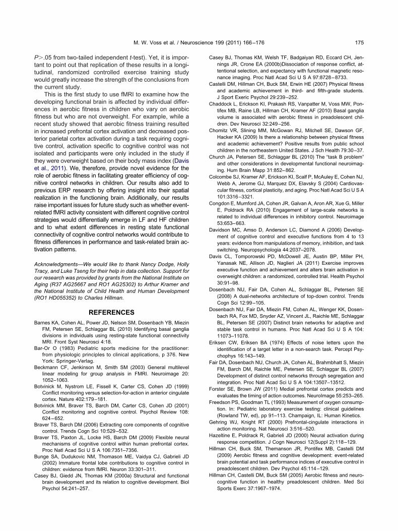
Functional MRI. During fMRI scanning, participants per-ormed a version of a flanker paradigm in which they were pre-ented with a horizontal array of five fish that faced either right (�)
or left (�). Participants were asked to identify the orientation of thecentral fish while ignoring the orientation of the peripheral fish (seeFig. 1). The stimuli were presented for 1 s on each trial followed bya fixed interstimulus interval (ISI) of 1 s. In the magnet, beforescanning began for the task, all participants completed 12 practicetrials with feedback. This number of practice trials was sufficientfor children to understand the task, as very few participantsneeded to repeat the practice trials due to misunderstanding ornon-response. Practice trials presented all trial types in a random
Table 1B. Mean (SD) values for demographic, KBIT, and fitness data
Variable Young adults (YA) Performanc
Lower-fit (L
N 26 14ge 21.7 (2.1) 9.9 (.5)male (%) 9 (34.6%) 5 (35.7
anner — 1.6 (.5)BIT composite 108.1 (6.9) 118.4 (13.1O2max (ml/kg/min) 40.5 (6.2) 37.0 (4.1)O2max percentile 40.25 (24.7) 16.7 (19.5
order, but otherwise resembled the event timing of real trials. Task
trials during scanning were presented in a blocked design suchthat there were three blocks of congruent (Con) fish and threeblocks of incongruent (Inc) fish trials interleaved with 30 s blocksof fixation baseline. In each Con and Inc block, there were 12 Conand Inc trials, respectively, and six neutral (e.g. - -�- -) trialsrandomly interspersed, for a total of 36 trials for each of Con, Inc,and neutral conditions. The presence of neutral trials ensured thatsubjects retained a strategy of focusing on the central target whiletrying to ignore peripheral distractors. Given the block design,activation specific to neutral trials could not be modeled sepa-rately. Since there were equal numbers of neutral trials per block,their effect on task-related activation would presumably cancelout. For this reason, only the Con and Inc conditions are pre-sented for behavioral and neuroimaging results. Functional imag-ing parameters were TR�2000 ms, TE�25 ms, flip angle�80°,blood oxygen level dependent (BOLD) volume acquisitions�230volumes; the order of blocks for all participants was Con, Inc, Inc,Con, Inc, Con.
Functional MRI preprocessing was carried out using FSL4.1.2 (FMRIB’s Software Library, www.fmrib.ox.ac.uk/fsl). The fol-lowing pre-statistics processing were applied: rigid body motioncorrection using MCFLIRT (Jenkinson et al., 2002), removal ofnon-brain structures using BET (Smith, 2002), spatial smoothingusing a Gaussian kernel of FWHM 6.0 mm, grand-mean intensitynormalization of the entire 4D dataset by a single multiplicativefactor, and temporal filtering with a high-pass frequency cut-off of220 s. Regression-based analysis of fMRI data was carried outusing FSL’s FEAT Version 5.98. For individual-level analyses, thehemodynamic response to each block was convolved with a dou-ble-gamma HRF function. Of primary interest to this study was thecontrast of Inc�Con. This contrast was computed for each subjectat the individual level and carried forward to a mixed-effects groupanalysis. Higher-level mixed-effects analyses were carried outusing FLAME (Beckmann et al., 2003). For all GLM activation
g adults and performance-matched children groups
ed children P-value (2-tailed)
Higher-fit (HF) Young-children LF-HF children
14 — —9.8 (.6) �.001 NS
8 (57.1%) NS NS1.6 (.5) — NS
117.9 (6.6) �.001 NS53.6 (5.6) .05 �.00183.9 (5.6) NS �.001
Fig. 1. Example task stimuli for a response indicating central targetstimulus is pointing to the right. For interpretation of the references tocolor in this figure legend, the reader is referred to the Web version of
for youn
e-match
F)
%)
)
this article.

5ica
S�P�.0
M. W. Voss et al. / Neuroscience 199 (2011) 166–176 169
analyses the, statistical threshold for significant activation was setto Z�2.33 voxel, P�.05 cluster-correction.
Whole-brain analyses were followed by region-of-interest(ROI) analysis of fMRI activation. When statistically significantclusters of activation from contrasts of interest extended intomultiple neuroanatomical regions, fMRI clusters were maskedwith the Harvard-Oxford cortical and subcortical structural atlasespackaged with the FSL software. Separable brain regions in fMRIactivation maps were defined as the conjunction of voxels that hada greater than 25% probability of representing a labeled atlasregion and statistically significant fMRI activation on the whole-brain group statistical map.
RESULTS
Task performance
A repeated measures ANOVA was conducted to examinegroup differences in behavioral performance, with condi-tion (Con, Inc) as the within-subjects factor and group(LF, HF, YA) as the between-subjects factor. Sex wasentered as a covariate for all analyses. For reaction time(RT), there was a significant effect of condition, F(1,56)�70.24, P�.001; a significant effect of group, F(2,56)�
9.35, P�.001; and a non-significant group�conditionnteraction (P�.05). These effects reflected that RT wasonsistently longer for the Inc condition, and the youngdults had shorter RT than children. Based on a priori
hypotheses concerning fitness effects on cognition, thetwo groups of children were compared with an ANOVA.This analysis demonstrated that there was a significanteffect of condition, F(1,33)�43.18, P�.001, and non-significant effects of group and group�condition inter-action (P�.05). Thus, regardless of group assignment,children responded slower on Inc trials compared withCon trials, see Fig 2A.
For accuracy, there was a significant effect of condi-tion, F(1,56)�4.23, P�.05; a significant effect of group,F(2,56)�12.42, P�.001; and a significant group�condi-tion interaction, F(2,56)�6.15, P�.01. These effects re-
Fig. 2. (A) Main effect of condition for reaction time, (B) LF children shby independent samples t-test, (C) accuracy percent cost, * P�.05, N
flected that participants were less accurate on the Inc
compared with the Con condition, that young adults weremore accurate than children, and that accuracy differencesoccurred with a preferential decrement in the Inc condition(see Fig. 2B). A priori analyses aimed at fitness differencesin the child groups yielded a significant effect of condition,F(1,33)�5.32, P�.05; a significant effect of group, F(1,33)�5.41, P�.05; and a marginal group�condition interac-tion, F(1,33)�3.00, P�.09. These effects reflected thatchildren were less accurate overall on the Inc comparedwith the Con condition, that LF children were less accu-rate than HF children, and that this difference tended tobe stronger for the Inc condition, see Fig 2B. For exam-ple, based on separate independent samples t-tests forthe Con and Inc condition, LF children were less accuratethan HF for the Inc (t(34)�2.60, P�.05), but not the Con(t(34)�2.07, P�.05) condition (see Fig. 2B). Further, wheninterference cost was computed ((Con accuracy—Inc ac-curacy)/Inc accuracy), a Mann–Whitney non-parametrictest (percent accuracy cost was not normally distributed)showed that LF children had greater cost than HF children(U�95.5, Z��2.11, P�.05, r��.35), see Fig 2C. Sex wasnon-significant in each of the previous ANOVAs; therefore,it was not a likely confound in the non-parametric groupcomparison.
fMRI activation
Behavioral performance results thus indicate that theflanker task captures a cognitive ability that is poorer inchildren compared with young adults, but which is alsosensitive to aerobic fitness differences in children suchthat HF children outperform LF children and do not differin accuracy from young adults. Thus, to further under-stand how aerobic fitness moderates brain function inchildren, fMRI analysis focused on examining differ-ences in task-related brain activation between LF andHF children. However, before we infer group differencesin activation based solely on individual differences in
ater cost specifically for the incongruent condition, * P�.05 as tested5, as tested by Mann–Whitney non-parametric test.
owed gre
fitness, we must address the confound of group differ-

M. W. Voss et al. / Neuroscience 199 (2011) 166–176170
ences in behavioral performance (Church et al., 2010).Therefore, LF and HF groups matched on group-levelperformance in percent accuracy cost were used for allfMRI analyses (see Table 1b). Sex was also used as acovariate in fMRI analyses.
Significant brain activation clusters for both groups forall task contrasts are listed in Table 2. For the currentstudy, we focus on group comparisons of the Inc and Concondition, as this contrast best targets cognitive controlprocesses associated with development and fitness. Forthe Inc�Con fMRI comparison, results indicated that LFchildren activated brain regions associated with attentionalcontrol and inhibition to a greater extent than HF children.Significant brain activation clusters for this group�condi-tion interaction are listed in Table 2c, d, and included threeextended areas of activation.
Table 2. Summary of group differences in fMRI activation for LF anchildren, (c) LF�HF children, and (d) HF�LF childrenTable 2a. (a) Task activation foci for lower-fit children
Cluster anatomical description lower-fit children task contrast
Congruent�baselineL occipital pole, extending into L lateral inferior occipital cortexR occipital pole, extending into R lateral occipital cortex, R lateral i
occipital cortex, R occipital fusiform gyrusIncongruent �baseline
L occipital pole, extending into R occipital poleL thalamus, extending into L caudateL frontal poleL supplementary motor area, extending into L dorsal anterior cingu
Congruent�incongruentNone
Incongruent�congruentL thalamus, extending into L inferior pre-central gyrus, L putamen,L middle frontal gyrus, extending into L frontal pole, L inferior frontaR anterior superior temporal gyrus, extending into R central opercu
R inferior pre-central gyrus, R inferior frontal gyrus
Statistical parametric map threshold set at Z�2.33 voxel, P�.05 cstatistical peaks in cluster, and Z score is for corresponding coordinatP-value is the P-value significance for the cluster.
Table 2b. Task activation foci for higher-fit children
Cluster anatomical description higher-fit children task contrast
Congruent�baselineL supplementary motor area, extending into R supplementary moto
R pre-central gyrus, L superior frontal gyrusR occipital pole, extending into R occipital fusiform gyrusL occipital pole, extending into L lateral occipital cortexL thalamus, extending into L putamen, L insular cortex, L caudate
Incongruent�baselineR occipital pole, extending into R occipital fusiform gyrusL occipital pole, extending into L inferior lateral occipital cortex
Congruent�incongruentNone
Incongruent�congruentNone
Statistical parametric map threshold set at Z�2.33 voxel, P�.05 cstatistical peaks in cluster, and Z score is for corresponding coordinat
P-value is the P-value significance for the cluster.The first cluster included areas of the left and right pre-and post-central gyrus, supplementary motor area, dorsalanterior cingulate gyrus, and left superior parietal lobule.Activation of the supplementary motor area and bilateralpre-central gyrus has been associated with poorer re-sponse inhibition, presumably because the activation rep-resents motor activity that is not fully suppressed and,therefore, leads to increased response competition (Con-gdon et al., 2010). The second cluster included the leftinsular cortex, extending into the central opercular cortex,left anterior temporal lobe, left putamen, and left thalamus.The insular, opercular, thalamus, and putamen regions arepart of the cingulo-opercular network, hypothesized to beinvolved in prolonged task maintenance during cognitivecontrol (Dosenbach et al., 2007). The third cluster includedareas of the left anterior prefrontal cortex such as the
ups matched on behavioral performance for (a) LF children, (b) HF
MNI coordinates (x,y,z) Z score Voxels P-value
�22, �98, �10 6.18 1859 7.38�10�5
22, �94, �10 5.98 1626 2.31�10�4
�12, �102, �6 5.83 5838 1.58�10�11
�14, �16, 14 4.12 3442 5.96�10�8
�38, 44, 2 3.66 1035 .005x �2, �6, 68 4.12 909 .01
e �8, �8, 10 3.66 2607 2.38�10�7
�28, 30, 30 3.72 1046 .002, 60, 2, 2 3.50 595 .04
rrection; R�right, L�left brain hemisphere, MNI coordinates are forn; voxels refer to the number of standard space voxels in the cluster;
MNI coordinates (x,y,z) Z score Voxels P-value
�10, �2, 56 4.50 2493 4.11�10�6
22, �94, �10 5.70 1487 4.67�10�4
�22, �100, �12 5.59 1137 .003�14, �20, 18 4.18 714 .04
24, �94, �10 4.79 1287 .001�20, �98, �12 4.96 891 .01
rrection; R�right, L�left brain hemisphere, MNI coordinates are forn; voxels refer to the number of standard space voxels in the cluster;
d HF gro
nferior
late corte
L caudatl gyruslar cortex
luster-coe locatio
r area,
luster-coe locatio

e locatio
M. W. Voss et al. / Neuroscience 199 (2011) 166–176 171
middle frontal gyrus and frontal pole. The lateral anteriorprefrontal cortex is hypothesized to work with the anteriorcingulate to engage and adjust top-down modulation ofcognitive control (Botvinick et al., 2001). To further char-acterize group differences in activation in these three ex-tended clusters, we conducted ROI analyses based on theconjunction of fMRI activation and neuranatomical regionsdelineated in the Harvard-Oxford cortical and subcorticalatlases packaged with FSL.
The ROI results indicated that fitness was associatedwith differences in regional activation in the Con and Incconditions, and suggest that the two groups are usingdifferent strategies during task performance. To confirmthat each ROI showed the group�condition interactionrepresentative of the larger cluster, separate repeatedmeasures ANOVAs for each ROI were conducted to ex-amine group differences in task-related activation, withcondition (Con, Inc) as the within-subjects factor and group(LF, HF) as the between-subjects factor. All 13 ROIs hada significant group�condition interaction at P�.01 (seeTable 3). To further break down the group�condition in-
Table 2c. Task activation foci for LF�HF
Cluster anatomical description lower-fit�higher-fit children task contra
Congruent�baselineNone
Incongruent�baselineNone
Congruent�incongruentNone
Incongruent�congruentL post-central gyrus, extending into L pre-central gyrus, L
supplementary motor area, R post-central gyrus, R pre-centralgyrus, R dorsal anterior cingulate gyrus, L superior parietal lobul
L insular cortex, extending into L central operculum, L temporal poL anterior superior temporal gyrus, L putamen, L thalamus
L middle frontal gyrus, extending into L frontal pole
Statistical parametric map threshold set at Z�2.33 voxel, P�.05 cstatistical peaks in cluster, and Z score is for corresponding coordinatP-value is the P-value significance for the cluster.
Table 2d. Task activation foci for HF�LF children
Cluster anatomical description higher-fit�lower-fit children task contra
Congruent�baselineNone
Incongruent�baselineNone
Congruent�incongruentL post-central gyrus, extending into L pre-central gyrus, L
supplementary motor area, R post-central gyrus, R pre-centralgyrus, R dorsal anterior cingulate gyrus, L superior parietal lobul
L insular cortex, extending into L central operculum, L temporal poL anterior superior temporal gyrus, L putamen, L thalamus
L middle frontal gyrus, extending into L frontal poleIncongruent�congruent
None
Statistical parametric map threshold set at Z�2.33 voxel, P�.05 cstatistical peaks in cluster, and Z score is for corresponding coordinat
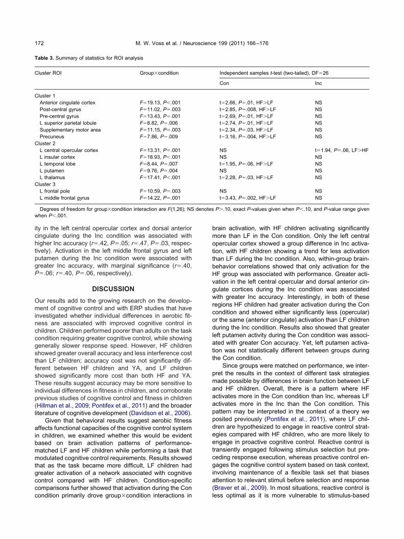
P-value is the P-value significance for the cluster.teractions, interaction analyses were followed by two-tailedindependent t-tests that compared group activation differ-ences in Con and Inc conditions. Results from this analysisare listed in Table 3 and illustrated in Fig 3; they indicatethat interactions were largely driven by activation differ-ences during the Con condition, with the exception of theleft central opercular cortex.
If there were strategy differences between fitnessgroups, we would expect different ROIs to be related todifferent aspects of behavioral performance. Thus, to ex-amine how differences in task-related brain activity may berelated to task strategy, we assessed the association be-tween regional brain activation and task performancewithin groups. For this analysis, due to small group sizes,conservative estimates of brain-behavior associationswere examined with two-tailed non-parametric Kendall’stau correlations; we report only correlations that were sig-nificant at P�.05, and results are summarized in Fig 4.Only activation for the HF group was correlated with taskperformance. Activity in the left putamen was associatedwith greater Con accuracy (r�.44, P�.04), whereas activ-
MNI coordinates (x,y,z) Z score Voxels P-value
�24, �32, 62 3.64 4895 1.18�10�11
�38, �2, �6 3.37 1406 1.71�10�4
�32, 30, 32 3.39 605 .04
rrection; R�Right, L�left brain hemisphere, MNI coordinates are forn; voxels refer to the number of standard space voxels in the cluster;
MNI coordinates (x,y,z) Z score Voxels P-value
�24, �32, 62 3.64 4895 1.18�10�11
�38, �2, �6 3.37 1406 1.71�10�4
�32, 30, 32 3.39 605 .04
rrection; R�right, L �left brain hemisphere, MNI coordinates are forn; voxels refer to the number of standard space voxels in the cluster;
st
ele,
luster-co
st
ele,
luster-coe locatio
tpgP
aibmmtgccc
bmottbHvgwrcodlatt
pmaaappdeetcgia(
M. W. Voss et al. / Neuroscience 199 (2011) 166–176172
ity in the left central opercular cortex and dorsal anteriorcingulate during the Inc condition was associated withhigher Inc accuracy (r�.42, P�.05; r�.47, P�.03, respec-ively). Activation in the left middle frontal gyrus and leftutamen during the Inc condition were associated withreater Inc accuracy, with marginal significance (r�.40,�.06; r�.40, P�.06, respectively).
DISCUSSION
Our results add to the growing research on the develop-ment of cognitive control and with ERP studies that haveinvestigated whether individual differences in aerobic fit-ness are associated with improved cognitive control inchildren. Children performed poorer than adults on the taskcondition requiring greater cognitive control, while showinggenerally slower response speed. However, HF childrenshowed greater overall accuracy and less interference costthan LF children; accuracy cost was not significantly dif-ferent between HF children and YA, and LF childrenshowed significantly more cost than both HF and YA.These results suggest accuracy may be more sensitive toindividual differences in fitness in children, and corroborateprevious studies of cognitive control and fitness in children(Hillman et al., 2009; Pontifex et al., 2011) and the broaderliterature of cognitive development (Davidson et al., 2006).
Given that behavioral results suggest aerobic fitnessffects functional capacities of the cognitive control system
n children, we examined whether this would be evidentased on brain activation patterns of performance-atched LF and HF children while performing a task thatodulated cognitive control requirements. Results showed
hat as the task became more difficult, LF children hadreater activation of a network associated with cognitiveontrol compared with HF children. Condition-specificomparisons further showed that activation during the Con
Table 3. Summary of statistics for ROI analysis
Cluster ROI Group�condition
Cluster 1Anterior cingulate cortex F�19.13, P�.001Post-central gyrus F�11.02, P�.003Pre-central gyrus F�13.43, P�.001L superior parietal lobule F�8.82, P�.006Supplementary motor area F�11.15, P�.003Precuneus F�7.86, P�.009
Cluster 2L central opercular cortex F�13.31, P�.001L insular cortex F�18.93, P�.001L temporal lobe F�8.44, P�.007L putamen F�9.76, P�.004L thalamus F�17.41, P�.001
Cluster 3L frontal pole F�10.59, P�.003L middle frontal gyrus F�14.22, P�.001
Degrees of freedom for group�condition interaction are F(1,26); NSwhen P�.001.
ondition primarily drove group�condition interactions in l
rain activation, with HF children activating significantlyore than LF in the Con condition. Only the left centralpercular cortex showed a group difference in Inc activa-ion, with HF children showing a trend for less activationhan LF during the Inc condition. Also, within-group brain-ehavior correlations showed that only activation for theF group was associated with performance. Greater acti-ation in the left central opercular and dorsal anterior cin-ulate cortices during the Inc condition was associatedith greater Inc accuracy. Interestingly, in both of these
egions HF children had greater activation during the Conondition and showed either significantly less (opercular)r the same (anterior cingulate) activation than LF childrenuring the Inc condition. Results also showed that greater
eft putamen activity during the Con condition was associ-ted with greater Con accuracy. Yet, left putamen activa-ion was not statistically different between groups duringhe Con condition.
Since groups were matched on performance, we inter-ret the results in the context of different task strategiesade possible by differences in brain function between LFnd HF children. Overall, there is a pattern where HFctivates more in the Con condition than Inc, whereas LFctivates more in the Inc than the Con condition. Thisattern may be interpreted in the context of a theory weosited previously (Pontifex et al., 2011), where LF chil-ren are hypothesized to engage in reactive control strat-gies compared with HF children, who are more likely tongage in proactive cognitive control. Reactive control isransiently engaged following stimulus selection but pre-eding response execution, whereas proactive control en-ages the cognitive control system based on task context,
nvolving maintenance of a flexible task set that biasesttention to relevant stimuli before selection and responseBraver et al., 2009). In most situations, reactive control is
Independent samples t-test (two-tailed), DF�26
Con Inc
t�2.66, P�.01, HF�LF NSt�2.85, P�.008, HF�LF NSt�2.69, P�.01, HF�LF NSt�2.74, P�.01, HF�LF NSt�2.34, P�.03, HF�LF NSt�3.16, P�.004, HF�LF NS
NS t�1.94, P�.06, LF�HFNS NSt�1.95, P�.06, HF�LF NSNS NSt�2.28, P�.03, HF�LF NS
NS NSt�3.43, P�.002, HF�LF NS
P�.10, exact P-values given when P�.10, and P-value range given
denotesess optimal as it is more vulnerable to stimulus-based

dg
dpar Web vers
M. W. Voss et al. / Neuroscience 199 (2011) 166–176 173
interference. Pontifex et al. (2011) suggested that LF chil-ren adopt a reactive control strategy by continually en-
Fig. 3. ROI task activation summary for Con and Inc condition for Lclusters that were significant for group�condition interaction (see Tabepicted in radiological orientation (R�L, L�R). ROIs for cluster onre-central gyrus, superior parietal lobule, supplementary motor area, anterior temporal lobe, putamen, and thalamus; ROIs for cluster threeeferences to color in this figure legend, the reader is referred to the
aging the anterior cingulate to try to initiate top-down
control from the prefrontal cortex. Whereas the reactivecontrol strategy may be beneficial for LF children in the
F children; Harvard Oxford anatomical ROIs derived from activation* P�.05, † P�.05 � .10; error bars represent SE of mean; all brains
e (left to right): dorsal anterior cingulate cortex, post-central gyrus,neus; ROIs for cluster two include: left central opercular cortex, insula,left frontal pole and left middle frontal gyrus. For interpretation of theion of this article.
F and Hle 2c, d);e includnd precuinclude
Con condition, they are unable to upregulate the system to

ac(c to color in
M. W. Voss et al. / Neuroscience 199 (2011) 166–176174
efficiently implement a reactive strategy in the more diffi-cult Inc condition. In contrast, HF children may adopt aproactive strategy according to increased demand for cog-nitive control in the Inc condition, resulting in an increasedcapacity for the anterior cingulate to initiate top-down con-trol from the lateral prefrontal cortex. The difference instrategy may be driven by the inability of LF children toeffectively engage the anterior prefrontal cortex to maintaina sustained task set (Fair et al., 2007), as this has beensuggested as a hypothesis for why older adults tend to alsouse a reactive versus a proactive control strategy (Paxtonet al., 2008).
In this context, activation during the Con condition mayreflect enhanced reactive control for the LF children and lackof engagement of proactive control for the HF children. Incontrast, during the Inc condition, LF children are unable toefficiently engage the reactive strategy, which results ingreater activation of the network, reflective of inefficient reg-ulation of cognitive control and, in turn, worse performance asthe task becomes more difficult. In contrast, HF childrensufficiently upregulate the proactive strategy in the more dif-ficult condition, which results in more efficient processingand, in turn, less brain activation coupled with better taskperformance. Thus, more efficient activation from HF childrenduring difficult task conditions would explain the seeminglyparadoxical effect where they activate less; yet, within thesmaller range of brain activity, more activation is associatedwith better performance. Also in line with this theoreticalaccount was the marginal association between increased leftmiddle frontal gyrus activation and Inc accuracy for HF, butnot LF children. Together, the interaction pattern and differ-ences in within-group brain-behavior correlations suggestthat HF children are utilizing a different strategy that is madepossible by more efficient cognitive control brain networks.To more directly test this hypothesis, however, future re-search could utilize an fMRI experimental design that permitsdissociating cue and target stimulus processing (Braver et al.,2009) to examine whether the timecourse of preparatory(proactive) versus reactive brain activity differs between fit-ness groups or whether strategy shifts occur following aero-
Fig. 4. Summary of associations between functional activity and perfoctivation for congruent and incongruent conditions, respectively; �Coonditions, respectively. Statistical maps are shown in neurological orientahttp://brainvis.wustl.edu); activations are significant clusters for fitnesluster-correction threshold of P�.05. For interpretation of the references
bic training.
As predicted, in this study, fitness was associated withdifferent brain activation patterns in regions associated withcognitive control processes. HF children demonstrated moreefficient activation of brain regions involved in the cingulo-opercular network. Yet, one study that has shown this brainnetwork continues to develop throughout childhood exam-ined this network during the resting state, a measure offunctional brain activity during quiet rest (Fair et al., 2007).This suggests that fitness may not only affect the extent towhich these brain regions are evoked during task demands,but that fitness may also affect the fundamental functionalarchitecture of cognitive control brain systems. The two pos-sibilities are not mutually exclusive, as the cohesion of brainnetworks during the resting state is thought to directly affecttheir active recruitment. Nevertheless, it will be important forfuture research to characterize how the resting functionalarchitecture of the developing brain is impacted by individualdifferences in fitness, and the impact of such differences ondifferences in task-related brain activity and performance.
The extent to which brain volume differences contrib-ute to our results is unknown. A recent study has shownthat HF children have greater dorsal striatum volume com-pared with LF children, which was in turn related to im-proved conflict resolution (Chaddock et al., 2010). How-ever, in this study, although HF children had greater vol-ume of the left putamen, volume in this region was notassociated with Con accuracy. Thus, it is unlikely thatvolumetric differences played a role in the left putamenfunctional activation differences in the current study. How-ever, it will be important for future research to integratehow individual differences in developmental structural andfunctional plasticity interact to affect performance in chil-dren. Another limitation of this study is the cross-sectionaldesign, and it is, therefore, possible that genetic or lifestylefactors other than aerobic fitness level contributed to ourresults. To address this concern, information about avera-ge hours of television viewing on weekday and weekend,average computer hours on weekday and weekend, avera-ge hours of videogame play on weekday and weekend,and socioeconomic status was collected. However, LF and
LF�lower-fit, HF�higher-fit; �Con act. and �Inc act. indicate greaterd �Inc acc indicate greater accuracy for congruent and incongruentand L�L), surface visualization is on the PALS-B12 atlas using CARET
on interaction (see Table 2c, d), at voxel threshold of Z�2.33, andthis figure legend, the reader is referred to the Web version of this article.
rmance;n acc antion (R�R
s�conditi
HF groups did not differ on any of these variables (all
M. W. Voss et al. / Neuroscience 199 (2011) 166–176 175
P�.05 from two-tailed independent t-test). Yet, it is impor-tant to point out that replication of these results in a longi-tudinal, randomized controlled exercise training studywould greatly increase the strength of the conclusions fromthe current study.
This is the first study to use fMRI to examine how thedeveloping functional brain is affected by individual differ-ences in aerobic fitness in children who vary on aerobicfitness but who are not overweight. For example, while arecent study showed that aerobic fitness training resultedin increased prefrontal cortex activation and decreased pos-terior parietal cortex activation during a task requiring cogni-tive control, activation specific to cognitive control was notisolated and participants were only included in the study ifthey were overweight based on their body mass index (Daviset al., 2011). We, therefore, provide novel evidence for therole of aerobic fitness in facilitating greater efficiency of cog-nitive control networks in children. Our results also add toprevious ERP research by offering insight into their spatialrealization in the functioning brain. Additionally, our resultsraise important issues for future study such as whether event-related fMRI activity consistent with different cognitive controlstrategies would differentially emerge in LF and HF childrenand to what extent differences in resting state functionalconnectivity of cognitive control networks would contribute tofitness differences in performance and task-related brain ac-tivation patterns.
Acknowledgments—We would like to thank Nancy Dodge, HollyTracy, and Luke Tseng for their help in data collection. Support forour research was provided by grants from the National Institute onAging (R37 AG25667 and RO1 AG25302) to Arthur Kramer andthe National Institute of Child Health and Human Development(RO1 HD055352) to Charles Hillman.
REFERENCES
Barnes KA, Cohen AL, Power JD, Nelson SM, Dosenbach YB, MiezinFM, Petersen SE, Schlaggar BL (2010) Identifying basal gangliadivisions in individuals using resting-state functional connectivityMRI. Front Syst Neurosci 4:18.
Bar-Or O (1983) Pediatric sports medicine for the practitioner:from physiologic principles to clinical applications, p 376. NewYork: Springer-Verlag.
Beckmann CF, Jenkinson M, Smith SM (2003) General multilevellinear modeling for group analysis in FMRI. Neuroimage 20:1052–1063.
Botvinick M, Nystrom LE, Fissell K, Carter CS, Cohen JD (1999)Conflict monitoring versus selection-for-action in anterior cingulatecortex. Nature 402:179–181.
Botvinick MM, Braver TS, Barch DM, Carter CS, Cohen JD (2001)Conflict monitoring and cognitive control. Psychol Review 108:624–652.
Braver TS, Barch DM (2006) Extracting core components of cognitivecontrol. Trends Cogn Sci 10:529–532.
Braver TS, Paxton JL, Locke HS, Barch DM (2009) Flexible neuralmechanisms of cognitive control within human prefrontal cortex.Proc Natl Acad Sci U S A 106:7351–7356.
Bunge SA, Dudukovic NM, Thomason ME, Vaidya CJ, Gabrieli JD(2002) Immature frontal lobe contributions to cognitive control inchildren: evidence from fMRI. Neuron 33:301–311.
Casey BJ, Giedd JN, Thomas KM (2000a) Structural and functionalbrain development and its relation to cognitive development. Biol
Psychol 54:241–257.Casey BJ, Thomas KM, Welsh TF, Badgaiyan RD, Eccard CH, Jen-nings JR, Crone EA (2000b)Dissociation of response conflict, at-tentional selection, and expectancy with functional magnetic reso-nance imaging. Proc Natl Acad Sci U S A 97:8728–8733.
Castelli DM, Hillman CH, Buck SM, Erwin HE (2007) Physical fitnessand academic achievement in third- and fifth-grade students.J Sport Exeric Psychol 29:239–252.
Chaddock L, Erickson KI, Prakash RS, Vanpatter M, Voss MW, Pon-tifex MB, Raine LB, Hillman CH, Kramer AF (2010) Basal gangliavolume is associated with aerobic fitness in preadolescent chil-dren. Dev Neurosci 32:249–256.
Chomitz VR, Slining MM, McGowan RJ, Mitchell SE, Dawson GF,Hacker KA (2009) Is there a relationship between physical fitnessand academic achievement? Positive results from public schoolchildren in the northeastern United States. J Sch Health 79:30–37.
Church JA, Petersen SE, Schlaggar BL (2010) The “task B problem”and other considerations in developmental functional neuroimag-ing. Hum Brain Mapp 31:852–862.
Colcombe SJ, Kramer AF, Erickson KI, Scalf P, McAuley E, Cohen NJ,Webb A, Jerome GJ, Marquez DX, Elavsky S (2004) Cardiovas-cular fitness, cortical plasticity, and aging. Proc Natl Acad Sci U S A101:3316–3321.
Congdon E, Mumford JA, Cohen JR, Galvan A, Aron AR, Xue G, MillerE, Poldrack RA (2010) Engagement of large-scale networks isrelated to individual differences in inhibitory control. Neuroimage53:653–663.
Davidson MC, Amso D, Anderson LC, Diamond A (2006) Develop-ment of cognitive control and executive functions from 4 to 13years: evidence from manipulations of memory, inhibition, and taskswitching. Neuropsychologia 44:2037–2078.
Davis CL, Tomporowski PD, McDowell JE, Austin BP, Miller PH,Yanasak NE, Allison JD, Naglieri JA (2011) Exercise improvesexecutive function and achievement and alters brain activation inoverweight children: a randomized, controlled trial. Health Psychol30:91–98.
Dosenbach NU, Fair DA, Cohen AL, Schlaggar BL, Petersen SE(2008) A dual-networks architecture of top-down control. TrendsCogn Sci 12:99–105.
Dosenbach NU, Fair DA, Miezin FM, Cohen AL, Wenger KK, Dosen-bach RA, Fox MD, Snyder AZ, Vincent JL, Raichle ME, SchlaggarBL, Petersen SE (2007) Distinct brain networks for adaptive andstable task control in humans. Proc Natl Acad Sci U S A 104:11073–11078.
Eriksen CW, Eriksen BA (1974) Effects of noise letters upon theidentification of a target letter in a non-search task. Percept Psy-chophys 16:143–149.
Fair DA, Dosenbach NU, Church JA, Cohen AL, Brahmbhatt S, MiezinFM, Barch DM, Raichle ME, Petersen SE, Schlaggar BL (2007)Development of distinct control networks through segregation andintegration. Proc Natl Acad Sci U S A 104:13507–13512.
Forster SE, Brown JW (2011) Medial prefrontal cortex predicts andevaluates the timing of action outcomes. NeuroImage 55:253–265.
Freedson PS, Goodman TL (1993) Measurement of oxygen consump-tion. In: Pediatric laboratory exercise testing: clinical guidelines(Rowland TW, ed), pp 91–113. Champaign, IL: Human Kinetics.
Gehring WJ, Knight RT (2000) Prefrontal-cingulate interactions inaction monitoring. Nat Neurosci 3:516–520.
Hazeltine E, Poldrack R, Gabrieli JD (2000) Neural activation duringresponse competition. J Cogn Neurosci 12(Suppl 2):118–129.
Hillman CH, Buck SM, Themanson JR, Pontifex MB, Castelli DM(2009) Aerobic fitness and cognitive development: event-relatedbrain potential and task performance indices of executive control inpreadolescent children. Dev Psychol 45:114–129.
Hillman CH, Castelli DM, Buck SM (2005) Aerobic fitness and neuro-cognitive function in healthy preadolescent children. Med Sci
Sports Exerc 37:1967–1974.
M. W. Voss et al. / Neuroscience 199 (2011) 166–176176
Jenkinson M, Bannister P, Brady M, Smith S (2002) Improved optimi-zation for the robust and accurate linear registration and motioncorrection of brain images. Neuroimage 17:825–841.
Kaufman AS, Kaufman NL (1990) Kaufman brief intelligence testmanual. Circle Pines, MN: American Guidance Service.
Oldfield RC (1971) The assessment and analysis of handedness: theEdinburgh inventory. Neuropsychologia 9:97–113.
Paxton JL, Barch DM, Racine CA, Braver TS (2008) Cognitive control,goal maintenance, and prefrontal function in healthy aging. CerebCortex 18:1010–1028.
Pontifex MB, Raine LB, Johnson CR, Chaddock L, Voss MW, CohenNJ, Kramer AF, Hillman CH (2011) Cardiorespiratory fitness andthe flexible modulation of cognitive control in preadolescent chil-
dren. J Cogn Neurosci 23:1332–1345.Shvartz E, Reibold RC (1990) Aerobic fitness norms for males andfemales aged 6-75: A review. Aviat Space Environ Med 61:3–11.
Smith SM (2002) Fast robust automated brain extraction. Hum BrainMapp 17:143–155.
Taylor SJ, Whincup PH, Hindmarsh PC, Lampe F, Odoki K, CookDG (2001) Performance of a new pubertal self-assessmentquestionnaire: a preliminary study. Paediatr Perinat Epidemiol15:88 –94.
Utter AC, Robertson RJ, Nieman DC, Kang J (2002) Children’s OMNIscale of perceived exertion: walking/running evaluation. Med SciSports Exerc 34:139–144.
Whaley MH, Brubaker PH, Otto RM (2006) ACSM’s Guidelines forexercise testing and prescription, 7th ed, pp 93–114. New York,
NY: Lippincott Williams & Wilkins.(Accepted 6 October 2011)(Available online 14 October 2011)