advancing parental age and risk of solid tumors in...
TRANSCRIPT

Research ArticleAdvancing Parental Age and Risk of Solid Tumors in Children:A Case-Control Study in Peru
Ligia Rios , Liliana Vásquez , Mónica Oscanoa, IvánMaza , and Jenny Gerónimo
Pediatric Oncology Unit, Edgardo Rebagliati Martins Hospital, 490 Domingo Cueto Avenue, Lima 11, Peru
Correspondence should be addressed to Ligia Rios; [email protected]
Received 20 November 2017; Revised 6 March 2018; Accepted 20 May 2018; Published 12 June 2018
Academic Editor: Thomas R. Chauncey
Copyright © 2018 Ligia Rios et al.This is an open access article distributed under theCreative CommonsAttribution License, whichpermits unrestricted use, distribution, and reproduction in any medium, provided the original work is properly cited.
Background. The causes of childhood cancer are not well known, but the advanced age of the parents has been suggested as a riskfactor for childhood cancer in several observational studies. In this study, we examine a possible link between parental age andchildhood solid tumors. Methods. We conducted a hospital-based case-control study (310 cases and 620 controls, matched by ageand gender) at Rebagliati Hospital, Lima, Peru. Odd ratio was used to compare categories of advancing maternal and paternal agewith and without adjusting for possible confounding factors were calculated. Results. The risk of childhood retinoblastoma wassignificantly higher among children of mothers aged> 35 years (adjusted OR 1.21; 95% CI, 1.09-6.08) and fathers aged> 35 years(OR 1.17; 1.01-16.33). A significant trend with increasing mother's age (p = 0.037) and father's age (p = 0.005) was found. Therewere more risks to development of non-Hodgkin's lymphoma (p = 0.047) and gonadal germ cell tumors (p = 0.04) for advancedpaternal age. There was a strong protective effect of increasing parity on risk of solid tumors in children (p=0.0015). Conclusion.Our results suggest that advanced parental age is associated with the risk for the development of retinoblastoma. Advanced paternalage increases the risk of non-Hodgkin lymphoma and gonadal germ cell tumor. The higher the order of birth of the children, theless the chance of developing any neoplasm.
1. Introduction
Although the childhood cancer is a rare disease, during thelast years, appearance of children’s cancer had a higher inci-dence rate, affecting approximately one in 435 children underage 15 years [1]. Several associated factors have been describedin its development, like biological aspects (chromoso-mal anomalies, immunological alterations), environmentalaspects (exposure to radiation, viral infections, socioeco-nomic status, and parental occupation), maternal aspects(breastfeeding, the mother’s consumption levels of alcohol ortobacco, nutritional supplements [2–4]), and familial features(family history of cancer, advance maternal and/or paternalage, and number of previous siblings); however the etiologyfor the most of childhood cancer is still unknown.
Family structures have changed in relation to past gen-erations since nowadays, when maternity/paternity occurs,advancing maternal or paternal age and a lower number ofchildren are also more likely to occur. Additionally, advancedmaternal age has been positively linked with higher risk of
having a child with Down syndrome, among other congenitaldisorders, which in turn has a higher incidence of acutemyeloid and lymphoid leukaemia [5]. Advanced paternal age(as a factor independent of maternal age) could mean risk ofdisorders associated germ cell mutation [6].
The link between advanced maternal and paternal ageand a higher incidence of the appearance of children’s cancerhas been revealed in studies conducted among patients withleukaemia [7–9], lymphomas, brain tumors [10], germ celltumors, and malignant neoplasms in general [11–13] eventhough, in many other studies, such link has not beencorroborated with consistent results [14–18]. Likewise, somestudies show an increase in the risk of childhood cancer in thefirst child [13, 14, 19] although, in many others, contradictoryfindings have been made [7, 20].
To date, it has not been made clear if advanced parentalage is linked with higher risk of children’s cancer; hence, thepresent study aims to determine whether advancing parentalage is associated with an increased risk of childhood cancerin offspring.
HindawiJournal of OncologyVolume 2018, Article ID 3924635, 9 pageshttps://doi.org/10.1155/2018/3924635
2 Journal of Oncology
2. Materials and Methodology
2.1. Participants and Recruitment. A case-control study basedon hospital records (Rebagliati Hospital, Lima, Peru) wasconducted. Both, cases and controls, included children andadolescents younger than 18. For every case, 2 age-and sex-comparable controls were assigned (in the case of the age-comparable control, the margin was +/- 6 months).
Cases had an anatomical pathological diagnosis ofHodgkin and Non-Hodgkin lymphoma, brain tumors, germcell tumors, and any solid tumors, between the years 2012and 2015. Controls were children who were hospitalizedfor nononcological diseases (ICD: A09, B01.9, G40.9, K35.9,J18.0, J21.9, J45.9, L03.9, S52, andT29) during the sameperiod.
Data collection was conducted via questionnaires toparents about their medical histories and their families’medical histories, together with a revision of every patient orcontrol patient’s medical record.
2.2. Statistical Analysis. The data was analysed using con-ditional logistic regression for studies involving the casesand controls. Relative risks were estimated by odd ratios(OR) with a 95% confidence interval (CI) with a primaryanalysis between the link between maternal and paternalage as categorical variable according to age group (<20, 20-24, 25-29, 30-35, and >35) and as quantitative variable andchildhood cancer diagnosis. We established raw OR and ORadjusted to confounding variables; these were chosen basedon their prior observed association with childhood cancers[11–14]: paternal and maternal education (elementary, highschool, and higher education), birth order of siblings in thefamily, and origin. Unfortunately due to missing data incontrol group, it was not possible to include birth weightand prematurity as confounding variables. Data was analysedwith STATA statistical package (Small STATA Version 13.0,STATA Corporation, College Station, Texas, USA).
2.3. Ethical Aspects. This study has been evaluated andapproved by the Ethics Commission of our institution priorto project execution. All clinical and sociodemographic datacomply with confidentiality norms to protect the identity ofpatients.
3. Results
Between 2012 and 2015, 310 cases of childhood cancer (solidtumors and lymphomas) were identified; we compared thosecases with 620 controls, matched according to age (with themargin of six months), sex, and geographical area of origin(Table 1).
The presence of childhood cancer was slightly higheramong males. Parental age was grouped into a 5-year group.Themain age of casemothers at birthwas slightly older (28.92[SD=6.30]) than control mothers (28.47[6.16]); contrarypattern was observed with the main age of fathers, slightlyyounger in cases (31.74 [6.82]) than controls (32.37 [7.82]).The only statistically significant difference between controlsand cases was in the order of birth, since the difference
observed in the level of education of the father is influenceby the number of lost data.
Table 2 shows the distribution of solid tumors andlymphomas in the cases. Brain tumors (20%), non-Hodgkinlymphoma (13.87%), osteosarcoma (13.55%), and Wilms'tumor (11.29%) were the most frequent cancers. It shouldbe mentioned that in our study Wilms tumor presented ahigher prevalence than neuroblastoma (WT 11% versus NB4%), unlike those occurring in North America and Europe,but similar to that observed in other studies in Latin America[21].
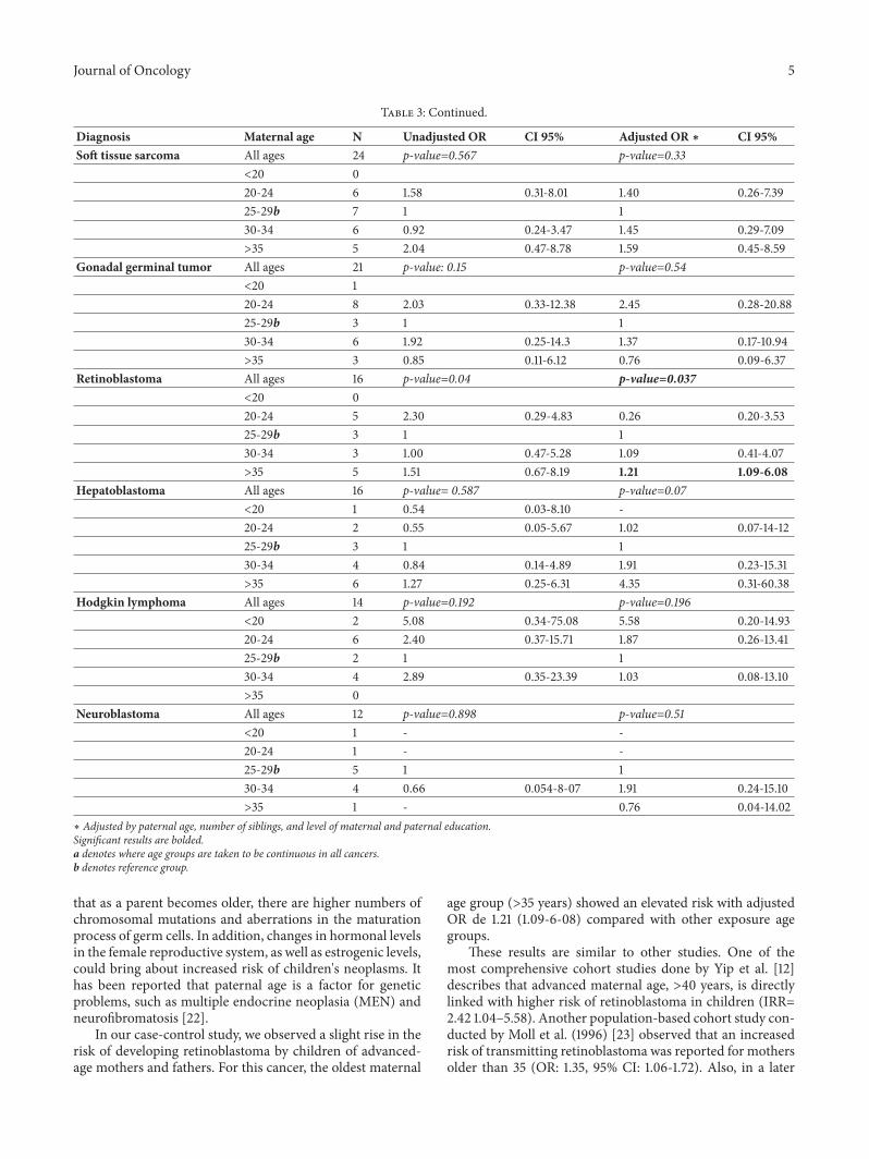
In our case-control study, maternal age shows posi-tive linear trends (age groups are taken to be continuous)for 2 childhood cancer groups: osteosarcoma (p=0.019)and retinoblastoma (p=0.04); however after adjustment forcovariates (paternal age, number of siblings, and level ofparental education), maternal age only indicated a positivelinear trend for retinoblastoma (p=0.037). In addition, therisk of development retinoblastoma was significantly higheramong children of mothers older than 35 years old (adjustedOR: 1.21; 95% CI: 1.9-6.8); such association did not occur inother neoplasms. (Table 3).
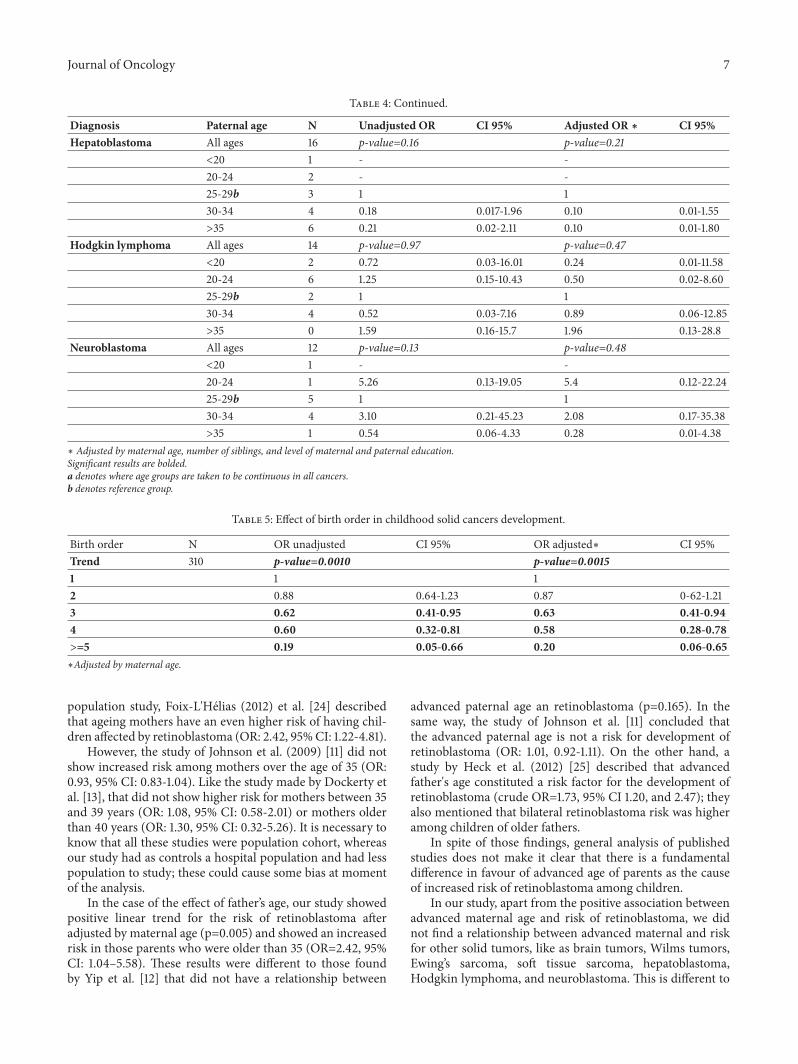
Similarly, paternal age demonstrated positive lineartrends (age groups are taken to be continuous) for 2 child-hood cancer groups: gonadal germinal tumor (p=0.03) andretinoblastoma (p=0.03). After adjustment for covariates(maternal age, number of siblings, and level of parentaleducation), paternal age continued showing a positive lin-ear trend for non-Hodgkin lymphoma (p=0.047), gonadalgerminal tumor (p=0.04), and retinoblastoma (p=0.037).Additionally, the risk of development retinoblastoma wassignificantly higher among children of fathers older than 35years old (adjusted OR: 1.17; 95% CI: 1.0-16.33). (Table 4).
Furthermore, a protective effect against risk of solidtumors when parity increased (p=0.0015) was found. It wasalso observed that there was a significant decrease in the riskof children's neoplasms as the number of siblings in a familyincreased; there is a protective effect from the third child (ORadjusted: 0.63, 0.41-0.94). We decided to do the analysis ofall the neoplasias together, due to having few cases to do aseparated analysis. (Table 5).
4. Discussion
This work represents the first published study focused onadvanced parental age (maternal or paternal) as a risk factorassociated with the appearance of solid tumors in children inPeru.
Advanced parental age, considered in some studies asolder than 35 or 40 years old, has been frequently linkedwith higher risk of children's malignant neoplasms [7–13],with results in other studies being contradictory [14–17].Incidence rate of paediatric cancer is on the rise worldwide;likewise, advanced-age parity between mothers and fathershas become more common. The mechanism causing cancerin children of advanced-age mothers or fathers has not beenclearly described. Reproductive age could affect risk of cancerin children through several processes, especially considering

Journal of Oncology 3
Table 1: Characteristics of children diagnosed with cancer and controls (2012-2015).
Characteristic Cases Controls P valueN % N %
Age, years 0.119<1 16 5.2 42 6.801-04 83 26.8 145 23.405-09 76 24.5 167 26.910-14 87 28.1 171 27.615-18 48 15.5 95 15.3
Sex 0.963Male 175 56.5 349 56.3Female 135 43.6 271 43.7
Origin 0.349Coast 233 75.2 485 78.2Andean 59 19.0 95 15.3Forest 18 5.8 40 6.5
Maternal age at birth, years 0.239<20 17 5.5 36 5.820-24 88 28.4 138 22.325-29 72 23.2 194 31.330-34 81 26.1 135 21.8>35 52 16.8 117 18.9Mean (SD) 28.92 (6.30) 28.47 (6.16)Missing 1 8
Paternal age at birth, years 0.520<20 4 1.3 15 2.820-24 40 12.9 67 12.625-29 85 27.5 128 24.130-34 81 26.2 122 23.0>35 99 32.0 199 37.5Mean (SD) 31.74 (6.82) 32.37 (7.82)Missing 1 89
Education level of the mother 0.801Elementary school 26 8.5 22 9.6High school 116 37.8 90 39.3Higher education 165 53.8 117 51.1Missing 3 391
Education level of the father 0.039Elementary school 9 2.9 14 6.2High school 100 32.7 87 38.5Higher education 197 64.4 125 55.3Missing 4 394
Birth order 0.0021 188 60.8 354 57.12 84 27.2 175 28.23 26 8.4 63 10.24 10 3.2 15 2.4>=5 1 0.3 13 2.1Missing 1 0
Twins 0.090Yes 2 0.6 7 1.1No 308 99.4 613 98.9
TOTAL 310 100.0 620 100.0

4 Journal of Oncology
Table 2: Distribution of solid tumors and lymphomas.
Characteristic CasesN %
DiagnosisBrain tumor 62 20.0Non-Hodgkin lymphoma 43 13.9Osteosarcoma 42 13.6Wilms tumor 35 11.3Ewing's sarcoma 24 7.7Soft tissue sarcoma 24 7.7Gonadal germinal tumor 21 6.8Retinoblastoma 16 5.2Hepatoblastoma 16 5.2Hodgkin lymphoma 14 4.5Neuroblastoma 12 3.9Extragonadal germinal tumor 1 0.3
TOTAL 310 100.0
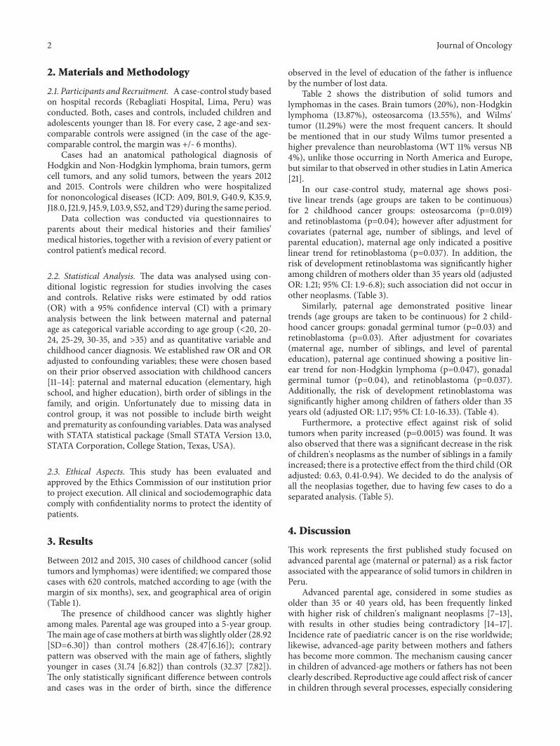
Table 3: Effect of maternal age in childhood solid cancers development.
Diagnosis Maternal age N Unadjusted OR CI 95% Adjusted OR ∗ CI 95%Brain tumor All ages 62 p-value=0.99a p-value=0.97
<20 5 1.06 0.32-3.46 1.06 0.71-1.5520-24 16 2.06 0.80-5.29 1.92 0.27-4.1625-29b 14 1 130-34 18 2.10 0.83-5.27 1.42 0.53-3.08>35 9 1.26 0.44-3.55 0.88 0.26-2.97
Non-Hodgkin lymphoma All ages 43 p-value=0.56 p-value=0.80<20 1 0.33 0.03-3.08 0.45 0.34-4.5020-24 11 1.29 0.47-3.51 1.53 0.54-4.3025-29b 13 1 130-34 13 1.69 0.63-4.48 1.61 0.50-4.57>35 5 1.00 0.29-3-37 0.91 0.22-3.81
Osteosarcoma All ages 42 p-value=0.019 p-value=0.054<20 3 0.85 0.34-2.56 0.60 0.40-3.0920-24 16 0.70 0.24-2.04 0.58 0.16-2.0825-29b 10 1 130-34 8 1.38 0.52-3.64 0.99 0.62-2.30>35 5 1.91 1.12-3.09 1.35 0.81-2.33
Wilms tumor All ages 35 p-value=0.954 p-value=0.346<20 1 - -20-24 10 2.47 0.73-8.39 2.76 0.75-10.1725-29b 5 1 130-34 9 3.71 0.96-14.30 3.18 0.73-13.83>35 10 1.52 0.43-5.39 0.91 0.22-3.73
Ewing’s sarcoma All ages 24 p-value=0.713 p-value=0.543<20 2 0.78 0.11-5.26 0.65 0.07-5.3520-24 7 1.24 0.32-4.76 1.51 0.31-7.3725-29b 7 1 130-34 3 0.35 0-07-1.60 2.73 0.51-1.33>35 5 1.46 0.26-8.11 1.05 0.18-6.06
Journal of Oncology 5
Table 3: Continued.
Diagnosis Maternal age N Unadjusted OR CI 95% Adjusted OR ∗ CI 95%So� tissue sarcoma All ages 24 p-value=0.567 p-value=0.33
<20 020-24 6 1.58 0.31-8.01 1.40 0.26-7.3925-29b 7 1 130-34 6 0.92 0.24-3.47 1.45 0.29-7.09>35 5 2.04 0.47-8.78 1.59 0.45-8.59
Gonadal germinal tumor All ages 21 p-value: 0.15 p-value=0.54<20 120-24 8 2.03 0.33-12.38 2.45 0.28-20.8825-29b 3 1 130-34 6 1.92 0.25-14.3 1.37 0.17-10.94>35 3 0.85 0.11-6.12 0.76 0.09-6.37
Retinoblastoma All ages 16 p-value=0.04 p-value=0.037<20 020-24 5 2.30 0.29-4.83 0.26 0.20-3.5325-29b 3 1 130-34 3 1.00 0.47-5.28 1.09 0.41-4.07>35 5 1.51 0.67-8.19 1.21 1.09-6.08
Hepatoblastoma All ages 16 p-value= 0.587 p-value=0.07<20 1 0.54 0.03-8.10 -20-24 2 0.55 0.05-5.67 1.02 0.07-14-1225-29b 3 1 130-34 4 0.84 0.14-4.89 1.91 0.23-15.31>35 6 1.27 0.25-6.31 4.35 0.31-60.38
Hodgkin lymphoma All ages 14 p-value=0.192 p-value=0.196<20 2 5.08 0.34-75.08 5.58 0.20-14.9320-24 6 2.40 0.37-15.71 1.87 0.26-13.4125-29b 2 1 130-34 4 2.89 0.35-23.39 1.03 0.08-13.10>35 0
Neuroblastoma All ages 12 p-value=0.898 p-value=0.51<20 1 - -20-24 1 - -25-29b 5 1 130-34 4 0.66 0.054-8-07 1.91 0.24-15.10>35 1 - 0.76 0.04-14.02
∗ Adjusted by paternal age, number of siblings, and level of maternal and paternal education.Significant results are bolded.a denotes where age groups are taken to be continuous in all cancers.b denotes reference group.
that as a parent becomes older, there are higher numbers ofchromosomal mutations and aberrations in the maturationprocess of germ cells. In addition, changes in hormonal levelsin the female reproductive system, as well as estrogenic levels,could bring about increased risk of children's neoplasms. Ithas been reported that paternal age is a factor for geneticproblems, such as multiple endocrine neoplasia (MEN) andneurofibromatosis [22].
In our case-control study, we observed a slight rise in therisk of developing retinoblastoma by children of advanced-age mothers and fathers. For this cancer, the oldest maternal
age group (>35 years) showed an elevated risk with adjustedOR de 1.21 (1.09-6-08) compared with other exposure agegroups.
These results are similar to other studies. One of themost comprehensive cohort studies done by Yip et al. [12]describes that advanced maternal age, >40 years, is directlylinked with higher risk of retinoblastoma in children (IRR=2.42 1.04–5.58). Another population-based cohort study con-ducted by Moll et al. (1996) [23] observed that an increasedrisk of transmitting retinoblastoma was reported for mothersolder than 35 (OR: 1.35, 95% CI: 1.06-1.72). Also, in a later

6 Journal of Oncology
Table 4: Effect of paternal age in childhood solid cancers development.
Diagnosis Paternal age N Unadjusted OR CI 95% Adjusted OR ∗ CI 95%Brain tumor All ages 62 p-value=0.94a p-value=0.98
<20 5 0.44 0.02-4.57 0.33 0.02-3.8920-24 16 1.12 0-42-3.01 1.01 0.36-2.8325-29b 14 1 130-34 18 0.90 0.33-2.4 0.91 0.30-2.73>35 9 0.90 0.36-2.19 0.99 0.34-2.85
Non-Hodgkin lymphoma All ages 43 p-value=0.09 p-value=0.047<20 1 - -20-24 11 0.24 0.06-0.96 0.19 0.03-0.9725-29b 13 1 130-34 13 0.54 0.17-1.73 0.37 0.09-1.43>35 5 0.48 0.17-1.34 0.36 0.82-1.64
Osteosarcoma All ages 42 p-value=0.10 p-value=0.23<20 3 - -20-24 16 5.70 0.98-33.0 3.10 0.94-8.625-29b 10 1 130-34 8 1.80 0.55-5.24 1.15 0.32-4.05>35 5 1.35 0.46-3.97 0.92 0.27-3.1
Wilms tumor All ages 35 p-value=0.31 p-value=0.39<20 1 - -20-24 10 0.41 0.06-2.61 0.36 0.05-2.5125-29b 5 1 130-34 9 1.05 0.26-4.17 1.30 0.30-5.59>35 10 1.43 0.42-4.92 2.06 0.41-10.16
Ewing’s sarcoma All ages 24 p-value=0.36 p-value=0.52<20 2 - -20-24 7 1.01 0.14-7.12 1.00 0.13-7.2225-29b 7 1 130-34 3 0.89 0.24-3.23 0.66 0.13-3.37>35 5 0.38 0.07-1.88 0.25 0.03-2.01
So� tissue sarcoma All ages 24 p-value=0.43 p-value=0.38<20 0 - -20-24 6 0.33 0.28-4.11 0.91 0.4-18.325-29b 7 1 130-34 6 0.48 0.11-2.02 0.76 0.13-4.3>35 5 0.42 0.12-1.45 0.25 0.02-1.86
Gonadal germinal tumor All ages 21 p-value=0.03 p-value=0.04<20 1 - -20-24 8 2.74 0-35-20.41 2.09 0.16-26.0325-29b 3 1 130-34 6 1.52 1.29-19.4 5.71 1.76-14.41>35 3 1.37 0.22-8.53 5.82 0.32-10.36
Retinoblastoma All ages 16 p-value=0-03 p-value=0.005<20 020-24 5 1.96 0.39-4.34 3.37 0.24-4.8125-29b 3 1 130-34 3 2.11 0.28-15.75 1.37 0.59-12.19>35 5 2.10 1.21-15.04 1.17 1.01-16.33
Journal of Oncology 7
Table 4: Continued.
Diagnosis Paternal age N Unadjusted OR CI 95% Adjusted OR ∗ CI 95%Hepatoblastoma All ages 16 p-value=0.16 p-value=0.21
<20 1 - -20-24 2 - -25-29b 3 1 130-34 4 0.18 0.017-1.96 0.10 0.01-1.55>35 6 0.21 0.02-2.11 0.10 0.01-1.80
Hodgkin lymphoma All ages 14 p-value=0.97 p-value=0.47<20 2 0.72 0.03-16.01 0.24 0.01-11.5820-24 6 1.25 0.15-10.43 0.50 0.02-8.6025-29b 2 1 130-34 4 0.52 0.03-7.16 0.89 0.06-12.85>35 0 1.59 0.16-15.7 1.96 0.13-28.8
Neuroblastoma All ages 12 p-value=0.13 p-value=0.48<20 1 - -20-24 1 5.26 0.13-19.05 5.4 0.12-22.2425-29b 5 1 130-34 4 3.10 0.21-45.23 2.08 0.17-35.38>35 1 0.54 0.06-4.33 0.28 0.01-4.38
∗ Adjusted by maternal age, number of siblings, and level of maternal and paternal education.Significant results are bolded.a denotes where age groups are taken to be continuous in all cancers.b denotes reference group.
Table 5: Effect of birth order in childhood solid cancers development.
Birth order N OR unadjusted CI 95% OR adjusted∗ CI 95%Trend 310 p-value=0.0010 p-value=0.00151 1 12 0.88 0.64-1.23 0.87 0-62-1.213 0.62 0.41-0.95 0.63 0.41-0.944 0.60 0.32-0.81 0.58 0.28-0.78>=5 0.19 0.05-0.66 0.20 0.06-0.65∗Adjusted by maternal age.
population study, Foix-L'Helias (2012) et al. [24] describedthat ageing mothers have an even higher risk of having chil-dren affected by retinoblastoma (OR: 2.42, 95%CI: 1.22-4.81).
However, the study of Johnson et al. (2009) [11] did notshow increased risk among mothers over the age of 35 (OR:0.93, 95% CI: 0.83-1.04). Like the study made by Dockerty etal. [13], that did not show higher risk for mothers between 35and 39 years (OR: 1.08, 95% CI: 0.58-2.01) or mothers olderthan 40 years (OR: 1.30, 95% CI: 0.32-5.26). It is necessary toknow that all these studies were population cohort, whereasour study had as controls a hospital population and had lesspopulation to study; these could cause some bias at momentof the analysis.
In the case of the effect of father’s age, our study showedpositive linear trend for the risk of retinoblastoma afteradjusted by maternal age (p=0.005) and showed an increasedrisk in those parents who were older than 35 (OR=2.42, 95%CI: 1.04–5.58). These results were different to those foundby Yip et al. [12] that did not have a relationship between
advanced paternal age an retinoblastoma (p=0.165). In thesame way, the study of Johnson et al. [11] concluded thatthe advanced paternal age is not a risk for development ofretinoblastoma (OR: 1.01, 0.92-1.11). On the other hand, astudy by Heck et al. (2012) [25] described that advancedfather's age constituted a risk factor for the development ofretinoblastoma (crude OR=1.73, 95% CI 1.20, and 2.47); theyalso mentioned that bilateral retinoblastoma risk was higheramong children of older fathers.
In spite of those findings, general analysis of publishedstudies does not make it clear that there is a fundamentaldifference in favour of advanced age of parents as the causeof increased risk of retinoblastoma among children.
In our study, apart from the positive association betweenadvanced maternal age and risk of retinoblastoma, we didnot find a relationship between advanced maternal and riskfor other solid tumors, like as brain tumors, Wilms tumors,Ewing’s sarcoma, soft tissue sarcoma, hepatoblastoma,Hodgkin lymphoma, and neuroblastoma. This is different to
8 Journal of Oncology
the report of Johnson et al. [11], who found positive lineartrends for childhood cancer groups: lymphoma (1.06 perfive-year increase [95% CI =1.01–1.12]), central nervoussystem tumors (1.07 [1.00–1.10]), neuroblastoma (1.09 [1.04–1.15]), Wilms tumor (1.16 [1.09–1.22]), bone tumors (1.10[1.00–1.20]), and soft tissue sarcomas (1.10 [1.04–1.17]). Inmodels that adjusted to paternal age and other covariates,maternal age remained associated with childhood cancersoverall, central nervous system tumors, neuroblastoma,Wilms tumor, and soft tissue sarcomas. Similar to ourwork, in other studies, there is no relationship between theparental age and risk of hepatoblastoma [11, 26, 27]. On theother hand, in our study we observed an increased risk ofnon-Hodgkin lymphoma and advanced paternal age (p =0.047), unlike that described by Yip [12] et al., who did notfind a significant association. In addition, we found a positivelinear trend for the risk of gonadal germ tumor, after beingadjusted by maternal age (p=0.005).
Interestingly, our study showed that single children hada higher risk of developing childhood cancer than childrenwho had siblings. That is, the greater the order of birth of achild, the lower the risk of cancer (p=0.0015), and we couldsee a protective effect from the third child (OR adjusted: 0.63,0.41-0.94), which continued for the fourth (OR: 0.58, 0.28-0.78) and fifth child (OR=0.20, 0.06-0.65).These results weresimilar to those reported by Von Behren et al. (2011) [21], whofound that, for all combined cancers, there was a protectiveeffect from the third child (OR=0.90, 0.85-0.96) and thatit continued with the fourth child (OR=0.87, 0.81-0.93).Another study showed a rise in the risk of childhood cancer inthe first-born child [19]; however, many other studies presentcontradictory findings [7, 12, 18, 20]. This information iscrucial, since it could generate a main hypothesis in futurestudies.
Due to its observational nature, this study presentsstrengths and weaknesses. One of its main strengths is thatit is the first study of its kind conducted in our countryand region. Its main weakness lies in the number of casesincluded in the study, because these pathologies representdiseases, which are relatively infrequent. Further limitationsalso include employing hospital-type controls and missingdata in some variables; therefore, the results obtained inthis study cannot be generalized to the Peruvian populationand are only useful in the hospital setting in which thestudy was carried out. Nowadays in Peru, it is virtuallyimpossible to apply population studies into children's cancerbecause Peruvian hospitals do not have registration systemsof optimal quality.
5. Conclusions
Our study suggests that advanced parental (maternal andpaternal) age, ≥ 35 years old, increases the risk of devel-oping retinoblastoma in children; at advanced paternal ageincreases the risk of developing non-Hodgkin lymphoma andgonadal germ cell tumors; and finally, this study observedthat the higher the order of birth of the children, the lesschance of developing any neoplasm. Additional studies with
larger population-based samples are needed to confirm ourhypothesis.
Disclosure
The results of this study were previously presented like partof the abstract “Advancing Parental Age and Risk of SolidTumors in Children: Evidence from a Case-Control Studyand a Meta-Analysis of Epidemiological Studies,” in the 48thCongress of the International Society of Paediatric Oncology(SIOP) 2016 [28].
Conflicts of Interest
The authors declare that there are no conflicts of interestregarding the publication of this paper.
Acknowledgments
The authors would like to acknowledge the Departmentof Epidemiology, Health Technology and Research Institute(IETSI–ESSALUD), Lima, Peru, for their technical supportin this study.
References
[1] B. Myriam Campbell, C. Myriam Ferreiro, M. AlessandroBronda et al., “Tumores abdominales malignos enla infancia. Orientacion diagnostica,” Revista Chilenade Pediatrıa, vol. 70, no. 6, pp. 464–469, 1999,http://dx.doi.org/10.4067/S0370-41061999000600003.
[2] E. Milne, J. A. Royle, M. Miller et al., “Maternal folate and othervitamin supplementation during pregnancy and risk of acutelymphoblastic leukemia in the offspring,” International Journalof Cancer, vol. 126, no. 11, pp. 2690–2699, 2010.
[3] Y. I. Goh, E. Bollano, T. R. Einarson, and G. Koren, “Prenatalmultivitamin supplementation and rates of pediatric cancers: Ameta-analysis,” Clinical Pharmacology & Therapeutics, vol. 81,no. 5, pp. 685–691, 2007.
[4] J. Schuz, T. Weihkopf, and P. Kaatsch, “Medication use duringpregnancy and the risk of childhood cancer in the offspring,”European Journal of Pediatrics, vol. 166, no. 5, pp. 433–441, 2007.
[5] J. A. Ross, L. G. Spector, L. L. Robison, and A. F. Olshan,“Epidemiology of leukemia in children with down syndrome,”Pediatric Blood & Cancer, vol. 44, no. 1, pp. 8–12, 2005.
[6] A. J.Wilcox, D. P. Sandler, and R. B. Everson, “Using father’s ageto explore the role of germ cell mutation as a cause of humancancer,” International Journal of Epidemiology, vol. 17, no. 2, pp.469–471, 1988.
[7] P. Reynolds, J. V. Behren, and E. P. Elkin, “Birth characteristicsand leukemia in young children,” American Journal of Epidemi-ology, vol. 155, no. 7, pp. 603–613, 2002.
[8] S. A. Kaye, L. L. Robison, W. A. Smithson, P. Gunderson, F. L.King, and J. P. Neglia, “Maternal reproductive history and birthcharacteristics in childhood acute lymphoblastic leukemia,”Cancer, vol. 68, no. 6, pp. 1351–1355, 1991.
[9] M. M. Maule, L. Vizzini, F. Merletti, C. Magnani, G. Pastore,and L. Richiardi, “Parental age and risk of acute lymphocyticleukaemia and embryonal tumours in the Piedmont Region,

Journal of Oncology 9
Italy,” International Journal of Epidemiology, vol. 36, no. 3, pp.691-692, 2007.
[10] K. Hemminki and P. Kyyronen, “Parental age and risk ofsporadic and familial cancer in offspring: Implications for germcell mutagenesis,” Epidemiology, vol. 10, no. 6, pp. 747–751, 1999.
[11] K. J. Johnson, S. E. Carozza, E. J. Chow et al., “Parental age andrisk of childhood cancer,” Epidemiology, vol. 20, no. 4, pp. 475–483, 2009.
[12] B. H. Yip, Y. Pawitan, and K. Czene, “Parental age and riskof childhood cancers: A population-based cohort study fromSweden,” International Journal of Epidemiology, vol. 35, no. 6,pp. 1495–1503, 2006.
[13] J. D. Dockerty, G. Draper, T. Vincent, S. D. Rowan, and K. J.Bunch, “Case-control study of parental age, parity and socioe-conomic level in relation to childhood cancers,” InternationalJournal of Epidemiology, vol. 30, no. 6, pp. 1428–1437, 2001.
[14] L. L. Hjalgrim, K. Rostgaard, H. Hjalgrim et al., “Birth weightand risk for childhood leukemia in Denmark, Sweden, Norway,and Iceland,” Journal of the National Cancer Institute, vol. 96, no.20, pp. 1549–1556, 2004.
[15] E. Roman, J. Simpson, P. Ansell, T. Lightfoot, C. Mitchell,and T. Eden, “Perinatal and reproductive factors: a reporton haematological malignancies from the UKCCS,” EuropeanJournal of Cancer, vol. 41, no. 5, pp. 749–759, 2005.
[16] J. Schuz, P. Kaatsch, U. Kaletsch, R. Meinert, and J. Michaelis,“Association of childhood cancer with factors related to preg-nancy and birth,” International Journal of Epidemiology, vol. 28,no. 4, pp. 631–639, 1999.
[17] L. Murray, P. McCarron, K. Bailie et al., “Association of earlylife factors and acute lymphoblastic leukaemia in childhood:Historical cohort study,” British Journal of Cancer, vol. 86, no.3, pp. 356–361, 2002.
[18] X. Ma, C. Metayer, M. B. Does, and P. A. Buffler, “Maternalpregnancy loss, birth characteristics, and childhood leukemia(United States),” Cancer Causes & Control, vol. 16, no. 9, pp.1075–1083, 2005.
[19] T.Westergaard,M. Frisch, J. B. Pedersen et al., “Birth Character-istics, Sibling Patterns, and Acute Leukemia Risk in Childhood:A Population-Based Cohort Study,” Journal of the NationalCancer Institute, vol. 89, no. 13, pp. 939–947, 1997.
[20] P. A. McKinney, E. Juszczak, E. Findlay, K. Smith, and C.S. Thomson, “Pre- and perinatal risk factors for childhoodleukaemia and other malignancies: A Scottish case controlstudy,” British Journal of Cancer, vol. 80, no. 11, pp. 1844–1851,1999.
[21] J. VonBehren, L.G. Spector, B.A.Mueller et al., “Birth order andrisk of childhood cancer: A pooled analysis from fiveUS States,”International Journal of Cancer, vol. 128, no. 11, pp. 2709–2716,2011.
[22] K. M. Carlson, J. Bracamontes, C. E. Jackson et al., “Parent-of-origin effects in multiple endocrine neoplasia type 2B,”American Journal of Human Genetics, vol. 55, no. 6, pp. 1076–1082, 1994, Pubmed PMCID: PMC453.
[23] A. C. Moll, S. M. Imhof, D. J. Kuik et al., “High parental age isassociated with sporadic hereditary retinoblastoma: The Dutchretinoblastoma register 1862-1994,”HumanGenetics, vol. 98, no.1, pp. 109–112, 1996.
[24] L. Foix-Lhlias, I. Aerts, L. Marchand et al., “Are children bornafter infertility treatment at increased risk of retinoblastoma?”Human Reproduction, vol. 27, no. 7, pp. 2186–2192, 2012.
[25] J. E. Heck, C. A. Lombardi, T. J. Meyers, M. Cockburn, M.Wilhelm, and B. Ritz, “Perinatal characteristics and retinoblas-toma,” Cancer Causes & Control : CCC, vol. 23, no. 9, pp. 1567–1575, 2012, Pubmed PMCID: PMC3429932.
[26] C. C. McLaughlin, M. S. Baptiste, M. J. Schymura, P. C. Nasca,and M. S. Zdeb, “Maternal and infant birth characteristics andhepatoblastoma,” American Journal of Epidemiology, vol. 163,no. 9, pp. 818–828, 2006.
[27] P. Reynolds, K. Y.Urayama, J. VonBehren, and J. Feusner, “Birthcharacteristics and hepatoblastoma risk in young children,”Cancer, vol. 100, no. 5, pp. 1070–1076, 2004.
[28] L. Rios, L. Vasquez, M. Oscanoa, I. Maza, and J. Geronimo,“Advancing parental age and risk of solid tumors in children:evidence from a case-control study and a meta-analysis ofepidemiological studies (P-0290),” in 48th Congress of theInternational Society of Paediatric Oncology, SIOP 2016 ScientificProgramme+Index. Pediatr Blood Cancer, vol. 63, pp. S5–S321,2016.

Stem Cells International
Hindawiwww.hindawi.com Volume 2018
Hindawiwww.hindawi.com Volume 2018
MEDIATORSINFLAMMATION
of
EndocrinologyInternational Journal of
Hindawiwww.hindawi.com Volume 2018
Hindawiwww.hindawi.com Volume 2018
Disease Markers
Hindawiwww.hindawi.com Volume 2018
BioMed Research International
OncologyJournal of
Hindawiwww.hindawi.com Volume 2013
Hindawiwww.hindawi.com Volume 2018
Oxidative Medicine and Cellular Longevity
Hindawiwww.hindawi.com Volume 2018
PPAR Research
Hindawi Publishing Corporation http://www.hindawi.com Volume 2013Hindawiwww.hindawi.com
The Scientific World Journal
Volume 2018
Immunology ResearchHindawiwww.hindawi.com Volume 2018
Journal of
ObesityJournal of
Hindawiwww.hindawi.com Volume 2018
Hindawiwww.hindawi.com Volume 2018
Computational and Mathematical Methods in Medicine
Hindawiwww.hindawi.com Volume 2018
Behavioural Neurology
OphthalmologyJournal of
Hindawiwww.hindawi.com Volume 2018
Diabetes ResearchJournal of
Hindawiwww.hindawi.com Volume 2018
Hindawiwww.hindawi.com Volume 2018
Research and TreatmentAIDS
Hindawiwww.hindawi.com Volume 2018
Gastroenterology Research and Practice
Hindawiwww.hindawi.com Volume 2018
Parkinson’s Disease
Evidence-Based Complementary andAlternative Medicine
Volume 2018Hindawiwww.hindawi.com
Submit your manuscripts atwww.hindawi.com