advances of stem cell therapeutics in cutaneous wound ...downloads.hindawi.com › journals › mi...
TRANSCRIPT

Review ArticleAdvances of Stem Cell Therapeutics in Cutaneous WoundHealing and Regeneration
Suman Kanji1 and Hiranmoy Das2
1Cardiovascular Stem Cell Research Laboratory, Wexner Medical Center, The Ohio State University, Columbus, OH, USA2Vascular Biology and Stem Cell Research Laboratory, Department of Biomedical Sciences, School of Pharmacy, Texas TechUniversity Health Sciences Center, Amarillo, TX 79106, USA
Correspondence should be addressed to Hiranmoy Das; [email protected]
Received 13 May 2017; Revised 14 August 2017; Accepted 13 September 2017; Published 29 October 2017
Academic Editor: Juarez A. S. Quaresma
Copyright © 2017 Suman Kanji and Hiranmoy Das. This is an open access article distributed under the Creative CommonsAttribution License, which permits unrestricted use, distribution, and reproduction in any medium, provided the original workis properly cited.
Cutaneous wound healing is a complex multiple phase process, which overlaps each other, where several growth factors, cytokines,chemokines, and various cells interact in a well-orchestrated manner. However, an imbalance in any of these phases and factorsmay lead to disruption in harmony of normal wound healing process, resulting in transformation towards chronic nonhealingwounds and abnormal scar formation. Although various therapeutic interventions are available to treat chronic wounds, currentwound-care has met with limited success. Progenitor stem cells possess potential therapeutic ability to overcome limitations ofthe present treatments as it offers accelerated wound repair with tissue regeneration. A substantial number of stem cell therapiesfor cutaneous wounds are currently under development as a result of encouraging preliminary findings in both preclinical andclinical studies. However, the mechanisms by which these stem cells contribute to the healing process have yet to be elucidated.In this review, we emphasize on the major treatment modalities currently available for the treatment of the wound, role ofvarious interstitial stem cells and exogenous adult stem cells in cutaneous wound healing, and possible mechanisms involved inthe healing process.
1. Introduction
Skin, the largest organ of the body, has multiple importantfunctions, such as acts as a barrier to foreign pathogens,regulates body temperature, supplies sensation, and preventsdehydration of the body [1]. An open wound could bedefined as a type of injury in which the skin is torn, cut, orpunctured resulting in disruption of normal anatomic struc-ture and function [2]. Normal wound healing process iscomposed of a well-orchestrated process of cell migration,proliferation, and extracellular matrix deposition undergoingthree overlapping but distinct phases of inflammation, prolif-eration, and maturation [3] and is a critical survival factor foran individual. Disruption of the cellular and molecular sig-nals in conditions such as diabetes, infection, or radiationexposure may result in an inefficient healing. The skin woundmight be of different nature and varies from surgical to acci-dental lacerations, burns, pressure ulcers, diabetic ulcers, and
venous ulcers. In current medical practice, chronic cutaneouswound healing often demands a major, long-term medicalattention and consumes a substantial amount of expenses[4]. The cost of treatment related to wounds and associatedcomplications exceed $20 billion annually in the US [5]. Forexample, a diabetic foot ulcer typically costs around $50,000to treat due to its refractory nature and continuous care [6].Thus, enormous effort has been invested in developinginnovative and efficient therapies to improve wound healing.
Current wound care has limited success and is veryexpensive. Thus, the approach of regenerative medicine hasemerged as an alternative to improve the outcome of healingand has potential in reducing continuous economic burden.Regenerative therapy mainly focuses on stem cells that havethe ability to self-renew and differentiate into multiple celltypes and is crucial for physiologic tissue renewal and forregeneration after injury. As the understanding of stem cellbiology grows through basic research, including preclinical
HindawiMediators of InflammationVolume 2017, Article ID 5217967, 14 pageshttps://doi.org/10.1155/2017/5217967

models, stem cell-based therapies are increasingly evidentin translational medicine. Current review emphasizes theunderstanding of the role of different endogenous andadult stem cells in cutaneous wound repair.
2. Events in Normal Wound Healing Process
The skin consists of three layers such as epidermis, der-mis, and hypodermis. The epidermis, most outer layer,consists of multilayered epithelium extending from thebasement membrane, which separates the dermis to theair. It is devoid of extracellular matrix (ECM) except thebasement membrane. The basement membrane containsprogenitor cells, which undergo continuous self-renewaland differentiate into keratinocytes. The keratinocytesmigrate towards the surface of the skin where they even-tually undergo terminal differentiation and maturation [3].These keratinocytes form a keratinized layer of dead cellsat the skin surface, which provides the main barrier [7].The dermis is the thickest of the three layers of skin,which is present just below the epidermis. The dermis isa connective tissue comprised of fibroblasts, ECM, vascu-lar endothelial cells, and skin appendages (hair follicles,sweat glands) [7]. Fibroblasts secrete molecules like colla-gen and elastin, which provide mechanical strength andelasticity to the skin. The hypodermis underneath the der-mis is composed of adipose tissue, which provides insula-tion and cushioning between the skin and other skeletalstructures, like bone and muscle [7]. Cutaneous woundhealing process is imperative to restore a skin defect andto regain lost integrity, tensile strength, and barrier func-tion of the skin [8]. Cutaneous wound repair is a multi-faceted process involving inflammation, proliferation, andtissue remodeling [9].
2.1. Inflammation. The wound healing process starts withcoagulation and fibrin clot formation called hemostasis.Platelets from damaged cutaneous blood vessels areexposed to ECM upon injury and damage. Fibrin bindsto monocytes and neutrophils through integrin CD11b/CD18 receptor and participates in the inflammatory phase.Fibrin also binds to endothelial and fibroblast cells viaαvβ3 integrin [10] and stimulates angiogenesis. Plateletsand mast cells release diffusible factors, such as tumornecrosis factor- (TNF-) α and platelet-derived growth fac-tor (PDGF), and exert inflammatory response [11]. Localinflammatory agents, such as activated complement andhistamine, cause redness and swelling. This matrix is rap-idly invaded by neutrophils, followed by monocytes, andother immunocompetent cells to remove dead tissues andcontrol infection. Polymorphonuclear cells (PMNs) arethe first inflammatory cells to arrive at the site of a cuta-neous wound in large numbers between 24–48 hours[12]. Several growth factors and cytokines, such as inter-leukin (IL)-8, PDGF, and growth-related oncogene(GRO)-α/CXCL1 chemokine (C-X-C motif) ligand, areinvolved in drawing PMNs to a wound bed [9]. ThesePMNs are the major source of proinflammatory cyto-kines, such as IL-1α, IL-1β, IL-6, and TNF-α, and exert
cascades of inflammatory reactions and prevent infection(Figure 1(b)). PMNs are removed by macrophages throughapoptosis, called PMN debridement via slough eschar[9, 12]. Monocytes come to the wound bed after PMN andtransform into macrophages, which are abundant duringday 2 and 3 but remain there for weeks. Different factors,such as macrophage chemoattractant protein- (MCP-) 1,macrophage inflammatory protein- (MIP-) 1α, vascularendothelial growth factor (VEGF), PDGF, and transforminggrowth factor- (TGF-) β, attract monocytes to the woundbed, and activated macrophages secrete IL-1α, IL-1β, IL-6,and TNF-α to perpetuate inflammatory reactions [9]. Thisinflammatory phase lasts for the first 4 days in normalwound healing process [13]. Besides eliminating microbesand debris, these inflammatory cells also initiate repairand mediate angiogenesis as the wound exits its inflam-matory phase.
2.2. Tissue Remodeling. Inflammatory cells promote therecruitment and proliferation of fibroblasts, vascular endo-thelial cells, and keratinocytes during the proliferative phase[14]. Approximately 4 days after injury, the provisionalECM begins to be replaced by the granulation tissue (GT).GT is composed of fibroblasts, collagen, blood vessels, andmacrophages. Fibroblasts are one of the most important celltypes in the wound healing process. Several matrix metallo-proteinases (MMPs), such as MMP-1, -2, and -3, playimportant roles in migration of fibroblasts into the provi-sional wound matrix. Fibroblasts secrete collagen, increasedthe amount of deposited collagens, especially collagen-I,and enhance cross-linking, which resulted in an increasein mechanical strength of the wound. Collagen productionbegins approximately 3 to 5 days after tissue injury and isstimulated by a number of growth factors, including PDGF,TGF-β, epidermal growth factor (EGF), insulin-like growthfactor (IGF)-1, and fibroblast growth factor- (FGF-) 2 [9].Fibroblasts differentiate into myofibroblasts, which pro-motes wound contraction and results in reduction of thewound area.
2.3. Proliferation. Neovascularization also occurs in concertwith the help of invaded capillaries, recruited vascular endo-thelial cells, and endothelial progenitor cells to support thenewly formed tissue and to transport circulatory cells to thewound [4]. Endothelial cell migration is initiated on day 2of postwounding and stimulated by VEGF, FGF, angiopoie-tin, and TGF-β. Several MMPs including MMP-1, MMP-2,MMP-9, MMP-19, and membrane associated MT-MMPsplay crucial a role in various aspects of angiogenesis. Deposi-tion of GT mediates reepithelialization to the provisionalwound bed. Keratinocytes migrate from the wound edgesand proliferate on the surface of the GT [13]. For the progres-sion of wound healing, bidirectional interactions betweenkeratinocytes and fibroblasts are necessary by creating aparacrine loop [9, 15]. Occurrence of GT usually observedbetween 5 to 20 days of postwounding [7]. In the maturationphase, the wound becomes reepithelialized and the dermisregains most of its tensile strength. After complete woundclosure, tissue remodeling takes place below the epidermis
2 Mediators of Inflammation

and may take up to a year or longer to complete [3]. In adults,a mature, nonerythematous flat linear scar formation is thehallmark of an ideal wound healing [16].
3. Acute and Chronic Wounds
Acute cutaneous wounds resulted from a trauma, whichundergo a repair process and lead to a benign scar whenthe repair process is orderly and timely [2]. Failure of thisprocess may lead to an undesirable scar or a nonhealingwound due to the extended wound area or the depthexceeds the patient’s ability to heal (Figure 1). The abilityto heal diminishes in different pathological conditions.Patients with chronic wounds (most notably diabetic footulcers) have underlying conditions, such as high bloodsugar level and obesity, that impair wound healing. Pressureulcers and venous ulcers are also some of the most com-mon forms of chronic wounds. Chronic wounds are fre-quently linked to old age [17] and correlates with a poorreservoir of fully functional stem cells [18–20]. It is alsolinked with the age-related decreased strength and elasticityof skin and decreased blood flow to the extremities due tosedentary lifestyle and smoking [7]. Several studies suggestthat psychological stress have a negative impact on woundhealing [21, 22].
4. Current Treatments for Wound Healing
To achieve a complete healing of the wound, an appropriatewound care is critical, and standard treatment modalities areused to improve the wound bed. Therapy for chronic woundsmainly focuses on the identification and correction of theprecipitating and perpetuating factors. This approachincludes the use of antibiotics for accompanying cellulitis,revascularization of ischemic limbs, and compression devicesfor venous ulcers and rigorous off-loading for decubitus(pressure) ulcers [23, 24]. Despite the advancement in cur-rent wound care, chronic wounds do not heal or heal veryslowly in the majority of the cases. Therefore, in recent years,efforts have been made to develop more and more advancedtreatment strategies such as application of growth factors andcytokines [25], skin grafting [26, 27], and hyperbaric oxygen(HBO2) therapy [28].
4.1. Growth Factors and Cytokines. Therapeutic effects of var-ious growth factors and cytokines were tested in the clinicalmanagement of nonhealing wounds. Among these growthfactors, PDGF, VEGF, bFGF, and granulocyte-macrophagecolony stimulating factor (GM-CSF) were tested extensively[29]. PDGF-BB was the most popular and approved by theFood and Drug Administration (FDA) for the treatment ofdiabetic neuropathic ulcers of the foot in the United Statesof America. However, later, the FDA announced the malig-nancy risk associated with this product [29, 30]. Hence, thejourney of finding appropriate therapeutic growth factor forchronic wounds still continues.
4.2. Skin Graft. Efforts have also been devoted into tissueengineering in making appropriate skin grafts to heal refrac-tory wounds successfully. The skin is the first tissue, which
was successfully engineered in the laboratory for clinicalapplication. There were two approaches to develop bioengi-neered skin, matrix-based product, where biodegradablematrix was used and the cell-based products, where cells wereused for the application. There are bioengineered skin con-structs, which are currently available and approved for clini-cal practice for the treatment of diabetic neuropathic ulcers.A bilayer living skin construct is also approved for venousand diabetic ulcers. Integra® is the first commercially avail-able engineered skin substitute used for deep burn wound.Cross-linked collagen and chondroitin-6-sulfate copolymerare mixed together to form the dermal matrix. A silicon sheetis used which acts as a temporary epidermal layer [26]. Allo-derm® is another skin substitute, specifically a dermal substi-tute, used for both wound repair and reconstructive surgery.This dermal substitute is made up of human cadaver dermisand used successfully for a full-thickness burn. Alloderm hasreduced angiogenic components due to the risk of graft rejec-tion [27]. Epicel™ is an example of a cultured autologous epi-dermis made up of human keratinocytes and used as anepidermal substitute for burned wounds, acute wounds, andchronic wounds [27]. Although Epicel has a little risk ofrejection for large area wound coverage, this graft has limita-tion due to its short half-life and fragile nature. Instead ofhaving novelty, artificially engineered skin is having certaindisadvantages. In order to apply onto a patient, a skin biopsynot only takes several weeks to be expanded into sufficientcultured epidermis but also the product is very costly [26].
4.3. Hyperbaric Oxygen Therapy. Oxygen therapy underpressure also called as hyperbaric oxygen (HBO2) has beentried to improve wound healing for the last forty years withlimited clinical benefits. HBO2 uses in wound healing onthe basis of the fact that oxygen under certain pressurewhen applied to wounds can stimulate angiogenesis, pro-mote fibroblast proliferation, and enhance immune func-tion. There are very few evidences from clinical studiesthat demonstrate the efficacy of HBO2 therapy in any kindof foot ulcers or refractory wounds [28]. However, theapplication of HBO2 is currently not in clinical practicebecause this therapy could lead to significant side effectsincluding myopia, oxygen toxicity in the brain leading toseizures, and pneumothorax [31].
Hence, approximately 50% of the patients with chroniculcers do not heal when their ulcers were previously resistantto conventional therapy [32]. It is more and more evidentfrom the wound healing experience of the last decade thatmore radical steps, such as stem cell therapy, need to be takento propel the treatment of chronic wounds in a direction thatwill not only take care the external complexities of the woundbut also will act on multiple modalities of wound healingsystemically. For example, adult stem cells, which are multi-potent and angiogenic, might be a suitable candidate forthis purpose. Additionally, these stem cells can also be usedas a vehicle for gene therapy, such as VEGF, and PDGF-BB[33, 34], which will add an extra dimension in treatingchronic wounds such as diabetic ulcer. However, selectionof a suitable cell type as a clinical candidate for wound heal-ing therapy would be a great challenge.
3Mediators of Inflammation

5. Role of Stem Cells in Wound Repair(Endogenous and Exogenous)
The epithelium of the skin has a remarkable ability of self-renewal over the lifetime and also produces daughter cellsthat differentiate into one or multiple lineages. Cutaneouswound healing is the natural response but in case of severeconditions such as burn or diabetes, the repair process isinsufficient to achieve an effective cure. In these chronic con-ditions, the result is neither aesthetically nor functionallyperfect with the loss of epidermal appendages and the gener-ation of connective tissue scar. Although epidermal stem cellsin the basal layer, as an endogenous source of stem cells, canregenerate skin, but these cells are not sufficient to provideperfect repair after deep and extensive skin damage. Thus,exogenous supply of stem cells in traumatic conditions maybe one of the novel therapeutic strategies to achieve perfectskin repair.
6. Endogenous Stem Cells
6.1. Hair Follicle and Interfollicular Epidermal Stem Cells.Three major compartments of the epidermis, such as inter-follicular epidermis, sebaceous gland, and hair follicle, arecapable of self-renewal (Figure 1). Among these compart-ments, interfollicular epidermis and sebaceous glandsundergo constant self-renewal, whereas hair follicles undergocycles of phases such as resting, growth, and involution [35].In physiological condition, these compartments of the epi-dermis are rejuvenated by the differentiation of their ownstem cells. However, during injury, these epidermal compart-ments are capable of repopulating one another [36, 37]. Incase of full-thickness wounds, where the hair follicle isobliterated, wound healing occurs slowly from the woundedge; whereas, in case of partial thickness, wound healingis accelerated and relies on reepithelialization with themigration of cells from the hair follicle and sebaceousgland [38, 39]. The hair follicle bulge to epidermal stemcells in partial thickness wound regeneration, which istransient and bulge-derived cells are replaced eventuallyby interfollicular epidermal stem cell progeny as the injuryis recovered or stress is relieved [37, 40]. Although bulgeepidermal stem cells are not essential for wound closure[39], these cells significantly expedite closure in the earlystages of wound healing [41]. Thus, hair follicle and its con-nective tissue sheath are attractive targets for the develop-ment of regenerative therapies due to its accessibility andrichness of stem cells.
6.2. Endothelial Progenitor Cells. Endothelial progenitor cellsplay an important role in wound healing process via angio-genesis and facilitate wound closure. These progenitor cellsmight be tissue resident or originate from the bonemarrow. Bone marrow-derived endothelial progenitor cellshome to the site of cutaneous injury in response tohypoxia-inducible factor (HIF)-1-induced stromal cell-derived factor (SDF)-1 in hypoxic milieu [42, 43]. However,these phenomena are impaired in pathophysiological condi-tions such as diabetes and with the age [44]. Thus, diabetic
wound fails to heal, especially with the increasing age. Hence,appropriate exogenous stem cell transplantation might be analternative strategy to cure chronic wounds. It is also believedthat resident endothelial progenitor cells in the skin also con-tribute to wound neovascularization through angiogenesis[45]. Isolated tissue resident endothelial progenitor cells con-tribute to angiogenesis by differentiating into blood vesselsupon transplantation [46].
7. Cell-Based Therapy for Wounds
Human stem cells may offer considerable opportunities pro-viding both undifferentiated and differentiated cells for genetherapy, drug discovery, and regenerative medicine [47]. Inaddition, stem cells could be transduced ex vivo and manipu-lated cells reintroduced into the host. Manipulated stem cellscould also offer new therapeutic approaches for specific dis-eases conditions. Wound repair is a complex process and isinfluenced by numerous secreted factors, including cytokines,chemokines, and growth factors. In theory, application ofstem cells to wounds is advantageous over administrationof a single agent because stem cells have a unique featureof interacting with wound environment and modulatetheir activity to release multiple factors, which may facili-tate wound healing process (Figure 1). Stem cells can alsopotentially serve as a source of cells for providing skinsubstitutes in applications for tissue engineering. Thus,the selection of a suitable stem cell is a challenge in orderto achieve a desirable efficacy in wound healing. Embry-onic stem cells could be the most favorable over adultstem cells for the repair and regeneration of skin tissuesdue to their capacity of self-renewal and unlimited supplyof differentiated keratinocytes or keratinocyte progenitorsfor treating cutaneous injuries. However, embryonic stemcell-related research has raised difficult ethical issues andhas evoked a great public interest and controversy.
Moreover, embryonic stem cells have a potential togenerate tumors. The development of therapies using stemcells in the context of injury and wound healing has pri-marily relied on adult stem cells. Adult stem cells derivedfrom the bone marrow, peripheral blood, umbilical cordblood, or adipose tissue with their limited capacity of self-renewal and proliferation would be more acceptable for ther-apeutic application in human skin tissues. Thus, an immenseamount of research is going on to prove the efficacy andmechanisms of action of these stem cells for skin regenera-tion. There are already some encouraging results fromhuman studies using multipotent adult stem cells as thera-peutic agents for tissue repair [44, 48–50]. Endogenous stemcell populations are thought to play an important role in dif-ferent aspects of skin wound healing including inflammation,reepithelialization, neovascularization, and tissue remodeling[51].However, inpathological conditions, it hasbeenobservedthat administration of exogenous adult stem cell acceleratedwound healing through various mechanisms such as accel-eration of reepithelialization, stimulation of neovasculariza-tion in a paracrine manner, or directly differentiating intovarious cell types such as keratinocyte, fibrocytes, endothelialcells, and pericytes (Table 1).
4 Mediators of Inflammation

7.1. Embryonic Stem Cells. Embryonic stem cells (ESCs) arepluripotent in nature which reside within the blastocyst.These cells have a potential to differentiate into any of thethree primary germ layers namely endoderm, mesoderm,or ectoderm [52]. Embryonic stem cells can be differentiatedinto keratinocytes in presence of selected medium contain-ing specific growth factors. These keratinocytes are capableof forming multilayered epidermis in culture, making thema key cell type for bioengineered skin [53]. However,the use of embryonic stem cells remains controversial,
as ethical concerns exist regarding the harvest of cells fromlive embryos. Moreover, the potential for immune rejec-tion and teratoma formation remains as other concerns.Hence, focus has been redirected towards adult stem cellsas an alternative source with potential to apply in variousdisease conditions.
7.2. Induced Pluripotent Stem Cells. Induced pluripotent stemcells (iPSCs) are the multipotent cells with self-renewal prop-erties, which are engineered from differentiated adult somatic
Dermis
Epidermis
Adipose tissue
Phase I
Refractory wound
A B(+) Treatment (‒) Treatment
vvvvvv
vv
vvvv vvvv vv
vv
vv
Exogenousstem cells
vv
Eschar
Fibroblast
Resident and circulatoryendothelial progenitor cell
Myofibroblast
regulated
Exogenous stem cells
Hair follicle and interfollicularepidermal stem cells
Adipose cell
Neutrophil
Late macrophage
Early macrophage
Collagen
Fibrin clot
Platelet plug
vv
(a)
Dermis
Epidermis
Adipose tissue
Phase II
Refractory wound
A B(+) Treatment (‒) Treatment
vvvv
vvvv
vv
vv
Exogenousstem cells
Eschar
IL-1, and 6, TNF�훼, and MMP-1, 2, 3, and 13
IL-1, and 6, TNF�훼, and MMP-1, 2, 3, and 13
IL-10, VEGF, PDGF, and TGF-�훽IL-10, VEGF, PDGF, and TGF-�훽
Fibroblast
Resident and circulatoryendothelial progenitor cell
Myofibroblast
regulated
Exogenous stem cells
Hair follicle and interfollicularepidermal stem cells
Adipose cell
Neutrophil
Late macrophage
Early macrophage
Collagen
Fibrin clot
Platelet plug
vv
(b)
Dermis
Epidermis
Adipose tissue
Phase III
Refractory wound
A B(+) Treatment (‒) Treatment
vvvv
vvvv
vv
vv
Exogenousstem cells
IL-1, and 6, TNF�훼, and MMP-1, 2, 3, and 13
IL-1, and 6, TNF�훼, and MMP-1, 2, 3, and 13
IL-10, VEGF, PDGF, TGF-�훽
Fibroblast
Resident and circulatoryendothelial progenitor cell
Myofibroblast
regulated
Exogenous stem cells
Hair follicle and interfollicularepidermal stem cells
Adipose cell
Neutrophil
Late macrophage
Early macrophage
Collagen
Fibrin clot
Platelet plug
Eschar
vv
IL-10, VEGF, PDGF, TGF-�훽
(c)
Figure 1: Graphical presentation of stem cell-mediated effect on refractory wound healing process. (a) Phase I: in inflammatory phase,the wound bed contains a large number of neutrophils, early phase macrophages, platelet plugs, and fibrin clots. Initiation of healingprocess occurs at this phase. (b) Phase II, A: systemic or local administration of stem cell homed to the wound bed. Exogenous stemcells mobilize host resident stem cells to take part in the healing process in GT formation by facilitating angiogenesis. Exogenous stemcells also directly take part in this healing process. The surrounding mobilized fibroblasts also differentiate into myofibroblasts and withcollagen deposition facilitate reepithelialization process. B: in the absence of stem cell therapy, inflammatory cells such as neutrophilsand macrophages still remain within the wound bed and impaired recruitment of endogenous stem cells occurs, which mediate animbalance in the orchestrated harmony. GT formation is hindered due to the lack of angiogenesis, myofibroblast differentiation, collagendeposition, and reepithelialization. (c) Phase III: stem cell therapy generates scar tissue within the wound by replacing the provisionalmatrix. However, without stem cell therapy, refractory condition remains. The wound bed remains enriched with inflammatory cells andtheir proinflammatory secretory products. IL: interleukin; VEGF: vascular endothelial growth factor; PDGF: platelet-derived growth factor;TGF-β: transforming growth factor beta; MMP: matrix metalloproteinase.
5Mediators of Inflammation
cells, such as fibroblasts and keratinocytes, using transcrip-tion factors (e.g., Oct-3/4, Sox2, c-Myc, and KLF4) [54–56].Unlike ESCs, iPSCs not only eliminate ethical issues but alsoreduce the chances of immune rejection while using it thera-peutically [57]. A negligible immune response was alsoobserved in iPSCs derived from human skin fibroblasts[58]. The unique reprogramming of iPSC technology madeit possible to generate genetically diverse patient-specific celllines from genetic skin disorders or chronic wounds whichhave tremendous potential for disease modeling and drugscreening [59, 60]. During the last decade, significant prog-ress has been made in the differentiation of the mouse,human iPSCs in to dermal stem cells and hair follicle lineages[58, 61], mesenchymal cells with the potential of formingdermal papilla [62], fibroblasts [63], melanocytes [64], kera-tinocytes [65, 66], among others. The multipotent capacitywith limited immunoreactivity of iPSCs makes them a pro-spective agent for treating chronic skin disorders and unre-solved wounds [67]. iPSCs generated from patients alsocould be modified and have the potential for cell therapy thathave been shown in several studies as a proof of concept[66, 68]. However, application of iPSCs in human patientsneed further extensive analyses for safety and reliability ofthe reprogramming technology due to the risk of teratoge-nicity, mutagenesis, among others [69]. iPSCs can also pro-vide a foundation for modeling a complex human organlike skin tissue due to their ability to be differentiated intomultiple cell types in the body, and their unlimited growthpotential was also demonstrated in various in vivo models[70, 71]. iPSCs therefore hold a great promise in the field ofwound repair and regenerative medicine.
7.3. Mesenchymal Stem Cells. Mesenchymal stromal cells,also known as mesenchymal stem cells (MSCs), are adultstem cells capable of self-renewal and multipotential differ-entiation [72, 73]. MSCs can be obtained from the bone
marrow and other tissues such as adipose tissue, nerve tissue,umbilical cord blood, and dermis with phenotypic heteroge-neity [74–79]. In regenerative medicine, unlike embryonicstem cells, the use of mesenchymal stem cells could avoidethical issues. Also, allogeneic MSC transplantation mayinduce little immunoreactivity to the host [80, 81]. Thus,MSCs have received considerable attention for modulatingwound repair [82]. MSCs have been tested for skin repairand regeneration in various acute and chronic skin injurieslike acute incisional and excisional wounds, diabetic skinulcers, radiation, and thermal burns [76, 83, 84]. Inflamma-tion and oxidative stress generated during wound healingnot only attract bone marrow-derived mesenchymal stemcells at the wound area and conducive to self-renewal andproliferation [85] but also support wound healing throughdifferentiation and the promotion of blood vessel formation.MSC therapy has shown enhanced wound healing throughincreased angiogenesis, reepithelialization, and tissue granu-lation. In clinical settings, MSC also showed a great promisein treating refractory wounds. In clinical studies, after MSCtreatment, patients showed improvement of their woundswithin days following administration, characterized by adecrease in wound size, an increase in the vascularity of thedermis, and increased dermal thickness of the wound bed[48, 86]. Additionally, coadministration of MSC at thewound site along with an autologous graft composed ofautologous skin fibroblasts on biodegradable collagen mem-branes also decreased wound size and increased vascularityand dermal thickness in chronic diabetic foot ulcers [84].All these findings from preclinical and clinical studies dem-onstrated that MSCs can contribute to wound repair andmay be a resource for regenerative therapy.
7.4. Adipose-Derived Stem Cells. Adipose-derived stem cells(ASCs) are the precursor cells that are present within thestromal-vascular fraction of an enzymatically digested fat
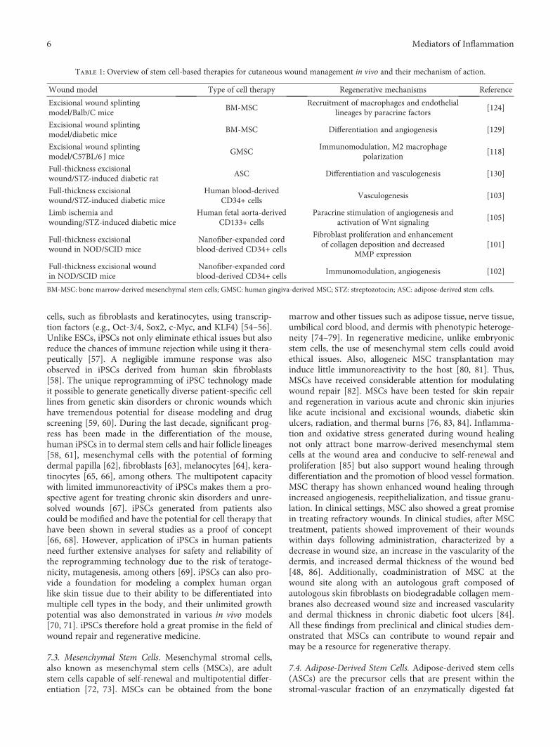
Table 1: Overview of stem cell-based therapies for cutaneous wound management in vivo and their mechanism of action.
Wound model Type of cell therapy Regenerative mechanisms Reference
Excisional wound splintingmodel/Balb/C mice
BM-MSCRecruitment of macrophages and endothelial
lineages by paracrine factors[124]
Excisional wound splintingmodel/diabetic mice
BM-MSC Differentiation and angiogenesis [129]
Excisional wound splintingmodel/C57BL/6 J mice
GMSCImmunomodulation, M2 macrophage
polarization[118]
Full-thickness excisionalwound/STZ-induced diabetic rat
ASC Differentiation and vasculogenesis [130]
Full-thickness excisionalwound/STZ-induced diabetic mice
Human blood-derivedCD34+ cells
Vasculogenesis [103]
Limb ischemia andwounding/STZ-induced diabetic mice
Human fetal aorta-derivedCD133+ cells
Paracrine stimulation of angiogenesis andactivation of Wnt signaling
[105]
Full-thickness excisionalwound in NOD/SCID mice
Nanofiber-expanded cordblood-derived CD34+ cells
Fibroblast proliferation and enhancementof collagen deposition and decreased
MMP expression[101]
Full-thickness excisional woundin NOD/SCID mice
Nanofiber-expanded cordblood-derived CD34+ cells
Immunomodulation, angiogenesis [102]
BM-MSC: bone marrow-derived mesenchymal stem cells; GMSC: human gingiva-derived MSC; STZ: streptozotocin; ASC: adipose-derived stem cells.
6 Mediators of Inflammation
tissue. Minimal invasive nature of tissue harvest has madethese stem cells more attractive for regenerative medicine.ASCs are multipotent in nature and can be differentiated intodifferent lineages such as bone, fat, cartilage, and muscle [75,87]. ASCs can be characterized while in culture dish asCD73+/CD90+/CD105+/CD44+/CD45−/CD31− cells, whichcan be distinguished from the bone marrow-derived MSCsby their expressions of CD36 and negative for CD106 mole-cules on their cell surface [88]. Although both of these celltypes share surface markers, biologically they are differentin terms of proliferation rate and differentiation, cytokinesecretion, and chemokine expressions [89–91]. Thus, ASCsand MSCs may contribute to the wound healing differently.The capability of ASCs to secrete growth factors, to differen-tiate into multiple cell types, and to promote angiogenesisrenders them a viable skin substitute [92, 93]. The ability ofASCs for soft tissue reconstruction makes them attractivefor wound healing [94].
7.5. Hematopoietic Stem Cells. The possible role of hemato-poietic stem cells (HSC) in skin regeneration is evident inmany occasions. HSC can be isolated from the bone marrow(BM), umbilical cord blood, and peripheral blood by using itssurface markers. In several occasions, skin “chimerism”(identification of epithelial cells of donor genotype) has beenobserved after clinical HSC transplantations such as BM orperipheral blood mononuclear cells (PBMC) [95–97]. Thefindings of donor-derived contribution of HSC to epitheliallineages in the host offer the broad-spectrum plasticity ofHSC and indicate the possibility of skin regeneration bytransplantation of HSC in chronic wound disorders. In amurine excisional wound model, a significant number of dif-ferentiated green fluorescent protein (GFP) positive cellswere found in the hair follicles, sebaceous glands, and epider-mis in host skin 21 days after transplantation of syngeneicGFP+bone marrow cells [48]. Additionally, a study has alsoshown that the differentiation potential of human umbilicalcord blood stem cells into keratinocytes in vitro [98]. Apartfrom plasticity, the role of HSC in angiogenesis is also evidentin myocardial infarction model, which is important and maybe ascribable for the perfect and functional repair of skin tis-sue [99]. An emerging concept, epithelial and mesenchymalcell interaction is supposed to be a vital phenomenon in ker-atinocyte proliferation and differentiation, might play a cru-cial role in cutaneous wound healing and reepithelialization[4, 92]. The expression of CD34 and CD133 cells in dermalfibroblast and follicular matrix during embryogenesis pro-vides an indication for the role of HSC in the molecular con-trol of epithelial-mesenchymal cell interactions [100].
Peripheral blood, fetal aorta, and umbilical cord bloodare also enriched with stem and progenitor cells, whichexpress CD34 and CD133markers. These cells are also multi-potent and have shown a neovascularization potential in pre-clinical ischemic models [33, 34]. In preclinical woundhealing models, we and others reported that CD34+ orCD133+ cells accelerate wound closure. We have demon-strated the wound healing ability of nanofiber-expanded cordblood-derived CD34+ cells in a mouse excisional woundmodel and an in vitro cellular model. We have shown that
after systemic administration, these stem cells reached to thewound bed and facilitated wound healing. Our study revealedthat nanofiber-expanded cord blood-derivedCD34+ cell ther-apy accelerates wound healing by inhibiting several matrixmetalloproteinases at the wound bed which prevents collagendegradation and increased the abundance of collagencomponents, procollagen1A1 at the wound bed [101]. Unlikeprevious experiments, we demonstrated for the first time thatnanofiber-expanded cord blood-derived CD34+ stem cellsaccelerated wound closure by secreting collagen and therebypositively contributed to extracellular matrix [101], indicat-ing that CD34+ stem cell treatment is having a potential totreat the refractory wounds resulting from diabetes or trau-matic skin injuries. We further extended this work to explorethe regulation of inflammatory response by CD34+ celltherapy using the same mouse wound model. Overall, ourstudy demonstrated that CD34+ cell therapy mediated sup-pression of prolonged inflammation, positively contributedto increased angiogenesis, and accelerated wound closurecompared to nontreated wounds [102]. These data provideda valuable information regarding the benefits of CD34+ stemcell-mediated wound healing and cell therapetic mechanismbehind accelerated wound closure. In another study, treat-ment with human CD34+ peripheral blood mononuclearcells also accelerates healing of full-thickness skin woundsin diabetic mice by accelerated revascularization and epider-mal healing [103]. A similar observation was also found in areport where cord blood-derived CD34+ cell treatment accel-erated diabetic wound closure by stimulating keratinocytes,fibroblast proliferation, and neovascularization in a paracrinemanner [104]. In another study, human fetal aorta-derivedCD133+ progenitor cells and their conditioned mediumtreatment accelerated healing in ischemic diabetic ulcer bystimulating angiogenesis with activation of theWnt signalingpathway in the host [105]. These findings indicate that blood-derived progenitors may have a therapeutic potential in thetreatment of skin lesions in complex pathological condi-tions such as diabetes.
8. Mechanisms of Stem Cell-Mediated WoundHealing
8.1. Immunomodulation, Resolution of Inflammation, andFibrosis. An imbalance in regulation of inflammation at thewound bed leads to defective healing. Sustained unresolvedinflammation leads to chronic wound. Prolonged inflam-mation even leads to fibrotic scar formation. In chronicwounds, unresolved inflammation leads to increased prote-ase activity and deregulated fibroblast activity, which resultedin decreased collagen deposition and ECM formation. Hence,resolution of inflammation is a big challenge in diabeticwound healing. In recent years, several studies have demon-strated the immunomodulatory function of cultured adultstem cells in laboratory conditions obtained from varioussources like the umbilical cord blood, amniotic fluid, andbone marrow. Thus, allogeneic stem cell therapy inducesimmunomodulation in the wound bed and facilitates woundhealing by resolving inflammation, as well as helping inreducing scar formation [106].
7Mediators of Inflammation

A substantial number of studies have demonstrated thattreatment of MSCs has significant immunomodulatoryeffects during wound healing and in other inflammatory con-ditions [107]. This immunomodulatory effect on the host notonly makes them a suitable candidate for allogeneic trans-plantation [108] but also makes them an attractive cell ther-apeutic agent to treat chronic wounds [106]. Studies havedemonstrated that MSCs obtained from various sources suchas the umbilical cord and bone marrow showed an anti-inflammatory effect in rat cutaneous wound and in vitrofibroblast model. MSC treatment demonstrated a signifi-cantly lower number of inflammatory cells and proinflamma-tory cytokines such as IL-1 and TNF-αwith an increased levelof IL-10 at the cutaneous wound bed in a rat model [109]. Inaddition, when murine BM-MSC were cocultured withhuman fibroblasts, the mRNA levels of intercellular adhesionmolecule 1 (ICAM1) has decreased [110]. These studies sug-gest the potential of MSC in attenuating wound inflamma-tion and inducing healing in chronic inflammatory stage.
Allogeneic transplantation of cord blood and cord blood-derived stem cells is also regarded as less immunogenic [111].Our results demonstrated that nanofiber-expanded cordblood-derived CD34+ cells might have an immunomodula-tory effect in vitro and in vivo wound healing models [102].Systemically transplanted CD34+ cells accelerated woundclosure, which was correlated with decreased inflammatoryactivity at the wound bed characterized by reduced inflam-matory gene expression such as, IL-1β, TNF-α, IL-6, andNOS2A. At the same time, expression of anti-inflammatorymolecule IL-10 was significantly increased indicating thatCD34+ cell therapy has the potential to control the inflam-mation during wound healing process. To further elucidatethe mechanism, we showed that CD34+ cells secrete IL-10,an anti-inflammatory molecule, and suppress NF-κB acti-vation in a human primary fibroblast cell model. In thesimilar in vitro model, when human primary fibroblastswere cocultured with CD34+ cells in presence of inflam-matory stimulus with TNF-α, NF-κB activation was signif-icantly decreased by upregulation of IL-10 [102]. Sustainedor unresolved inflammation prevents wound healing byinhibiting angiogenesis and catabolizes extracellular matrixin the wound bed. Our results suggest that nanofiber-expanded cord blood-derived CD34+ cell therapy mightbe a potential candidate to treat chronic wounds to resolveinflammation in a timely manner that will facilitate furtherangiogenesis and ECM formation for accelerated healing.Other studies have also suggested that ASCs also modulatethe immune system and downregulate the inflammationby releasing growth factors critical for healing which aredescribed in the reviews [112].
Antimicrobial activity is critical for wound clearancefrom infection. Studies have shown that MSCs have anti-microbial activities, which may also be helpful for chronicwound resolution. Antimicrobial activities of MSCs wereshown directly by the secretion of antimicrobial factorssuch as LL-37 [113]. In another study, it was shown thatMSCs could secrete immune-modulating factors, whichwill upregulate bacterial killing and phagocytosis by immunecells [114].
Tissue resident and peripheral macrophages play a signif-icant role in initiation of inflammation after injury andresolving inflammation in a timely manner during the heal-ing process. Macrophages shift gears between proinflamma-tory M1 and anti-inflammatory alternatively activated M2states. Studies have shown in various models that MSCsmay influence macrophage M1/M2 polarization after contactwith macrophage [115–117]. M2 polarized macrophages playan important role in the resolution of inflammation andclearance of dead cells from the wound environment foraccelerated healing. In a mouse wound healing model, itwas shown that the human gingiva-derived MSC treatmentin vivo promoted an M2 macrophage polarization, whichwas correlated well with anti-inflammatory wound environ-ment and accelerated cutaneous wound healing [118]. Theability of MSCs to resolve inflammation might be useful intreating chronic unresolved wounds.
Moreover, dysregulated fibrosis or scarring is caused byexcessive deposition of ECM. Inflammation largely regulatesfibrosis process. The immunomodulatory activity of MSCsmight regulate fibrosis and therefore anti-inflammatoryactivity of MSCs reduces the scar formation. In an in vivomurine wounding model, it showed that BM-MSCs atten-uated development in skin fibrosis [119].
8.2. Differentiation. In several studies, it has shown that adultstem cells are multipotential and able to contribute to woundhealing by differentiating into several tissue lineages startingfrom the inflammatory cells to myofibroblasts. During theinflammatory phase, HSCs from the bone marrow undergoesmyelopoiesis with the help of MSC and supply the leukocytesto the wound region [92]. Also, tissue resident stem cellsundergo differentiation in response to various stimuli duringthe wound healing process [120, 121]. From the regenerativeperspective, differentiation is one of the key phenomenon bywhich exogenously applied adult stem cells exert therapeuticefficacy in cutaneous wound models. MSC is one of the mostwidely studied in this regard. Several studies have demon-strated that transplanted MSCs can differentiate into epider-mal keratinocytes, endothelial cells, and pericytes directlyparticipating in the structural repair of a wound. MSC trans-plantation led to accelerated cutaneous wound closure inboth normal and diabetic mice, where MSCs expresskeratinocyte-specific markers suggesting their role to pro-mote wound healing by differentiation [76]. Similarly,another study demonstrated that MSCs also transdifferenti-ate into keratinocytes, endothelial cells, and pericytes in cuta-neous wounds after intravenous injection in mice [122].Adipose-derived stromal cells are also capable of differentiat-ing into epithelial, endothelial, and fibroblast lineages in vivowhen applied to wounds by means of a seeded scaffold [92].A similar kind of low-level transdifferentiation phenomenonwas also observed in human skin after HSC transplantationwhere the mesodermal origin of HSC contributes to epitheliallineage in the host [95, 96].
8.3. Angiogenesis. Optimal repair and restoration offunctional vasculature is crucial to achieve ideal healing ofwounds. The process of neovascularization primarily
8 Mediators of Inflammation
accomplishes revascularization of the wound bed. Neovascu-larization is achieved through two independent processescalled angiogenesis and vasculogenesis, which lead to thedevelopment of functional microvascular networks. Tradi-tionally, vasculogenesis is the de novo synthesis of new ves-sels by endothelial progenitor cells whereas angiogenesis isthe development of new vessels from existing capillaries[123]. In a refractory wound-like diabetic wound, woundrevascularization is affected due to an imbalance in therelease of soluble mediators and improper function of endog-enous progenitor and stem cells which is essential for idealwound healing, leading to a hindered and orchestrated heal-ing process. Several other and our own published studies[101, 102] have demonstrated that transplantation of adultstem or progenitor cells contribute to angiogenesis or vascu-logenesis through directly differentiating into cell typesessential for blood vessel formation or by stimulating endog-enous mediators or cells in a paracrine manner. Studies havefound that MSC expresses high levels of vascular endothelialgrowth factor (VEGF) and angiopoeitin-1, which indicatethat the MSC-mediated accelerated wound healing is due tothe release of proangiogenic factors and induction of angio-genesis [76]. Additionally, paracrine signaling and the releaseof soluble factors (e.g., VEGF) by MSC are found to promoteangiogenesis at the wound bed after cutaneous injury in nor-mal and diabetic mice [124, 125]. Another interestinghypothesis about MSC is that these cells may also act as peri-cytes, which stabilizes the blood vessel formation [126].Future studies will further bolster this claim. However, therole of MSC in promoting angiogenesis is firmly evident byseveral instances in normal and refractory wound healingmodels [127, 128].
In other stem cells like ASC, it was noticed that ASCtreatment also promotes angiogenesis and accelerates woundhealing by producing VEGF [93]. Thus, induction of angio-genesis to accelerate wound healing by stem cell therapy isnot only evident in MSC and ASC but is also found in severalother types of stem and progenitor cells [103, 105]. Thus,increased angiogenesis by stem cell therapy not onlysupports GT formation but also supplies nutrients andclear the apoptotic cells from the wound bed and helpsin wound resolution.
9. Conclusions and Future Directions
It is evident that stem cells have a tremendous potential forcutaneous tissue regeneration, as these cells not only canregenerate lost tissue but also promote wound repair throughparacrine manner. Several cell types, such as embryonic stemcells, iPSCs, mesenchymal stem cells, resident tissue stemcells, epithelial stem cells, adipose-derived stem cells, andhematopoietic stem cells, are currently under intense investi-gation. Recent data on autologous MSC therapy in cutaneousrepair showed a great promise as a therapeutic agent in clin-ical practice. Despite rapid progress in evaluating the efficacyof MSC transplantation for wound healing, several questionsstill need to be addressed. Using specific markers to charac-terize and isolate a distinct pool of MSC, which is homoge-neous and functional, is one of the key points to be
addressed. Further studies are necessary to characterize theniche of MSC, which helps MSCs to be effective in the woundhealing process. Further investigation on experimental andclinical application of stem cells in wound healing is neces-sary to identify the ideal source of stem cells and the mostefficacious mode of cell delivery.
Human umbilical cord blood is rich in stem and progen-itor cells and is easily accessible for blood collection. Theregenerative potential of CD34+ and CD133+ cell therapyin cutaneous tissue repair obtained from peripheral or umbil-ical cord blood opens up a possibility of providing cheap andaffordable care for refractory wounds. Thus, a suitable tech-nology like ex vivo expansion technique would be useful toget a large number of stem cells for the clinical application.Our group and others have shown promising results inexpansion of umbilical cord blood-derived stem cells. Theexpanded stem cells were characterized (CD34+) and haveshown their multipotential and angiogenic capabilities inpreclinical ischemic models as well as in murine cutaneouswound models [33, 34, 101, 102] where CD34+ stem celltherapy accelerated wound closure by resolving inflamma-tion with concurrent inhibition of MMP expressions. Addi-tionally, these cells also can be manipulated in vitro withproangiogenic factors like VEGF and PDGF, which are effica-cious in improving ischemia-related complications in pre-clinical peripheral and cardiac ischemic models. Thus,looking at their angiogenic and anti-inflammatory potential,this pool of stem cells may have a very promising future intreating refractory wounds. Moreover, nanofiber-expandedstem cells coupled with the innovative biotechnologiesmay open a new direction for plastic and reconstructivesurgeons. Finally, the use of stem cells to induce cutaneoustissue regeneration holds a great promise for modernregenerative medicine.
Conflicts of Interest
No competing financial interests exist.
Acknowledgments
This work was supported in part by the National Instituteof Health (NIH) Grant nos. R01AR068279 (NIAMS), STTRR41EY024217 (NEI), and R41EY024217-01A1S1 (NEI)and the Texas Tech University Health Sciences Centerstart-up fund.
References
[1] E. Proksch, J. M. Brandner, and J. M. Jensen, “The skin: anindispensable barrier,” Experimental Dermatology, vol. 17,no. 12, pp. 1063–1072, 2008.
[2] B. S. Atiyeh, J. Ioannovich, C. A. Al-Amm, and K. A.El-Musa, “Management of acute and chronic open wounds:the importance of moist environment in optimal woundhealing,” Current Pharmaceutical Biotechnology, vol. 3,no. 3, pp. 179–195, 2002.
[3] G. C. Gurtner, S. Werner, Y. Barrandon, and M. T. Longaker,“Wound repair and regeneration,” Nature, vol. 453, no. 7193,pp. 314–321, 2008.
9Mediators of Inflammation
[4] A. J. Boulton, L. Vileikyte, G. Ragnarson-Tennvall, andJ. Apelqvist, “The global burden of diabetic foot disease,”Lancet, vol. 366, no. 9498, pp. 1719–1724, 2005.
[5] M. Braddock, C. J. Campbell, and D. Zuder, “Currenttherapies for wound healing: electrical stimulation, biolog-ical therapeutics, and the potential for gene therapy,”International Journal of Dermatology, vol. 38, no. 11,pp. 808–817, 1999.
[6] K. Beckrich and S. A. Aronovitch, “Hospital-acquired pres-sure ulcers: a comparison of costs in medical vs. surgicalpatients,” Nursing Economics, vol. 17, no. 5, pp. 263–271,1999.
[7] C. Harvey, “Wound healing,” Orthopaedic Nursing, vol. 24,no. 2, pp. 143–157, 2005.
[8] A. J. Singer, H. C. Thode Jr., and S. A. McClain, “Develop-ment of a histomorphologic scale to quantify cutaneous scarsafter burns,” Academic Emergency Medicine, vol. 7, no. 10,pp. 1083–1088, 2000.
[9] S. Werner and R. Grose, “Regulation of wound healing bygrowth factors and cytokines,” Physiological Reviews,vol. 83, no. 3, pp. 835–870, 2003.
[10] M. W. Mosesson, K. R. Siebenlist, and D. A. Meh, “The struc-ture and biological features of fibrinogen and fibrin,” Annalsof the New York Academy of Sciences, vol. 936, pp. 11–30,2006.
[11] A. M. Szpaderska, E. I. Egozi, R. L. Gamelli, and L. A.DiPietro, “The effect of thrombocytopenia on dermal woundhealing,” The Journal of Investigative Dermatology, vol. 120,no. 6, pp. 1130–1137, 2003.
[12] W. T. Lawrence, “Physiology of the acute wound,” Clinics inPlastic Surgery, vol. 25, no. 3, pp. 321–340, 1998.
[13] J. M. Rhett, G. S. Ghatnekar, J. A. Palatinus, M. O'Quinn,M. J. Yost, and R. G. Gourdie, “Novel therapies for scarreduction and regenerative healing of skin wounds,” Trendsin Biotechnology, vol. 26, no. 4, pp. 173–180, 2008.
[14] M. Maurer, T. Theoharides, R. D. Granstein et al., “What isthe physiological function of mast cells?,” ExperimentalDermatology, vol. 12, no. 6, pp. 886–910, 2003.
[15] H. Smola, G. Thiekotter, and N. E. Fusenig, “Mutual induc-tion of growth factor gene expression by epidermal-dermalcell interaction,” The Journal of Cell Biology, vol. 122, no. 2,pp. 417–429, 1993.
[16] K. J. Stewart, “A quantitative ultrastructural study of collagenfibrils in human skin, normal scars, and hypertrophic scars,”Clinical Anatomy, vol. 8, no. 5, pp. 334–338, 1995.
[17] P. J. Franks, C. J. Moffatt, D. C. Doherty, R. Smithdale, andR. Martin, “Longer-term changes in quality of life in chronicleg ulceration,” Wound Repair and Regeneration, vol. 14,no. 5, pp. 536–541, 2006.
[18] A. Nijnik, L. Woodbine, C. Marchetti et al., “DNA repair islimiting for haematopoietic stem cells during ageing,”Nature, vol. 447, no. 7145, pp. 686–690, 2007.
[19] C. C. Zouboulis, J. Adjaye, H. Akamatsu, G. Moe-Behrens,and C. Niemann, “Human skin stem cells and the ageing pro-cess,” Experimental Gerontology, vol. 43, no. 11, pp. 986–997,2008.
[20] G. Van Zant and Y. Liang, “The role of stem cells in aging,”Experimental Hematology, vol. 31, no. 8, pp. 659–672, 2003.
[21] J. Walburn, K. Vedhara, M. Hankins, L. Rixon, andJ. Weinman, “Psychological stress and wound healing in
humans: a systematic review and meta-analysis,” Journal ofPsychosomatic Research, vol. 67, no. 3, pp. 253–271, 2009.
[22] A. Cole-King and K. G. Harding, “Psychological factors anddelayed healing in chronic wounds,” Psychosomatic Medicine,vol. 63, no. 2, pp. 216–220, 2001.
[23] D. L. Steed, C. Attinger, T. Colaizzi et al., “Guidelines for thetreatment of diabetic ulcers,” Wound Repair and Regenera-tion, vol. 14, no. 6, pp. 680–692, 2006.
[24] M. C. Robson and A. Barbul, “Guidelines for the best care ofchronic wounds,” Wound Repair and Regeneration, vol. 14,no. 6, pp. 647-648, 2006.
[25] M. Gharaee-Kermani and S. H. Phan, “Role of cytokines andcytokine therapy in wound healing and fibrotic diseases,”Current Pharmaceutical Design, vol. 7, no. 11, pp. 1083–1103, 2001.
[26] Y. M. Bello, A. F. Falabella, and W. H. Eaglstein, “Tissue-engineered skin. Current status in wound healing,” AmericanJournal of Clinical Dermatology, vol. 2, no. 5, pp. 305–313,2001.
[27] B. K. Sun, Z. Siprashvili, and P. A. Khavari, “Advances in skingrafting and treatment of cutaneous wounds,” Science,vol. 346, no. 6212, pp. 941–945, 2014.
[28] M. Londahl, P. Katzman, A. Nilsson, and C. Hammarlund,“Hyperbaric oxygen therapy facilitates healing of chronic footulcers in patients with diabetes,” Diabetes Care, vol. 33, no. 5,pp. 998–1003, 2010.
[29] S. Barrientos, H. Brem, O. Stojadinovic, andM. Tomic-Canic,“Clinical application of growth factors and cytokines inwound healing,” Wound Repair and Regeneration, vol. 22,no. 5, pp. 569–578, 2014.
[30] J. M. Smiell, T. J. Wieman, D. L. Steed, B. H. Perry, A. R.Sampson, and B. H. Schwab, “Efficacy and safety of becapler-min (recombinant human platelet-derived growth factor-BB)in patients with nonhealing, lower extremity diabetic ulcers: acombined analysis of four randomized studies,” WoundRepair and Regeneration, vol. 7, no. 5, pp. 335–346, 1999.
[31] S. C. Wu, W. Marston, and D. G. Armstrong, “Wound care:the role of advanced wound-healing technologies,” Journalof the American Podiatric Medical Association, vol. 100,no. 5, pp. 385–394, 2010.
[32] V. Falanga and M. Sabolinski, “A bilayered living skinconstruct (APLIGRAFR) accelerates complete closure ofhard-to-heal venous ulcers,” Wound Repair and Regenera-tion, vol. 7, no. 4, pp. 201–207, 1999.
[33] H. Das, N. Abdulhameed, M. Joseph, R. Sakthivel, H. Q. Mao,and V. J. Pompili, “Ex vivo nanofiber expansion and geneticmodification of human cord blood-derived progenitor/stemcells enhances vasculogenesis,” Cell Transplantation, vol. 18,no. 3, pp. 305–318, 2009.
[34] H. Das, J. C. George, M. Joseph et al., “Stem cell therapy withoverexpressed VEGF and PDGF genes improves cardiacfunction in a rat infarct model,” PLoS One, vol. 4, no. 10,article e7325, 2009.
[35] L. Alonso and E. Fuchs, “The hair cycle,” Journal of CellScience, vol. 119, Part 3, pp. 391–393, 2006.
[36] E. Fuchs and V. Horsley, “More than one way to skin,” Genes& Development, vol. 22, no. 8, pp. 976–985, 2008.
[37] V. Levy, C. Lindon, Y. Zheng, B. D. Harfe, and B. A. Morgan,“Epidermal stem cells arise from the hair follicle afterwounding,” The FASEB Journal, vol. 21, no. 7, pp. 1358–1366, 2007.
10 Mediators of Inflammation
[38] B. E. Zawacki and R. J. Jones, “Standard depth burns in therat: the importance of the hair growth cycle,” British Journalof Plastic Surgery, vol. 20, no. 4, pp. 347–354, 1967.
[39] A. K. Langton, S. E. Herrick, and D. J. Headon, “Anextended epidermal response heals cutaneous wounds inthe absence of a hair follicle stem cell contribution,” TheJournal of Investigative Dermatology, vol. 128, no. 5,pp. 1311–1318, 2008.
[40] M. Ito, Y. Liu, Z. Yang et al., “Stem cells in the hair folliclebulge contribute to wound repair but not to homeostasis ofthe epidermis,” Nature Medicine, vol. 11, no. 12, pp. 1351–1354, 2005.
[41] J. A. Nowak, L. Polak, H. A. Pasolli, and E. Fuchs, “Hairfollicle stem cells are specified and function in early skinmorphogenesis,” Cell Stem Cell, vol. 3, no. 1, pp. 33–43,2008.
[42] O. M. Tepper, J. M. Capla, R. D. Galiano et al., “Adult vascu-logenesis occurs through in situ recruitment, proliferation,and tubulization of circulating bone marrow-derived cells,”Blood, vol. 105, no. 3, pp. 1068–1077, 2005.
[43] D. J. Ceradini, A. R. Kulkarni, M. J. Callaghan et al., “Progen-itor cell trafficking is regulated by hypoxic gradients throughHIF-1 induction of SDF-1,” Nature Medicine, vol. 10, no. 8,pp. 858–864, 2004.
[44] J. M. Capla, R. H. Grogan, M. J. Callaghan et al., “Diabetesimpairs endothelial progenitor cell-mediated blood vesselformation in response to hypoxia,” Plastic and ReconstructiveSurgery, vol. 119, no. 1, pp. 59–70, 2007.
[45] J. E. Bluff, M. W. Ferguson, S. O'Kane, and G. Ireland, “Bonemarrow-derived endothelial progenitor cells do not contrib-ute significantly to new vessels during incisional wound heal-ing,” Experimental Hematology, vol. 35, no. 3, pp. 500–506,2007.
[46] G. Grenier, A. Scime, F. Le Grand et al., “Resident endothelialprecursors in muscle, adipose, and dermis contribute to post-natal vasculogenesis,” Stem Cells, vol. 25, no. 12, pp. 3101–3110, 2007.
[47] C. Roh, Q. Tao, C. Photopoulos, and S. Lyle, “In vitrodifferences between keratinocyte stem cells and transit-amplifying cells of the human hair follicle,” The Journalof Investigative Dermatology, vol. 125, no. 6, pp. 1099–1105,2005.
[48] E. V. Badiavas, M. Abedi, J. Butmarc, V. Falanga, andP. Quesenberry, “Participation of bone marrow derived cellsin cutaneous wound healing,” Journal of Cellular Physiology,vol. 196, no. 2, pp. 245–250, 2003.
[49] H. A. Navsaria, N. O. Ojeh, N. Moiemen, M. A. Griffiths, andJ. D. Frame, “Reepithelialization of a full-thickness burn fromstem cells of hair follicles micrografted into a tissue-engineered dermal template (Integra),” Plastic and Recon-structive Surgery, vol. 113, no. 3, pp. 978–981, 2004.
[50] M. J. Schurr, K. N. Foster, J. M. Centanni et al., “Phase I/IIclinical evaluation of StrataGraft: a consistent, pathogen-free human skin substitute,” The Journal of Trauma, vol. 66,no. 3, pp. 866–873, 2009, discussion 873-864.
[51] K. Lau, R. Paus, S. Tiede, P. Day, and A. Bayat, “Exploring therole of stem cells in cutaneous wound healing,” ExperimentalDermatology, vol. 18, no. 11, pp. 921–933, 2009.
[52] J. A. Thomson, J. Itskovitz-Eldor, S. S. Shapiro et al., “Embry-onic stem cell lines derived from human blastocysts,” Science,vol. 282, no. 5391, pp. 1145–1147, 1998.
[53] D. Aberdam, “Derivation of keratinocyte progenitor cells andskin formation from embryonic stem cells,” The Interna-tional Journal of Developmental Biology, vol. 48, no. 2-3,pp. 203–206, 2004.
[54] K. Takahashi and S. Yamanaka, “Induction of pluripotentstem cells from mouse embryonic and adult fibroblastcultures by defined factors,” Cell, vol. 126, no. 4, pp. 663–676, 2006.
[55] K. Takahashi, K. Tanabe, M. Ohnuki et al., “Induction of plu-ripotent stem cells from adult human fibroblasts by definedfactors,” Cell, vol. 131, no. 5, pp. 861–872, 2007.
[56] T. Aasen, A. Raya, M. J. Barrero et al., “Efficient and rapidgeneration of induced pluripotent stem cells from humankeratinocytes,” Nature Biotechnology, vol. 26, no. 11,pp. 1276–1284, 2008.
[57] P. Guha, J. W. Morgan, G. Mostoslavsky, N. P. Rodrigues,and A. S. Boyd, “Lack of immune response to differentiatedcells derived from syngeneic induced pluripotent stem cells,”Cell Stem Cell, vol. 21, no. 1, pp. 144–148, 2017.
[58] Q. Lu, M. Yu, C. Shen et al., “Negligible immunogenicityof induced pluripotent stem cells derived from humanskin fibroblasts,” PLoS One, vol. 9, no. 12, article e114949,2014.
[59] H. Ohnishi, T. Kawasaki, T. Deguchi, and S. Yuba, “Gen-eration of xeroderma pigmentosum-a patient-derivedinduced pluripotent stem cell line for use as future diseasemodel,” Cellular Reprogramming, vol. 17, no. 4, pp. 268–274, 2015.
[60] J. Liu, M. V. Joglekar, H. Sumer, A. A. Hardikar, H. Teede,and P. J. Verma, “Integration-free human induced pluripo-tent stem cells from type 1 diabetes patient skin fibroblastsshow increased abundance of pancreas-specific microRNAs,”Cell Medicine, vol. 7, no. 1, pp. 15–24, 2014.
[61] Y. Sugiyama-Nakagiri, T. Fujimura, and S. Moriwaki,“Induction of skin-derived precursor cells from humaninduced pluripotent stem Cells,” PLoS One, vol. 11, no. 12,article e0168451, 2016.
[62] O. Veraitch, Y. Mabuchi, Y. Matsuzaki et al., “Induction ofhair follicle dermal papilla cell properties in human inducedpluripotent stem cell-derived multipotent LNGFR(+)THY-1(+) mesenchymal cells,” Scientific Reports, vol. 7, article42777, 2017.
[63] K. J. Hewitt, Y. Shamis, R. B. Hayman et al., “Epigeneticand phenotypic profile of fibroblasts derived from inducedpluripotent stem cells,” PLoS One, vol. 6, no. 2, articlee17128, 2011.
[64] S. Ohta, Y. Imaizumi, Y. Okada et al., “Generation of humanmelanocytes from induced pluripotent stem cells,” PLoS One,vol. 6, no. 1, article e16182, 2011.
[65] G. Bilousova, J. Chen, and D. R. Roop, “Differentiation ofmouse induced pluripotent stem cells into a multipotent ker-atinocyte lineage,” The Journal of Investigative Dermatology,vol. 131, no. 4, pp. 857–864, 2011.
[66] M. Itoh, M. Kiuru, M. S. Cairo, and A. M. Christiano, “Gen-eration of keratinocytes from normal and recessive dystro-phic epidermolysis bullosa-induced pluripotent stem cells,”Proceedings of the National Academy of Sciences of the UnitedStates of America, vol. 108, no. 21, pp. 8797–8802, 2011.
[67] J. Zhang, J. Guan, X. Niu et al., “Exosomes released fromhuman induced pluripotent stem cells-derived MSCs facili-tate cutaneous wound healing by promoting collagen
11Mediators of Inflammation

synthesis and angiogenesis,” Journal of Translational Medi-cine, vol. 13, p. 49, 2015.
[68] V. Sebastiano, H. H. Zhen, B. Haddad et al., “HumanCOL7A1-corrected induced pluripotent stem cells for thetreatment of recessive dystrophic epidermolysis bullosa,”Science Translational Medicine, vol. 6, no. 264, article264ra163, 2014.
[69] K. Okita, T. Ichisaka, and S. Yamanaka, “Generation ofgermline-competent induced pluripotent stem cells,” Nature,vol. 448, no. 7151, pp. 313–317, 2007.
[70] R. Takagi, J. Ishimaru, A. Sugawara et al., “Bioengineering a3D integumentary organ system from iPS cells using anin vivo transplantation model,” Science Advances, vol. 2,no. 4, article e1500887, 2016.
[71] K. Gledhill, Z. Guo, N. Umegaki-Arao, C. A. Higgins,M. Itoh, and A. M. Christiano, “Melanin transfer inhuman 3D skin equivalents generated exclusively frominduced pluripotent stem cells,” PLoS One, vol. 10, no. 8,article e0136713, 2015.
[72] H. Nakagawa, S. Akita, M. Fukui, T. Fujii, and K. Akino,“Human mesenchymal stem cells successfully improve skin-substitute wound healing,” The British Journal of Derma-tology, vol. 153, no. 1, pp. 29–36, 2005.
[73] D. S. Krause, N. D. Theise, M. I. Collector et al., “Multi-organ,multi-lineage engraftment by a single bone marrow-derivedstem cell,” Cell, vol. 105, no. 3, pp. 369–377, 2001.
[74] O. K. Lee, T. K. Kuo, W. M. Chen, K. D. Lee, S. L. Hsieh, andT. H. Chen, “Isolation of multipotent mesenchymal stem cellsfrom umbilical cord blood,” Blood, vol. 103, no. 5, pp. 1669–1675, 2004.
[75] P. A. Zuk, M. Zhu, P. Ashjian et al., “Human adipose tissue isa source of multipotent stem cells,” Molecular Biology of theCell, vol. 13, no. 12, pp. 4279–4295, 2002.
[76] Y. Dai, J. Li, J. Li et al., “Skin epithelial cells in mice fromumbilical cord blood mesenchymal stem cells,” Burns,vol. 33, no. 4, pp. 418–428, 2007.
[77] C. K. Perng, H. H. Ku, S. H. Chiou et al., “Evaluation ofwound healing effect on skin-defect nude mice by usinghuman dermis-derived mesenchymal stem cells,” Transplan-tation Proceedings, vol. 38, no. 9, pp. 3086-3087, 2006.
[78] D. T. Shih, D. C. Lee, S. C. Chen et al., “Isolation andcharacterization of neurogenic mesenchymal stem cells inhuman scalp tissue,” Stem Cells, vol. 23, no. 7, pp. 1012–1020, 2005.
[79] E. A. Jones, S. E. Kinsey, A. English et al., “Isolation and char-acterization of bone marrow multipotential mesenchymalprogenitor cells,” Arthritis and Rheumatism, vol. 46, no. 12,pp. 3349–3360, 2002.
[80] E. Mansilla, G. H. Marin, F. Sturla et al., “Human mesenchy-mal stem cells are tolerized by mice and improve skin andspinal cord injuries,” Transplantation Proceedings, vol. 37,no. 1, pp. 292–294, 2005.
[81] V. Falanga, S. Iwamoto, M. Chartier et al., “Autologous bonemarrow-derived cultured mesenchymal stem cells deliveredin a fibrin spray accelerate healing in murine and humancutaneous wounds,” Tissue Engineering, vol. 13, no. 6,pp. 1299–1312, 2007.
[82] S. Bajada, I. Mazakova, J. B. Richardson, andN. Ashammakhi,“Updates on stem cells and their applications in regenerativemedicine,” Journal of Tissue Engineering and RegenerativeMedicine, vol. 2, no. 4, pp. 169–183, 2008.
[83] J. J. Lataillade, C. Doucet, E. Bey et al., “New approach toradiation burn treatment by dosimetry-guided surgery com-bined with autologous mesenchymal stem cell therapy,”Regenerative Medicine, vol. 2, no. 5, pp. 785–794, 2007.
[84] J. Vojtassak, L. Danisovic, M. Kubes et al., “Autologous bio-graft and mesenchymal stem cells in treatment of the diabeticfoot,” Neuro Endocrinology Letters, vol. 27, Supplement 2,pp. 134–137, 2006.
[85] W. L. Grayson, F. Zhao, B. Bunnell, and T. Ma, “Hypoxiaenhances proliferation and tissue formation of human mes-enchymal stem cells,” Biochemical and Biophysical ResearchCommunications, vol. 358, no. 3, pp. 948–953, 2007.
[86] J. Cha and V. Falanga, “Stem cells in cutaneous wound heal-ing,” Clinics in Dermatology, vol. 25, no. 1, pp. 73–78, 2007.
[87] P. A. Zuk, M. Zhu, H. Mizuno et al., “Multilineage cells fromhuman adipose tissue: implications for cell-based therapies,”Tissue Engineering, vol. 7, no. 2, pp. 211–228, 2001.
[88] P. Bourin, B. A. Bunnell, L. Casteilla et al., “Stromal cells fromthe adipose tissue-derived stromal vascular fraction and cul-ture expanded adipose tissue-derived stromal/stem cells: ajoint statement of the International Federation for AdiposeTherapeutics and Science (IFATS) and the InternationalSociety for Cellular Therapy (ISCT),” Cytotherapy, vol. 15,no. 6, pp. 641–648, 2013.
[89] L. Danisovic, I. Varga, S. Polak et al., “Comparison ofin vitro chondrogenic potential of human mesenchymalstem cells derived from bone marrow and adipose tissue,”General Physiology and Biophysics, vol. 28, no. 1, pp. 56–62,2009.
[90] J. S. Elman, M. Li, F. Wang, J. M. Gimble, and B. Parekkadan,“A comparison of adipose and bone marrow-derived mesen-chymal stromal cell secreted factors in the treatment ofsystemic inflammation,” Journal of Inflammation, vol. 11,no. 1, p. 1, 2014.
[91] S. T. Hsiao, A. Asgari, Z. Lokmic et al., “Comparative analysisof paracrine factor expression in human adult mesenchymalstem cells derived from bone marrow, adipose, and dermaltissue,” Stem Cells and Development, vol. 21, no. 12,pp. 2189–2203, 2012.
[92] A. M. Altman, N. Matthias, Y. Yan et al., “Dermal matrix as acarrier for in vivo delivery of human adipose-derived stemcells,” Biomaterials, vol. 29, no. 10, pp. 1431–1442, 2008.
[93] T. G. Ebrahimian, F. Pouzoulet, C. Squiban et al., “Cell ther-apy based on adipose tissue-derived stromal cells promotesphysiological and pathological wound healing,” Arterioscle-rosis, Thrombosis, and Vascular Biology, vol. 29, no. 4,pp. 503–510, 2009.
[94] M. Cherubino and K. G. Marra, “Adipose-derived stem cellsfor soft tissue reconstruction,” Regenerative Medicine, vol. 4,no. 1, pp. 109–117, 2009.
[95] M. Korbling, R. L. Katz, A. Khanna et al., “Hepatocytes andepithelial cells of donor origin in recipients of peripheral-blood stem cells,” The New England Journal of Medicine,vol. 346, no. 10, pp. 738–746, 2002.
[96] H. Murata, A. Janin, C. Leboeuf et al., “Donor-derived cellsand human graft-versus-host disease of the skin,” Blood,vol. 109, no. 6, pp. 2663–2665, 2007.
[97] S. Jiang, L.Walker, M. Afentoulis et al., “Transplanted humanbone marrow contributes to vascular endothelium,” Proceed-ings of the National Academy of Sciences of the United Statesof America, vol. 101, no. 48, pp. 16891–16896, 2004.
12 Mediators of Inflammation
[98] L. P. Kamolz, A. Kolbus, N.Wick et al., “Cultured human epi-thelium: human umbilical cord blood stem cells differentiateinto keratinocytes under in vitro conditions,” Burns, vol. 32,no. 1, pp. 16–19, 2006.
[99] R. Sackstein, “The bone marrow is akin to skin: HCELL andthe biology of hematopoietic stem cell homing,” The Journalof Investigative Dermatology, vol. 122, no. 5, pp. 1061–1069,2004.
[100] X. Fu and X. Sun, “Can hematopoietic stem cells be an alter-native source for skin regeneration?,” Ageing ResearchReviews, vol. 8, no. 3, pp. 244–249, 2009.
[101] S. Kanji, M. Das, R. Aggarwal et al., “Nanofiber-expandedhuman umbilical cord blood-derived CD34+ cell therapyaccelerates cutaneous wound closure in NOD/SCID mice,”Journal of Cellular and Molecular Medicine, vol. 18, no. 4,pp. 685–697, 2014.
[102] S. Kanji, M. Das, R. Aggarwal et al., “Nanofiber-expandedhuman umbilical cord blood-derived CD34+ cell therapyaccelerates murine cutaneous wound closure by attenuatingpro-inflammatory factors and secreting IL-10,” Stem CellResearch, vol. 12, no. 1, pp. 275–288, 2014.
[103] E. Sivan-Loukianova, O. A. Awad, V. Stepanovic,J. Bickenbach, and G. C. Schatteman, “CD34+ blood cellsaccelerate vascularization and healing of diabetic mouse skinwounds,” Journal of Vascular Research, vol. 40, no. 4,pp. 368–377, 2003.
[104] J. Y. Kim, S. H. Song, K. L. Kim et al., “Human cord blood-derived endothelial progenitor cells and their conditionedmedia exhibit therapeutic equivalence for diabetic woundhealing,” Cell Transplantation, vol. 19, no. 12, pp. 1635–1644, 2010.
[105] L. S. Barcelos, C. Duplaa, N. Krankel et al., “Human CD133+
progenitor cells promote the healing of diabetic ischemiculcers by paracrine stimulation of angiogenesis and activationof Wnt signaling,” Circulation Research, vol. 104, no. 9,pp. 1095–1102, 2009.
[106] A. Nuschke, “Activity of mesenchymal stem cells in therapiesfor chronic skin wound healing,” Organogenesis, vol. 10,no. 1, pp. 29–37, 2014.
[107] N. G. Singer and A. I. Caplan, “Mesenchymal stem cells:mechanisms of inflammation,” Annual Review of Pathology,vol. 6, pp. 457–478, 2011.
[108] N. K. Satija, V. K. Singh, Y. K. Verma et al., “Mesenchymalstem cell-based therapy: a new paradigm in regenerativemedicine,” Journal of Cellular and Molecular Medicine,vol. 13, no. 11-12, pp. 4385–4402, 2009.
[109] L. Liu, Y. Yu, Y. Hou et al., “Human umbilical cord mesen-chymal stem cells transplantation promotes cutaneouswound healing of severe burned rats,” PLoS One, vol. 9,no. 2, article e88348, 2014.
[110] A. N. Smith, E. Willis, V. T. Chan et al., “Mesenchymalstem cells induce dermal fibroblast responses to injury,”Experimental Cell Research, vol. 316, no. 1, pp. 48–54,2010.
[111] M. J. Laughlin, “Umbilical cord blood for allogeneic trans-plantation in children and adults,” Bone Marrow Transplan-tation, vol. 27, no. 1, pp. 1–6, 2001.
[112] L. E. Kokai, K. Marra, and J. P. Rubin, “Adipose stem cells:biology and clinical applications for tissue repair and regen-eration,” Translational Research, vol. 163, no. 4, pp. 399–408, 2014.
[113] A. Krasnodembskaya, Y. Song, X. Fang et al., “Antibacte-rial effect of human mesenchymal stem cells is mediatedin part from secretion of the antimicrobial peptide LL-37,”Stem Cells, vol. 28, no. 12, pp. 2229–2238, 2010.
[114] S. H. Mei, J. J. Haitsma, C. C. Dos Santos et al., “Mesenchymalstem cells reduce inflammation while enhancing bacterialclearance and improving survival in sepsis,” American Jour-nal of Respiratory and Critical Care Medicine, vol. 182,no. 8, pp. 1047–1057, 2010.
[115] D. I. Cho, M. R. Kim, H. Y. Jeong et al., “Mesenchymal stemcells reciprocally regulate the M1/M2 balance in mouse bonemarrow-derived macrophages,” Experimental & MolecularMedicine, vol. 46, article e70, 2014.
[116] S. Dameshghi, A. Zavaran-Hosseini, S. Soudi, F. J. Shirazi,S. Nojehdehi, and S. M. Hashemi, “Mesenchymal stem cellsalter macrophage immune responses to Leishmania majorinfection in both susceptible and resistance mice,” Immu-nology Letters, vol. 170, pp. 15–26, 2016.
[117] Y. Hu, C. Qin, G. Zheng et al., “Mesenchymal stem cell-educated macrophages ameliorate LPS-induced systemicresponse,” Mediators of Inflammation, vol. 2016, Article ID3735452, 12 pages, 2016.
[118] Q. Z. Zhang, W. R. Su, S. H. Shi et al., “Human gingiva-derived mesenchymal stem cells elicit polarization of M2macrophages and enhance cutaneous wound healing,” StemCells, vol. 28, no. 10, pp. 1856–1868, 2010.
[119] Y. Wu, S. Huang, J. Enhe et al., “Bone marrow-derivedmesenchymal stem cell attenuates skin fibrosis developmentin mice,” International Wound Journal, vol. 11, no. 6,pp. 701–710, 2014.
[120] T. Kumamoto, D. Shalhevet, H. Matsue et al., “Hair folliclesserve as local reservoirs of skin mast cell precursors,” Blood,vol. 102, no. 5, pp. 1654–1660, 2003.
[121] M. Lako, L. Armstrong, P. M. Cairns, S. Harris, N. Hole, andC. A. Jahoda, “Hair follicle dermal cells repopulate the mousehaematopoietic system,” Journal of Cell Science, vol. 115,Part 20, pp. 3967–3974, 2002.
[122] M. Sasaki, R. Abe, Y. Fujita, S. Ando, D. Inokuma, andH. Shimizu, “Mesenchymal stem cells are recruited intowounded skin and contribute to wound repair by transdiffer-entiation into multiple skin cell type,” Journal of Immunol-ogy, vol. 180, no. 4, pp. 2581–2587, 2008.
[123] S. Kassmeyer, J. Plendl, P. Custodis, and M. Bahramsoltani,“New insights in vascular development: vasculogenesis andendothelial progenitor cells,” Anatomia, Histologia, Embryo-logia, vol. 38, no. 1, pp. 1–11, 2009.
[124] L. Chen, E. E. Tredget, P. Y. Wu, and Y. Wu, “Paracrine fac-tors of mesenchymal stem cells recruit macrophages andendothelial lineage cells and enhance wound healing,” PLoSOne, vol. 3, no. 4, article e1886, 2008.
[125] M. Gnecchi, Z. Zhang, A. Ni, and V. J. Dzau, “Paracrinemechanisms in adult stem cell signaling and therapy,”Circulation Research, vol. 103, no. 11, pp. 1204–1219,2008.
[126] M. Crisan, S. Yap, L. Casteilla et al., “A perivascular origin formesenchymal stem cells in multiple human organs,” CellStem Cell, vol. 3, no. 3, pp. 301–313, 2008.
[127] V. I. Shumakov, N. A. Onishchenko, M. F. Rasulov, M. E.Krasheninnikov, and V. A. Zaidenov, “Mesenchymal bonemarrow stem cells more effectively stimulate regenerationof deep burn wounds than embryonic fibroblasts,” Bulletin
13Mediators of Inflammation

of Experimental Biology and Medicine, vol. 136, no. 2,pp. 192–195, 2003.
[128] D. S. Kwon, X. Gao, Y. B. Liu et al., “Treatment with bonemarrow-derived stromal cells accelerates wound healing indiabetic rats,” International Wound Journal, vol. 5, no. 3,pp. 453–463, 2008.
[129] Y. Wu, L. Chen, P. G. Scott, and E. E. Tredget, “Mesenchymalstem cells enhance wound healing through differentiationand angiogenesis,” Stem Cells, vol. 25, no. 10, pp. 2648–2659, 2007.
[130] C. Nie, D. Yang, J. Xu, Z. Si, X. Jin, and J. Zhang, “Locallyadministered adipose-derived stem cells accelerate woundhealing through differentiation and vasculogenesis,” CellTransplantation, vol. 20, no. 2, pp. 205–216, 2011.
14 Mediators of Inflammation

Submit your manuscripts athttps://www.hindawi.com
Stem CellsInternational
Hindawi Publishing Corporationhttp://www.hindawi.com Volume 2014
Hindawi Publishing Corporationhttp://www.hindawi.com Volume 2014
MEDIATORSINFLAMMATION
of
Hindawi Publishing Corporationhttp://www.hindawi.com Volume 2014
Behavioural Neurology
EndocrinologyInternational Journal of
Hindawi Publishing Corporationhttp://www.hindawi.com Volume 2014
Hindawi Publishing Corporationhttp://www.hindawi.com Volume 2014
Disease Markers
Hindawi Publishing Corporationhttp://www.hindawi.com Volume 2014
BioMed Research International
OncologyJournal of
Hindawi Publishing Corporationhttp://www.hindawi.com Volume 2014
Hindawi Publishing Corporationhttp://www.hindawi.com Volume 2014
Oxidative Medicine and Cellular Longevity
Hindawi Publishing Corporationhttp://www.hindawi.com Volume 2014
PPAR Research
The Scientific World JournalHindawi Publishing Corporation http://www.hindawi.com Volume 2014
Immunology ResearchHindawi Publishing Corporationhttp://www.hindawi.com Volume 2014
Journal of
ObesityJournal of
Hindawi Publishing Corporationhttp://www.hindawi.com Volume 2014
Hindawi Publishing Corporationhttp://www.hindawi.com Volume 2014
Computational and Mathematical Methods in Medicine
OphthalmologyJournal of
Hindawi Publishing Corporationhttp://www.hindawi.com Volume 2014
Diabetes ResearchJournal of
Hindawi Publishing Corporationhttp://www.hindawi.com Volume 2014
Hindawi Publishing Corporationhttp://www.hindawi.com Volume 2014
Research and TreatmentAIDS
Hindawi Publishing Corporationhttp://www.hindawi.com Volume 2014
Gastroenterology Research and Practice
Hindawi Publishing Corporationhttp://www.hindawi.com Volume 2014
Parkinson’s Disease
Evidence-Based Complementary and Alternative Medicine
Volume 2014Hindawi Publishing Corporationhttp://www.hindawi.com