advanced thyroid follicular carcinoma in a pregnant woman
TRANSCRIPT

Case ReportAdvanced Thyroid Follicular Carcinoma in a Pregnant Woman
Victor Rocha Pinheiro,1 Bruno Minoru Miamoto,2 Júlia Thalita Queiróz Rocha,2 Carlos Segundo Paiva Soares,3 José Vicente Tagliarini,4 Roberto Antônio de Araujo Costa,5 Glaucia Maria F. da Silva Mazeto ,6 Mariangela Esther Alencar Marques,7 and Cristiano Claudino Oliveira 8
1Student of Medicine, Botucatu School of Medicine, São Paulo State University (FMB UNESP), Botucatu, São Paulo, Brazil2Pathologist and Postgraduate Student, Botucatu School of Medicine, São Paulo State University (FMB UNESP), Botucatu, São Paulo, Brazil
3Otolaryngologist and Head and Neck Surgeon, Department of Ophthalmology, Otorhinolaryngology and Head and Neck Surgery, Botucatu School of Medicine, São Paulo State University (FMB UNESP), Botucatu, São Paulo, Brazil
4Professor and Otolaryngologist and Head and Neck Surgeon, Department of Ophthalmology, Otorhinolaryngology and Head and Neck Surgery, Botucatu School of Medicine, São Paulo State University (FMB UNESP), Botucatu, São Paulo, Brazil
5Professor and Obstetrician, Department of Obstetrician and Gynecology, Botucatu School of Medicine, São Paulo State University (FMB UNESP), Botucatu, São Paulo, Brazil
6Professor and Endocrinologist, Department of Internal Medicine, Botucatu School of Medicine, São Paulo State University (FMB UNESP), Botucatu, São Paulo, Brazil
7Professor and Pathologist, Department of Pathology, Botucatu School of Medicine, São Paulo State University (FMB UNESP), Botucatu, São Paulo, Brazil
8Pathologist, Department of Pathology, Botucatu School of Medicine, São Paulo State University (FMB UNESP) and São Luiz/D’Or Hospital, São Paulo, Brazil
Correspondence should be addressed to Cristiano Claudino Oliveira; [email protected]
Received 1 August 2019; Accepted 27 November 2019; Published 28 December 2019
Academic Editor: Achille Pich
Copyright © 2019 Victor Rocha Pinheiro et al. is is an open access article distributed under the Creative Commons Attribution License, which permits unrestricted use, distribution, and reproduction in any medium, provided the original work is properly cited.
e diagnostic and therapeutic approach for pregnant women with thyroid nodules can present a challenge, especially concerning surgical procedures. In the context of malignant diagnosis, by �ne needle aspiration (FNA), during pregnancy, the uncertainty lies in performing surgery. is article reports the case of a 41-year-old pregnant woman in her �rst gestation, who sought medical care complaining of right shoulder pain. Imaging workup depicted the destruction of the humeral head and involvement of the surrounding so� tissue. She was 20 weeks pregnant. e histological report favored the diagnosis of malignancy and the thyroid as the primary site. At 30 weeks of gestation, the patient underwent a cesarean section, a total thyroidectomy, and total resection of the metastasis. e child was born healthy, but one year a�er the diagnosis, the patient died. Bone and so� tissue metastasis of thyroid neoplasms are not very common and indicate poor prognosis.
1. Introduction
yroid nodules are common entities, and malignancy is found in 5% to 10% of them [1]. e �nding of a thyroid nodule in a cervical ultrasonographic examination, depending on the size and radiological characteristics standardized by the yroid
Imaging Reporting and Data System (TIRADS), may require further investigation with �ne needle aspiration (FNA) and, pos-sible surgery depending on the cytological �ndings. is scenario can be challenging to manage during pregnancy [1, 2]. is arti-cle presents the clinical and pathological features of advanced-stage thyroid carcinoma diagnosed in a pregnant woman.
HindawiCase Reports in PathologyVolume 2019, Article ID 3064624, 5 pageshttps://doi.org/10.1155/2019/3064624

Case Reports in Pathology2
2. Case Presentation
A 41-year-old pregnant woman, in her �rst gestation, sought medical care complaining of pain in her right shoulder for the last two months, associated with the impairment of moving her arm. She was 20 weeks pregnant and did not attend any prenatal consultation. Her medical history included smoking, correctly treated syphilis, and pulmonary thromboembolism. e patient did not have any family history of thyroid disease.
Right arm radiography (Figure 1) demonstrated a lytic bone lesion on her shoulder, apparently involving the sur-rounding so� tissues. e initial working diagnosis was a so� tissue or bone tumor with di�erential secondary lesions, for example, metastatic carcinomas. e patient was submitted to a biopsy of the lesion (Figure 2), which revealed a high-grade malignant neoplasm with an epithelioid aspect on the histo-logical examination. e predominant pattern was solid with sheets of atypical cells and a sparse microfollicular-like arrangement. ere were many mitoses and, occasionally,
Figure 1: Right arm radiographic. ere is a lytic bone lesion on her shoulder with in�ltration of local so� tissue. Core biopsies were performed from this site.
(a) (b)
(c)
Figure 2: (a) (H&E, 400x and 20x). Right arm lesion biopsy. ere are two images. e small one is a panoramic view with many fragments of the sample. e large one, with a high magni�cation, demonstrates the follicular aspect of the atypical cells. (b) (400x, TTF1). e neoplastic cells are positive for TTF1, indicating thyroid as a possibility of primary site for this case. (c) (400x, thyroglobulin). Besides the positivity for TTF1, the thyroglobulin pattern of positivity indicates thyroid as primary site of the cancer.
3Case Reports in Pathology
nuclear hyperchromasia. e morphological hypothesis was a metastatic carcinoma and, more remotely, primitive neuro-ectodermal neoplasms.
e immunohistochemistry (IHC) was di�use and strongly positive for cytokeratin (AE1/AE3) and TTF1. CD45, CD99, chromogranin-A, and synaptophysin were negative. Since the morphology was favorable for thyroid di�erentia-tion, the IHC panel was extended to thyroglobulin, calcitonin, and surfactant with a positive result only for thyroglobulin. us, the diagnosis of metastatic thyroid carcinoma was made. Also, the thoracic computerized tomography (CT) failed to show any pulmonary lesion. e thyroid examination revealed an increased cervical volume, with some �rm and painful areas, in the paramedian region. It is important to mention that until that moment the patient had no previous medical care. e cervical enlargement observed in the anterior por-tion of the neck was not reported by the patient. When asked, the patient reported that there might have been some increase in the region.
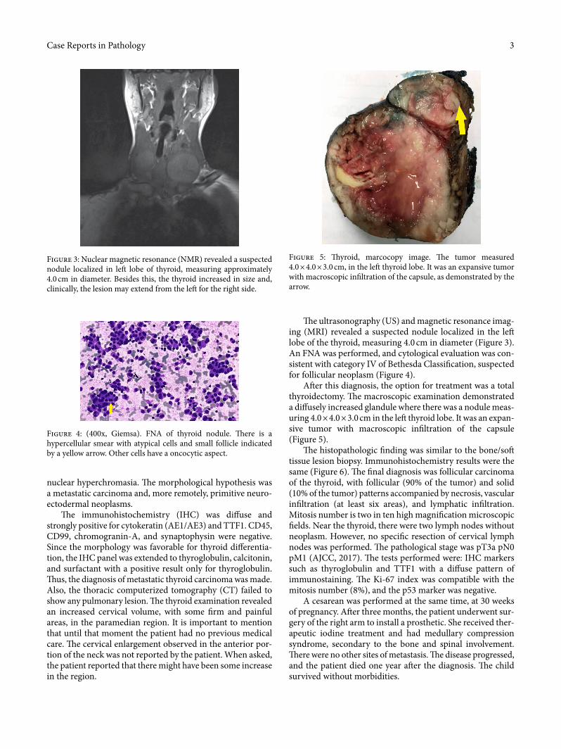
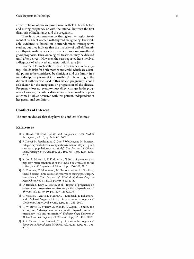
e ultrasonography (US) and magnetic resonance imag-ing (MRI) revealed a suspected nodule localized in the le� lobe of the thyroid, measuring 4.0 cm in diameter (Figure 3). An FNA was performed, and cytological evaluation was con-sistent with category IV of Bethesda Classi�cation, suspected for follicular neoplasm (Figure 4).
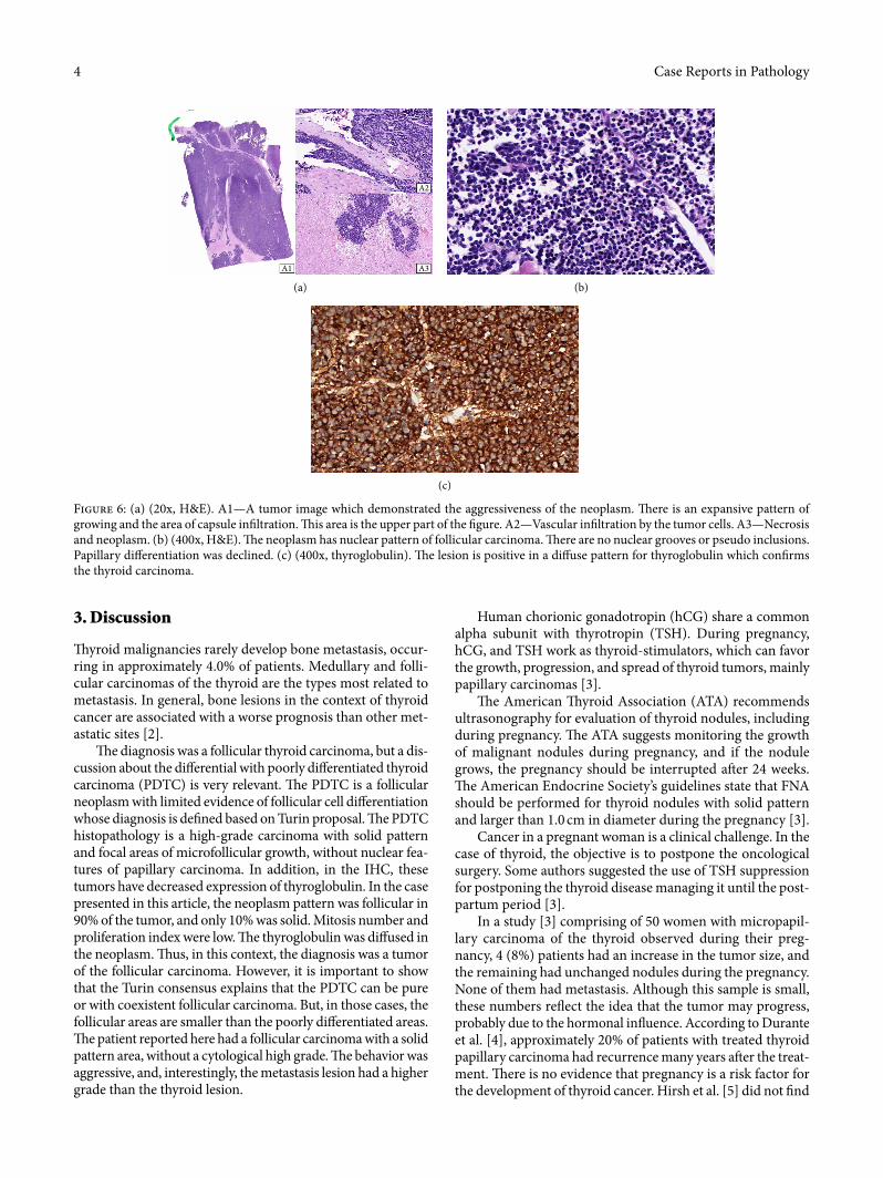
A�er this diagnosis, the option for treatment was a total thyroidectomy. e macroscopic examination demonstrated a di�usely increased glandule where there was a nodule meas-uring 4.0 × 4.0 × 3.0 cm in the le� thyroid lobe. It was an expan-sive tumor with macroscopic in�ltration of the capsule (Figure 5).
e histopathologic �nding was similar to the bone/so� tissue lesion biopsy. Immunohistochemistry results were the same (Figure 6). e �nal diagnosis was follicular carcinoma of the thyroid, with follicular (90% of the tumor) and solid (10% of the tumor) patterns accompanied by necrosis, vascular in�ltration (at least six areas), and lymphatic in�ltration. Mitosis number is two in ten high magni�cation microscopic �elds. Near the thyroid, there were two lymph nodes without neoplasm. However, no speci�c resection of cervical lymph nodes was performed. e pathological stage was pT3a pN0 pM1 (AJCC, 2017). e tests performed were: IHC markers such as thyroglobulin and TTF1 with a di�use pattern of immunostaining. e Ki-67 index was compatible with the mitosis number (8%), and the p53 marker was negative.
A cesarean was performed at the same time, at 30 weeks of pregnancy. A�er three months, the patient underwent sur-gery of the right arm to install a prosthetic. She received ther-apeutic iodine treatment and had medullary compression syndrome, secondary to the bone and spinal involvement. ere were no other sites of metastasis. e disease progressed, and the patient died one year a�er the diagnosis. e child survived without morbidities.
Figure 3: Nuclear magnetic resonance (NMR) revealed a suspected nodule localized in le� lobe of thyroid, measuring approximately 4.0 cm in diameter. Besides this, the thyroid increased in size and, clinically, the lesion may extend from the le� for the right side.
Figure 4: (400x, Giemsa). FNA of thyroid nodule. ere is a hypercellular smear with atypical cells and small follicle indicated by a yellow arrow. Other cells have a oncocytic aspect.
Figure 5: yroid, marcocopy image. e tumor measured 4.0 × 4.0 × 3.0 cm, in the le� thyroid lobe. It was an expansive tumor with macroscopic in�ltration of the capsule, as demonstrated by the arrow.
Case Reports in Pathology4
Human chorionic gonadotropin (hCG) share a common alpha subunit with thyrotropin (TSH). During pregnancy, hCG, and TSH work as thyroid-stimulators, which can favor the growth, progression, and spread of thyroid tumors, mainly papillary carcinomas [3].
e American yroid Association (ATA) recommends ultrasonography for evaluation of thyroid nodules, including during pregnancy. e ATA suggests monitoring the growth of malignant nodules during pregnancy, and if the nodule grows, the pregnancy should be interrupted a�er 24 weeks. e American Endocrine Society’s guidelines state that FNA should be performed for thyroid nodules with solid pattern and larger than 1.0 cm in diameter during the pregnancy [3].
Cancer in a pregnant woman is a clinical challenge. In the case of thyroid, the objective is to postpone the oncological surgery. Some authors suggested the use of TSH suppression for postponing the thyroid disease managing it until the post-partum period [3].
In a study [3] comprising of 50 women with micropapil-lary carcinoma of the thyroid observed during their preg-nancy, 4 (8%) patients had an increase in the tumor size, and the remaining had unchanged nodules during the pregnancy. None of them had metastasis. Although this sample is small, these numbers re¶ect the idea that the tumor may progress, probably due to the hormonal in¶uence. According to Durante et al. [4], approximately 20% of patients with treated thyroid papillary carcinoma had recurrence many years a�er the treat-ment. ere is no evidence that pregnancy is a risk factor for the development of thyroid cancer. Hirsh et al. [5] did not �nd
3. Discussion
yroid malignancies rarely develop bone metastasis, occur-ring in approximately 4.0% of patients. Medullary and folli-cular carcinomas of the thyroid are the types most related to metastasis. In general, bone lesions in the context of thyroid cancer are associated with a worse prognosis than other met-astatic sites [2].
e diagnosis was a follicular thyroid carcinoma, but a dis-cussion about the di�erential with poorly di�erentiated thyroid carcinoma (PDTC) is very relevant. e PDTC is a follicular neoplasm with limited evidence of follicular cell di�erentiation whose diagnosis is de�ned based on Turin proposal. e PDTC histopathology is a high-grade carcinoma with solid pattern and focal areas of microfollicular growth, without nuclear fea-tures of papillary carcinoma. In addition, in the IHC, these tumors have decreased expression of thyroglobulin. In the case presented in this article, the neoplasm pattern was follicular in 90% of the tumor, and only 10% was solid. Mitosis number and proliferation index were low. e thyroglobulin was di�used in the neoplasm. us, in this context, the diagnosis was a tumor of the follicular carcinoma. However, it is important to show that the Turin consensus explains that the PDTC can be pure or with coexistent follicular carcinoma. But, in those cases, the follicular areas are smaller than the poorly di�erentiated areas. e patient reported here had a follicular carcinoma with a solid pattern area, without a cytological high grade. e behavior was aggressive, and, interestingly, the metastasis lesion had a higher grade than the thyroid lesion.
A1 A3
A2
(a) (b)
(c)
Figure 6: (a) (20x, H&E). A1—A tumor image which demonstrated the aggressiveness of the neoplasm. ere is an expansive pattern of growing and the area of capsule in�ltration. is area is the upper part of the �gure. A2—Vascular in�ltration by the tumor cells. A3—Necrosis and neoplasm. (b) (400x, H&E). e neoplasm has nuclear pattern of follicular carcinoma. ere are no nuclear grooves or pseudo inclusions. Papillary di�erentiation was declined. (c) (400x, thyroglobulin). e lesion is positive in a di�use pattern for thyroglobulin which con�rms the thyroid carcinoma.
5Case Reports in Pathology
any correlation of disease progression with TSH levels before and during pregnancy or with the interval between the first diagnosis of malignancy and the pregnancy.
�ere is no consensus on the timing for the surgical treat-ment of pregnant women with thyroid malignancy. �e avail-able evidence is based on nonrandomized retrospective studies, but they indicate that the majority of well-differenti-ated thyroid malignancies in pregnancy have slow growth and good prognosis. �us, oncological treatment may be delayed until a�er delivery. However, the case reported here involves a diagnosis of advanced and metastatic disease [6].
Treatment for metastatic disease in pregnancy is challeng-ing. It holds risks for both mother and child, which are essen-tial points to be considered by clinicians and the family, in a multidisciplinary team, if it is possible [7]. According to the different authors discussed in this article, pregnancy is not a risk factor for the neoplasm or progression of the disease. Pregnancy does not seem to cause direct changes in the prog-nosis. However, metastatic disease is a relevant marker of poor outcome [7, 8], as occurred with this patient, independent of her gestational condition.
Conflicts of Interest
�e authors declare that they have no conflicts of interest.
References
[1] S. Rosas, “�yroid Nodule and Pregnancy,” Acta Médica Portuguesa, vol. 16, pp. 341–342, 2003.
[2] P. Choksi, M. Papaleontiou, C. Guo, F. Worden, and M. Banerjee, “Megan haymart; skeletal complications and mortality in thyroid cancer: a population-based study,” �e Journal of Clinical Endocrinology & Metabolism, vol. 102, no. 4, pp. 1254–1260, 2017.
[3] Y. Ito, A. Miyauchi, T. Kudo et al., “Effects of pregnancy on papillary microcarcinomas of the thyroid re-evaluated in the entire patient,” �yroid, vol. 26, no. 1, pp. 156–160, 2016.
[4] C. Durante, T. Montesano, M. Torlontano et al., “Papillary thyroid cancer: time course of recurrence during postsurgery surveillance,” �e Journal of Clinical Endocrinology & Metabolism, vol. 98, no. 2, pp. 636–642, 2013.
[5] D. Hirsch, S. Levy, G. Tsvetov et al., “Impact of pregnancy on outcome and prognosis of survivors of papillary thyroid cancer,” �yroid, vol. 20, no. 10, pp. 1179–1185, 2010.
[6] C. Modesti, P. Aceto, L. Masini, C. P. Lombardi, R. Bellantone, and L. Sollazzi, “Approach to thyroid carcinoma in pregnancy,” Updates in Surgery, vol. 69, no. 2, pp. 261–265, 2017.
[7] C. W. Rowe, K. Murray, A. Woods, S. Gupta, R. Smith, and K. Wynne, “Management of metastatic thyroid cancer in pregnancy: risk and uncertainty,” Endocrinology, Diabetes & Metabolism Case Reports, vol. 2016, no. 1, pp. 16–0071, 2016.
[8] S. S. Yu and L. A. Bischoff, “�yroid cancer in pregnancy,” Seminars in Reproductive Medicine, vol. 34, no. 6, pp. 351–355, 2016.

Stem Cells International
Hindawiwww.hindawi.com Volume 2018
Hindawiwww.hindawi.com Volume 2018
MEDIATORSINFLAMMATION
of
EndocrinologyInternational Journal of
Hindawiwww.hindawi.com Volume 2018
Hindawiwww.hindawi.com Volume 2018
Disease Markers
Hindawiwww.hindawi.com Volume 2018
BioMed Research International
OncologyJournal of
Hindawiwww.hindawi.com Volume 2013
Hindawiwww.hindawi.com Volume 2018
Oxidative Medicine and Cellular Longevity
Hindawiwww.hindawi.com Volume 2018
PPAR Research
Hindawi Publishing Corporation http://www.hindawi.com Volume 2013Hindawiwww.hindawi.com
The Scientific World Journal
Volume 2018
Immunology ResearchHindawiwww.hindawi.com Volume 2018
Journal of
ObesityJournal of
Hindawiwww.hindawi.com Volume 2018
Hindawiwww.hindawi.com Volume 2018
Computational and Mathematical Methods in Medicine
Hindawiwww.hindawi.com Volume 2018
Behavioural Neurology
OphthalmologyJournal of
Hindawiwww.hindawi.com Volume 2018
Diabetes ResearchJournal of
Hindawiwww.hindawi.com Volume 2018
Hindawiwww.hindawi.com Volume 2018
Research and TreatmentAIDS
Hindawiwww.hindawi.com Volume 2018
Gastroenterology Research and Practice
Hindawiwww.hindawi.com Volume 2018
Parkinson’s Disease
Evidence-Based Complementary andAlternative Medicine
Volume 2018Hindawiwww.hindawi.com
Submit your manuscripts atwww.hindawi.com