advanced separation methods for collagen parent a - natureblink
TRANSCRIPT
Journal of Chromatography B, 739 (2000) 3–31www.elsevier.com/ locate /chromb
Review
Advanced separation methods for collagen parent a-chains,their polymers and fragments
* ˇ´Z. Deyl , I. Miksık´ ´Institute of Physiology, Academy of Sciences of the Czech Republic, Vıdenska 1083, 14220 Prague, Czech Republic
Abstract
Current techniques used for collagen a-chains and their CNBr fragments are reviewed. Ion exchange, gel permeation,reversed-phase and affinity chromatography are discussed mainly from the preparative aspects as these are both thetechniques of choice to remove biological matrix contaminants always present in collagen preparations and techniquesroutinely used for preparative purposes. Among electromigration procedures gel electrophoresis is widely used both forintact collagen a-chains and their fragments. Recently this technique was applied also for miniaturised preparations.Immunoblotting techniques serve more specific detection of otherwise hard to distinguish different collagen polypeptidechains. Capillary electromigration techniques brought recently new aspects of understanding the behaviour of collagenproteins upon different separation modes and seem to represent a smart perspective for better quantitation of individualcollagen species. 2000 Elsevier Science B.V. All rights reserved.
Keywords: Reviews; Collagen parent a-chains
Contents
1. Introduction ............................................................................................................................................................................ 42. Standard separation procedures (isolation of fibril forming collagens from tissues)....................................................................... 5
2.1. Examples of multiple chromatographic procedures ............................................................................................................ 63. High performance chromatography ........................................................................................................................................... 64. Electromigration methods ........................................................................................................................................................ 10
4.1. Polyacrylamide gel electrophoresis................................................................................................................................... 104.2. Micropreparative gel electrophoresis ................................................................................................................................ 174.3. Detection of collagen zones by immunoblotting ................................................................................................................ 184.4. Capillary electrophoresis ................................................................................................................................................. 18
4.4.1. Separation of parent a-chains ............................................................................................................................... 185. Specific features of collagen CNBr fragments separation............................................................................................................ 246. Conclusions ............................................................................................................................................................................ 29Acknowledgements ...................................................................................................................................................................... 30References .................................................................................................................................................................................. 30
*Corresponding author. Tel.: 1420-2-475-2534; fax: 1420-2-475-2558.E-mail address: [email protected] (Z. Deyl)
0378-4347/00/$ – see front matter 2000 Elsevier Science B.V. All rights reserved.PI I : S0378-4347( 99 )00515-0
ˇ´4 Z. Deyl, I. Miksık / J. Chromatogr. B 739 (2000) 3 –31
1. Introduction which brings about a considerable internal homo-geneity of the proteins. Individual parent a-chains
The term collagen stands for a series of at least 19 can be either identical (as in type III collagen) orproteins of which the most abundant ones form different (as in type I collagen) or a mixture of bothextracellular fibrils (network structures). Additional (as in type V collagen). In type I collagen theten proteins contain collagen-like domains (for re- individual parent a-chains are called a and a ; a1 2
view see [1]). Mutations in the six different collagen triple helical molecule of type I collagen is consti-types are the cause of a number of human diseases tuted of two a and one a chains having a chain1 2
like osteogenesis imperfecta, chondrodisplasias, structure of [a (I)] a and is called g-collagen (g-1 2 2
some forms of osteoporosis, some types of osteoar- fraction) while dimers of a-chains are designed asthritis, the renal disease called Alport syndrome etc. b-fraction (composed e.g. of a a or a a chains).1 2 1 1
Fibrotic reactions, tissue remodelling and wound There are additional specific features of fibril form-healing represent additional biomedically important ing collagens: they contain hydroxyproline and hy-areas of collagen chemistry [2,3]. Interactions with droxylysine, one third of all amino acids present isreactive metabolites result in a number of storage formed by glycine residues, they are extremelyeffects of which glycation appears the most studied resistant to common proteases (except tissue andphenomenon in connection with diabetes and aging bacterial collagenases) and they are more soluble in(see e.g. [4–6]). the random coil than in native conformation. Fibril
It is beyond the scope of this review to describe in forming collagens are devoid of tryptophan, containdetail individual collagen types (their structures are little if any tyrosine residues and, except collagenschematically presented in Fig. 1) [1]. type III are practically devoid of cysteine. Also the
Briefly, the collagen superfamily can be divided proportion of methionine is low which makes itinto several classes (each containing several collagen possible to use CNBr fragmentation for the identifi-types) on the basis of structural features of individual cation of a particular collagen a-chain.members, namely fibrils forming collagens (type I, Individual collagen a-chains can be polymerisedII, III and V), collagens forming networks (type IV, either by physiological cross-links (lysinonorleucine,VIII and X), collagens found on the surface of fibrils hydroxylysinonorleucine, aldol condensation prod-(fibril associated collagens FACIT) with interrupted uct, pyridinoline (see e.g. [8–11])) or by non-physio-triple helices that include collagens type IX, XII, logical cross-linking agents (typically aldehydes (seeXIV and XIX), collagens forming beaded filaments e.g. [12–14])). Pathological crosslinking can be(type VI), collagens forming anchoring fibrils for typically caused by the interaction with reducingbasement membranes (type VII), collagens with sugars or oxidation products of unsaturated lipidstransmembrane domain (types XIII and XVII) and [13] (also monotopical interactions are possiblecollagens not yet fully characterised (types XV and [15]).XVIII). Polymerised fibril forming collagens (no matter
This review is devoted mainly to advances in whether physiologically or non-physiologically) areseparation techniques of fibril forming collagens, insoluble and their solubilization is routinely doneparticularly collagen type I, which is the best known either by mild pepsinization by which short terminalof this series of proteins, abundant in vertebrate regions possessing the polymerisation sites (cross-tissues where it constitutes a considerable proportion links) are cleaved off, or by CNBr cleavage whichof all proteins present. The building scheme of fibril results in a limited fragmentation of the parent a-forming collagens comprises a triple helical structure chains as mentioned already. Tissue collagenasescomposed of parent a-chains winding one around the (which unfortunately are difficult to obtain) split theother in a superhelical manner. Individual a-chains, triple-helical structure in two thirds from its N-end;the sequence of which in case of type I, II, III and V bacterial collagenases (Clostridium histolyticum) areis completely known [7], are of molecular mass far less specific, cleave the sequence in small frag-around 100 000–150 000 (see Table 1). About one ments (mostly tripeptides) and are therefore of littlethird of the sequence is formed by gly-pro-x repeats use in structural studies.

ˇ´Z. Deyl, I. Miksık / J. Chromatogr. B 739 (2000) 3 –31 5
Fig. 1. Schematic structures of various collagens. Reprinted from Prockop and Kivirikko [1] with permission, from the Annual Review ofBiochemistry, Vol. 64, 1995, by Annual Reviews http: / /www.annualreviews.org.
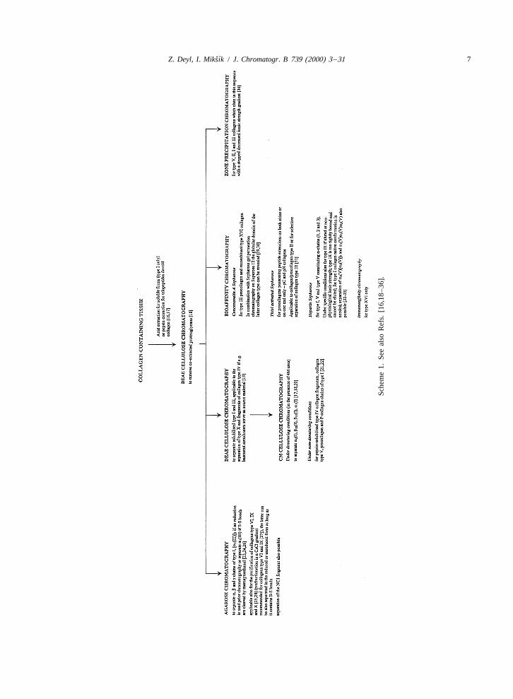
2. Standard separation procedures (isolation of review see Ref. [17]) and frequently smart combina-fibril forming collagens from tissues) tions of these are used exploiting e.g. the presence or
absence of S–S bonds in the terminal region (orSeparation of classical (low-pressure) chromatog- along the whole molecule as is the case of collagen
raphy involves quite a large number of methods. type III), charge, molecular size, the presence ofPractically all types of chromatographic operational glycosidic residues or differences in physico-chemi-modes have been exploited for this purpose (for a cal properties of individual collagen species in the
ˇ´6 Z. Deyl, I. Miksık / J. Chromatogr. B 739 (2000) 3 –31
native and denatured stage. The fundamental strategy respect to trifluoroacetic acid). The result was theabout the application of individual analytical meth- separation of a (V)a (V)a (V) and [a (V)] a (V)1 2 3 1 2 2
ods is presented in Scheme 1, details about actual [37].applications exceed the scope of this survey and canbe found in the quoted references and informationabout fractional precipitation (which may serve as a 3. High performance chromatographypre-purification step) is summarised in Table 1.
A number of rigid sorbents varying in polarity orcapable of molecular sieving have been explored
2.1. Examples of multiple chromatographic over the years.procedures Cyanopropyl bonded packings were shown to
allow separations of human type I, II and IIIExamples of the purposeful combination of in- collagens as early as in 1981 [38]. Another possi-
dividual chromatographic modes are abundant. They bility explored was the use of the so-called glycoph-frequently combine not only several chromatographic ases like LiChrosorb Diol, TSK-SW gels and last butbut almost regularly also at least one electromi- not least Separon HEMA 1000 Glc (a copolymer ofgration step. A nice example for many is the 2-hydroxyethyl methacrylate with ethylene dimeth-isolation of different species of collagen type V acrylate covalently bonded with glucose) [39–42].a-chains. In the reported procedure the first step is While the former sorbents were successfully applied
2chromatography on Fractogel EMD SO 650(S) to the separation of a number of proteins, only the3
column with 0.04 M Tris–HCl buffer pH 8.2 con- last one was shown to be applicable to collagentaining 2 M urea and 0.05 M NaCl with linear NaCl separations, in particular because of its good molecu-gradient to the final concentration of 1 M. Next lar sieving properties with relatively little of otherBakerbond PEI Butyl Scout column was used with types of interactions (though some are present asan NaCl gradient from 0 to 0.35 M over 20 min. discussed later). Standard chromatographic columnsRemoval of non-volatile buffer components was (50038 mm) packed with the Separon HEMA 1000done in the third step, a reversed-phase chromatog- Glc sorbent, particle size 12–17 mm were used withraphy, carried out with a water–acetonitrile gradient 0.05 M Tris–HCl (pH 7.5) made 2 M with respect to(10–80% acetonitrile, solvents made 0.1% with urea as mobile phase. Skin collagen type I poly-
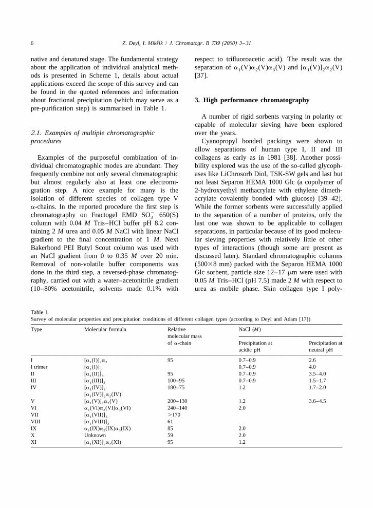
Table 1Survey of molecular properties and precipitation conditions of different collagen types (according to Deyl and Adam [17])
Type Molecular formula Relative NaCl (M)molecular massof a-chain Precipitation at Precipitation at
acidic pH neutral pH
I [a (I)] a 95 0.7–0.9 2.61 2 2
I trimer [a (I)] 0.7–0.9 4.01 3
II [a (II)] 95 0.7–0.9 3.5–4.01 3
III [a (III)] 100–95 0.7–0.9 1.5–1.71 3
IV [a (IV)] 180–75 1.2 1.7–2.01 3
[a (IV)] a (IV)1 2 2
V [a (V)] a (V) 200–130 1.2 3.6–4.51 2 2
VI a (VI)a (VI)a (VI) 240–140 2.01 2 3
VII [a (VII)] .1701 3
VIII [a (VIII)] 611 3
IX a (IX)a (IX)a (IX) 85 2.01 2 3
X Unknown 59 2.0XI [a (XI)] a (XI) 95 1.21 2 2
ˇ´Z. Deyl, I. Miksık / J. Chromatogr. B 739 (2000) 3 –31 7
Sche
me
1.Se
eal
soR
efs.
[16,
18–3
6].
ˇ´8 Z. Deyl, I. Miksık / J. Chromatogr. B 739 (2000) 3 –31
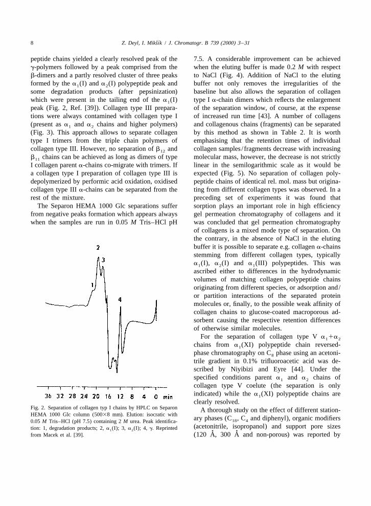
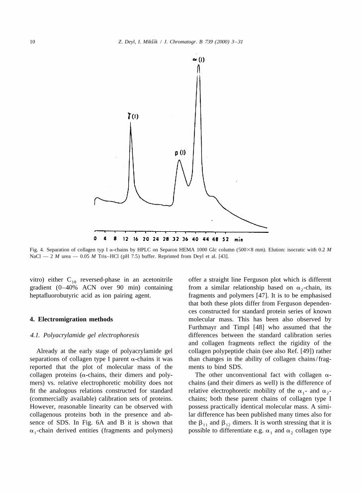
peptide chains yielded a clearly resolved peak of the 7.5. A considerable improvement can be achievedg-polymers followed by a peak comprised from the when the eluting buffer is made 0.2 M with respectb-dimers and a partly resolved cluster of three peaks to NaCl (Fig. 4). Addition of NaCl to the elutingformed by the a (I) and a (I) polypeptide peak and buffer not only removes the irregularities of the1 2
some degradation products (after pepsinization) baseline but also allows the separation of collagenwhich were present in the tailing end of the a (I) type I a-chain dimers which reflects the enlargement1
peak (Fig. 2, Ref. [39]). Collagen type III prepara- of the separation window, of course, at the expensetions were always contamined with collagen type I of increased run time [43]. A number of collagens(present as a and a chains and higher polymers) and collagenous chains (fragments) can be separated1 2
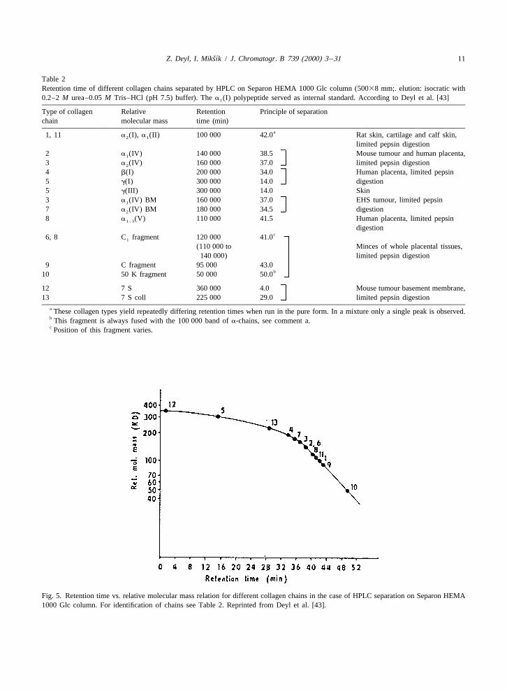
(Fig. 3). This approach allows to separate collagen by this method as shown in Table 2. It is worthtype I trimers from the triple chain polymers of emphasising that the retention times of individualcollagen type III. However, no separation of b and collagen samples / fragments decrease with increasing12
b chains can be achieved as long as dimers of type molecular mass, however, the decrease is not strictly11
I collagen parent a-chains co-migrate with trimers. If linear in the semilogarithmic scale as it would bea collagen type I preparation of collagen type III is expected (Fig. 5). No separation of collagen poly-depolymerized by performic acid oxidation, oxidised peptide chains of identical rel. mol. mass but origina-collagen type III a-chains can be separated from the ting from different collagen types was observed. In arest of the mixture. preceding set of experiments it was found that
The Separon HEMA 1000 Glc separations suffer sorption plays an important role in high efficiencyfrom negative peaks formation which appears always gel permeation chromatography of collagens and itwhen the samples are run in 0.05 M Tris–HCl pH was concluded that gel permeation chromatography
of collagens is a mixed mode type of separation. Onthe contrary, in the absence of NaCl in the elutingbuffer it is possible to separate e.g. collagen a-chainsstemming from different collagen types, typicallya (I), a (I) and a (III) polypeptides. This was1 2 1
ascribed either to differences in the hydrodynamicvolumes of matching collagen polypeptide chainsoriginating from different species, or adsorption and/or partition interactions of the separated proteinmolecules or, finally, to the possible weak affinity ofcollagen chains to glucose-coated macroporous ad-sorbent causing the respective retention differencesof otherwise similar molecules.
For the separation of collagen type V a 1a1 2
chains from a (XI) polypeptide chain reversed-1
phase chromatography on C phase using an acetoni-8
trile gradient in 0.1% trifluoroacetic acid was de-scribed by Niyibizi and Eyre [44]. Under thespecified conditions parent a and a chains of1 2
collagen type V coelute (the separation is onlyindicated) while the a (XI) polypeptide chains are1
clearly resolved.Fig. 2. Separation of collagen typ I chains by HPLC on Separon A thorough study on the effect of different station-HEMA 1000 Glc column (50038 mm). Elution: isocratic with
ary phases (C , C and diphenyl), organic modifiers18 40.05 M Tris–HCl (pH 7.5) containing 2 M urea. Peak identifica-(acetonitrile, isopropanol) and support pore sizestion: 1, degradation products; 2, a (I); 3, a (I); 4, g. Reprinted1 2
˚ ˚from Macek et al. [39]. (120 A, 300 A and non-porous) was reported by

ˇ´Z. Deyl, I. Miksık / J. Chromatogr. B 739 (2000) 3 –31 9
Fig. 3. Separation of collagen chains present in typ III before (A) and after (B) performic acid treatment. Conditions: column, SeparonHEMA 1000 Glc (50038 mm); elution, isocratic with 0.05 M Tris–HCl (pH 7.5) containing 2 M urea. Peak identification (A): 1, a (I); 2,1
a (I); 3, [a (I)] a ; 4, g; (B): 1, a (III); 2, a (I); 3, a (I); 4, [a (I)] a . Reprinted from Macek et al. [39].2 1 2 2 1 1 2 1 2 2
Fields et al. [45]. It was demonstrated that large pore peptide bonds. The best sorbents of those examinedsupports gave distorted peaks with small collagens were diphenyl or non-porous C reversed-phases; as18
and triple helical peptides, resulting in poor res- mobile phases standard water–acetonitrile gradientsolution. The formation of broad peaks has been were recommended.ascribed to conformational instability of the sepa- Reiser et al. [46] recommended for the separationrated solutes and slow cis-trans isomerisation of the of CNBr peptides of type I collagen (glycated in
ˇ´10 Z. Deyl, I. Miksık / J. Chromatogr. B 739 (2000) 3 –31
Fig. 4. Separation of collagen typ I a-chains by HPLC on Separon HEMA 1000 Glc column (50038 mm). Elution: isocratic with 0.2 MNaCl — 2 M urea — 0.05 M Tris–HCl (pH 7.5) buffer. Reprinted from Deyl et al. [43].
vitro) either C reversed-phase in an acetonitrile offer a straight line Ferguson plot which is different18
gradient (0–40% ACN over 90 min) containing from a similar relationship based on a -chain, its2
heptafluorobutyric acid as ion pairing agent. fragments and polymers [47]. It is to be emphasisedthat both these plots differ from Ferguson dependen-ces constructed for standard protein series of known
4. Electromigration methods molecular mass. This has been also observed byFurthmayr and Timpl [48] who assumed that the
4.1. Polyacrylamide gel electrophoresis differences between the standard calibration seriesand collagen fragments reflect the rigidity of the
Already at the early stage of polyacrylamide gel collagen polypeptide chain (see also Ref. [49]) ratherseparations of collagen type I parent a-chains it was than changes in the ability of collagen chains / frag-reported that the plot of molecular mass of the ments to bind SDS.collagen proteins (a-chains, their dimers and poly- The other unconventional fact with collagen a-mers) vs. relative electrophoretic mobility does not chains (and their dimers as well) is the difference offit the analogous relations constructed for standard relative electrophoretic mobility of the a - and a -1 2
(commercially available) calibration sets of proteins. chains; both these parent chains of collagen type IHowever, reasonable linearity can be observed with possess practically identical molecular mass. A simi-collagenous proteins both in the presence and ab- lar difference has been published many times also forsence of SDS. In Fig. 6A and B it is shown that the b and b dimers. It is worth stressing that it is11 12
a -chain derived entities (fragments and polymers) possible to differentiate e.g. a and a collagen type1 1 2
ˇ´Z. Deyl, I. Miksık / J. Chromatogr. B 739 (2000) 3 –31 11
Table 2Retention time of different collagen chains separated by HPLC on Separon HEMA 1000 Glc column (50038 mm;. elution: isocratic with0.2–2 M urea–0.05 M Tris–HCl (pH 7.5) buffer). The a (I) polypeptide served as internal standard. According to Deyl et al. [43]1
Type of collagen Relative Retention Principle of separationchain molecular mass time (min)
a1, 11 a (I), a (II) 100 000 42.0 Rat skin, cartilage and calf skin,2 1
limited pepsin digestion2 a (IV) 140 000 38.5 Mouse tumour and human placenta,1
3 a (IV) 160 000 37.0 limited pepsin digestion2
4 b(I) 200 000 34.0 Human placenta, limited pepsin5 g(I) 300 000 14.0 digestion5 g(III) 300 000 14.0 Skin3 a (IV) BM 160 000 37.0 EHS tumour, limited pepsin1
7 a (IV) BM 180 000 34.5 digestion2
8 a (V) 110 000 41.5 Human placenta, limited pepsin1–3
digestionc6, 8 C fragment 120 000 41.01
(110 000 to Minces of whole placental tissues,140 000) limited pepsin digestion
9 C fragment 95 000 43.0b10 50 K fragment 50 000 50.0
12 7 S 360 000 4.0 Mouse tumour basement membrane,13 7 S coll 225 000 29.0 limited pepsin digestion
a These collagen types yield repeatedly differing retention times when run in the pure form. In a mixture only a single peak is observed.b This fragment is always fused with the 100 000 band of a-chains, see comment a.c Position of this fragment varies.
Fig. 5. Retention time vs. relative molecular mass relation for different collagen chains in the case of HPLC separation on Separon HEMA1000 Glc column. For identification of chains see Table 2. Reprinted from Deyl et al. [43].

ˇ´12 Z. Deyl, I. Miksık / J. Chromatogr. B 739 (2000) 3 –31
Fig. 6. Relation between mobility (migration distance) in polyacrylamide gel electrophoresis and molecular mass for collagen cyanogen´bromide fragments and chains: (a) in the presence of of SDS, and (b) in the absence of SDS. Reprinted from Svojtkova et al. [47].
I parent chains both in the presence or absence of the and Rodbard [50] that for the separation of sub-surfactant and this difference persists also with the stances with parallel lines on a Ferguson plot theb and b dimers. The suggestion [48] that it is the optimal separation should be obtained at zero gel11 12
rigidity of the collagen polypeptide chains which is concentration, which in practice is approximated bythe cause of the different migration of collagen 3% gel with 20% crosslinking.a-chains, polymers and fragments can be accepted There are two conclusions that emerge from thewhen comparing the behaviour of these entities with above consideration. First, dilute gels are likely tothe standard proteins calibration set, however, they give the chance to separate chain polymers higherare unlikely to explain the mobility difference be- than trimers which in the conventional gels (5–tween the a and a chains and their polymers as it 7.5%) remain on the start (Fig. 8). The other aspect,1 2
emerges both from the amino acid composition and as proven experimentally, is that for polymers com-the total sequence of these polypeptides that they are posed of a - and a -chains the dependence of the1 2
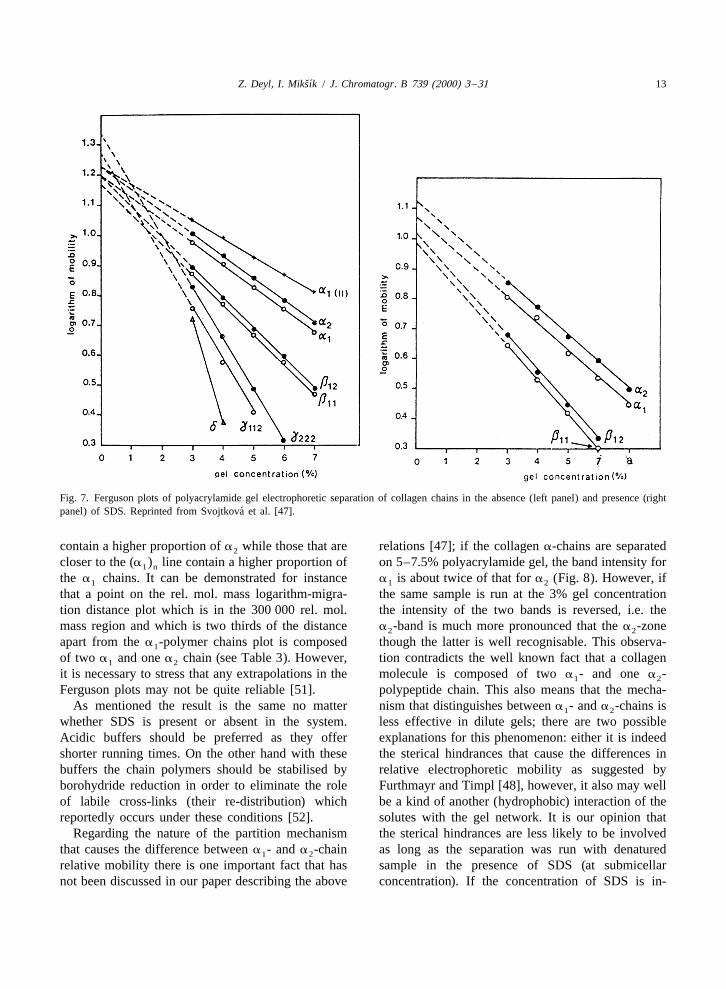
very much alike indeed. relative electrophoretic mobility on the logarithm ofThe plots shown in Fig. 7, left panel (constructed the molecular mass falls between the extreme two
in the absence of SDS in the system) indicate that the lines given by the polymers consisting of a - and1
straight lines corresponding to a - and a -chains a -chains only, e.g. the point for the b fraction lies1 2 2 12
(and their polymers / fragments) are parallel. It can mid-way between the extreme lines representing thetherefore be concluded that the retardation coeffi- homodimers.cients (the slopes of these lines) are identical. This is The above results allow a simple way for thein concert with the fact that the molecular mass of determination of collagen polymeric structures inboth these polypeptide chains is nearly identical. terms of constituting a-chains using 3% poly-
A similar conclusion was drawn from data ob- acrylamide, no matter whether the separation istained when the separations were run in the presence carried out in the presence or absence of SDS.of SDS (Fig. 7, right panel). It appeared therefore Provided that the Ferguson plot of (a ) and (a )1 n 2 n
that the difference in electrophoretic mobility stems can be constructed, the polymers containing differentfrom the difference in a physical property of these proportion of either chain fall in between the linespolypeptide chains the nature of which is so far obtained with pure polymers (containing a singleunknown. It has been demonstrated by Chrambach chain only). Those closer to the (a ) polymer line2 n
ˇ´Z. Deyl, I. Miksık / J. Chromatogr. B 739 (2000) 3 –31 13
Fig. 7. Ferguson plots of polyacrylamide gel electrophoretic separation of collagen chains in the absence (left panel) and presence (right´panel) of SDS. Reprinted from Svojtkova et al. [47].
contain a higher proportion of a while those that are relations [47]; if the collagen a-chains are separated2
closer to the (a ) line contain a higher proportion of on 5–7.5% polyacrylamide gel, the band intensity for1 n
the a chains. It can be demonstrated for instance a is about twice of that for a (Fig. 8). However, if1 1 2
that a point on the rel. mol. mass logarithm-migra- the same sample is run at the 3% gel concentrationtion distance plot which is in the 300 000 rel. mol. the intensity of the two bands is reversed, i.e. themass region and which is two thirds of the distance a -band is much more pronounced that the a -zone2 2
apart from the a -polymer chains plot is composed though the latter is well recognisable. This observa-1
of two a and one a chain (see Table 3). However, tion contradicts the well known fact that a collagen1 2
it is necessary to stress that any extrapolations in the molecule is composed of two a - and one a -1 2
Ferguson plots may not be quite reliable [51]. polypeptide chain. This also means that the mecha-As mentioned the result is the same no matter nism that distinguishes between a - and a -chains is1 2
whether SDS is present or absent in the system. less effective in dilute gels; there are two possibleAcidic buffers should be preferred as they offer explanations for this phenomenon: either it is indeedshorter running times. On the other hand with these the sterical hindrances that cause the differences inbuffers the chain polymers should be stabilised by relative electrophoretic mobility as suggested byborohydride reduction in order to eliminate the role Furthmayr and Timpl [48], however, it also may wellof labile cross-links (their re-distribution) which be a kind of another (hydrophobic) interaction of thereportedly occurs under these conditions [52]. solutes with the gel network. It is our opinion that
Regarding the nature of the partition mechanism the sterical hindrances are less likely to be involvedthat causes the difference between a - and a -chain as long as the separation was run with denatured1 2
relative mobility there is one important fact that has sample in the presence of SDS (at submicellarnot been discussed in our paper describing the above concentration). If the concentration of SDS is in-
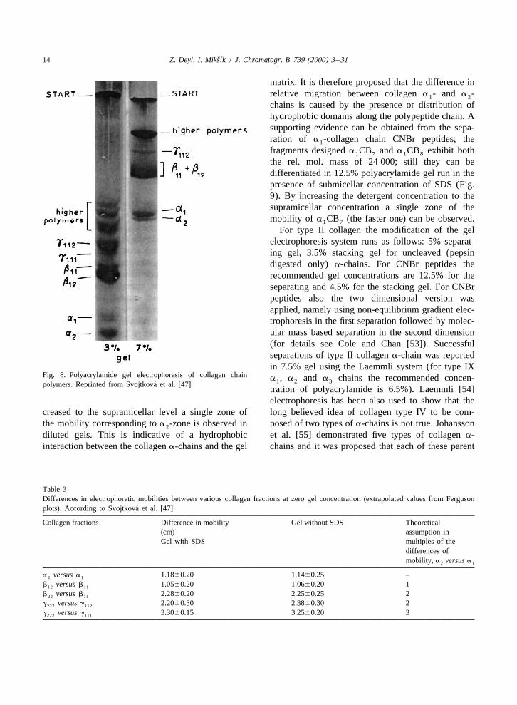
ˇ´14 Z. Deyl, I. Miksık / J. Chromatogr. B 739 (2000) 3 –31
matrix. It is therefore proposed that the difference inrelative migration between collagen a - and a -1 2
chains is caused by the presence or distribution ofhydrophobic domains along the polypeptide chain. Asupporting evidence can be obtained from the sepa-ration of a -collagen chain CNBr peptides; the1
fragments designed a CB and a CB exhibit both1 7 1 8
the rel. mol. mass of 24 000; still they can bedifferentiated in 12.5% polyacrylamide gel run in thepresence of submicellar concentration of SDS (Fig.9). By increasing the detergent concentration to thesupramicellar concentration a single zone of themobility of a CB (the faster one) can be observed.1 7
For type II collagen the modification of the gelelectrophoresis system runs as follows: 5% separat-ing gel, 3.5% stacking gel for uncleaved (pepsindigested only) a-chains. For CNBr peptides therecommended gel concentrations are 12.5% for theseparating and 4.5% for the stacking gel. For CNBrpeptides also the two dimensional version wasapplied, namely using non-equilibrium gradient elec-trophoresis in the first separation followed by molec-ular mass based separation in the second dimension(for details see Cole and Chan [53]). Successfulseparations of type II collagen a-chain was reportedin 7.5% gel using the Laemmli system (for type IX
Fig. 8. Polyacrylamide gel electrophoresis of collagen chain a , a and a chains the recommended concen-1 2 3´polymers. Reprinted from Svojtkova et al. [47].tration of polyacrylamide is 6.5%). Laemmli [54]electrophoresis has been also used to show that the
creased to the supramicellar level a single zone of long believed idea of collagen type IV to be com-the mobility corresponding to a -zone is observed in posed of two types of a-chains is not true. Johansson2
diluted gels. This is indicative of a hydrophobic et al. [55] demonstrated five types of collagen a-interaction between the collagen a-chains and the gel chains and it was proposed that each of these parent
Table 3Differences in electrophoretic mobilities between various collagen fractions at zero gel concentration (extrapolated values from Ferguson
´plots). According to Svojtkova et al. [47]
Collagen fractions Difference in mobility Gel without SDS Theoretical(cm) assumption inGel with SDS multiples of the
differences ofmobility, a versus a2 1
a versus a 1.1860.20 1.1460.25 –2 1
b versus b 1.0560.20 1.0660.20 112 11
b versus b 2.2860.20 2.2560.25 222 11
g versus g 2.2060.30 2.3860.30 2222 112
g versus g 3.3060.15 3.2560.20 3222 111

ˇ´Z. Deyl, I. Miksık / J. Chromatogr. B 739 (2000) 3 –31 15
Type X collagen parent chains were separated bythe nearly standard SDS–PAGE Laemmli procedureusing 8% polyacrylamide gel under reducing or non-reducing conditions [56]. Type XI collagen of thestoichiometry a (XI), a (XI) and a (XI) represents1 2 3
a minor constituents in type II collagen chain and itis typical in undergoing complex post-transcriptionalmodifications. For electrophoretic separation 6%separating and 4% stacking gel are used; the samplebuffer reported was 125 mM Tris–HCl pH 6.8, 10%glycerol, 4% SDS, 10 mM dithiothreitol and 0.004%Bromphenol blue. In order to allow for betteridentification of high molecular mass components thestandard acrylamide:bisacrylamide ratio in blottingexperiments 1:0.027 was changed to 1:0.006 [57].
Procollagens of type I collagen are accessible toseparation in the standard PAGE arrangement(Laemmli) with 5% separating and 3% stacking gels.Two-dimensional separations of a(I) chains weredescribed by Debey et al. [58]. Polyacrylamide gelelectrophoresis can serve also for revealing a (I) and1
a (I) chains shortened by brief proteolysis (e.g. 52
min at 388C) or even fragments partly treated withvertebrate collagenase (this approach was applied toreveal aminoacid substitution in a wild type ofosteogenesis imperfecta with the conclusion thatsuch replacements function as core sites stabilisingthe collagen helix). A similar approach of gel
Selectrophoresis of shortened a-chains, typically a1
type I from human bone refers to standard conditionsused for intact a-chains [59]. It is also possible toseparate individual a-chains (shown for collagentype I) in e.g. 5% polyacrylamide gel, cut out theseparated zones, use them for cyanogen bromideFig. 9. Separation of CNBr peptides in 12% polyacrylamide gel;
3note that peptides of identical molecular mass (24.10 –a CB and cleavage and separate the CNBr fragments in the1 7
a CB ).1 8 second dimension on a 12.5% gel. For detaileddescription of this kind of procedures see Refs.
type IV collagen a-chains forms a specific subclass [60,61]. The method described by Bonadio et al. [61]with a specific chain composition. It was concluded ran as follows: collagen proteins were separated inthat type IV collagen can be categorised into two the first dimension on 5% SDS–PAGE under reduc-subclasses, one containing a (IV) and a (IV) chains ing conditions followed by the separation of CNBr1 2
while the other is composed of a (IV) and a (IV) peptides as described in the legend to the Fig. 10.3 4
polypeptide species. The final idea is that between Alternatively, CNBr peptides could be mapped in amolecules of the same chain composition polymeri- two-dimensional system which employed isoelectricsation proceeds rather easily while cross-linking focusing in the first dimension. Collagens werebetween different a-chains is restricted setting thus incubated for 5 h at 378C in 70% formic acid thatdefinite limits upon their collagen type polymeri- contained CNBr, 20 mg/ml, under N atmosphere.2
sation. To terminate the reaction, samples were diluted with

ˇ´16 Z. Deyl, I. Miksık / J. Chromatogr. B 739 (2000) 3 –31
a (I)CB cyanogen bromide fragment which results1 6
in partial recovery of a (I) chains in the form of1
disulfide bonded dimers [62].Polyacrylamide gel electrophoresis combined with
immunoblotting is widely used in protein electro-phoresis to improve detection specifity. It is notsurprising that examples of this technique can befound also in the area of collagen separation. Forinstance identification of a and a chains of1A 1B
collagen type XII and a collagen chain and its1
dimer from collagen type XIV can be done in thisway [63].
Two dimensional polyacrylamide gel electropho-resis has been applied to reveal a new form oftumour and foetal collagen typical in its capability tobind laminin. Non equilibrium polyacrylamide gelelectrophoresis followed the procedure of O’Farell etal. [64] using pH 3–10 and 5–7 ampholytes with 4%gel. Denatured samples were investigated. In foetaland tumour tissue acidic components were foundwhich upon isolation from the 2 D gels and CNBrcleavage gave a pattern differing in at least two
Fig. 10. Autoradiogram of medium collagens synthesized by fractions of apparent mol. mass between 24 andosteogenesis imperfecta, parental, and control cells after cleavage 329?10 . It is noticeable that a (I)CB , a (I)CB and1 6 1 8with cyanogen bromide. For CNBr peptide mapping, a chains
a (I)CB peptides were completely absent in thefrom medium (after pepsin) were separated in the first dimension 1 7
on 5% SDS–PAGE, the lanes were cut out and proteins digested acidic components [65].in the gel with CNBr, and the CNBr peptides were separated by SDS gel electrophoresis (6.5%) under reducingSDS–PAGE in the second dimension. A schematic diagram of the conditions has been used also for revealing chaina1(I) chain with its CNBr peptides indicated by numbers is
heterogeneity in type VI collagen from confluentpresented at the bottom of the figure. The relative electrophoreticcultures of nuchal ligament fibroblasts. 35-Smobilities of of a(I) CNBr peptides from normal a1(I) chains (s)
and overmodified a1(I) chains (d) is shown. Reprinted from methionine labelled medium proteins were concen-Bonadio et al. [61] with permission. trated by precipitation with (NH ) SO to 30%4 2 4
saturation (fluorographic detection). There were twointeresting observations with this experiment, namelythat the a (VI) and a (VI) species were more1 2
water and lyophilized (32). The peptides were abundant in the skin samples investigated, whiledissolved in 4 M urea, 2.5% Triton X-100, and 4% ligamentum nuchae samples were consistent with theampholines and then separated by isoelectric focus- idea of a heterotrimeric structure and, second, that ining on 23115 mm cylindrical polyacrylamide gels. spite of being different in their primary structure theyThe gels were equilibrated after electrophoresis in comigrated in the gels used (rel. mol. masses of0.5% ammonium persulfate and then sample buffer 140 000 for a 1a (VI), 260–280 000 for a (VI). It1 2 3
lacking SDS and urea then overlaid on 12.5% gels has, however, to be borne in mind that type VIthat contained no urea. Second-dimension electro- monomer contains a relatively short triple helicalphoresis was performed as described above. Pro- region (105 nm) with large globular domains at bothcedures like these have been successfully applied to its C- and N-end [66]. The three polypeptidesdetect the biochemical background of some inherited representing the major constituents of type VI col-diseases. Typically in lethal osteogenesis imperfecta lagen from bovine lens capsule can be separated byan additional cysteine residue is present in the standard Laemmli procedure and identified by elec-

ˇ´Z. Deyl, I. Miksık / J. Chromatogr. B 739 (2000) 3 –31 17
trotransfer blotting and immunoreaction according to acid) and destained in 40% methanol, 10% aceticthe procedure described by Kyhse-Andersen [67]. acid. Proteinase activity was revealed as cleared
For gel electrophoretic examination of collagen (unstained) zones.type XIII the samples were boiled with SDS–PAGEsample buffer (with or without reduction with 10 4.2. Micropreparative gel electrophoresismM 2-mercaptoethanol) followed by electrophoresisand transferred onto nitrocellulose membranes [68]. A simple method for semipreparative (microscale)The anti type XIII collagen antisera and mouse isolation of type I, II, III and V a-chains by sodiumanti-human b-1 integrin antibodies were applied at dodecyl sulfate electrophoresis followed by electro-dilution 1:1000 to the filters, and the affinity purified elution was developed by Acil et al. [72]: 4%antibodies were used at concentration 5 mg/ml. The polyacrylamide gels were used (6% for the stackingfilters were washed thoroughly after 1 h of primary gel) and the very electrophoresis was run in 63 mMantibody incubation at room temperature, and then Tris, 2% SDS and 10% glycerol containing runincubated with horseradish peroxidase conjugate buffer which was made 0.01% with respect toanti-rabbit or anti-mouse secondary antibody at a bromphenol blue (pH 6.8). Following the electro-dilution of 1:5000–1:10 000. The immunosignal was phoretic separation the protein bands were blotted ondetected after washing using the enhanced PVDF [poly(vinylidene difluoride)] Immobilon Pchemiluminiscence system and film (Amersham membrane 0.45 mm pore size. The gels were washedPharmacia Biotech.). in a transfer buffer (40 mM 6-amino-N-hexanacid,
SDS gel electrophoresis in dilute gels (4%) can be 5% methanol, 0.1% SDS pH 7.2) for 5 min (toused also to reveal a-chain aggregates. Crude col- reduce the amount of bicine and Tris. During thislagen preparations display two a, two b and two period the PVDF membrane was rinsed with 100%g-bands (which are believed to represent in- methanol for 2 min and stored in deionized water.tramolecularly cross-linked a-chains). In addition The gels were sandwiched between a sheet of PVDFfour other bands with rather slow mobility (slower membrane and several sheets of blotting paper andthan the g-bands) were ascribed to the presence of assembled in the blotting apparatus. Blotting wasintermolecular cross-links [69,70]. done for 2.5 h at 100 mA (48C). After electroblotting
Polyacrylamide gel electrophoresis is also applic- the Immobilon (PVDF) membrane was washed withable for tissue collagenase activity assays; an easily deionized water for 5 min and stained with 0.1%applicable version is that published in [71]. BioRad Coomassie blue in 50% methanol for 5 min followedmini protean system was used in this case with 10% by destaining in 50% methanol–10% acetic acid forpolyacrylamide gels of 10 cm high and 1.5 mm 5–10 min and air dried. The blotted protein bandsthickness. Duracryl from Millipore (containing 1 mg/ were cut off, pooled in a test tube, soaked in 1 mlml gelatine or 0.2–1 mg/ml collagen) was dispersed elution buffer (50 mM Tris, 2% SDS, 1% Tritonin buffered solution containing 2.5 ml gel, 1.5 M X-100, pH 9) and shaken overnight. After elution ofTris–HCl pH 8.8, 100 ml of 10% SDS, 4 ml the a-chains the membrane slurry was removed bypolyacrylamide in 0.125 M Tris pH 6.8. The gels filtration (30 000 rel. mol. mass cut off) and cen-were polymerised by adding 50 ml of 10% am- trifuged.monium persulfate and 10 ml 0.1% TEMED. Sam- For electroelution the protein bands visualised byples were diluted half in 1 M Tris pH 6.8 (containing Coomassie Brilliant Blue were cut off, placed into an50% glycerol and 0.4% Bromphenol blue). The very Eppendorf tube, soaked in the defixing buffer (6 mMseparation followed the Laemmli conditions. After urea, 192 mM bicine, 25 mM Tris, 0.2% SDS and 5the electrophoresis was brought to an end the gels mM dithiothreitol) for 0.5 h at room temperature towere washed twice in 200 ml of 2.5% Triton X100 bring the collagen a-chains into solution. The slices(300 ml each) and incubated in 100 mM Tris–HCl, 5 were then equilibrated in electrophoresis buffer (192mM CaCl , 0.005% Brij, 0.01% NaN pH at 378C mM bicine, 25 mM Tris and 1.73 mM SDS pH 8.5)2 3
for 6–48 h. Gels were then stained with Coomassie for 5 min, cut into small pieces and placed into anBrilliant Blue G 280 (50% methanol, 10% acetic electroelution glass tube; electroelution was done at
ˇ´18 Z. Deyl, I. Miksık / J. Chromatogr. B 739 (2000) 3 –31
10 mA per tube for 2 h at room temperature. The the analytes involved. Therefore for quantitation ofdifferent a-chains were transferred from the gel slice individual collagen types evaluation by the presencethrough a frit into the membrane cap and retained by of CNBr marker peptides is preferred [78]. Thedialysis (15 000 rel. mol. mass cut off). advantage of this approach is further stressed by the
fact that insoluble collagens present in tissues can be4.3. Detection of collagen zones by released (and assayed) by CNBr cleavage.immunoblotting
4.4.1. Separation of parent a-chainsLocalisation of the individual collagen zones to be Successful separations of collagen a-chains can be
eluted from the polyacrylamide gel can be done by obtained (i) in very diluted buffers (typically 2.5 mMthe blotting technique. To increase specificity of sodium borate pH 9.2) [77], (ii) in acid buffersdetection, immunoblotting procedures are gaining (pH|2.5, about 25 mM buffers), (iii) in the presencepopularity, mainly because there is no chance to of a neutral surfactant in the sample (but not in thedistinguish e.g. between individual collagen poly- background electrolyte) [80] or (iv) in acid buffers ofpeptide a-chains by direct staining procedures. moderate ionic strength (pH 2.5; |75 mM buffers) in
Immunoblotting has been described in detail for the presence of copolymeric liquid crystals (Pluronictype II, V and XI collagens by Ricard-Blum et al. F-127) [79] in the run buffer.[73] and Hartmann et al. [74]. Native collagen In an early investigation [77] it was demonstratedmolecules were transferred to nitrocellulose (0.45 that collagen type I, II, V, IX and XI constitutingmm pore size, Schleicher and Schuell, Dassel) and a-chains and their chain polymers can be separatedwere blotted at 0.4 A for 25 min with 0.7% acetic in 2.5 mM sodium tetraborate buffer at pH 9.2 in lessacid as transfer buffer. than 15 min. in a 50 cm3100 mm fused-silica
A more detailed protocol has already been re- capillary. The separations are relatively insensitive toported originally by Hartmann et al. [74]. After the voltage applied. Although separations weretransfer, the nitrocellulose membrane was incubated routinely made at 18 kV per 50 cm capillary,for 1 h at 378C to prevent non-specific binding, and satisfactory results were obtained at 10–15 kV asprobed overnight with the respective collagen type well; naturally the run times were proportionallypolyclonal antibody diluted 1:50 in phosphate-buf- increased. On the other hand these separations arefered saline–3% bovine serum albumin. Immuno- extremely sensitive to capillary overloading. Thisdetection was achieved using a biotinylated sec- should be positively avoided as otherwise poorlyondary antibody using avidin-biotinylated horserad- reproducible electropherograms are obtained. It wasish peroxidase complexes. 4-Chloro-1-naphthol was argued that one of the reasons may be the length ofadded as colour substrate. the sample applied. In the view of later results it is
clear that the main reason for such irregularities is4.4. Capillary electrophoresis the protein adherence to the capillary wall [80].
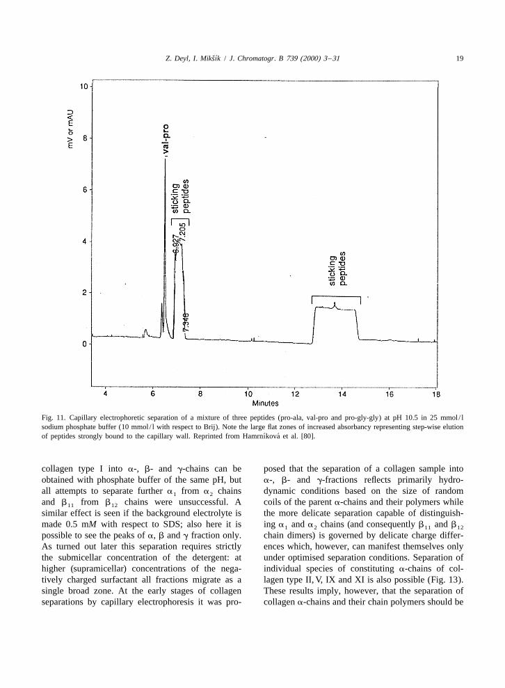
Clusters of adhering protein are under such circum-Successful separation of proteins by capillary stances released at a later stage of the separation
electrophoresis stimulated attempts to use this tech- owing to the endoosmotic flow, and irregular spikesnique also for the separation of collagens and their emerge (as proven by separating model peptidesfragments. In spite of the wide perspectives only two possessing sequences similar to those found inpapers of other authors have been published to our collagens, Fig. 11; if however the amount of thebest knowledge so far [75,76]. Though attempts to sample applied is kept within specific limits and ifseparate different collagen constituting a-chains [77] the separation is run at pH sufficiently high (9.6)have been quite successful at least as far as the these effects can be abolished. Selectivity withqualitative analysis is concerned, it turned out soon respect to the separation a and a chains is1 2
that direct quantitation of parent a-chains of fibrous considerably influenced by the presence or absenceforming collagens suffers from strong interactions of (submicellar) concentration of SDS in the back-between the inner surface of bare silica capillary and ground electrolyte (Fig. 12). The separation of
ˇ´Z. Deyl, I. Miksık / J. Chromatogr. B 739 (2000) 3 –31 19
Fig. 11. Capillary electrophoretic separation of a mixture of three peptides (pro-ala, val-pro and pro-gly-gly) at pH 10.5 in 25 mmol / lsodium phosphate buffer (10 mmol / l with respect to Brij). Note the large flat zones of increased absorbancy representing step-wise elution
´ ´of peptides strongly bound to the capillary wall. Reprinted from Hamrnıkova et al. [80].
collagen type I into a-, b- and g-chains can be posed that the separation of a collagen sample intoobtained with phosphate buffer of the same pH, but a-, b- and g-fractions reflects primarily hydro-all attempts to separate further a from a chains dynamic conditions based on the size of random1 2
and b from b chains were unsuccessful. A coils of the parent a-chains and their polymers while11 12
similar effect is seen if the background electrolyte is the more delicate separation capable of distinguish-made 0.5 mM with respect to SDS; also here it is ing a and a chains (and consequently b and b1 2 11 12
possible to see the peaks of a, b and g fraction only. chain dimers) is governed by delicate charge differ-As turned out later this separation requires strictly ences which, however, can manifest themselves onlythe submicellar concentration of the detergent: at under optimised separation conditions. Separation ofhigher (supramicellar) concentrations of the nega- individual species of constituting a-chains of col-tively charged surfactant all fractions migrate as a lagen type II, V, IX and XI is also possible (Fig. 13).single broad zone. At the early stages of collagen These results imply, however, that the separation ofseparations by capillary electrophoresis it was pro- collagen a-chains and their chain polymers should be

ˇ´20 Z. Deyl, I. Miksık / J. Chromatogr. B 739 (2000) 3 –31
positively influenced by adding a polymer withmolecular sieving properties to the run buffer.
As shown in Fig. 14, SDS electrophoresis incapillaries containing linear polyacrylamide offersseparations of constituting a-polypeptide chains aswell as their polymers which are at least comparablewith slab gel electrophoresis [81]. Moreover thisapproach offers the possibility to quantitate the areapercentage of individual peaks, a procedure whichwith slab gel separations is less convenient.
Moreover capillary electrophoresis in non-cross-linked gels offers the possibility of separating chainpolymers of rel. mol. mass 300 000 and higher. Thisis of considerable importance because no methodoffering sufficient sensitivity is available for analys-ing such polymers (as a matter of fact in most casesthese emerge as a single broad peak). In diluted slabgels these can be separated as shown in Fig. 8;chromatographic methods don’t offer sufficientselectivity.Fig. 12. Capillary zone electrophoretic analysis (2.5 mM sodium
tetraborate buffer, pH 9.2) of (A) insoluble collagen solubilized The practical applicability of electrophoresis inunder denaturing conditions (428C, 30 min), (B) isolated collagen linear polyacrylamide gels is shown in Fig. 15,type I and (C) same sample as in B but run in the presence of 0.5
where the separation of a glycated collagen sample ismM SDS. Reprinted from Deyl et al. [77].shown. There are two features in which such profiles
Fig. 13. Capillary zone electrophoretic separation (2.5 mM sodium tetraborate buffer, pH 9.2) of collagens (A) type II, (B) type V, (C) typeIX and (D) type XI. Reprinted from Deyl et al. [77].

ˇ´Z. Deyl, I. Miksık / J. Chromatogr. B 739 (2000) 3 –31 21
Fig. 15. Capillary gel electrophoresis profiles of collagen chains(4% polyacrylamide, Tris–glycine buffer pH 8.8) after incubationwith glucose for 4 days (A) and 7 days (B). Reprinted from Deyl
ˇ´and Miksık [81].
differ from untreated collagen electropherograms,namely in splitting the a-peaks into two each, and inthe increased peaks of the g-chain polymers andhigher. Both these effects can be ascribed to theinteraction of free lysine amino groups with glucose.It has been shown by Reiser et al. [46,82] that suchinteractions are non-specific, affecting several lysineresidues in the collagen molecule.
Based on the above observation it is feasible toassume that splitting of the two a-chain peaks intofour upon incubation may reflect monotopical bind-ing of glucose to several sites along the collagen
Fig. 14. Capillary gel electrophoresis profiles of collagen chains a-chain, which should result in a decrease of positive(4% polyacrylamide, Tris–glycine buffer pH 8.8). (A) Complete charges of these polypeptide chains (it does notsample, (B) a-region proteins sampled from a preceding slab gel
exclude changes in the hydrophobic properties of therun, (C) as (B), but b-region excised from slab gel. Slab gelmolecules involved and possibly some conformation-electrophoresis is shown on top of the figure (cathode on the right
ˇ´side of the gel). Reprinted from Deyl and Miksık [81]. al alterations of the polypeptides as well). The higher

ˇ´22 Z. Deyl, I. Miksık / J. Chromatogr. B 739 (2000) 3 –31
proportion of g- and higher a-chain polymers could comparing these profiles with those obtained bybe explained on the basis of glucose-polymerising standard reversed-phase chromatography (for morereactions, which eventually lead to the decreased details see the part on CNBr peptide separations).solubility of the treated collagen samples (see These profiles exhibit a striking similarity: the larger[46,82]). peptides migrate more slowly than the smaller ones.
To explain splitting of the a-chain corresponding This was interpreted in such a way that longer CNBrpeaks in more detail is at the moment difficult. The peptides, owing to the large internal homogeneity inanomalous behaviour of collagen a-chains in slab gel the collagen polypeptide chains should possess moreelectrophoresis has been already discussed (see hydrophobic domains than the small ones and if it isabove). Glycation of collagen a-chains not only the hydrophobic interactions that are involved in thedecreases the number of positive charges per a- partition mechanism the larger peptides must inchain, but also introduces a relatively large sugar electrophoresis come later before the detector’smolecule into the structure which may further alter window. To bring this mechanism to usable resultsthe number of ionizable side chain groups (OH very delicate optimisation is needed (otherwise allfunctionalities in the sugar moiety) and/or change solutes emerge in a single peak). Encouraged by thethe hydrophobic properties of the a-chain. It is also work of Quirino et al. [84] (on stacking of reversenot clear whether or not glycated and non-glycated moving micelles and a water plug and by the work of
¨collagen a-chains bind the same amount of SDS; any Lloyd and Watzig [85] who showed that introducingalteration in this respect will result in changes of the an SDS wash can effectively remove all peptides /electromigration properties of the protein molecules proteins sticking to the capillary wall) we finallyin the polyacrylamide gel matrix. It has been docu- developed a system in which no detergent wasmented that at low gel concentrations (below 7.5) present in the background electrolyte but the sampleconsiderably higher molecular masses are revealed was dissolved in the background buffer containingby Ferguson’s plots for glycoproteins if the gels are 33 mM Brij. As shown in Fig. 16 a completecalibrated with the standard protein sets and such separation of a and a chains, b and g polymers1 2
effects can be expected for glycated proteins as well was obtained.[46,83]. The results reported so far bring about strong
Though electrophoresis in linear polyacrylamide evidence that in capillary electrophoresis of parentgels is well acceptable and offers results superior to collagen a-chains and chain polymers the partitionall other methods, it still suffers one considerable mechanism involves both ionic and hydrophobicdisadvantage, namely the gels don’t survive more interactions and bears all the features of a mixed-than five runs, before being clogged and a systematic mode separation process. As a matter of fact becauseerror in decreasing the peak sizes was observed with the separation exploits also the properties of thesubsequent runs even if the separations were run capillary wall, it can be considered a special case ofwith heat denatured samples and at elevated tempera- electrochromatography in open tubes (capillaries).tures (508C) to minimise renaturation and possible Taking this into consideration it can be predicted thatassociation of individual a-chains into higher poly- a molecular sieve capable of exerting synergisticmers. hydrophobic and molecular sieving effects would
It has been mentioned already several times that offer good separations: such separations can bealso hydrophobic interactions with the capillary wall materialised by using e.g. Pluronic liquid crystalsmay contribute to the successful separation of col- (Pluronic copolymers exhibit the general formulalagen constituting chains. The evidence that it is [poly(ethylene oxide)] [poly(propylene oxide)]x y
indeed so, came from the experiments in which [poly(ethylene oxide)] [86]). Basically these co-x
collagen chain fragments released were separated by polymers are surfactants, which associate into largecapillary electrophoresis in bare silica capillaries at micelles; this self-association is favoured by in-pH 2.5 (separations of parent collagen a-chains at creased concentration and temperature. The lessthis pH is impossible as no peaks can be seen in the polar poly(propyleneoxide) chain segments are de-detector window in within 1.5 h run time) and solvated and segregate into a hydrophobic core

ˇ´Z. Deyl, I. Miksık / J. Chromatogr. B 739 (2000) 3 –31 23
Fig. 16. Capillary electrophoretic separation of collagen type I. Constituting a-chains, a (I) and a (I), their dimers (b) and trimers (g)1 2
along with high molecular mass aggregates. These aggregates disappear if the sample is shortly boiled (10 min) before application and if thesample is run at 508C. Corresponding increase of a, b and g-chains was observed. Sample applied in 33 mmol / l Brij, background electrolyte
´ ´25 mmol / l sodium phosphate at pH 10.5. Reprinted from Hamrnıkova et al. [80].

ˇ´24 Z. Deyl, I. Miksık / J. Chromatogr. B 739 (2000) 3 –31
surrounded by highly hydrated poly(oxyethylene) hydroxymethyl cellulose, however, it was demon-chains. Indeed with collagen type I intact a-chains strated that if the separation is run in a plain 25–50reasonable separations were possible [79]. mM phosphate buffer, the separation is practically
the same as shown with the commercial buffer. Theonly difference reported was that the large
5. Specific features of collagen CNBr fragments a (I)CB peptide (resulting from an incomplete2 3,5
separation cleavage) did not emerge within 90 min of runningtime. The separations run in 50 mM buffer lasted
Electrokinetic separation of CNBr peptides of slightly over half-an-hour, the marker peptidesdifferent collagen types has been described in the emerging between 32 and 38 min, however, for thissection on gel electrophoresis which differs from rather short running times it was necessary to dilutesystems used for the separation of intact a-chains in the background electrolyte to 25 mM phosphatethe gel concentration. In capillary electrophoresis the concentration.situation is different. The results can be expressed as the proportion of a
While capillary electrophoresis of intact a-chains given collagen type (I, III and V), based on areaof collagen can be under specified conditions changes of the marker peptides using them asmaterialised in both acid and alkaline pH, separation internal standards. The proportion of individualof collagen CNBr fragments is possible at acid pH collagen types results from peak areas of a (I)CB ,2 4
only. Separations at pH 2.5 result in a complex a (III)CB and a (V)CB peptides. The applicabili-1 2 1 1
profile in which small-molecular mass peptides are ty of this approach is demonstrated in Table 4 werewell resolved with short migration times. It was the proportions of the three collagen types in variousreported [78] that the experimental conditions may tissues obtained by the method described are com-vary considerably; for instance any buffer concen- pared with literary data. Spiking can be done eithertration between 25 and 100 mM, temperature be- with purified peptides or with CNBr digests oftween 25 and 508C and voltage between 10 and 25 accompanying collagen types. Obviously the latterkV all give still satisfactory results. A typical elec- approach simplifies the method considerably as ittropherogram is presented in Fig. 17. However, the does not require tedious purification of spikingincrease of migration time with increasing molecular peptides.mass holds for peptides with rel. mol. mass 13 500 The described separation of CNBr fragments of(149 amino acids) and larger only. Peptides having parent a-chains of fibre forming collagens by capil-rel. mol. mass less than 4600 did not follow the rel. lary electrophoresis appears satisfactory for quan-mol. mass-migration dependence. The same holds (or titating marker peptides, however, the separation isis even more distinct) with the low molecular mass incomplete with other members of this peptidepeptides from type III and type V collagens (typically family. There are the same types of interactionsa (III)CB , 40 AA residues, a (V)CB , 54 res- involved as with the complete a-chain polypeptides,1 2 1 1
idues). Fig. 17 shows collagen type I CNBr peptides namely ionic forces and hydrophobic interactions.profile spiked with a (I)CB , a (III)CB and The profiles obtained with CZE and reversed-phase1 1 1 2
a (V)CB . As long as the peptides separated are chromatography are very much alike, supporting1 1
pure and they occur in each collagen type only once thereby the idea of the crucial role of hydrophobicper molecule, they can be used as quantitation domains.markers (spiking was done on the level that corres- To get a deeper insight into the mechanismsponded to the expected concentration of other than governing partition of glycine and proline /hydroxy-type I collagen in natural samples). proline rich peptides it is, perhaps, worth while to
In the original paper of Deyl et al. [78] a BioRad focus on model peptides of this type. As reportedpH 2.5 buffer was used the composition of which recently [79] there are several possibilities how towas not disclosed at the time when the paper was improve such separations, namely (i) by adjustingpublished, but it was claimed that it contained a carefully the pH of the background electrolyte, (ii) tomolecular sieving polymer. Later this turned to be decrease the polarity of the run buffer by using the

ˇ´Z. Deyl, I. Miksık / J. Chromatogr. B 739 (2000) 3 –31 25
Fig. 17. Separation of collagen type I CNBr peptides obtained from a commercial preparation (Sigma) spiked with a (V)CB and1 1
a (III)CB . Background electrolyte used was Bio-Rad pH 2.5 phosphate buffer (with polymeric modifier) diluted 1:1 with Milli-Q water.1 2
Peak identification: 1-a (I)CB , 2-a (I)CB , 3-a (V)CB , 4-a (I)CB , 5-a (III)CB , 6-a (I)CB , 7-a (I)CB , 8-a (I)CB , 9-a (I)CB ,1 2 1 4 1 1 2 1 1 2 1 5 2 2 1 3 1 6
10-a (I)CB , 11-a (I)CB , 12-a (I)CB , 13-a (I)CB . Reprinted from Deyl et al. [78].1 7 1 8 2 4 2 3,5
addition of organic solvents (acetonitrile, methanol), buffer (Fig. 18) [88]. The best separation of type I(iii) by exploiting the hydrophobic domains of the collagen cyanogen bromide peptides has been ob-separated entities using a surfactant containing run tained with 7.5% Pluronic F127 in Tris–phosphate

ˇ´26 Z. Deyl, I. Miksık / J. Chromatogr. B 739 (2000) 3 –31
Table 4The proportion of collagen type I and III in artificial mixtures as determined by capillary zone electrophoresis (50 mM phosphate buffer pH2.5). According to Deyl et al. [78]
Test mixture Reveal after spiking with After spiking with a1(I)CB21 After spiking with a2(I)CB4 for
collagen type I and III CNBr a1(I)CB4 equimolar mixture for collagen type I and a1(III)CB2 for
peptide mixture collagen type I and a1(III)CB2 for collagen type III
collagen type III
Collagen Collagen Collagen Collagen Collagen Collagen Collagen Collagen Collagen Collagen Collagen Collagen
type I type III type III (%) type I type III type III (%) type I type III type III (%) type I type III type III (%)
(mg/ml) (mg/ml) (mg/ml) (mg/ml) (mg/ml) (mg/ml) (mg/ml) (mg/ml)
400 0 0 420 0 0 430 0 0 420 0 0
300 40 10 378 (620) 54 (63) 12 381 (615) 38 (67) 9 387 (617) 42 (66) 9
320 80 20 311 75 19 308 87 22 296 89 23
280 120 30 273 134 32 285 122 29 280 128 31
240 160 40 237 155 39 262 158 37 259 165 39
200 200 50 190 (617) 197 (612) 50 187 (621) 203 (618) 52 179 (623) 204 (612) 53
buffer (10 and 75 mmol / l respectively) at pH 2.5 intermediate pHs. Optimum separation can be ob-(Fig. 19). Pluronic liquid crystals exhibit simul- tained either at pH 2.5 or pH 8.0 in 25 mmol / ltaneously a molecular sieving effect and act as phosphate buffer at 15 kV. Addition of either 10%surfactants as well (they are rich is spatially arranged acetonitrile or 20% methanol to the pH 8.0 bufferhydrophobic domains) which leads to a smart syner- results in a complete separation of the three membersgistic effect of these two properties and facilitates of the test set. While addition of bases (hexylamineseparation of some peptides in the set (Fig. 19) [79]. or triethylamine) leads only to a partial improvementHowever, as demonstrated by the comparison of a (decreased sticking of the gly–gly dipeptide atprofile obtained by reversed-phase chromatography, alkaline pH), making the background electrolyte 5some peaks are displaced indicating that the sepa- mmol / l with respect to cetyltrimethylammoniumration mechanism is quite complex. Obviously there bromide offers another possibility how to separateare considerable differences in selectivity not only the tested peptide mixture (reversed polarity mode).between HPLC and different electromigration meth- For more complex peptide mixtures, however, theods but also within the set of electromigration above rather traditional approaches are not applic-separations as well. The critical set of peptides to be able. The best results were obtained at alkaline pHseparated are those of rel. mol. mass around 20 000. (10.5, 25 mmol / l phosphate buffer) if the sample is
An additional possibility is to use a surfactant plug dissolved in the presence of a detergent (17.5 mmol /that would prevent adsorption of the peptides /pro- l Brij 35 or 33 mmol / l SDS). If either detergent isteins to the capillary wall and establish partition present in the background electrolyte, depending onequilibrium between the capillary wall and the the surfactant concentration the analytes emerge in apassing by micelles in the pseudophase. The latter single peak (high concentrations) or are separated inspeculation was based not only on the early com- more (two) broad sections (plateaus). The separationparison with reversed-phase chromatography but also of the 10 component test mixture was never com-on the fact that either hydrophobic coating of the plete, however the selectivity can be adjusted bycapillary or using SDS micelles in the run buffer manipulating the pH of the background electrolyte orprevented binding of the peptides /proteins to the by changing the surfactant used. Generally bettercapillary wall. results were obtained with Brij 35 as compared to
With the short glycine and proline containing SDS. The main profit of these experiments is visual-dipeptides the electrophoretic behaviour is rather ised in the fact that by dissolving proline and glycinesimple [80]. Switching from acid to alkaline pH the rich proteins, typically a-chains of collagen and theirorder of migrating peptides is reversed following polymers, separation not only of the a and a1 2
their pKa values; no separation is observed at the chains but also of their dimers, trimers and higher

ˇ´Z. Deyl, I. Miksık / J. Chromatogr. B 739 (2000) 3 –31 27
Fig. 18. Capillary electrophoretic separation of rat tail tendon collagen CNBr peptides in the presence of 50 mmol / l SDS in 50 mmolphosphate buffer (pH 2.5). Insert: the same sample run in submicellar detergent concentration (0.1%¯3.5 mmol / l SDS). Both runs inreversed polarity mode. Note changes in the peak sequence when high surfactant concentration in the background electrolyte is used (inserttaken from our previous report – see Ref. [87]). Peak identification: A: 1, a (I)CB ; 2, a (I)CB 3, a (III)CB ; 4, a (I)CB ; 5,1 2 1 4; 1 4 ’1 6
a (I)CB 1a (I)CB ; 6, a (I)CB ; 7, a (I)CB 1(a (III)CB ) ; B: 1, a (I)CB , precise location unknown; 213 as ad A; 4 as ad A; 5,1 7 1 8 2 4 2 3,5 1 9 3 1 2
contains a (I)CB 1a (I)CB 1incompletely cleaved products, 617 ad A. None of the peaks shown in B represents a pure peptide except1 7 1 8
ˇ´No. 4 (a (I)CB ). Reprinted from Miksık and Deyl [88].1 6

ˇ´28 Z. Deyl, I. Miksık / J. Chromatogr. B 739 (2000) 3 –31
Fig. 19. Separation of rat tail tendon collagen CNBr peptides by (A) capillary electrophoresis in the Pluronic copolymer media (10 mM Trisand 75 mM phosphate buffer, pH 2.5, containing 7.5% Pluronic F127 copolymer) and by (B) reversed-phase high-performance liquidchromatography (for details see Ref. [79]). Identification of individual fractions: 1, a (I)CB , a (I)CB , a (I)CB ; 2, a (I)CB ,2 2 1 2 1 5 1 4
a (III)CB , a (III)CB , a (I)CB , a (III)CB ; 3, a (I)CB ; 4, a (III)CB , a (I)CB , a (I)CB , a (I)CB and incomplete cleavage1 3 1 6 1 3 1 4 1 6 1 5 1 7 1 8 2 4
ˇ´products; 5, a (I)CB , (a (III)CB ) . Reprinted from Miksık and Deyl [79].2 3,5 1 9 3

ˇ´Z. Deyl, I. Miksık / J. Chromatogr. B 739 (2000) 3 –31 29
polymers is possible as mentioned in the preceding detergent is used, association of the peptides may notsection of this review. follow strictly the number of hydrophobic domains
In analogy to the separation of complete parent in the molecule (e.g. for sterical reasons). Anothera-chains and chain polymers the separation mecha- reason could be that the peptide may be simply toonism apparently involves interaction of the solutes small to contain distinct hydrophobic domains (thiswith the surfactant decreasing thereby their sticking is the case of small CNBr peptides as discussed into the capillary wall. connection with quantitation of different collagen
The third possibility of separating fibre forming types in tissues using marker peptides a (III)CB1 2
collagen (type I) CNBr peptides is to run the and a (V)CB ). Consequently the sequence of1 1
separation in very acid buffers (pH 2.5) at a high emerging peaks need not necessarily follow thesurfactant concentration. The separations are con- number of such domains and the general patternsveniently materialised in 50 mM phosphate buffer cannot be interpreted in terms of the molecular massmade 50 mmol / l with respect to SDS. With this of the analyte as some of the peaks will be displaced.system at least for some peptides plate counts of As long as a considerable amount of evidence is100 000 can be achieved. The main feature of this available about the involvement of both ionic andsystem is that the peptide-micelle associates move hydrophobic interactions in the separation of col-rapidly to the anode. Owing to the practically lagen derived CNBr peptides (and intact parent a-negligible endoosmotic flow at this pH the system chains as well) it may be plausible to investigate thehas to be run in the negative polarity mode. At the possibility of exploiting gel matrices which aresupramicellar concentration of the detergent the capable of introducing still another separation princi-partition mechanism apparently reflects interaction ple in the system, namely molecular sieving. Ran-(sticking) of the individual peptides to the capillary dom polymers of the poly(acrylamide) type favourwall and their elution by the passing by buffer. the molecular sieving mechanism as discussed al-Supporting evidence can be drawn from an experi- ready. Therefore other sieving media should bement with surface treated capillary in which the looked for: with Pluronic, poly(ethylene oxide)-poly-separation is virtually lost. If the separation is carried (propylene oxide) liquid crystals such sieving mediaout at low (submicellar) concentration of the surfac- have become available; they exhibit the moleculartant [87], the solutes (peptides) associate with the sieving effect, they exert hydrophobic domains ca-detergent molecules by which the selectivity of the pable of hydrophobic interactions (they act in a waysystem is increased and, perhaps, the peptide-de- similar to surfactants) and they are capable oftergent associates are at least partly prevented from hydrophilic (ionic) interactions as well. Indeed CNBrthe interaction with the capillary wall. At sup- fragments can be separated in these matrices usingramicellar concentration of the detergent association Tris–phosphate (10 and 75 mM, respective) bufferof the adsorbed peptides occurs with the surfactant pH 2.5 containing 7.5% Pluronic copolymer asclusters (micelles) which in turn results in more easy shown in Fig. 19. The separation mechanism here isdetachment and faster anodic movement, however, a typical multimode interaction and therefore it is notthe selectivity is worse than in the previous case. In surprising that the order of emerging peaks is notsystems using either sub or supramicellar concen- strictly that of following the increase in rel. mol.tration of the detergent (SDS), the separation is more mass. This means that the individual separationrugged and less dependent on the amount of sample mechanisms are not completely synergistic, thoughinjected (the capillary is less prone to clogging as generally good separations can be obtained.seen in standard CZE without detergents). As theanodic movement of individual peptides in systemsexploiting SDS in the background electrolyte is quite 6. Conclusionsfast, electrophoretic separations must be run with thereversed polarity. A broad armamentum of techniques has developed
However, it has to be stressed that in the pro- over the years in parallel to the broadening of thecedure in which supramicellar concentration of the family of collagen proteins. Generally these tech-
ˇ´30 Z. Deyl, I. Miksık / J. Chromatogr. B 739 (2000) 3 –31
[3] J. Engel, D.J. Prockop, Annu. Rev. Biophys. Biophys. Chem.niques involve ion-exchange chromatography, gel20 (1991) 137.permeation, reversed-phase separations, gel electro-
¨ ¨[4] R. Veijola, M. Knip, L. Risteli, M.-L. Kaar, R. Puuka, J.phoresis (one- and two-dimensional) and recently Ilonen, Pediatric Res. 33 (1993) 501.also capillary electromigration methods. As far as [5] L. Robert, Gerontology 44 (1998) 307.high-performance liquid chromatography is [6] P. Odetti, I. Aragno, S. Garibaldi, S. Valentini, M.A. Pron-
zato, R. Rolandi, Gerontology 44 (1998) 187.concerned, a rapid fractionation strategy has been[7] S. Ayad, R. Boot-Handford, M.J. Humphries, K.E. Kadler,developed about 13 years ago, which, though a little
A. Shuttleworth, The Extracellular Matrix FactBook, Aca-outdated today can serve as a guidance for the moredemic Press, London, 1994.
recently discovered collagen species as well [89]. [8] N.D. Light, A.J. Bailey, in: A. Viidik, J. Vuust (Eds.),Some of the methods like gel electrophoresis of Biology of Collagen, Academic Press, London, 1980, pp.
15–38.parent a-chains and their CNBr fragments are[9] M. Yamauchi, E.P. Katz, G.L. Mechanic, Biochemistry 25routinely used, though with minor variations. Still
(1986) 4907.the long time known fact that e.g. a (I) and a (I)1 2 [10] D.R. Eyre, M.A. Paz, P.M. Gallop, Annu. Rev. Biochem. 53chains possessing large internal homogeneity and (1984) 717.very close in their aminoacid sequence can be [11] T.J. Sims, A.J. Bailey, J. Chromatogr. 582 (1992) 49.
[12] A.J. Bailey, T.J. Sims, N.C. Avery, E.P. Halligan, Biochem.separated by conventional polyacrylamide gel elec-J. 305 (1995) 385.trophoresis has not yet been satisfactorily explained.
[13] P. Odetti, M.A. Pronzato, G. Noberasco et al., Lab. Invest. 70Some light on the separational problems was shed by(1994) 61.
using capillary electromigration techniques. Appar- [14] S. Tanaka, G. Avigad, E.F. Eikenberry, B. Brodsky, J. Biol.ently all separations of parent collagen a-chains and Chem. 263 (1988) 17650.their fragments are based on multiple (multimodal) ˇ´[15] Z. Deyl, I. Miksık, J. Zicha, J. Chromatogr. A, in press
[16] E.J. Miller, R.K. Rhodes, Methods Enzymol. 82A (1982) 33.interactions, apparently ionic and hydrophobic in[17] Z. Deyl, M. Adam, J. Chromatogr. 488 (1989) 161.nature; smart combinations of appropriate conditions,[18] E.J. Miller, Biochemistry 10 (1971) 1652.involving molecular sieving represent a challenge for[19] T. Kirsch, K. von der Mark, Biochem. J. 265 (1990) 453.
more perfect and possibly also validated quantitative [20] S.N. Dixit, R.W. Glanville, in: H. Furthmayr (Ed.), Immuno-determinations of different collagen a-chains and chemistry of the Extracellular Matrix, CRC Press, Boca
Raton, FL, 1982, p. 75.their fragments. Gel filled capillaries prepared in situ[21] R.W. Glanville, A. Rauter, P. Fietzek, Eur. J. Biochem. 95with compounds that (after polymerisation) aside to
(1979) 383.molecular sieving exhibit also e.g. surfactant prop-[22] C. Welsh, S. Gay, R.K. Rhodes, P.R. Pfister, E.J. Miller,
erties may open a new separation pathway in which Biochim. Biophys. Acta 625 (1980) 78.the strictly electrokinetic mechanisms merge with [23] H. Sage, P. Bornstein, Methods Enzymol. 82A (1982) 96.those of micellar electrokinetic chromatography (sur- [24] E. Chung, E.M. Keele, E.J. Miller, Biochemistry 13 (1974)
3459.face active agents) and capillary electrochromatog-[25] D.E. Mechling, J.E. Gambee, N.P. Morris, L.Y. Sakai, D.R.raphy (gel filled capillaries).
¨Keene, R. Mayne, H.P. Bachinger, J. Biol. Chem. 271 (1996)13781.
[26] R.A. Condell, V.P. Hanko, E.A. Larenas, G. Wallace, K.A.McCullough, Anal. Biochem. 212 (1993) 436.Acknowledgements
[27] S. Ayad, A. Marriott, K. Morgan, M. Grant, Biochem. J. 262(1989) 753.This work was supported by Grant Agency of the
[28] T. Kirsch, K. von der Mark, Eur. J. Biochem. 196 (1991)Czech Republic (Grants Nos. 203/96/K128 and 575.203/99/0191). [29] J. Uitto, B.A. Booth, K.L. Polak, Biochim. Biophys, Acta
624 (1980) 545.[30] J. Wu, D.R. Eyre, J. Biol. Chem. 270 (1995) 18865.[31] B.C. Sykes, FEBS Lett. 61 (1976) 180.
References ¨[32] K.M. Keller, J.M. Keller, K. Kuhn, Biochim. Biophys. Acta882 (1986) 1.
[33] Y. Yaoi, K. Hashimoto, H. Koitabashi, K. Takahara, M. Ito, I.[1] D.J. Prockop, K.I. Kivirikko, Annu. Rev. Biochem. 64Kato, Biochim. Biophys. Acta 1035 (1990) 139.(1995) 403.
´ [34] G. Smith Jr., K.D. Brandt, Collagen Rel. Res. 7 (1987) 315.[2] J. Novotna, J. Herget, Life Sci. 62 (1998) 1.

ˇ´Z. Deyl, I. Miksık / J. Chromatogr. B 739 (2000) 3 –31 31
[35] K. Mizuno, T. Hayashi, J. Biochem. (Tokyo) 116 (1994) [64] P.H. O’Farell, J. Biol. Chem. 250 (1975) 4007.1257. [65] I. Pucci-Minafra, C. Luparello, M. Andriolo, L. Basirico, A.
[36] H.P. Ehrlich, Prep. Biochem. 9 (1979) 407. Aquino, S.A. Minafra, Biochemistry 32 (1993) 7421.[37] K. Sato, T. Taira, R. Takayama, K. Ohtsuki, M. Kawabata, J. [66] G.J. Gibson, K.T. Francki, J.J. Hopwood, B.K. Foster,
Chromatogr. B 663 (1995) 25. Biochem. J. 277 (1991) 513.[38] A. Fallon, R.V. Lewis, K.D. Gibson, Anal. Biochem. 110 [67] J. Kyhse-Andersen, J. Biochem Biophys. Methods 10 (1984)
(1981) 318. 203.´[39] K. Macek, Z. Deyl, J. Coupek, J. Sanitrak, J. Chromatogr. [68] M. Muraoka, T. Hayashi, J. Biochem. (Tokyo) 114 (1993)
222 (1981) 284. 358.[40] P. Roumeliotis, K.K. Unger, J. Chromatogr. 185 (1979) 445. [69] G.C. Na, L.J. Phillips, E.I. Freire, Biochemistry 28 (1989)[41] C.T. Wehr, S.R. Abbott, J. Chromatogr. 185 (1979) 453. 7153.[42] K. Filka, J. Coupek, J. Kocourek, Biochim. Biophys. Acta [70] G.C. Na, Biochemistry 28 (1989) 7161.
539 (1978) 518. [71] B. Gogly, N. Groult, W. Hornebeck, G. Godeau, B. Pellat,´ ´[43] Z. Deyl, K. Macek, M. Adam, M. Horakova, J. Chromatogr. Anal. Biochem 255 (1998) 211.
230 (1982) 409. ¨ ¨[72] Y. Acil, J. Brinckmann, P. Behrens, P.K. Muller, B. Batge, J.[44] C. Niyibizi, D.R. Eyre, Eur. J. Biochem. 224 (1994) 943. Chromatogr. A 758 (1997) 313.[45] C.G. Fields, B. Grab, J.L. Lauer, G.B. Fields, Anal. Bio-
[73] S. Ricard-Blum, D.J. Hartmann, G. Ville, J. Chromatogr. 530chem. 231 (1995) 57.
(1990) 432.[46] K.M. Reiser, M.A. Amigable, J.A. Last, J. Biol. Chem. 267
[74] D.J. Hartmann, J.C. Trinchet, S. Ricard-Blum, M. Beau-(1992) 24207.
grand, P. Callard, G. Ville, Clin. Chem. 36 (1990) 421.´[47] E. Svojtkova, Z. Deyl, M. Adam, J. Chromatogr. 84 (1973)
[75] O. Harada, S. Sumita, M. Sugita, T. Yamamoto, Bull. Che.147.
Soc. Jpn. 69 (1996) 3375.[48] H. Furthmayr, R. Timpl, Anal. Biochem. 41 (1971) 510.
[76] O. Harada, T. Sumida, K. Tsunoda, M. Sugita, Hyogo-[49] J.W. Freytag, M.E. Noelken, B.G. Hudson, Biochemistry 18kenritsu Kogyo Gijutsu Senta Kenkyu Hokokusho 6 (1996)(1979) 4761.112.[50] A. Chrambach, D. Rodbard, Science 172 (1971) 440.
´[77] Z. Deyl, V. Rohlıcek, M. Adam, J. Chromatogr. 480 (1989)[51] T. Takagi, Electrophoresis 18 (1997) 2239.371.[52] P.F. Davison, D.J. Cannon, L.P. Anderson, Connect. Tiss.
´ ˇ´ ´ ´ ´[78] Z. Deyl, J. Novotna, I. Miksık, D. Jelınkova, M. Uhrova, M.Res. 1 (1972) 205.´Suchanek, J. Chromatogr. B 689 (1997) 181.[53] W.G. Cole, D. Chan, Biochem. J. 197 (1981) 377.
ˇ´[79] I. Miksık, Z. Deyl, J. Chromatogr. B 739 (2000) 109.[54] U.K. Laemmli, Nature 227 (1970) 680.´ ´ ˇ´ ˇ[80] I. Hamrnıkova, I. Miksık, Z. Deyl, V. Kasicka, J. Chroma-[55] C. Johansson, R. Butkowski, J. Wieslander, J. Biol. Chem.
togr. A 838 (1999) 167.267 (1992) 24533.ˇ´[81] Z. Deyl, I. Miksık, J. Chromatogr. A 698 (1995) 369.[56] S.A. Jimenez, R. Yankowski, A.M. Reginato, Biochem. J.
[82] K. M Reiser, Proc. Soc. Exp. Biol. Med. 196 (1991) 17.233 (1996) 357.´[83] Z. Deyl, in: Z. Deyl, F.M. Everaerts, Z. Prusık, P.J. Sven-[57] J.-C. Rousseau, J. Farjanel, M.-M. Boutillon, D.J. Hartmann,
dsen, Electrophoresis, Part A: Techniques, Journal of Chro-´M. van der Rest, M. Moradi-Ameli, J. Biol. Chem. 271matography Library, Vol. 18A, Elsevier, Amsterdam, 1979,(1996) 23743.Ch. 4, p. 45.[58] P. Debey, C. Balny, P. Douzou, FEBS Lett. 35 (1979) 86.
[84] J.P. Quirino, K. Otsuka, S. Terabe, J. Chromatogr. B 714[59] B. Sykes, B. Puddle, M. Francis, R. Smith, Biochim.(1998) 29.Biophys. Res. Commun. 72 (1976) 1472.
¨[85] D.K. Lloyd, H. Watzig, J. Chromatogr. B 663 (1995) 400.[60] J. Bonadio, P.H. Byers, Nature 316 (1985) 363.[86] R.L. Rill, Y. Liu, D.H. Van Winckle, B.R. Locke, J. Chroma-[61] J. Bonadio, K.A. Holbrook, R.E. Gelinas, J. Jacob, P.H.
togr. A 817 (1998) 287.Byers, J. Biol. Chem. 260 (1985) 1734.´ ˇ´[87] Z. Deyl, J. Novotna, I. Miksık, J. Herget, J. Chromatogr. A[62] D. Kurosaka, S. Hattori, H. Hori, N. Yamaguchi, T.
796 (1998) 181.Hasegawa, H. Akimoto, Y. Nagai, J. Biochem. (Tokyo) 115ˇ´[88] I. Miksık, Z. Deyl, J. Chromatogr. A 852 (1999) 325.(1994) 853.
[63] L.Y. Sakai, D.R. Keene, E. Engvall, J. Cell Biol. 103 (1986) [89] J.F. Bateman, T. Mascara, D. Chan, W.G. Cole, Anal.2499. Biochem. 154 (1986) 338.