advanced materials for health monitoring with …download.xuebalib.com/xuebalib.com.29697.pdf ·...
TRANSCRIPT

REV
IEW
© 2017 WILEY-VCH Verlag GmbH & Co. KGaA, Weinheim wileyonlinelibrary.com (1 of 20) 1700024
Advanced Materials for Health Monitoring with Skin-Based Wearable Devices
Han Jin,* Yasmin Shibli Abu-Raya, and Hossam Haick*
DOI: 10.1002/adhm.201700024
remote and continuous healthcare moni-toring in non-clinical settings,[5,8] with the ability to detect developing diseases at intervals between routine examinations. The use of wearable healthcare devices also encourages people to take greater interest in their own healthcare in a more convenient and cheaper way, thereby improving their compliance.
Wearable devices are becoming smaller and more mobile with time, opening new alternatives to traditional ways that pro-viders have interacted with patients, car-ried out tests, collected data and delivered treatments.[9–11] Wearables come in many forms; there are smart wristbands,[12] watches,[13] shirts,[14] shoes,[15] headbands,[16] eyeglasses[17] and necklaces,[18] amongst others. Most of them contain sensors that gather raw data that is fed into a database or software application for analysis. Analysis typically triggers a response that would, e.g., alert a physician to contact a patient who is experiencing abnormal symptoms, or might
send message when an individual achieves a fitness or diet goal.[11]
Recent insights have resulted in a rapid development of var-ious wearable devices monitoring different parameters, such as pressure/strain, body vital signs (e.g. heart beat rate, respira-tion rate, and temperature) and biomarkers that can be found either in body fluids (e.g. saliva, sweat, tears), or in skin odor and breath.[19–23] Among these reported wearable healthcare settings, skin-based wearable devices have considerable advantage in allowing simultaneous monitoring of multi-physiological indexes and biomarkers.[19,21,22,24] By continuous or frequent detection of vital signs (e.g. pressure pulses and body temperature), wear-able devices can provide important feedback on human health status.[7,8,19,22,24–32] On the other hand, perspiration contains a diversity of chemical species (e.g. minerals, trace elements, lactic acid, urea, volatile organic compounds) that can be detected and monitored in sweat and, therefore, assist in determining the health status of a person. A good example of the potential of sweat diagnostics is the detection of cystic fibrosis,[33,34] currently in clinical use.[35] Table 1 summarizes the different physiological indexes and diagnostic biomarkers using wearable devices.
1.1. Wearable Device Opportunities in Modern Healthcare
As healthcare shifts towards a patient-centered outcome-based delivery model, wearable devices are being transformed into
Skin-based wearable devices have a great potential that could result in a revolutionary approach to health monitoring and diagnosing disease. With continued innovation and intensive attention to the materials and fabrica-tion technologies, development of these healthcare devices is progressively encouraged. This article gives a concise, although admittedly non-exhaustive, didactic review of some of the main concepts and approaches related to recent advances and developments in the scope of skin-based wearable devices (e.g. temperature, strain, biomarker-analysis werable devices, etc.), with an emphasis on emerging materials and fabrication techniques in the relevant fields. To give a comprehensive statement, part of the review presents and discusses different aspects of these advanced materials, such as the sensitivity, biocompatibility and durability as well as the major approaches proposed for enhancing their chemical and physical properties. A complementary section of the review linking these advanced materials with wearable device technologies is particularly specified. Some of the strong and weak points in development of each wearable material/device are highlighted and criticized. Several ideas regarding further improvement of skin-based wearable devices are also discussed.
Dr. H. JinDepartment of Chemical EngineeringTechnion – Israel Institute of TechnologyHaifa 3200003, IsraelE-mail: [email protected]. H. JinFaculty of Information Science and EngineeringNingbo UniversityNingbo 315211, P. R. ChinaProf. H. Haick, Dr. Y. S. Abu-RayaDepartment of Chemical Engineering and The Russell Berrie Nanotechnology InstituteTechnion – Israel Institute of TechnologyHaifa 3200003, IsraelE-mail: [email protected]
1. Introduction
Wearable devices are apparatuses that can be worn directly on the skin in different parts of the body. These devices have gained considerable attention owing to their ease of collecting crucial information in real time regarding a wearer’s health, both continuously and non-invasively.[1–7] In contrast to the traditional non-invasive methodologies (e.g., X-ray, Sonograph, nuclear magnetic resonance (MRI)), non-invasive diagnosis implemented by wearable devices creates new opportunities for
www.advhealthmat.de
Adv. Healthcare Mater. 2017, 6, 1700024
www.advancedsciencenews.com

REV
IEW
© 2017 WILEY-VCH Verlag GmbH & Co. KGaA, Weinheimwileyonlinelibrary.com1700024 (2 of 20)
important players in the healthcare system. As such, this trans-formation requires cross-disciplinary collaboration and should be driven by 3 main factors:
• A shift to disease prevention: there is increasing recognition that early diagnosis of a disease helps providing the best medical care and health outcomes for the patients.[38–41] In particular, there is a general agreement that damage to or-gans can be reversed if the disease is caught and treated at an early stage.[38,42] Hence, early diagnosis, in conjugation with prevention of disease, has attracted more attention and is considered a more cost-effective approach to bring better health outcomes, as well as improving the quality of life.[39,41] The principle of preventive medicine includes: prevention of disease before it occurs (primary prevention) and reduc-ing the intensity of a disease that has already arisen, thereby controlling progression or preventing relapse after recovery (secondary prevention).[38,40] With this in mind, highly sensi-tive wearable devices can replace routine tests (all or some), reducing health system overload and patient dependence on the system. On the other hand, wearable devices that have limited performance (evaluated by accuracy, sensitivity and/or specificity terms) can be “integrated” into routine follow-up. Devices that accumulate more statistics and repetition per user provide complementary information to existing di-agnostic/monitoring techniques that can be expected to in-crease the overall sensitivity and reduce false negative cases that might arise in the interval between regular tests.
• Personalization of medical care: effective early diagnosis re-lies on the application of routine monitoring of complex parameters of individuals, which involves the uninterrupted monitoring of human physiological indexes, such as the body temperature, heart-beat pulse or respiration rate, and skin moisture as well as some informative biomarkers.[8,25,43–48] Monitoring these informative physiological indexes and bi-omarkers seems not only to provide for timely diagnosis of disease (especially for high risk populations, such as carriers of hereditary and inevitable diseases), but is useful for gaug-ing pervasive and personalized physiological activity indexes
Han Jin received his PhD in Molecular and Materials Sciences from Kyushu University, Japan in 2012. He joined Ningbo University (China) as Senior Lecturer in 2013. From 2015 to 2016, he furthered his postdoctoral research in the Laboratory of Nanomaterial-based Devices, focusing on the develop-ment of novel electrochemical sensors for detecting volatile
organic compounds. Currently, he is an Associate Professor in the Gas Sensors and Sensing Technology Laboratory (Ningbo University) and member of “3315 Plan on Innovation Team”. His research interests include nano-materials, shape memory materials, electrochemistry and their applications in chemical sensing.
Yasmin Shibli Abu-Raya obtained her M.D. in 2003 from the Hebrew University of Jerusalem, Israel. She was certified as a specialist in Obstetrics, Gynecology and Infertility by the Israeli Medical association in 2010 and has been practicing as a specialist to date. She joined the group of Prof. Haick in March 2016 to further her Ph.D. studies
in non-invasive profiling of the embryo by volatolomics by miniaturized sensing tools.
Hossam Haick, Professor at the Technion – Israel Institute of Technology, is an expert in the field of nanotechnology and smart sensors. He is the founder and leader of several European consortiums for the development of advanced generations of nanosensors for disease diagnosis. His research interests include nanomaterial-based chemical (flexible)
sensors, electronic skin, nanoarray devices for screening, diagnosis, and monitoring of disease, breath analysis, vola-tile biomarkers, and molecular electronic devices.
and treatment to improve the quality of life and the efficacy of treatment.[2,11]
• Intelligent interpretation of a large volume of health-related data: big data tools collect billions of data points from wearable devices
www.advhealthmat.de
Adv. Healthcare Mater. 2017, 6, 1700024
www.advancedsciencenews.com
Table 1. Typical non-invasive skin-based wearable healthcare devices.
Sample source Physiological parameter Sensing devices Refs.
Temperature •Tbody(a) •Resistometric sensor
•FET(c)
[7,8]
Odor •VOCs(b) •Resistometric
•Colorimetric sensor
[19]
Pressure/strain •Heart/respiration pulse
•motion posture
•Resistometric sensor
•Capacitive sensor
•Piezoelectric sensor
[24]
Sweat •Metal ions
•Lactate
•Uric acid
•pH
•Ammonium
•hloride
•Skin humidity
•Potentiometric sensor
•Amperometric sensor
•Voltammetric sensor
•Resistometric sensor
•Colorimetric sensor
[27,29–
32,36,37]
(a)Tbody = Body temperature; (b)FET = Field Effect Transistor; (c)VOC = Volatile Organic Compound.
REV
IEW
© 2017 WILEY-VCH Verlag GmbH & Co. KGaA, Weinheim wileyonlinelibrary.com (3 of 20) 1700024
that can be used for health management in: (i) descriptive ana-lytics, measuring what has happened (e.g. frequency, costs, and resources); (ii) predictive analytics, that use the descriptive data to forecast likely outcomes; and (iii) prescriptive analytics, that provide the ability to make proactive decisions considering pre-empting predictions.[49,50] These analytics can help clinicians to make an accurate diagnosis, predict the health condition at an early stage, and intervene during the initial stages of an ill-ness.[51] They can also refer to historical medical data related to clinical assessments and lab data to create a continuous inflow to quickly implement changes.
1.2. Growth Segments of Wearable Devices in Healthcare
Several reviews and market reports have been published on different aspects of wearable devices currently available in the market and on the potential introduction of several innovative devices.[52,53] The following list a concise, although admittedly non-exhaustive overview, of the principle growth segments of wearable devices in healthcare applications:
• Paroxysmal (relapsing-remitting) diseases: defined as diseases with a sudden recurrence or intensification of symptoms, such as autoimmune diseases (systemic lupus erythemato-sus, multiple sclerosis, and inflammatory bowel disease), asthma and epilepsy.[54] On this front, wearable devices might help predict a sudden attack, and tailor the medication or life-style in an attempt to reduce or to prevent the upcoming risk.
• Hereditary cancer syndromes: defined as premalignant condi-tions with a genetic predisposition to certain types of cancer, often with onset at an early age and increased risk of devel-oping the disease.[55,56] Currently, ≈50 syndromes are linked with 5–10% of all cancers.[57] An obvious example is that of BRCA1 and BRCA2 gene mutations — a hereditary breast-ovarian cancer syndrome. Although several types of preven-tive surgeries and intensive cancer screening approaches (e.g. breast MRI, mammograms, breast ultrasonography, CT scans, PET scans, clinical pelvic exams, ultrasonography of the pelvic region, blood test of Ca-125 and others) substantial-ly reduce cancer risk,[58] a continued remote monitoring by wearable device(s) might reduce the risk of dismissing cases during their waiting for the examination. In some parts of the world, where traditional screening tests are not available, a simple, low-priced skin-related device may offer the main screening tool.
• Heart-beat rate: paroxysmal arrhythmias can induce a rapid increase in heart beat rate — tachycardia (e.g. paroxysmal atrial fibrillation and supraventricular tachycardia), or a rapid decrease in heart beat rate — bradycardia.[59,60] Non-invasive pulse-wave analysis was considered a useful method for screening early evidence of vascular events.[61] Because these events come and go suddenly, documentation may need to wear a 24 h Holter monitor. Wearable devices may be used for longer periods of time.
• Respiration rate: this is the rate at which breaths occur, usu-ally measured per minute. The typical respiratory rate for a healthy adult at rest is 12–20 breaths per min. Abnormal res-piration rate might indicate pulmonary disorders/diseases,
such as apnea, dyspnea, hyperpnea, tachypnea, hypopnea, bradypnea, orthopnea, platypnea, COPD, Biot’s respiration, Cheyne-Stokes respiration, Kussmaul breathing and others. Regular monitoring and documentation of the respiratory rate by wearable devices, along with education of appropri-ate action when the respiratory rate is abnormal, may help to identify and manage patients at risk and thereby reduce the incidence of serious adverse events.
• Temperature: body temperature is one of the most important vital signs owing to its close relation to the general health status. An abnormal rise in body temperature can be fever, which occurs when the body temperature regulating system is upset by disease or other physical disturbance. Infection is a common reason for fever.[62] Temperature also differs nor-mally at different points in the menstrual cycle, and measur-ing basal body temperature (BBT) can track ovulation both for aiding conception or avoiding pregnancy.[63] Wearable de-vices may, therefore, alleviate the monitoring of temperature during a fever disease and facilitate monitoring of BBTs.
• Body movements: health tracking devices and biosensing cloth-ing can provide detailed information on the part of the body that is experiencing discomfort and exerting high pressure. This helps athletes and sportpeople to avoid serious injuries.
Several technologies developed in the last few years signifi-cantly enhance the practicability of applying wearable devices in clinical use and academic research. One of the main reasons for that is the rapid emergence of new materials with interesting properties.[19,23,24] As a result, modern wearable devices are becoming cheaper, smaller and more reliable. Several reviews on these aspects have already been published.[52,53,64] The aim here is to present the current state of non-invasive skin-based wearable devices from the perspective of materials science and engineering. These emerging materials and related wearable devices should be compliant with the following basic criteria:
• Highly sensitivity to subtle changes in the physiological index es: subtle changes in the physiological indexes, e.g. body temper-ature, may be an early indicator of health problems. For the purpose of catching very slight changes in the human body, stimuli-responsive materials used in skin-based wearables should be very sensitive. For instance, thermal-responsive materials used in body temperature sensors should sense down to 0.1 °C variation, obviating missing minor signs of health risk.
• Adequate detection limit: stimuli and/or analytes obtained from human skin are generally at low level regime, e.g. pres-sure of pulsing artery is typically within several hundreds of Pa, which gives the benchmark for designing wearable pulsing sensors. Consequently, an adequate detection limit is particularly required by the responsive materials for these stimuli and/or analytes. Since the level of analytes coming from human skin is usually lower than that of analytes ob-tained directly from tissue or body fluid (excluding sweat), detection limit of these smart materials dedicated in the de-sign of skin-based wearables is even lower compared with those wearable counterparts developed for other uses (e.g. implantable devices, breath sample analyzer, etc.).
www.advhealthmat.de
Adv. Healthcare Mater. 2017, 6, 1700024
www.advancedsciencenews.com
REV
IEW
© 2017 WILEY-VCH Verlag GmbH & Co. KGaA, Weinheimwileyonlinelibrary.com1700024 (4 of 20)
• Compatible with human skin: materials that are featured as flexible, stretchable and less irritating are especially to be considered in these skin-based wearables, largely due to their unique bio-compatibility with soft and moist skin. Flexible and stretchable elastomers are more suitable for fabricating strain-responsive devices.
Owing to the continuous increase interest in the skin-based wearables, an update on the relevant materials is presented in this review. The review will put an emphasis on novel materials and sensing techniques, with regard to their advantages, limita-tions, challenges and future trends. A comprehensive descrip-tion of the unique technique in accessing health conditions by monitoring skin biomarkers and physiological parameters has been also particularly in focus.
2. Materials for Skin-based Wearable Healthcare Devices
2.1. Wearable Temperature Sensors
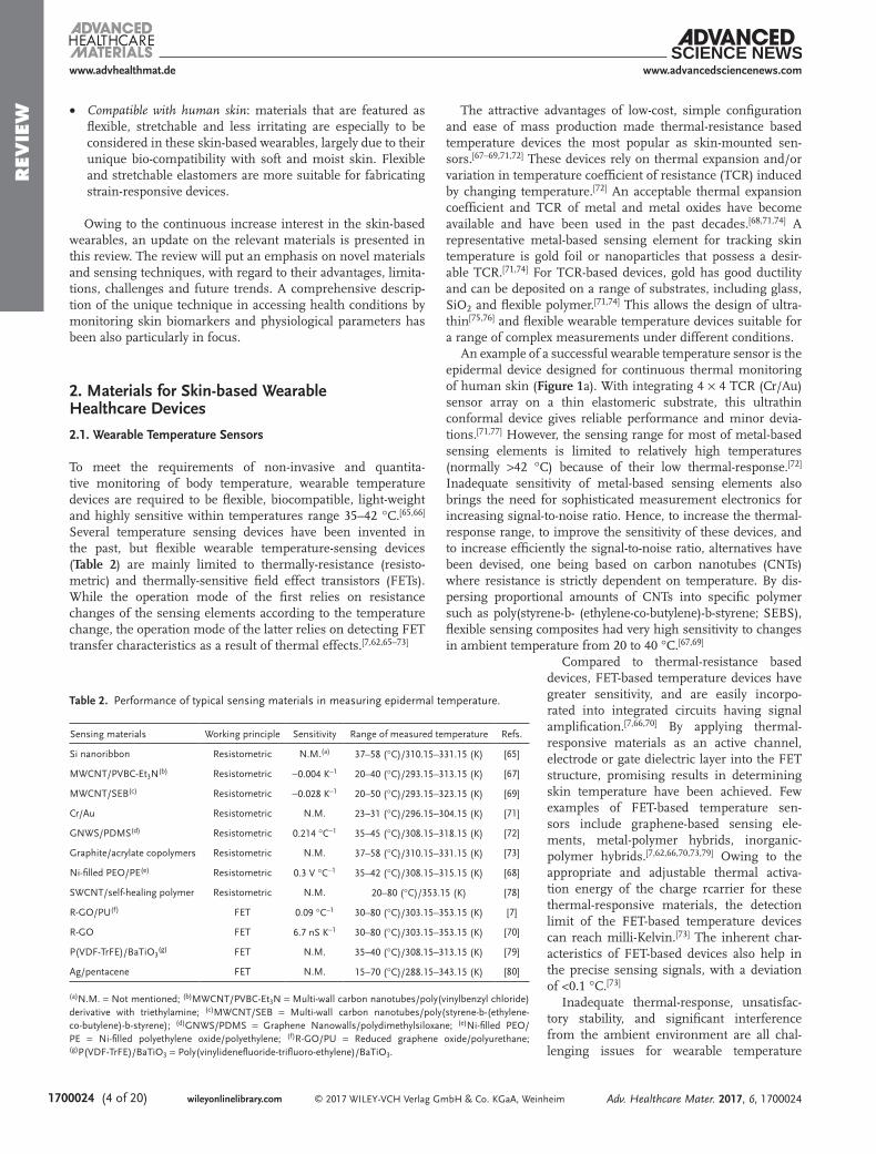
To meet the requirements of non-invasive and quantita-tive monitoring of body temperature, wearable temperature devices are required to be flexible, biocompatible, light-weight and highly sensitive within temperatures range 35–42 °C.[65,66] Several temperature sensing devices have been invented in the past, but flexible wearable temperature-sensing devices (Table 2) are mainly limited to thermally-resistance (resisto-metric) and thermally-sensitive field effect transistors (FETs). While the operation mode of the first relies on resistance changes of the sensing elements according to the temperature change, the operation mode of the latter relies on detecting FET transfer characteristics as a result of thermal effects.[7,62,65–73]
The attractive advantages of low-cost, simple configuration and ease of mass production made thermal-resistance based temperature devices the most popular as skin-mounted sen-sors.[67–69,71,72] These devices rely on thermal expansion and/or variation in temperature coefficient of resistance (TCR) induced by changing temperature.[72] An acceptable thermal expansion coefficient and TCR of metal and metal oxides have become available and have been used in the past decades.[68,71,74] A representative metal-based sensing element for tracking skin temperature is gold foil or nanoparticles that possess a desir-able TCR.[71,74] For TCR-based devices, gold has good ductility and can be deposited on a range of substrates, including glass, SiO2 and flexible polymer.[71,74] This allows the design of ultra-thin[75,76] and flexible wearable temperature devices suitable for a range of complex measurements under different conditions.
An example of a successful wearable temperature sensor is the epidermal device designed for continuous thermal monitoring of human skin (Figure 1a). With integrating 4 × 4 TCR (Cr/Au) sensor array on a thin elastomeric substrate, this ultrathin conformal device gives reliable performance and minor devia-tions.[71,77] However, the sensing range for most of metal-based sensing elements is limited to relatively high temperatures (normally >42 °C) because of their low thermal-response.[72] Inadequate sensitivity of metal-based sensing elements also brings the need for sophisticated measurement electronics for increasing signal-to-noise ratio. Hence, to increase the thermal-response range, to improve the sensitivity of these devices, and to increase efficiently the signal-to-noise ratio, alternatives have been devised, one being based on carbon nanotubes (CNTs) where resistance is strictly dependent on temperature. By dis-persing proportional amounts of CNTs into specific polymer such as poly(styrene-b- (ethylene-co-butylene)-b-styrene; SEBS), flexible sensing composites had very high sensitivity to changes in ambient temperature from 20 to 40 °C.[67,69]
Compared to thermal-resistance based devices, FET-based temperature devices have greater sensitivity, and are easily incorpo-rated into integrated circuits having signal amplification.[7,66,70] By applying thermal-responsive materials as an active channel, electrode or gate dielectric layer into the FET structure, promising results in determining skin temperature have been achieved. Few examples of FET-based temperature sen-sors include graphene-based sensing ele-ments, metal-polymer hybrids, inorganic-polymer hybrids.[7,62,66,70,73,79] Owing to the appropriate and adjustable thermal activa-tion energy of the charge rcarrier for these thermal-responsive materials, the detection limit of the FET-based temperature devices can reach milli-Kelvin.[73] The inherent char-acteristics of FET-based devices also help in the precise sensing signals, with a deviation of <0.1 °C.[73]
Inadequate thermal-response, unsatisfac-tory stability, and significant interference from the ambient environment are all chal-lenging issues for wearable temperature
www.advhealthmat.de
Adv. Healthcare Mater. 2017, 6, 1700024
www.advancedsciencenews.com
Table 2. Performance of typical sensing materials in measuring epidermal temperature.
Sensing materials Working principle Sensitivity Range of measured temperature Refs.
Si nanoribbon Resistometric N.M.(a) 37–58 (°C)/310.15–331.15 (K) [65]
MWCNT/PVBC-Et3N(b) Resistometric −0.004 K−1 20–40 (°C)/293.15–313.15 (K) [67]
MWCNT/SEB(c) Resistometric −0.028 K−1 20–50 (°C)/293.15–323.15 (K) [69]
Cr/Au Resistometric N.M. 23–31 (°C)/296.15–304.15 (K) [71]
GNWS/PDMS(d) Resistometric 0.214 °C−1 35–45 (°C)/308.15–318.15 (K) [72]
Graphite/acrylate copolymers Resistometric N.M. 37–58 (°C)/310.15–331.15 (K) [73]
Ni-filled PEO/PE(e) Resistometric 0.3 V °C−1 35–42 (°C)/308.15–315.15 (K) [68]
SWCNT/self-healing polymer Resistometric N.M. 20–80 (°C)/353.15 (K) [78]
R-GO/PU(f) FET 0.09 °C−1 30–80 (°C)/303.15–353.15 (K) [7]
R-GO FET 6.7 nS K−1 30–80 (°C)/303.15–353.15 (K) [70]
P(VDF-TrFE)/BaTiO3(g) FET N.M. 35–40 (°C)/308.15–313.15 (K) [79]
Ag/pentacene FET N.M. 15–70 (°C)/288.15–343.15 (K) [80]
(a)N.M. = Not mentioned; (b)MWCNT/PVBC-Et3N = Multi-wall carbon nanotubes/poly(vinylbenzyl chloride) derivative with triethylamine; (c)MWCNT/SEB = Multi-wall carbon nanotubes/poly(styrene-b-(ethylene-co-butylene)-b-styrene); (d)GNWS/PDMS = Graphene Nanowalls/polydimethylsiloxane; (e)Ni-filled PEO/PE = Ni-filled polyethylene oxide/polyethylene; (f)R-GO/PU = Reduced graphene oxide/polyurethane; (g)P(VDF-TrFE)/BaTiO3 = Poly(vinylidenefluoride-trifluoro-ethylene)/BaTiO3.

REV
IEW
© 2017 WILEY-VCH Verlag GmbH & Co. KGaA, Weinheim wileyonlinelibrary.com (5 of 20) 1700024
devices.[67,70,72] Latterly, graphene nanowalls (GNWs) in combi-nation with polydimethylsiloxane (PDMS) have been used as the sensing chip in a thermal-resistive sensor (Figure 1b).[72] In comparison to conventional temperature sensors, this device has a sensitivity over 108.6 Ω/°C between 35 and 45 °C, which is sufficient to monitor human body temperature. Further evidence proves that the high response of the sensor can be attributed to the excellent TCR (0.214 °C−1) of GNWs and large expansion coefficient of PDMS.[72] Stability of these tempera-ture devices has been many times reported, who found that the transition of morphology and phase of sensing elements during cooling-heating cycle is the main reason for irreproducibility of a thermal-response.[68,69] Adsorption of water vapor and/or ambient gas species is another crucial reason that generates hysteresis and causes significant interference.[70] Consequently, thermally stable materials are screened and extra encapsula-tion layers are added to decrease hysteresis, thereby improving stability and enhancing reproducibility (Figure 1c).[68–70] Taking these materials from the lab to the technological/industrial benchmark would require advanced printing techniques. A graphite-polymer based ultra-thin thermometer of 15 µm thick-ness that could be accomplished by such simple print and press method is a good example for fabrication of large-area stretch-able and transparent wearable temperature devices in the future. These fabricated thermometers should be able to measure tem-perature precisely between 25 and 50 °C (Figure 1d).[73]
2.2. Wearable Strain Sensors
Wearable strain sensors could be useful for the detection and monitoring of movement-based signals, such as heart-beat rate
and respiration rate. Generally, wearable strain devices should be lightweight, reliable, flexible and stretchable to match the mechanical properties of human skin. Sensitivity of these strain sensors must also be in line with their diverse healthcare appli-cations. For instance, mean value of feet pressure for adults mostly ranges from 140–868 kPa,[81] whereas systolic pres-sure (artery pulse) for healthy persons is within 0.67–5.32 kPa (5 to 40 mmHg).[82] This suggests that high sensitivity in a low-pressure regime (perhaps <10 kPa[83]) is required for strain sensor trying to detect arterial pulse. To date, many working models for strain devices have been developed.[84] Among them, flexible and stretchable devices based on piezoresistive, piezocapacitive and piezoelectric principles seem to be closer to practical implementation, due to their relatively simple sensory configuration, uncomplicated read-out systems and acceptable dynamic performance.[24,85–87]
2.2.1. Piezoresistive-Based Strain Sensors
Piezoresistive-based strain devices are typically composed of electrically conductive sensing films coupled with flexible sub-strates. When the structure of the device is deformed, changes in the microstructure of sensing films lead to changes in elec-trical resistance as a function of applied strain. Metal-based foil and graphitic-based sensing networks (e.g. InGaZn and graphene) are widely used in pressure/strain sensing.[88–90] In conductive networks, electrons can pass through overlapped nanomaterials within the percolation network. Stretching or bending in the conductive network results in disconnection between overlapped areas and thereby loss electrical con-nection; consequently, there is an increase of the electrical
www.advhealthmat.de
Adv. Healthcare Mater. 2017, 6, 1700024
www.advancedsciencenews.com
Figure 1. Typical materials and devices serve as or in temperature sensors. a) Image and the sensing performance of a 4 × 4 TCR sensor array after application to the skin using a water-soluble adhesive tape based on polyvinyl alcohol. Adapted with permission.[71] Copyright 2013, Nature Publishing Group. b) Cross-section SEM image of GNWs on PDMS and its “current and resistance versus temperature” curves. Adapted with permission.[72] Copyright 2013, Nature Publishing Group. c) Schematic of the TS-gated device and its response to temperature. Adapted with permission.[70] Copyright 2015, Royal Society of Chemistry. d) Photograph of a film of copolymer with graphite filler, with the temperature dependence of the resistivity of the temperature sensor. Adoated with permission.[73] Copyright 2015, National Academy of Science.
REV
IEW
© 2017 WILEY-VCH Verlag GmbH & Co. KGaA, Weinheimwileyonlinelibrary.com1700024 (6 of 20)
resistance of sensing film. Another known strain-responsive mechanism is crack propagation, i.e. cracks originate and propagate in brittle thin films coated on the top of soft pol-ymer layers upon stretching.[20] Generally speaking, the sensi-tivity (gauge factor) of the conventional metal-foil based strain device is in the range of 2–5 (a.u.) and semiconductor-based strain sensors might be 100 or greater. The value of sensi-tivity for flexible strain devices can be in the range of 1 to 100, depending on the sensing mechanisms, materials and micro/nanostructures.[20]
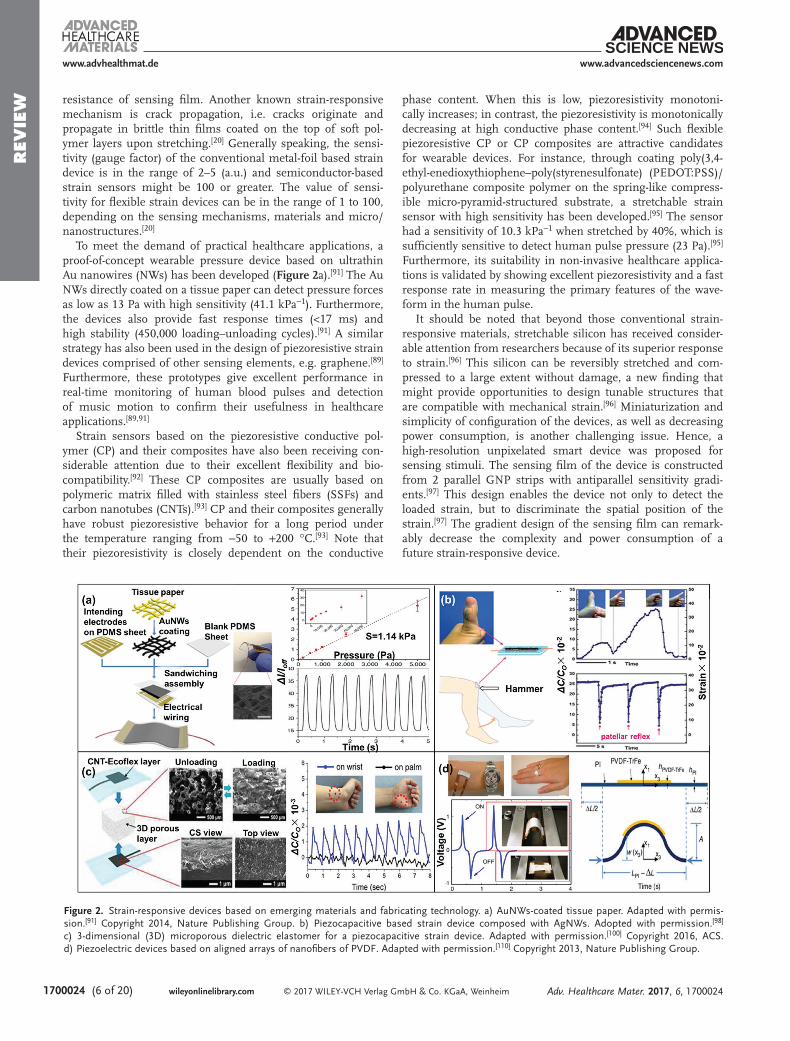
To meet the demand of practical healthcare applications, a proof-of-concept wearable pressure device based on ultrathin Au nanowires (NWs) has been developed (Figure 2a).[91] The Au NWs directly coated on a tissue paper can detect pressure forces as low as 13 Pa with high sensitivity (41.1 kPa−1). Furthermore, the devices also provide fast response times (<17 ms) and high stability (450,000 loading–unloading cycles).[91] A similar strategy has also been used in the design of piezoresistive strain devices comprised of other sensing elements, e.g. graphene.[89] Furthermore, these prototypes give excellent performance in real-time monitoring of human blood pulses and detection of music motion to confirm their usefulness in healthcare applications.[89,91]
Strain sensors based on the piezoresistive conductive pol-ymer (CP) and their composites have also been receiving con-siderable attention due to their excellent flexibility and bio-compatibility.[92] These CP composites are usually based on polymeric matrix filled with stainless steel fibers (SSFs) and carbon nanotubes (CNTs).[93] CP and their composites generally have robust piezoresistive behavior for a long period under the temperature ranging from −50 to +200 °C.[93] Note that their piezoresistivity is closely dependent on the conductive
phase content. When this is low, piezoresistivity monotoni-cally increases; in contrast, the piezoresistivity is monotonically decreasing at high conductive phase content.[94] Such flexible piezoresistive CP or CP composites are attractive candidates for wearable devices. For instance, through coating poly(3,4-ethyl-enedioxythiophene–poly(styrenesulfonate) (PEDOT:PSS)/polyurethane composite polymer on the spring-like compress-ible micro-pyramid-structured substrate, a stretchable strain sensor with high sensitivity has been developed.[95] The sensor had a sensitivity of 10.3 kPa−1 when stretched by 40%, which is sufficiently sensitive to detect human pulse pressure (23 Pa).[95] Furthermore, its suitability in non-invasive healthcare applica-tions is validated by showing excellent piezoresistivity and a fast response rate in measuring the primary features of the wave-form in the human pulse.
It should be noted that beyond those conventional strain-responsive materials, stretchable silicon has received consider-able attention from researchers because of its superior response to strain.[96] This silicon can be reversibly stretched and com-pressed to a large extent without damage, a new finding that might provide opportunities to design tunable structures that are compatible with mechanical strain.[96] Miniaturization and simplicity of configuration of the devices, as well as decreasing power consumption, is another challenging issue. Hence, a high-resolution unpixelated smart device was proposed for sensing stimuli. The sensing film of the device is constructed from 2 parallel GNP strips with antiparallel sensitivity gradi-ents.[97] This design enables the device not only to detect the loaded strain, but to discriminate the spatial position of the strain.[97] The gradient design of the sensing film can remark-ably decrease the complexity and power consumption of a future strain-responsive device.
www.advhealthmat.de
Adv. Healthcare Mater. 2017, 6, 1700024
www.advancedsciencenews.com
Figure 2. Strain-responsive devices based on emerging materials and fabricating technology. a) AuNWs-coated tissue paper. Adapted with permis-sion.[91] Copyright 2014, Nature Publishing Group. b) Piezocapacitive based strain device composed with AgNWs. Adopted with permission.[98] c) 3-dimensional (3D) microporous dielectric elastomer for a piezocapacitive strain device. Adapted with permission.[100] Copyright 2016, ACS. d) Piezoelectric devices based on aligned arrays of nanofibers of PVDF. Adapted with permission.[110] Copyright 2013, Nature Publishing Group.

REV
IEW
© 2017 WILEY-VCH Verlag GmbH & Co. KGaA, Weinheim wileyonlinelibrary.com (7 of 20) 1700024
2.2.2. Piezocapacitive-Based Strain Devices
Piezocapacitive-based strain devices use a highly compliant die-lectric layer sandwiched between a pair of stretchable electrodes. Applied strain changes the distance between 2 electrodes, which results in a change of capacitance.[24] For instance, a piezoca-pacitive strain sensor composed of silver nanowire (AgNWs), Ecoflex, liquid metal and PDMS was built for finger flexing and knee motions among other body movement tracking (Figure 2b).[98] Quick response (<40 ms), good pressure map-ping function and convinient wearability were revealed when mounted on the human body. Besides these conventional strain devices, a transparent flexible piezocapacitive pressure/strain sensor has been developed by Lipomi et al.[99] The piezocapaci-tive sensor, made by spraying transparent carbon nanotube on PDMS, gave a desirable performance in detecting pressure down to ≈50 kPa.[99] Note that this transparent piezocapacitive skin-based strain sensor is extremely compliant mechanically, physically robust and easily fabricated, although it is less sensi-tive than its other counterparts.
Piezocapacitive strain devices have high linearity, stretch-ability and low hysteresis, but suffer from low sensitivity. To overcome this challange, a 3D microporous dielectric elastomer with giant piezocapacitive effect was used to construct a pres-sure device (Figure 2c).[100] Due to the presence of micropores within the elastomeric dielectric layer, the resulting piezoca-pacitive pressure device is highly deformable by a minimal amount of pressure, leading to a marked increase in sensitivity. The gradual closure of micropores under compression also increased the effective dielectric constant, thereby enhancing sensitivity.The 3D microporous dielectric layer with serially stacked springs of elastomer bridges can cover a much wider range of pressure than those of previously reported micro-/nanostructured sensing materials. This proof-of-concept can help in monitoring both ultralow and high levels of human activity. Noteworthily, materials with unique microstructure can enhance their piezocapacitive performance. Compared with those piezocapactive materials with flat surface, the pressure sensor comprised of pyramidal dielectric elastomer significantly enhanced blood pulse monitoring and force tracking.[101] Similar improvements have also been investigated for piezoresistivie elastomer with hollow spheres,[102] thickness-gradient,[103] and pyramidal microstructures,[104] implying an efficient approach to improving the performance of strain sen-sors by well-designed strain-responsive materials.
2.2.3. Piezoelectric-Based Strain Sensors
Piezoelectric-based strain sensors detect changes in pressure or strain; in particular, sensing signal (voltage) is only gener-ated by dynamic changes of applied strain or force. Piezo-electric materials, especially certain crystals (e.g. ZnO and LiNbO3) and some ceramics (e.g. BaTiO3 and Pb(Zr11xTix)O3 (PZT)), generate a voltage potential when the crystal lattice is deformed.[105,106] The sensitivity of piezoelectric materials depends on their crystal structure and the voltage generated is directly proportional to the applied strain. These sensors have satisfying high-frequency responses, which makes them ideal
for measuring dynamic forces.[107] To match the mechanical properties of human skin, recent piezoelectric materials used in wearable devices are mechanically flexible.[106,108,109] These flexible piezoelectric materials can be used as either rigid piezo-electric material embedded in a flexible substrate or as a piezo-electric polymer film.
Compared with both piezoresistive- and piezocapacitive-based strain sensors, strain devices comprised of piezoelectric materials have greater dynamic durability. The nature of good high-frequency response to an applied strain makes this device more suitable for measuring vibrations. For example, piezoe-lectric-based strain sensors could be installed in a smart watch for monitoring heart-beat rate. However, the dynamic piezo-electric sensing mechanism disallows them from measuring static forces. A classic piezoelectric material commonly used in flexible strain devices is poly(vinylidenefluoride) (PVDF), a piezoelectric polymer that has considerable flexibility.[109] Its flexibility and high piezoelectric factor make PVDF an attrac-tive material for flexible strain devices. For example, Persano et al. (Figure 2d) reported on aligned arrays of nanofibers of PVDF.[110] Free-standing 3D architectures of aligned arrange-ments of PVDF fibers have been designed (Figure 2d); further-more, the polymer chains inside the fibers adopt strongly pref-erential orientations.[110] This microstructure enables ultra-high sensitivity in measuring pressure, even at exceptionally low values (0.1 Pa); in particular, exceptional piezoelectric character-istics offered by these devices indicate an application in moni-toring human motion.
2.3. Wearable Devices for Recording Electrical Conductivity Through the Skin
Wearable devices that detect tiny electrical changes on the skin could be helpful for monitoring the electrical activity of heart to locate an arrhythmia (abnormal heartbeat). These results can help one or one’s doctor to decide whether there is a need for medicine, a pacemaker, an implantable cardioverter defibrillator (ICD), cardiac neural ablation or some other surgery. For wear-able and accurate monitoring of electrophysiological signals in a variety of everyday conditions, reliable biopotential-capturing electrodes should be reusable, should have long-term stability, and should also not cause skin irritations or allergic reactions.
To date, disposable silver/silver chloride (Ag/AgCl)–gelled electrodes are most commonly used, but these electrodes have limited storage time (<1 year) and are not reusable.[111] The best possible alternatives are flexible dry electrodes, which are usu-ally made of polymeric- and textile-based materials.[111–114] For instance, after patterning a metal layer on a thin PDMS sub-strate, a biocompatible and flexible PDMS-based (polymetric) electrocardiogram (ECG) recording device can be obtained (Figure 3a).[112] In the case of textile-based electrodes, these are normally fabricated by knitting, weaving, embroidering and non-weaving methods. Figure 3b shows photographic images and comparison of the response of the textile-based dry electrode and (Ag/AgCl)–gelled electrode in wearable ECG recording devices. These flexible dry electrodes in general are more skin compatible compared with those of gelled elec-trodes (Figure 3).[111–114] However, disturbances to ECG signals
www.advhealthmat.de
Adv. Healthcare Mater. 2017, 6, 1700024
www.advancedsciencenews.com

REV
IEW
© 2017 WILEY-VCH Verlag GmbH & Co. KGaA, Weinheimwileyonlinelibrary.com1700024 (8 of 20)
caused by body movement can often appear. After wearing the polymeric dry electrodes for 7 days, no significant changes on skin were seen (Figure 3a); however, the skin under the (Ag/AgCl)–gelled electrode turned red and became itchy in 2 sub-jects. In conclusion, these newly proposed dry electrodes have comparatively good fidelity and are comfortable regarding body contact as well as being acceptable in long-term stability, indi-cating broad applicability to the ubiquitous field of biosignal monitoring.
2.4. Wearable Sensors for Analyzing Sweat Metabolites
Levels of sweat metabolites (such as lactate and uric acid) and electrolytes (metal ions, such as sodium and potassium), as well as skin humidity, are useful physiological parameter indirectly reflecting health status of an individual, and can potentially be used to probe body conditions by non-invasive monitoring.[21,23,33,34,37,115] Table 1 summarizes the use of sweat body fluids as physiological biomarkers.[27,29–32,36,37]
The concentration of uric acid and urea in sweat is much higher in uremic patients.[116] Levels of skin humidity and sweat pH are important indicators of dehydration and electrolyte loss during exercise.[117] Accordingly, sweat-based wearable devices are required for monitoring these non-invasive health states.
To date, sweat-based healthcare devices are mainly based on an electrochemical sensing principle, owing to the low-cost, high performance and excellent portability of electrochemical devices.[21,23] Figure 4a shows the configuration of a generally reported electrochemical sweat-based device.[118] Three elec-trodes are printed on the flexible substrate and, as might be necessary, a fourth electrode can be present, i.e. a working elec-trode (WE), a counter electrode (CE, sometimes as anode), a reference electrode (RE) and a cathode. WE and CE are nor-mally composed of conductive materials, such as carbon black, CNTs, Pt black, modified Ag or Au, etc.[21,118,119] Ag/AgCl is usually used as the RE. Conformal contact between the elec-trode surface and the skin is necessary to ensure efficient
functioning of the devices. Accordingly, substrates for these devices should be made of fabric (e.g. wool, cotton or nylon) or flexible plastics.[21]
Figure 4b–d shows 3 typical approaches[21,23,37,119–122] for the attachment of electrodes to a flexible substrate or skin.[21,23] The first approach relies on screen printed electrodes.[23] This approach has been massively employed in production due to its low-cost, industrial-scale fabrication of robust electrodes on different substrates. The geometries and thickness of the elec-trodes can be readily adjusted by changes of the screen mask to meet the design requirements of the wearable device. However, preparation of wearable devices by screen-printing involves sev-eral complicating issues; for instance, the influence of manu-facturing conditions and substrate properties, as well as the composition of the ink, need to be taken into consideration, since these factors significantly determine the performance of the devices. Generally, the natural properties of substrates must be compatible with the printing process and the specific opera-tional environment. Some necessary modification, e.g. doping of the noble catalyst, may be required for the inks to obtain specific functionality. The annealing temperature also requires optimization to avoid undesired deformation of the electrode and substrate. The left part of Figure 4b depicts the screen-printed microelectrodes on polyethylene naphthalate (PEN) for dopamine detection, suggesting the convenience of depositing responsive materials by this approach.
The second approach used for attachment of electrodes to a flexible substrate or skin relies on stamp transferred elec-trodes.[23] The pattern-transfer technique is very useful in fab-ricating sensing materials on non-planarr substrates. Figure 4c shows details of the stamp transfer process. It is known that, through pattern-transfer technique, responsive materials are highly compatible with irregular substrates possessing diverse surface morphologies (e.g. skin), without compromising the structural integrity of the pattern. Stamp transferred electrodes notably give better performance than those fabricated by screen-printing technique.[23] This fabricating technique provides a novel method to form the ink-based printable materials on
www.advhealthmat.de
Adv. Healthcare Mater. 2017, 6, 1700024
www.advancedsciencenews.com
Figure 3. Typical examples of flexible dry electrodes for ECG signal recording. Photographs of (a) polymeric-[111,112] and (b) textile-based electrodes, and their response to biosignals compared with those of commercial Ag/Agcl electrodes.[113,114] Adapted with permission.[111] Copyright 2011, Elsevier. Adapted with permission.[112] Copyright 2008, Elsevier. Adapted with permission.[113] Copyright 2008, Taylor & Francis. Adapted with permission.[114] Copyright 2010, IEEE.

REV
IEW
© 2017 WILEY-VCH Verlag GmbH & Co. KGaA, Weinheim wileyonlinelibrary.com (9 of 20) 1700024
non-planar and oversized surfaces incompatible with standard screen printing methods.
The third approach used for attachment of electrodes to a flexible substrate or skin relies on epidermal suspended elec-trodes.[21] The rationale behind this approach is to deal with the major problem for traditional wearable devices by restricted intimate contact with the skin at limited regions – something that can be solved if the sensing electrodes can be directly attached on the skin. Inspired by tattooing, the strategy of using elastomeric stamps or the tattoo technique to form electrodes directly on human epidermis has been proposed. The strategy includes steps of screen printing of conductive and insulating inks on commercial, temporary tattoo-base paper, to form the electrodes and/or devices, and then flip and apply the fabricated tattoo or printed electrodes to the skin. To adapt for mechanical stress and overcome the potential deformations due to bodily movements, it was suggested that finely dispersed carbon fibers should be incorporated into the tattoo-based devices. Numerous results showed the success and convenience of applying this technique in preparing electrodes and/or wearable devices on the skin. Among these examples, a classic prototype reported by Jia et al.[123] was noteworthy. In their prototype (Figure 4d), an enzymatic tattoo amperometric biosensor was built for continuously monitoring lactate levels in the perspiration as a biomarker of physical stress. This flexible, printed, temporary-transfer tattoo, electrochemical biosensor exhibited satisfying adherence to the skin and had acceptable selectivity to lactate, with linearity up to 20 mmol L−1. In particular, the device had commendable resilience against the continuous mechanical deformation expected from epidermal wear.
Simultaneous screening of target biomarkers is helpful in ensuring the accuracy of measurements. Consequently,
multifunctional wearable devices combining sensing and therapy have been particularly a focus of attention. Through pro-viding sufficient information of heater, temperature, humidity, glucose and pH, as well as several chemicals in sweat, these multifunctional wearable devices (e.g. graphene-based elec-trochemical device) give a comprehensive profile of health status.[27,124] On account of these attractive results, it is expected that sweat-based wearable devices could possibly alert traditional disease prevention.
Beyond the traditional electrochemical sweat-based healthcare devices, a novel colorimetric sensing patch reported recently shows its unique healthcare attractiveness during athletic or military training (Figure 5).[36] The strategy that microfluidic chip and colorimetric dyes can probably separate and respond to analytes in sweat has been introduced in designing the device. Therefore, in a combination of flexible microfluidic channel and colorimetric biochemical assays in the patch, lactate, glu-cose and chloride ion concentrations in sweat, with pH, can be detected in terms of image capture analysis.[36] After stinking the patch to the skin of individuals, reliable performance were obtained, and the sweat patches remained intact and functional even when used during an outdoor bicycle endurance race.
Spontaneously measuring chemical and ECG parameters can provide more comprehensive information about health status; however, most of the reported wearable healthcare sensors can only measure chemical or ECG parameters. To obtain fuller information of the human body, these monofunctional sen-sors need to be integrated into a sensor matrix, which increases the complexity of fabrication. A strategy to resolve this issue is to design sensors that can be used for monitoring chemical and ECG parameters spontaneously. Based on this strategy, a wearable chemical–ECG hybrid biosensing system is proposed
www.advhealthmat.de
Adv. Healthcare Mater. 2017, 6, 1700024
www.advancedsciencenews.com
Figure 4. General fabrication technologies of sweat-based wearable devices. a) Primary configuration of the sweat-based electrochemical wearable devices. b) Steps involved in screen printing thick-film electrochemical devices. Left of Figure b: representative design of flexible device fabricated by a screen-printing technique. Adapted with permission.[23] a,b) Adapted with permission.[21] Copyright 2014, Elsevier. c) Preparation progress of stamp transfer electrodes. d) Typical example of a tattoo-based wearable device. c,d) Adapted with permission.[23]

REV
IEW
© 2017 WILEY-VCH Verlag GmbH & Co. KGaA, Weinheimwileyonlinelibrary.com1700024 (10 of 20)
www.advhealthmat.de
Adv. Healthcare Mater. 2017, 6, 1700024
www.advancedsciencenews.com
Figure 5. Wearable flexible colorimetric sensing patch. a) Schematic illustrations, optical images and theoretical stress modelling of an epidermal microfluidic biosensor integrated with flexible electronics for sweat monitoring. b) Human trials of sweat monitoring in a temperature- and humidity-controlled conditions (35 °C at 50% relative humidity). Adapted with permission.[36] Copyright 2016, American Association for the Advancement of Science.
REV
IEW
© 2017 WILEY-VCH Verlag GmbH & Co. KGaA, Weinheim wileyonlinelibrary.com (11 of 20) 1700024
for real-time health and fitness monitoring.[125] The proposed system should give a desirable performance in simultaneously sensing lactate (0–28 mmol L−1) and monitoring ECG param-eters.[125] Such proof-of-concept opens a new way in designing flexible smart sweat-based healthcare devices.
2.5. Wearable Sensors for Detection of Volatile Biomarkers
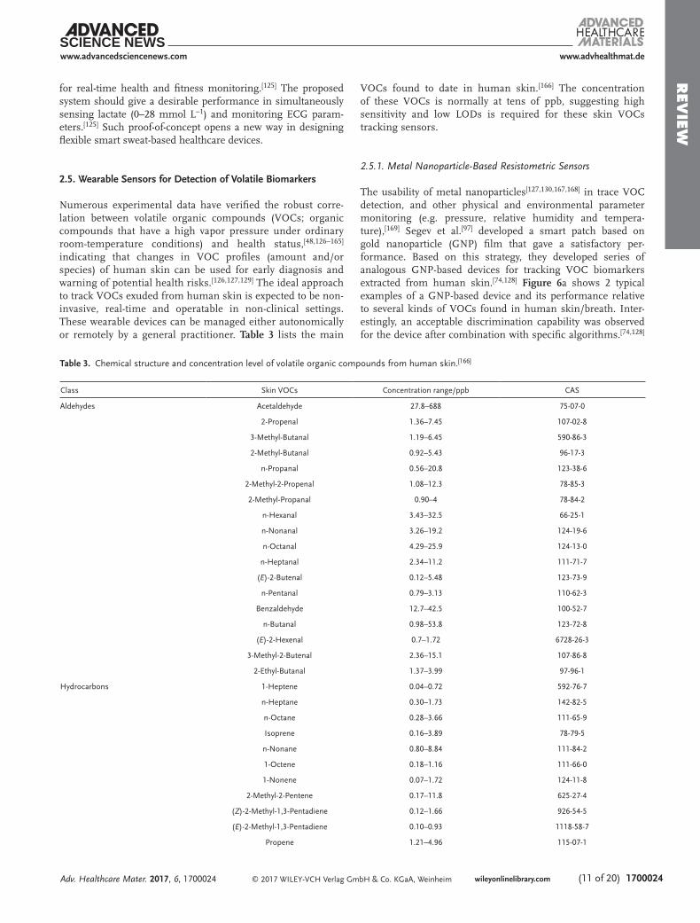
Numerous experimental data have verified the robust corre-lation between volatile organic compounds (VOCs; organic compounds that have a high vapor pressure under ordinary room-temperature conditions) and health status,[48,126–165] indicating that changes in VOC profiles (amount and/or species) of human skin can be used for early diagnosis and warning of potential health risks.[126,127,129] The ideal approach to track VOCs exuded from human skin is expected to be non-invasive, real-time and operatable in non-clinical settings. These wearable devices can be managed either autonomically or remotely by a general practitioner. Table 3 lists the main
VOCs found to date in human skin.[166] The concentration of these VOCs is normally at tens of ppb, suggesting high sensitivity and low LODs is required for these skin VOCs tracking sensors.
2.5.1. Metal Nanoparticle-Based Resistometric Sensors
The usability of metal nanoparticles[127,130,167,168] in trace VOC detection, and other physical and environmental parameter monitoring (e.g. pressure, relative humidity and tempera-ture),[169] Segev et al.[97] developed a smart patch based on gold nanoparticle (GNP) film that gave a satisfactory per-formance. Based on this strategy, they developed series of analogous GNP-based devices for tracking VOC biomarkers extracted from human skin.[74,128] Figure 6a shows 2 typical examples of a GNP-based device and its performance relative to several kinds of VOCs found in human skin/breath. Inter-estingly, an acceptable discrimination capability was observed for the device after combination with specific algorithms.[74,128]
www.advhealthmat.de
Adv. Healthcare Mater. 2017, 6, 1700024
www.advancedsciencenews.com
Table 3. Chemical structure and concentration level of volatile organic compounds from human skin.[166]
Class Skin VOCs Concentration range/ppb CAS
Aldehydes Acetaldehyde 27.8–688 75-07-0
2-Propenal 1.36–7.45 107-02-8
3-Methyl-Butanal 1.19–6.45 590-86-3
2-Methyl-Butanal 0.92–5.43 96-17-3
n-Propanal 0.56–20.8 123-38-6
2-Methyl-2-Propenal 1.08–12.3 78-85-3
2-Methyl-Propanal 0.90–4 78-84-2
n-Hexanal 3.43–32.5 66-25-1
n-Nonanal 3.26–19.2 124-19-6
n-Octanal 4.29–25.9 124-13-0
n-Heptanal 2.34–11.2 111-71-7
(E)-2-Butenal 0.12–5.48 123-73-9
n-Pentanal 0.79–3.13 110-62-3
Benzaldehyde 12.7–42.5 100-52-7
n-Butanal 0.98–53.8 123-72-8
(E)-2-Hexenal 0.7–1.72 6728-26-3
3-Methyl-2-Butenal 2.36–15.1 107-86-8
2-Ethyl-Butanal 1.37–3.99 97-96-1
Hydrocarbons 1-Heptene 0.04–0.72 592-76-7
n-Heptane 0.30–1.73 142-82-5
n-Octane 0.28–3.66 111-65-9
Isoprene 0.16–3.89 78-79-5
n-Nonane 0.80–8.84 111-84-2
1-Octene 0.18–1.16 111-66-0
1-Nonene 0.07–1.72 124-11-8
2-Methyl-2-Pentene 0.17–11.8 625-27-4
(Z)-2-Methyl-1,3-Pentadiene 0.12–1.66 926-54-5
(E)-2-Methyl-1,3-Pentadiene 0.10–0.93 1118-58-7
Propene 1.21–4.96 115-07-1

REV
IEW
© 2017 WILEY-VCH Verlag GmbH & Co. KGaA, Weinheimwileyonlinelibrary.com1700024 (12 of 20)
To prove the feasibility of the GNP-based devices in practical healthcare application, Broza et al.[100] used the developed approach to monitor VOCs of skin samples (Figure 6b); they included a heterogeneous group of 30 volunteers with no acute or active disease. Although their main goal was to associate VOCs from skin and breath of specific volunteers, the second goal was to achieve a chemical profiling based on the VOC samples from either skin or breath. This method successfully created a unique chemical barcoding enabling classification and clustering of the volunteers, e.g. clustering based on age-related VOCs.
2.5.2. Potential Materials for Tracking Skin VOCs
Several materials appear to be sensitive to VOCs from human breath and odor, as well as air pollutants. Part of these materials has not been used yet to track VOCs from the skin, but, argu-ably, they would have great potential for suchlike applications in the near future. A general review of these candidates is intro-duced in the following sub-sections.
2.5.2.1. Metal Oxides: Gas-sensitive metal oxides can be divided into transition- and non-transition-metal oxides.[170] It is gen-
Class Skin VOCs Concentration range/ppb CAS
1-Pentene 0.26–0.68 109-67-1
n-Pentane 0.49–2.86 109-66-0
(E)-2-Butene 0.05–0.9 624-64-6
2-Heptene 0.15–0.35 592-77-8
(Z)-2-Butene 0.11–0.76 590-18-1
2,3-Dimethyl-2-Butene 2.98–9.12 563-79-1
Ketones Acetone 86–808 67-64-1
2-Butanone 0.59–3.64 78-93-3
2-Pentanone 0.18–1.66 107-87-9
6-Methyl-5-Hepten-2-One 2.63–167 110-93-0
3-Buten-2-One 0.75–3.23 78-94-4
4-Methyl-3-Penten-2-One 3.81–42.63 141-79-7
2-Hexanone 0.34–0.53 591-78-6
2-Heptanone 1.59–1.66 110-43-0
Heterocycles 3-Methyl-Furan 0.08–0.75 930-27-8
2-Pentyl-Furan 0.23–1.03 3777-69-3
2-Methyl-Furan 0.13–1.01 534-22-5
2-Methyl-1,3-Dioxolane 0.11–3.15 497-26-7
2,5-Dimethyl-Furan 0.07–1.38 625-86-5
1,3-Dioxolane 2.81–21.85 646-06-0
Terpenes dl-Limonene 0.18–60.64 5989-27-5
p-Cymene 0.31–2.45 99-87-6
γ-Butyrolactone 0.97–18.90 96-48-0
β-Pinene 0.28–2.81 127-91-
p,α-Dimethyl-Styrene 0.76–10.17 1195-32-0
Eucalyptol 1.85–14.24 470-82-6
Esters Ethyl Acetate 3.74–81.05 141-78-6
Isopropyl Acetate 23.87–43.78 108-21-4
Isobutyl Acetate 11.63–15.52 110-19-0
n-Butyl Acetate 130.20–1409 123-86-4
Alcohols Ethanol 109–7377 64-17-5
2-Propanol 87.6 67-63-0
Sulfurs Dimethyl Sulfide 0.13–1.12 75-18-3
Allyl Methyl Sulfide 0.06–0.5 10152-76-8
Other Acetonitrile 0.47–89 1975-05-08
Table 3. Continued.
www.advhealthmat.de
Adv. Healthcare Mater. 2017, 6, 1700024
www.advancedsciencenews.com

REV
IEW
© 2017 WILEY-VCH Verlag GmbH & Co. KGaA, Weinheim wileyonlinelibrary.com (13 of 20) 1700024
erally believed that the former are more gas-sensitive than the non-transition-metal oxides because of their stronger active chemical structure. Primary factors that influence sensing behavior of metal oxides include chemical components, sur-face area (surface to volume ratio), microstructures of sensing layers, humidity and temperature.[171]
Generally, very few unitary metal oxides possess favorable properties for sensing VOCs, especially in tracing gas species at ppb level. For this level of sensitivity, recent studies have
focused mainly on composites, i.e. binary, ternary, quaternary or even more complex metal oxides (such as noble metal decorated metal oxides).[170,171] Details of the enhancement mechanism have been published.[170–172] Besides the chemical composition, materials with high surface to volume ratio pro-vide more reaction sites. Fabrication of nanofibers by electro-spinning (Figure 6c), generation of thin film by sputtering, as well as creating porous structures with help of pore forming agents, are the most popular ways of obtaining sensing
www.advhealthmat.de
Adv. Healthcare Mater. 2017, 6, 1700024
www.advancedsciencenews.com
Figure 6. Typical examples of the practical and potential sensing elements for skin VOC detection. a) Functional group (at different chain-length) modified GNPs-based flexible device. Adapted with permission.[74,128] Copyright Wiley and Copyright 2016, ACS. b) Combined volatolomics monitoring by nanomaterial-based devices. Adapted with permission.[100] Copyright 2016, ACS. c) Fiber-like SnO2 for tracking trace amounts of VOCs. Adapted with permission.[176] d) A colorimetric VOCs device composed of different odor-responsive pigments. Adopted with permission.[186] Copyright 2006, Asian Research Publishing Network.

REV
IEW
© 2017 WILEY-VCH Verlag GmbH & Co. KGaA, Weinheimwileyonlinelibrary.com1700024 (14 of 20)
candidates with a high surface area.[173–176] It has now been shown that, when the grain size is less than twice the thickness of surface charge layers, the grain is fully involved in the space-charge layer; consequently, gas sensitivity of the oxides can be significantly increased by adjusting grain size.[172,174] Syn-thesis of oxides with unique shape and morphology is a very desirable method of enhancing the sensitivity of metal oxide gas sensors, as the shape and morphology of the oxides deter-mines the crystallographic facets exposed on the surface of a nanocrystal, and will therefore determine the number of atoms located at the edges or corners.[174] By controlling the shape and morphology using different synthetic routes, the reactivity and selectivity of a nanocatalyst can be manually tailored.
One of the main challenges in metal-oxide sensors is that water adsorption appreciably lowers their sensitivity, since water adsorption on the metal oxide surface prevents donation of elec-trons to the sensing layers. Furthermore, prolonged exposure to humid environments leads to gradual formation of stable chemisorbed OH– on the surface, resulting in progressive deg-radation. However, surface hydroxyls start to desorb at ≈400 °C so that the hydroxyl ions can be removed by heating to tempera-tures >400 °C.[174,182] Thus, an optimal operating temperature will essentially eliminate humidity interference. As the balance of adsorption/desorption is markedly affected by temperature, optimization of operating temperature can improve the magni-tude of response/recovery speed and response. Table 4 lists sev-eral typical examples covering the above-mentioned strategies using SnO2 as the sensing element.[170,173,175,177–181]
2.5.2.2. Conducting Polymers: Conducting polymers (CPs) and their derivatives have been used as the active layers of gas sen-sors over the last century.[183] Compared to metal oxides sensors, devices made of conducting polymers have high sensitivities and short response durations at room temperature.[184] Con-ducting polymers are easily synthesized and their molecular chain structure can be conveniently modified by copolymeriza-tion or structural derivations.[171,184] Furthermore, conducting polymers have good mechanical properties, which allow easy fabrication of sensing devices.
Some of the earliest studies indicated that the sensing mech-anism of CPs to VOCs involves swelling, namely the adsorbed VOC leads to the separation of polymer chains and increases the hopping distance of charge career for charge transport. As a result, the electrical resistance (response signal) increases.[171,183,184] In some cases, polar VOC molecules behave as electron acceptors
or donors contributing a charge carrier to the polymer, and sub-sequently participate as dopants that increase or decrease carrier concentration.[171,183] Hybridization of CPs with inorganic and graphitic nanoscale building blocks, such as metal oxides, CNTs or graphene, are more advantageous in polymer processing.[171] These hybridized CPs can also provide tunable sensing per-formance to VOCs through hybridization of CPs with optimal nanoscale building blocks, furthering the design of advanced sensing devices for specific monitoring of VOC biomarkers.
2.5.2.3. Responsive Dyes: Theoretically, if a VOC molecule inter-acts with a responsive dye, a visual response can be obtained by a change in color. Based on this working principle, colorimetric VOC devices have been designed based on 2 fundamental requirements: (1) each chemically responsive dye must contain a center that strongly interacts with the VOC(s); (2) each inter-action center must be strongly coupled to an intense chromo-phore. Metalloporphyrins (e.g. Cu(II), Zn(II), Mn(II), Co(III), Cr(III) and Sn (IV)-based porphyrins, etc.) and some pH sen-sitive dyes (e.g. methyl red, bromophenol blue, chlorophenol red, etc.) are among the primary compositions for sensors based on responsive dyes.[185] Similarly, colorimetric devices showed their merits in identifying VOCs.[186] Following the interaction of VOCs with an array of responsive dyes, a unique color map was generated for each VOC and different color patterns could be seen at different concentrations of VOCs (Figure 6d).[186] Combined with a specific algorithm, excellent discrimination was obtained. LODs of the colorimetric devices made of respon-sive dyes have generally ranged from hundreds of ppb to several ppm. Due to the strong interaction between VOC molecules and responsive dyes, this kind of device can often be very sensitive to VOC biomarkers. In summary, these disposable responsive dye-based colorimetric devices have given interesting results for consideration in the future of skin-based wearable devices.
3. Emerging Technologies for Improved Sustainability and Multi-Functionality
In addition to high-performance (high sensitivity and supe-rior precision), future prospect of wearable healthcare devices should focus on multi-functionality and long-term sustain-ability, e.g. by ultra-low power consumption, long-term stability and multifunction integration. Emerging approaches to accom-plish these objectives are shown in Figure 7.
www.advhealthmat.de
Adv. Healthcare Mater. 2017, 6, 1700024
www.advancedsciencenews.com
Table 4. Comparison of ethanol sensing performance, based on different SnO2-based sensing materials.
Materials Synthesis routes Morphology BET surface area (m2 g−1)
Response to 100 ppm ethanol
Temperature (°C)
Response time (s)
Ref.
SnO2 Thermal evaporation Nanowire N.M. 2.1 400 N.M [177]
Coprecipitation Nonporous 19.5 13 300 13 [178]
Electrospinning Fiber N.M. 15 340 10 [175]
Hydrothermal Nanosheets 31 35 270 13 [179]
Coprecipitation Microcubes 42.7 58 280 N.M. [178,180]
SnO2/ZnO Thermal evaporation and hydrothermal hierarchical N.M. 6 400 N.M [177]
Ag-doped TiO2/SnO2 Wet impregnation and nanocasting Hierarchical flower 49 112.5 275 3.5 [181]

REV
IEW
© 2017 WILEY-VCH Verlag GmbH & Co. KGaA, Weinheim wileyonlinelibrary.com (15 of 20) 1700024
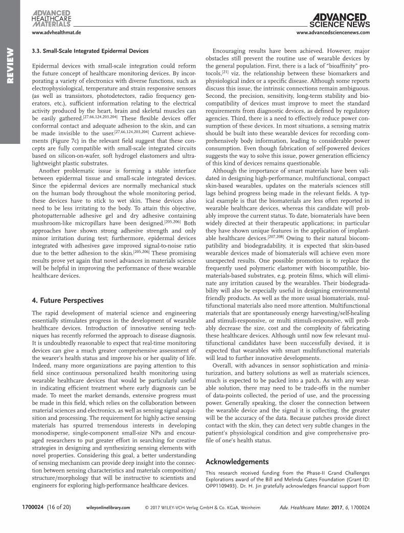
3.1. Self-Powered Healthcare Devices
Self-powered sensing devices act as power generator. The power generated by these devices can be stored in the capac-itor-like battery, which is then used for driving other elec-tronics.[105,187–191] These devices do not require external power to run the sensing activity or generate a read-out signal, obvi-ating the future need for a battery. Self-powered devices can therefore be miniaturized because a battery is not needed. At present, promising results for implementing self-power piezoelectric-, triboelectric-, thermoelectric- and near-field communication (NFC)-based devices have now primarily been achieved.[105,187,189,190,192–195] Particularly, by using piezo-electric, triboelectric and thermoelectric materials (e.g. ZnO NWS, BaTiO3 nanocomposites, P(VDF-TrFE) nanofibers and Bi2Te3), energy generated by mechanical or heat stimuli can be used to support the operation of these devices.[105,190,196,197] Among the piezoelectric materials available, ZnO-based materials (Figure 7a) have had more attention due to their excellent piezoelectric property, and in particular due to their gas-sensitivity and strain responsiveness.[190] With modifica-tion of ZnO NWS by unique ligands, acceptable selectivity to specific gas can be obtained.[190] However, these ZnO-based piezoelectric materials are relatively costly and inconvenient for integration into soft electronics compared with triboelec-tric materials. Hence, future self-powered sensing devices based on multi-functional triboelectric materials will be more attractive.
3.2. Self-Healing Healthcare Devices
The attractiveness of skin-related flexible healthcare devices can be attributed to their light weight and simple interac-tion with the human body that can continuously monitor health conditions. However, they can be incidentally scratched and/or mechanically cut, thereby destroying their sensing capa-bility. To avoid damage, self-healing flexible devices should be designed so that their mechanical/electrical/chemical proper-ties can be automatically restored. Several successful examples based on this concept have been reported using self-healing materials as their functional components, which have given acceptable sensing characteristics and complete healing after damage.[74,198,199] The positive results of the self-healing con-cept is a promising approach to the fabrication of smart wear-able devices with desirable long-term stability for future clinical applications (Figure 7b). Integration of self-healing polymers with deliberately controlled (semi) conducting inorganic nano-materials, such as molecularly modified Si nanowires, w/o car-rier donating or carrier withdrawing side-groups,[157,158,162,200,201] or TiO2
[202] have the potential to overcome several technological and cost barriers towards in-field use.
www.advhealthmat.de
Adv. Healthcare Mater. 2017, 6, 1700024
www.advancedsciencenews.com
Figure 7. Emerging technologies in wearable devices. a) A highly selec-tive and self-powered gas sensor using organic surface functionalization of a p-Si/n-ZnO diode. a) Adapted with permission.[190] b) Self-healing multi-functional sensing platform composed of self-healing polymer
and ligand functionalized GNPs. Adapted with permission.[74,128] Copy-right Wiley and Copyright 2016, ACS. c) Small-scale integrated epidermal devices. Adapted with permission.[124] Copyright 2016, Nature Publishing Group.
REV
IEW
© 2017 WILEY-VCH Verlag GmbH & Co. KGaA, Weinheimwileyonlinelibrary.com1700024 (16 of 20)
3.3. Small-Scale Integrated Epidermal Devices
Epidermal devices with small-scale integration could reform the future concept of healthcare monitoring devices. By incor-porating a variety of electronics with diverse functions, such as electrophysiological, temperature and strain responsive sensors (as well as transistors, photodetectors, radio frequency gen-erators, etc.), sufficient information relating to the electrical activity produced by the heart, brain and skeletal muscles can be easily gathered.[27,66,124,203,204] These flexible devices offer conformal contact and adequate adhesion to the skin, and can be made invisible to the user.[27,66,124,203,204] Current achieve-ments (Figure 7c) in the relevant field suggest that these con-cepts are fully compatible with small-scale integrated circuits based on silicon-on-wafer, soft hydrogel elastomers and ultra-lightweight plastic substrates.
Another problematic issue is forming a stable interface between epidermal tissue and small-scale integrated devices. Since the epidermal devices are normally mechanical stuck on the human body throughout the whole monitoring period, these devices have to stick to wet skin. These devices also need to be less irritating to the body. To attain this objective, photopatternable adhesive gel and dry adhesive containing mushroom-like micropillars have been designed.[205,206] Both approaches have shown strong adhesive strength and only minor irritation during test; furthermore, epidermal devices integrated with adhesives gave improved signal-to-noise ratio due to the better adhesion to the skin.[205,206] These promising results prove yet again that novel advances in materials science will be helpful in improving the performance of these wearable healthcare devices.
4. Future Perspectives
The rapid development of material science and engineering essentially stimulates progress in the development of wearable healthcare devices. Introduction of innovative sensing tech-niques has recently reformed the approach to disease diagnosis. It is undoubtedly reasonable to expect that real-time monitoring devices can give a much greater comprehensive assessment of the wearer’s health status and improve his or her quality of life. Indeed, many more organizations are paying attention to this field since continuous personalized health monitoring using wearable healthcare devices that would be particularly useful in indicating efficient treatment where early diagnosis can be made. To meet the market demands, extensive progress must be made in this field, which relies on the collaboration between material sciences and electronics, as well as sensing signal acqui-sition and processing. The requirement for highly active sensing materials has spurred tremendous interests in developing monodisperse, single-component small-size NPs and encour-aged researchers to put greater effort in searching for creative strategies in designing and synthesizing sensing elements with novel properties. Considering this goal, a better understanding of sensing mechanism can provide deep insight into the connec-tion between sensing characteristics and materials composition/structure/morphology that will be instructive to scientists and engineers for exploring high-performance healthcare devices.
Encouraging results have been achieved. However, major obstacles still prevent the routine use of wearable devices by the general population. First, there is a lack of “bioaffinity” pro-tocols,[21] viz. the relationship between these biomarkers and physiological index or a specific disease. Although some reports discuss this issue, the intrinsic connections remain ambiguous. Second, the precision, sensitivity, long-term stability and bio-compatibility of devices must improve to meet the standard requirements from diagnostic devices, as defined by regulatory agencies. Third, there is a need to effectively reduce power con-sumption of these devices. In most situations, a sensing matrix should be built into these wearable devices for recording com-prehensively body information, leading to considerable power consumption. Even though fabrication of self-powered devices suggests the way to solve this issue, power generation efficiency of this kind of devices remains questionable.
Although the importance of smart materials have been vali-dated in designing high-performance, multifunctional, compact skin-based wearables, updates on the materials sciences still lags behind progress being made in the relevant fields. A typ-ical example is that the biomaterials are less often reported in wearable healthcare devices, whereas this candidate will prob-ably improve the current status. To date, biomaterials have been widely directed at their therapeutic applications; in particular they have shown unique features in the application of implant-able healthcare devices.[207,208] Owing to their natural biocom-patibility and biodegradability, it is expected that skin-based wearable devices made of biomaterials will achieve even more unexpected results. One possible promotion is to replace the frequently used polymeric elastomer with biocompatible, bio-materials-based substrates, e.g. protein films, which will elimi-nate any irritation caused by the wearables. Their biodegrada-bility will also be especially useful in designing environmental friendly products. As well as the more usual biomaterials, mul-tifunctional materials also need more attention. Multifunctional materials that are spontaneously energy harvesting/self-healing and stimuli-responsive, or multi stimuli-responsive, will prob-ably decrease the size, cost and the complexity of fabricating these healthcare devices. Although until now few relevant mul-tifunctional candidates have been successfully devised, it is expected that wearables with smart multifunctional materials will lead to further innovative developments.
Overall, with advances in sensor sophistication and minia-turization, and battery solutions as well as materials sciences, much is expected to be packed into a patch. As with any wear-able solution, there may need to be trade-offs in the number of data-points collected, the period of use, and the processing power. Generally speaking, the closer the connection between the wearable device and the signal it is collecting, the greater will be the accuracy of the data. Because patches provide direct contact with the skin, they can detect very subtle changes in the patient’s physiological condition and give comprehensive pro-file of one’s health status.
AcknowledgementsThis research received funding from the Phase-II Grand Challenges Explorations award of the Bill and Melinda Gates Foundation (Grant ID: OPP1109493). Dr. H. Jin gratefully acknowledges financial support from
www.advhealthmat.de
Adv. Healthcare Mater. 2017, 6, 1700024
www.advancedsciencenews.com

REV
IEW
© 2017 WILEY-VCH Verlag GmbH & Co. KGaA, Weinheim wileyonlinelibrary.com (17 of 20) 1700024
the Kuancheng Wang Grant (Grant ID: 61301050), National Natural Science Foundation of China (Grant ID: 61631012), and a Scientific Research Grant supported by Enterprise (Grant ID: ZX2016000638, ZX201600068076). The authors also express their appreciation to Dr. V. Kloper for her help in organizing the figures in this review.
Received: January 6, 2017Revised: February 14, 2017
Published online: March 29, 2017
[1] M. Stoppa, A. Chiolerio, Sensors 2014, 14, 11957.[2] M. Tamsin, Int. J. Innov. Sci. Res. 2015, 13, 697.[3] W. Zeng, L. Shu, Q. Li, S. Chen, F. Wang, X. M. Tao, Adv. Mater.
2014, 26, 5310.[4] S. Ajami, F. Teimouri, J. Res. Med. Sci. 2015, 20, 1208.[5] G. Appelboom, E. Camacho, M. E. Abraham, S. S. Bruce,
E. L. Dumont, B. E. Zacharia, R. D. Amico, J. Slomian, J. Y. Reginster, O. Bruyère, E. S. Connolly Jr., Archives Pub. Health 2014, 72, 28.
[6] M. Esteban, A. Castaño, Environ. Int. 2009, 35, 438.[7] T. Q. Trung, S. Ramasundaram, B. U. Hwang, N. E. Lee,
Adv. Mater. 2016, 28, 502.[8] E. Kantoch, P. Augustyniak, Comput. Card. 2012, 325.[9] G. Comina, A. Suska, D. Filippini, Angew. Chem. Int. Ed. 2015, 54,
8708.[10] B. Munos, P. C. Baker, B. M. Bot, M. Crouthamel, G. D. Vries,
I. Ferguson, J. D. Hixson, L. A. Malek, J. J. Mastrototaro, V. Misra, A. Ozcan, L. Sacks, P. Wang, Ann. Ny. Acad. Sci. 2016, 1375, 3.
[11] S. Patel, H. Park, P. Bonato, L. Chan, M. Rodgers, J. Neuroeng. Rehabil. 2012, 9, 1.
[12] E. C. Nelson, T. Verhagen, M. L. Noordzij, Comput. Hum. Behav. 2016, 62, 364.
[13] H. W. Chuah, P. A. Rauschnabel, N. Krey, N. Bang, T. Ramayah, S. Lade, Comput. Hum. Behav. 2016, 65, 276.
[14] D. D. Finlay, C. D. Nugent, M. P. Donnelly, P. J. Mccullagh, N. D. Black, IEEE T. Inf. Technol. B. 2008, 12, 433.
[15] P. G. Jung, S. Oh, G. Lim, K. Kong, J. Dyn. Syst-T. ASME 2013, 136, 692.
[16] S. H. Kim, D. W. Ryoo, C. Bae, in In Proceedings of 30th Annual International IEEE Engineering in Medicine and Biology Society Con-ference, Vancouver, British Columbia, Canada, 2008, pp. 1557.
[17] N. Constant, O. Douglas-Prawl, S. Johnson, K. Mankodiya, IEEE International Conference on Wearable & Implantable Body Sensor Networks 2015, 1.
[18] R. M. Khandwalla, K. Birkeland, R. Zimmer, M. Banet, S. Pede, I. Kedan, J. Am. Coll. Cardiol. 2016, 67, 1296.
[19] G. Konvalina, H. Haick, Acc. Chem. Res. 2013, 47, 66.[20] M. Amjadi, K. U. Kyung, I. Park, M. Sitti, Adv. Funct. Mater. 2016,
26, 1678.[21] A. J. Bandodkar, J. Wang, Trends. Biotechnol. 2014, 32, 363.[22] S. K. Vashist, Anal. Chim. Acta 2012, 750, 16.[23] J. R. Windmiller, J. Wang, Electroanalysis 2012, 24, 1.[24] A. Chortos, Z. Bao, Mater. Today 2014, 17, 321.[25] N. S. Oliver, C. Toumazou, A. E. G. Cass, D. G. Johnston, Diabetic.
Med. 2009, 26, 197.[26] C. F. So, K. S. Choi, T. K. S. Wong, J. W. Y. Chung, Med. Devices:
Evidence Res. 2012, 5, 45.[27] R. Gao, S. Emaminejad, H. Y. Y. Nyein, S. Challa, K. Chen,
A. Peck, H. M. Fahad, H. Ota, H. Shiraki, D. Kiriya, D. H. Lien, G. A. Brooks, R. W. Davis, A. Javey, Nature 2016, 529, 509.
[28] J. R. Windmiller, A. J. Bandodkar, S. Parkhomovsky, J. Wang, Analyst 2012, 137, 1570.
[29] A. J. Bandodkar, V. W. S. Hung, W. Z. Jia, G. V. Ramírez, J. R. Windmiller, A. G. Martinez, J. L. Ramírez, G. Chan, K. Kerman, J. Wang, Analyst 2013, 138, 123.
[30] T. Guinovart, A. J. Bandodkar, J. R. Windmiller, F. J. Andrade, J. Wang, Analyst 2013, 138, 7031.
[31] J. G. Ruiz, R. Mas, C. Haro, E. Cabruja, R. Camero, M. A. A. Lomillo, F. J. Muñoz, Bionsens. Bioelectron. 2009, 24, 1788.
[32] D. Solovei, J. Žák, P. Majzlíková, J. Sedlácek, J. Hubálek, Sensors 2015, 15, 1479.
[33] S. Paliwal, B. H. Hwang, K. Y. Tsai, S. Mitragotri, Eur. J. Pharm. Sci. 2013, 50, 546.
[34] A. Mishra, R. Greaves, J. Massie, Clin. Biochem. Rev. 2005, 26, 135.[35] F. Vermeulen, P. Lebecque, K. D. Boeck, T. Leal, J. Cyst. Fibros
2016, 15, S38.[36] A. Koh, D. Kang, Y. G. Xue, S. Lee, R. M. Pielak, J. Kim,
T. Hwang, S. Min, A. Banks, P. Bastien, M. C. Manco, L. Wang, K. R. Ammann, K. I. Jang, P. Won, S. Han, R. Ghaffari, U. Paik, M. J. Slepian, G. Balooch, Y. G. Huang, J. A. Rogers, Sci. Transl. Med. 2016, 8, 1.
[37] B. Schazmann, D. Morris, C. Slater, S. Beirne, C. Fay, R. Reuveny, N. Moyna, D. Diamond, Anal. Methods 2010, 2, 342.
[38] H. O. Adami, N. E. Day, D. Trichopoulosc, W. C. Willett, Eur. J. Cancer 2001, 37, 118.
[39] J. E. Carrillo, J. E. Carrillo, H. R. Perez, D. S. Lopez, A. N. Pereira, A. T. Byron, J. Health Care Poor Underserved 2011, 22, 562.
[40] J. A. Rassi, J. C. P. Dias, J. A. M. Neto, A. Rassi, Heart 2009, 95, 524.
[41] H. R. Leavell, E. G. Clark, in Preventive Medicine for the Doctor in his Community (3rd ed.), Robert E. Krieger Publishing Company, Huntington, NY 1979.
[42] A. Lion, G. Bosser, G. C. Gauchard, K. Djaballah, J. P. Mallié, P. P. Perrin, J. Electromyogr. Kinesiol. 2010, 20, 1196.
[43] S. Ahmad, A. Tejuja, K. D. Newman, R. Zarychanski, A. J. Seely, Critical Care 2009, 13, 232.
[44] A. Green, Ann. Clin. Biochem. 2007, 44, 20.[45] S. Lee, H. Huang, M. Zelen, Stat. Methods Medres. 2004, 13, 443.[46] C. Lourenco, C. Turner, Metabolomics 2014, 4, 465.[47] E. E. V. Riet, A. W. Hoes, A. Limburg, M. A. Landman,
H. Kemperman, F. H. Rutten, BMJ Open 2016, 6, 1.[48] Y. Y. Broza, P. Mochalski, V. Ruzsanyi, A. Amann, H. Haick, Angew.
Chem. Int. Ed. 2015, 54, 11036.[49] T. S. Wells, R. J. Ozminkowski, K. Hawkins, G. R. Bhattarai,
D. G. Armstrong, Big Data Anal. 2016, 1, 1.[50] M. Almalki, K. Gray, F. M. Sanchez, Health Inf. Sci. Sys. 2015, 3, S1.[51] E. Veronese, U. Castellani, D. Peruzzo, M. Bellani, P. Brambilla,
Comput. Math. Method M. 2013, 2013, 585.[52] T. Page, Int. J. Tech. Diffusion 2015, 6, 12.[53] J. P. Sanders, A. Loveday, N. Pearson, C. Edwardson, T. Yates,
S. J. Biddle, D. W. Esliger, J. Med. Internet Res. 2016, 18, e90.[54] S. Narayanan, P. A. O’Meara, J. L. White, J. N. Chan, R. L. Hooper,
E. J. Baynton, Value Health 2015, 18, A365.[55] N. Bardeesy, R. A. DePinho, Nat. Rev. Cancer 2002, 2, 897.[56] C. Caldas, F. Carneiro, H. T. Lynch, J. Yokota, G. L. Wiesner,
S. M. Powell, F. R. Lewis, D. G. Huntsman, P. D. P. Pharoah, J. A. Jankowski, P. MacLeod, H. Vogelsang, G. Keller, K. G. M. Park, F. M. Richards, E. R. Maher, S. A. Gayther, C. Oliveira, N. Grehan, D. Wight, R. Seruca, F. Roviello, B. A. J. Ponder, C. E. Jackson, J. Med. Genet. 1999, 36, 873.
[57] R. Nagy, K. Sweet, C. Eng, Oncogene 2004, 23, 6445.[58] C. Lehman, C. Gatsonis, C. Isaacs, E. Pisano, S. Ascher,
P. Weatherall, D. Bluemke, M. Schnall, J. Clin. Oncol. 2004, 22, 9626.
[59] J. L. Smeets, M. A. Allessie, W. J. Lammers, F. I. Bonke, J. Hollen, Circ. Res. 1986, 58, 96.
www.advhealthmat.de
Adv. Healthcare Mater. 2017, 6, 1700024
www.advancedsciencenews.com

REV
IEW
© 2017 WILEY-VCH Verlag GmbH & Co. KGaA, Weinheimwileyonlinelibrary.com1700024 (18 of 20)
www.advhealthmat.de
Adv. Healthcare Mater. 2017, 6, 1700024
www.advancedsciencenews.com
[60] T. G. Farrell, Y. Bashir, T. Cripps, M. Malik, J. Poloniecki, E. D. Bennett, D. E. Ward, A. J. Camm, J. Am. Coll. Cardiol. 1991, 18, 687.
[61] J. N. Cohn, S. Finkelstein, G. McVeigh, D. Morgan, L. LeMay, J. Robinson, J. Mock, Hypertension 1995, 26, 503.
[62] Y. Hattori, L. Falgout, E. Lee, J. H. Jung, E. Poon, E. Lee, I. N. A. Geisler, D. Sadhwani, Y. H. Zhang, Y. W. Su, X. Q. Wang, Z. J. Liu, J. Xia, H. Y. Cheng, R. C. Webb, A. P. Bonifas, P. Won, J. W. Jeong, K. I. Jang, Y. M. Song, B. Nardone, M. Nodzenski, J. A. Fan, Y. G. Huang, D. P. West, A. S. Paller, M. Alam, W. H. Yeo, J. A. Rogers, Adv. Healthcare Mater. 2014, 3, 1597.
[63] K. S. Moghissi, Fertal. Steril. 1976, 27, 1415.[64] B. K. Paul, R. Panat, C. Mastrangelo, D. Kim, D. Johnson, J. Micro.
Nano-Manuf. 2016, 4, 044001.[65] J. Kim, M. Lee, H. J. Shim, R. Ghaffari, H. R. Cho, D. Son,
Y. H. Jung, M. Soh, C. Choi, S. Jung, K. Chu, D. Jeon, S. T. Lee, J. H. Kim, S. H. Choi, T. Hyeon, D. H. Kim, Nat. Commun. 2014, 5, 5747.
[66] M. Kaltenbrunner, T. Sekitani, J. Reeder, T. Yokota, K. Kuribara, T. Tokuhara, M. Drack, R. Schwödiauer, I. Graz, S. Bauer-Gogonea, S. Bauer, T. Someya, Nature 2013, 499, 458.
[67] A. Giuliani, M. Placidi, F. D. Francesco, A. Pucci, React. Funct. Polym. 2014, 76, 57.
[68] J. Jeon, H. B. R. Lee, Z. Bao, Adv. Mater. 2013, 25, 850.[69] G. Matzeu, A. Pucci, S. Savi, M. Romanelli, F. D. Francesco, Sens.
Actuators A 2012, 178, 94.[70] T. Q. Trung, N. Y. T. Tien, D. Kim, J. H. Jung, O. J. Yoon, N. E. Lee,
Adv. Mater. 2012, 24, 5254.[71] R. C. Webb, A. P. Bonifas, A. Behnaz, Y. H. Zhang, K. J. Yu,
H. Y. Cheng, M. X. Shi, Z. G. Bian, Z. J. Liu, Y. S. Kim, W. H. Yeo, J. S. Park, J. Z. Song, J. P. Li, Y. G. Huang, A. M. Gorbach, J. A. Rogers, Nat. Mater. 2013, 12, 938.
[72] J. Yang, D. P. Wei, L. L. Tang, J. Z. Song, W. Luo, J. Chu, T. P. Gao, D. L. Shi, C. L. Du, RSC Advances 2015, 5, 25609.
[73] T. Yokota, Y. Inoue, Y. Terakawa, J. Reeder, M. Kaltenbrunner, T. Ware, K. Yang, K. Mabuchi, T. Murakawa, M. Sekino, W. Voit, T. Sekitani, T. Someya, Proc. Natl. Acad. Sci. USA 2015, 112, 14533.
[74] T. P. Huynh, H. Haick, Adv. Mater. 2015, 28, 138.[75] H. Haick, M. Ambrico, J. Ghabboun, T. Ligonzo, D. Cahen, Phys.
Chem. Chem. Phys. 2004, 6, 4538.[76] H. Haick, J. Ghabboun, D. Cahen, Appl. Phys. Lett. 2005, 86,
042113/1.[77] R. C. Webb, R. M. Pielak, P. Bastien, J. Ayers, J. Niittynen,
J. Kurniawan, M. Manco, A. Lin, N. H. Cho, V. Malyrchuk, G. Balooch, J. A. Rogers, PloS One 2015, 10, e0118131.
[78] H. Yang, D. P. Qi, Z. Y. Liu, B. K. Chandran, T. Wang, J. C. Yu, X. D. Chen, Adv. Mater. 2016, 28, 9175.
[79] N. T. Tien, D. Jeon, D. I. Kim, D. D. Trung, J. S. Jang, B. H. Hwang, K. E. Byun, J. H. Bae, E. Lee, J. B. H. Tok, Z. Bao, N. E. Lee, J. J. Park, Adv. Mater. 2014, 26, 796.
[80] X. C. Ren, P. K. L. Chan, J. B. Lu, B. L. Huang, D. C. W. Leung, Adv. Mater. 2013, 25, 1291.
[81] P. R. Cavanagh, M. M. Rodgers, A. Iiboshi, Foot Ankle Int. 1987, 7, 262.
[82] H. Wald, M. Guernsey, F. H. Scott, Am. Heart J. 1937, 14, 319.[83] G. Schwartz, B. C.-K. Tee, J. Mei, A. L. Appleton, D. H. Kim,
H. Wang, Z. Bao, Nat. Commun. 2013, 4, 1859.[84] M. I. Tiwana, S. J. Redmond, N. H. Lovell, Sens. Actuators A 2012,
179, 17.[85] M. Amjadi, K. U. Kyung, I. Park, M. Sitti, Adv. Funct. Mater. 2016,
26, 1678.[86] Y. L. Tai, M. Mulle, I. A. Ventura, G. Lubineau, Nanoscale 2013, 7,
14766.[87] C. M. Boutry, A. Nguyen, Q. O. Lawal, A. Chortos,
S. Rondeau-Gagné, Z. Bao, Adv. Mater. 2015, 43, 6954.
[88] I. J. Park, C. Y. Jeong, I. T. Cho, J. H. Lee, E. S. Cho, S. J. Kwon, B. Kim, W. S. Cheong, S. H. Song, H. I. Kwon, Semicond. Sci. Technol. 2012, 27, 105019.
[89] Y. Wang, L. Wang, T. T. Yang, X. Li, X. B. Zang, M. Zhu, K. L. Wang, D. H. Wu, H. W. Zhu, Adv. Funct. Mater. 2014, 24, 4666.
[90] Y. Q. Li, Y. A. Samad, K. Liao, J. Mater. Chem. A 2014, 3, 2181.[91] S. Gong, W. Schwalb, Y. W. Wang, Y. Chen, Y. Tang, J. Si,
B. Shirinzadeh, W. L. Cheng, Nat. Commun. 2014, 5, 163.[92] S. Stassi, V. Cauda, G. Canavese, C. F. Pirri, Sensors 2014, 14, 5296.[93] L. H. Wang, Y. L. Li, IEEE. Trans. Instrum. Meas. 2013, 6, 495.[94] M. H. Zhang, J. K. Chen, C. S. Lu, Compos. Sci. Technol. 2016, 133,
1.[95] C. L. Choong, M. B. Shim, B. S. Lee, S. Jeon, D. S. Ko, T. H. Kang,
J. Bae, S. H. Lee, K. E. Byun, J. K. Im, Y. J. Jeong, C. E. Park, J. J. Park, U. I. Chung, Adv. Mater. 2014, 26, 3451.
[96] D. Y. Khang, H. Q. Jiang, Y. Huang, J. A. Rogers, Science 2006, 311, 208.
[97] M. Segev-Bar, G. Konvalina, H. Haick, Adv. Mater. 2015, 27, 1779.[98] S. S. Yao, Y. Zhu, Nanoscale 2014, 6, 2345.[99] D. J. Lipomi, M. Vosgueritchian, B. C.-K. Tee, S. L. Hellstrom,
J. A. Lee, C. H. Fox, Z. Bao, Nat. Nanotechol. 2011, 6, 788.[100] D. Kwon, T. I. Lee, J. Shim, S. Ryu, M. S. Kim, S. Kim, T. S. Kim,
I. Park, ACS Appl. Mater. Interfaces 2016, 8, 16922.[101] B. C.-K. Tee, A. Chortos, R. R. Dunn, G. Schwartz, E. Eason,
Z. Bao, Adv. Funct. Mater. 2014, 24, 5427.[102] L. J. Pan, A. Chortos, G. H. Yu, Y. Q. Wang, S. Isaacson, R. Allen,
Y. Shi, R. Dauskardt, Z. Bao, Nat. Commun. 2013, 5, 3002.[103] H. Y. Liu, D. P. Qi, P. Z. Guo, Y. Liu, B. W. Zhu, H. Yang, Y. Q. Liu,
B. Li, C. G. Zhang, J. C. Yu, B. Liedberg, X. D. Chen, Adv. Mater. 2015, 27, 6230.
[104] B. W. Zhu, Z. Q. Niu, H. Wang, W. R. Leow, H. Wang, Y. G. Li, L. Y. Zheng, J. Wei, F. W. Huo, X. D. Chen, Small 2014, 10, 3625.
[105] S. R. Anton, H. A. Sodano, Smart Mater. Struct. 2007, 16, R1.[106] K. T. Sun, H. Q. Zhang, L. Z. Hu, D. Q. Yu, S. S. Qiao, J. C. Sun,
J. M. Bian, X. Chen, L. X. Zhang, Q. Fu, Q. Q. Zhao, Phys. Status Solidi A 2010, 207, 488.
[107] C. Dagdeviren, Y. Shi, P. Joe, R. Ghaffari, G. Balooch, K. Usgaonkar, O. Gur, P. L. Tran, J. R. Crosby, M. Meyer, Y. W. Su, R. C. Webb, A. S. Tedesco, M. J. Slepian, Y. G. Huang, J. A. Rogers, Nat. Mater. 2015, 14, 728.
[108] H. Gullapalli, V. S. M. Vemuru, A. Kumar, A. B. Mendez, R. Vajtai, M. Terrones, S. Nagarajaiah, P. M. Ajayan, Small 2010, 6, 1641.
[109] T. Sharma, S. S. Je, B. Gill, J. X. J. Zhang, Sens. Actuators A 2012, 177, 87.
[110] L. Persano, C. Dagdeviren, Y. W. Su, Y. H. Zhang, S. Girardo, D. Pisignano, Y. G. Huang, J. A. Rogers, Nat. Commun. 2013, 4, 67.
[111] V. Marozas, A. Petrenas, S. Daukantas, A. Lukosevicius, J. Electro-cardiol. 2011, 44, 189.
[112] J. Y. Baek, J. H. An, J. M. Choi, K. S. Park, S. H. Lee, Sens. Actuators A 2008, 143, 423.
[113] P. J. Xu, H. Zhang, X. M. Tao, Text. Prog. 2008, 40, 183.[114] S. Park, S. Jayraman, IEEE Trans. Inf. Technol. Biomed. 2010, 14, 86.[115] G. Cizza, A. H. Marques, F. Eskandari, I. C. Christie, S. Torvik,
M. N. Silverman, T. M. Phillips, E. M. Sternberg, Biol. Psych. 2008, 64, 907.
[116] Y. Y. A. Tamer, E. A. Hadi, I. E. I. A. Badrani, Urol. Res. 1997, 25, 337.
[117] D. L. Costill, Clin. Sports. Med. 1984, 3, 639.[118] T. Guinovart, M. Parrilla, G. A. Crespo, F. X. Riusa, F. J. Andrade,
Analyst 2013, 138, 5208.[119] J. Weber, A. Kumar, A. Kumar, S. Bhansali, Sens. Actuat. B 2006, 17,
308.[120] J. Kim, I. Jeerapan, S. Imani, T. N. Cho, A. Bandodkar, S. Cinti,
P. P. Mercier, J. Wang, ACS Sensors 2016, 1, 1011.

REV
IEW
© 2017 WILEY-VCH Verlag GmbH & Co. KGaA, Weinheim wileyonlinelibrary.com (19 of 20) 1700024
www.advhealthmat.de
Adv. Healthcare Mater. 2017, 6, 1700024
www.advancedsciencenews.com
[121] J. R. Windmiller, A. J. Bandodkar, F. H. Ramirez, S. Parkhomovsky, A. G. Martinez, B. Wang, Chem. Commun. 2012, 48, 6794.
[122] Y. L. Yang, M. C. Chuang, S. L. Lou, J. Wang, Analyst 2012, 135, 1230.
[123] W. Z. Jia, A. J. Bandodkar, G. V. Ramírez, J. R. Windmiller, Z. J. Yang, J. L. Ramírez, G. Chan, J. Wang, Anal. Chem. 2013, 85, 6553.
[124] H. Lee, T. K. Choi, Y. B. Lee, H. R. Cho, R. Ghaffari, L. Wang, H. J. Choi, T. D. Chung, N. S. Lu, T. Hyeon, S. H. Choi, D. H. Kim, Nat. Nanotechnol. 2016, 11, 566.
[125] S. Imani, A. J. Bandodkar, A. M. V. Mohan, R. Kumar, S. F. Yu, J. Wang, P. P. Mercier, Nat. Commun. 2016, 7, 11650.
[126] Y. Y. Broza, L. Zuri, H. Haick, Sci. Rep. 2014, 4, 4611.[127] H. Haick, Y. Y. Broza, P. Mochalski, V. Ruzsanyi, A. Amann, Chem.
Soc. Rev. 2014, 43, 1423.[128] H. Jin, T. P. Huynh, H. Haick, Nano Lett. 2016, 16, 4194.[129] M. K. Nakhleh, H. Amal, R. Jeries, Y. Y. Broza, M. Aboud,
A. Gharra, H. Ivgi, S. Khatib, S. Badarneh, L. Har-Shai, L. Glass-Marmor, I. Lejbkowicz, A. Miller, S. Badarny, R. Winer, J. Finberg, S. Cohen-Kaminsky, F. Perros, F. Montani, B. Girerd, G. Garcia, G. Simonneau, F. Nakhoul, S. Baram, R. Salim, M. Hakim, M. Gruber, O. Ronen, T. Marshak, I. Doweck, O. Nativ, Z. Bahouth, D.-Y. Shi, W. Zhang, Q.-L. Hua, Y.-Y. Pan, L. Tao, H. Liu, A. Karban, E. Koifman, T. Rainis, R. Skapars, A. Sivins, G. Ancans, I. Liepniece-Karele, I. Kikuste, I. Lasina, I. Tolmanis, D. Johnson, S. Z. Millstone, J. Fulton, J. W. Wells, L. H. Wilf, M. Humbert, M. Leja, N. Peled, H. Haick, ACS Nano 2017, 11, 112.
[130] G. Peng, U. Tisch, O. Adams, M. Hakim, N. Shehada, Y. Y. Broza, S. Billan, R. A. Brotnyak, A. Kuten, H. Haick, Nat. Nanotechnol. 2009, 4, 669.
[131] H. Amal, M. Leja, K. Funka, I. Lasina, R. Skapars, A. Sivins, G. Ancans, I. Kikuste, A. Vanags, I. Tolmanis, A. Kirsners, L. Kupcinskas, H. Haick, Int. J. Cancer 2016, 138, 229.
[132] H. Amal, M. Leja, K. Funka, R. Skapars, A. Sivins, G. Ancans, I. Liepniece-Karele, I. Kikuste, I. Lasina, H. Haick, Gut 2016, 65, 400.
[133] H. Amal, D.-Y. Shi, R. Ionescu, W. Zhang, Q.-L. Hua, Y.-Y. Pan, L. Tao, H. Liu, H. Haick, Int. J. Cancer 2015, 136, E614.
[134] A. Amann, P. Mochalski, V. Ruzsanyi, Y. Y. Broza, H. Haick, J. Breath Res. 2014, 8, 016003.
[135] N. Bachar, L. Mintz, Y. Zilberman, R. Ionescu, X. Feng, K. Mullen, H. Haick, ACS Appl. Mater. Interfaces 2012, 4, 4960.
[136] O. Barash, N. Peled, U. Tisch, P. A. Bunn, F. R. Hirsch, H. Haick, Nanomedicine (New York, NY, US) 2012, 8, 580.
[137] O. Barash, W. Zhang, J. M. Halpern, Q. L. Hua, Y. Y. Pan, H. Kayal, K. Khoury, H. Liu, M. P. Davies, H. Haick, Oncotarget 2015, 6, 44864.
[138] S. Cohen-Kaminsky, M. Nakhleh, F. Perros, D. Montani, B. Girerd, G. Garcia, G. Simonneau, H. Haick, M. Humbert, Am. J. Respir. Crit. Care Med. 2013, 188, 756.
[139] M. P. Davies, O. Barash, R. Jeries, N. Peled, M. Ilouze, R. Hyde, M. W. Marcus, J. K. Field, H. Haick, Br. J. Cancer 2014, 111, 1213.
[140] M. Hakim, S. Billan, U. Tisch, G. Peng, I. Dvrokind, O. Marom, R. Abdah-Bortnyak, A. Kuten, H. Haick, Br. J. Cancer 2011, 104, 1649.
[141] M. Hakim, Y. Y. Broza, O. Barash, N. Peled, M. Phillips, A. Amann, H. Haick, Chem. Rev. 2012, 112, 5949.
[142] E. Homede, M. Abo Jabal, R. Ionescu, H. Haick, Adv. Funct. Mater. 2016, 26, 6359.
[143] R. Ionescu, Y. Broza, H. Shaltieli, D. Sadeh, Y. Zilberman, X. Feng, L. Glass-Marmor, I. Lejbkowicz, K. Mullen, A. Miller, H. Haick, ACS Chem. Neurosci. 2011, 2, 687.
[144] A. Karban, M. K. Nakhleh, J. C. Cancilla, R. Vishinkin, T. Rainis, E. Koifman, R. Jeries, H. Ivgi, J. S. Torrecilla, H. Haick, Adv. Health-care Mater. 2016, 5, 2339.
[145] G. Konvalina, H. Haick, Acc. Chem. Res. 2014, 47, 66.[146] O. Marom, F. Nakhoul, U. Tisch, A. Shiban, Z. Abassi, H. Haick,
Nanomedicine (London, UK) 2012, 7, 639.[147] M. Nakhleh, Y. Y. Broza, H. Haick, Nanomedicine (London, UK)
2014, 9, 1991.[148] M. K. Nakhleh, H. Amal, H. Awad, A. Gharra, N. Abu-Saleh,
R. Jeries, H. Haick, Z. Abassi, Nanomedicine (New York, NY, US) 2014, 10, 1767.
[149] M. K. Nakhleh, S. Badarny, R. Winer, R. Jeries, J. Finberg, H. Haick, Parkinsonism Relat. Disord. 2015, 21, 150.
[150] M. K. Nakhleh, S. Baram, R. Jeries, R. Salim, H. Haick, M. Hakim, Adv. Mater. Technol. 2016, 1, 1600132.
[151] M. K. Nakhleh, R. Jeries, A. Gharra, A. Binder, Y. Y. Broza, M. Pascoe, D. Keertan, H. Haick, Eur. Respir. J. 2014, 43, 1522.
[152] I. Nardi-Agmon, M. Abud-Hawa, O. Liran, N. Gai-Mor, M. Ilouze, A. Onn, J. Bar, D. Shlomi, H. Haick, N. Peled, J. Thorac. Oncol. 2016, 11, 827.
[153] N. Peled, O. Barash, U. Tisch, R. Ionescu, Y. Y. Broza, M. Ilouze, J. Mattei, P. A. Bunn Jr., F. R. Hirsch, H. Haick, Nanomedicine (New York, NY, US) 2013, 9, 758.
[154] N. Peled, M. Hakim, P. A. Bunn Jr., Y. E. Miller, T. C. Kennedy, J. Mattei, J. D. Mitchell, F. R. Hirsch, H. Haick, J. Thorac. Oncol. 2012, 7, 1528.
[155] G. Peng, M. Hakim, Y. Y. Broza, S. Billan, R. Abdah-Bortnyak, A. Kuten, U. Tisch, H. Haick, Br. J. Cancer 2010, 103, 542.
[156] G. Peng, U. Tisch, O. Adams, M. Hakim, N. Shehada, Y. Y. Broza, S. Billan, R. Abdah-Bortnyak, A. Kuten, H. Haick, Nat. Nanotechol. 2009, 4, 669.
[157] N. Shehada, G. Brönstrup, K. Funka, S. Christiansen, M. Leja, H. Haick, Nano Lett. 2014, 15, 1288.
[158] N. Shehada, J. C. Cancilla, J. S. Torrecilla, E. S. Pariente, G. Bronstrup, S. Christiansen, D. W. Johnson, M. Leja, M. P. Davies, O. Liran, N. Peled, H. Haick, ACS Nano 2016, 10, 7047.
[159] G. Shuster, Z. Gallimidi, A. H. Reiss, E. Dovgolevsky, S. Billan, R. Abdah-Bortnyak, A. Kuten, A. Engel, A. Shiban, U. Tisch, H. Haick, Breast Cancer Res. Treat. 2011, 126, 791.
[160] U. Tisch, H. Haick, MRS Bull. 2010, 35, 797.[161] R. Vishinkin, H. Haick, Small 2015, 11, 6142.[162] B. Wang, J. C. Cancilla, J. S. Torrecilla, H. Haick, Nano Lett. 2014,
14, 933.[163] B. Wang, T.-P. Huynh, W. Wu, N. Hayek, T. T. Do, J. C. Cancilla,
J. S. Torrecilla, M. M. Nahid, J. M. Colwell, O. M. Gazit, S. R. Puniredd, C. R. McNeill, P. Sonar, H. Haick, Adv. Mater. 2016, 28, 4012.
[164] Y. Broza, R. Kremer, U. Tisch, A. Gevorkyan, R. Abdah-Bortnyak, A. Shiban, L. A. Best, H. Haick, Nanomedicine (New York, NY, US) 2013, 9, 15.
[165] U. Tisch, I. Schlesinger, R. Ionescu, M. Nassar, N. Axelrod, D. Robertman, Y. Tessler, A. Marmur, J. Aharon-Peretz, H. Haick, Nanomedicine (London, UK) 2013, 8, 43.
[166] P. Mochalski, J. King, K. Unterkofler, H. Hinterhuber, A. Amann, J. Chromatogr. A. 2014, 959C, 62.
[167] E. Dovgolevsky, G. Konvalina, T. U. H. Haick, J. Phys. Chem. C 2010, 114, 14042.
[168] E. Dovgolevsky, U. Tisch, H. Haick, Small 2009, 5, 1158.[169] A. V. Zaretski, S. E. Root, A. Savchenko, E. Molokanova,
A. D. Printz, L. Jibril, G. Arya, M. Mercola, D. J. Lipomi, Nano Lett. 2016, 16, 1375.
[170] G. Korotcenkov, Mater. Sci. Eng. B 2007, 139, 1.[171] C. M. Hangarter, N. Chartuprayoon, S. C. Hernández, Y. H. Choa,
N. V. Myung, Nano Today 2013, 8, 39.

REV
IEW
© 2017 WILEY-VCH Verlag GmbH & Co. KGaA, Weinheimwileyonlinelibrary.com1700024 (20 of 20)
www.advhealthmat.de
Adv. Healthcare Mater. 2017, 6, 1700024
www.advancedsciencenews.com
[172] N. Yamazoe, G. Sakai, K. Shimanoe, Catal. Surv. Asia 2003, 7, 63.[173] Z. P. Li, Q. Q. Zhao, W. L. Fan, J. H. Zhan, Nanoscale 2011, 3,
1646.[174] C. X. Wang, L. W. Yin, L. Y. Zhang, D. Xiang, R. Gao, Sesnors 2010,
10, 2088.[175] Y. Zhang, J. P. Li, G. M. An, X. L. He, Sens. Actuat. B 2010, 144, 43.[176] J. S. Jang, J. M. Choi, S. J. Kim, I. D. Kim, Adv. Funct. Mater. 2016,
26, 4740.[177] N. D. Khoang, D. D. Trung, N. V. Duy, N. D. Hoa, N. V. Hieu, Sens.
Actuat. B 2012, 174, 594.[178] J. R. Huang, L. Y. Wang, C. P. Gu, Z. J. Wang, Y. F. Sun, J. J. Shim,
Sens. Actuat. B 2015, 207, 782.[179] P. Sun, X. D. Mei, Q. Y. Cai, J. Ma, J. C. Sun, X. H. Liang, C. J. Liu,
G. Y. Lu, Sens. Actuat. B 2013, 187, 301.[180] J. R. Huang, X. J. Xu, C. P. Gu, W. Z. Wang, B. Y. Geng, Y. F. Sun,
J. H. Liu, Sens. Actuat. B 2012, 173, 599.[181] V. K. Tomer, S. Duhan, J. Mater. Chem. A 2016, 4, 1033.[182] P. Shankar, J. B. B. Rayappa, Sci. Lett. J. 2015, 4, 1.[183] H. Bai, G. Q. Shi, Sesnors 2007, 7, 267.[184] A. Kolmakov, M. Moskovits, Annu. Rev. Mater. Res. 2004, 35, 151.[185] J. R. Askim, M. Mahmoudi, K. S. Suslick, Chem. Soc. Rev. 2013, 42,
8649.[186] F. Han, T. Inoue, T. Wasai, Y. Kurauchi, K. Ohga, J. Eng. Appl. Sci.
2006, 1, 1.[187] Z. L. Wang, Adv. Funct. Mater. 2008, 18, 3553.[188] S. Park, H. Kim, M. Vosgueritchian, S. Cheon, H. Kim, J. H. Koo,
D. H. Kim, S. Lee, G. Schwartz, H. Chang, Z. Bao, Adv. Mater. 2014, 26, 7324.
[189] W. Seung, M. K. Gupta, K. Y. Lee, K. S. Shin, J. H. Lee, T. Y. Kim, S. Kim, J. J. Lin, J. H. Kim, S. W. Kim, ACS Nano 2015, 9, 3501.
[190] M. W. G. Hoffmann, L. Mayrhofer, O. Casals, L. Caccamo, F. H. Ramirez, G. Lilienkamp, W. Daum, M. Moseler, A. Waag, H. Shen, J. D. Prades, Adv. Mater. 2014, 26, 8017.
[191] P. G. Jones, N. M. White, Sensor Rev. 2001, 21, 91.[192] M. Hyland, H. Hunter, J. Liu, E. Veety, D. Vashaee, Appl. Energy
2016, 182, 518.
[193] S. E. Jo, M. K. Kim, M. S. Kim, Y. J. Kim, Electron. Lett. 2012, 48, 1013.
[194] M. S. Thielen, L. M. Magno, C. Hierold, L. Benini, Energ. Convers. Manage. 2017, 131, 44.
[195] Y. J. Moon, J. Park, M. Jeong, S. H. Kim, J. G. Kang, D. Z. Kim, C. Yoo, Int. J. Circ. Theor. App. 2015, 44, 1483.
[196] X. Z. Wang, B. Yang, J. Q. Liu, Y. B. Zhu, C. S. Yang, Q. He, Sci. Rep. 2016, 6, 36409.
[197] Z. Lu, H. Zhang, C. Mao, C. M. Li, Appl. Energy 2016, 164, 57.[198] B. Tee, C. Wang, R. Allen, Z. Bao, Nat. Nanotechnol. 2012, 7,
825.[199] J. A. Carlson, J. M. English, D. J. Coe, Smart Mater. Struct. 2006,
15, N129.[200] M. Y. Bashouti, T. Stelzner, A. Berger, S. Christiansen, H. Haick,
J. Phys. Chem. C 2008, 112, 19168.[201] M. Y. Bashouti, T. Stelzner, A. Berger, S. Christiansen, H. Haick,
J. Phys. Chem. C 2009, 113, 14823.[202] S. Ghosh-Mukerji, H. Haick, Y. Paz, J. Photochem. Photobiol. A
2003, 160, 77.[203] D. Son, J. A. Lee, S. Qiao, R. Ghaffari, J. Kim, J. E. Lee, C. Y. Song,
S. J. Kim, D. J. Lee, S. W. Jun, S. X. Yang, M. Park, J. Shin, K. Do, M. Lee, K. Kang, C. S. Hwang, N. Lu, T. Hyeon, D. H. Kim, Nat. Nanotechnol. 2014, 9, 397.
[204] D. H. Kim, N. S. Lu, R. Ma, Y. S. Kim, R. H. Kim, S. D. Wang, J. Wu, S. M. Won, H. Tao, A. Islam, K. J. Yu, T. Kim, R. Chowdhury, M. Ying, L. Z. Xu, M. Li, H. J. Chung, H. Keum, M. McCormick, P. Liu, Y. W. Zhang, F. G. Omenetto, Y. G. Huang, T. Coleman, J. A. Rogers, Science 2011, 333, 838.
[205] S. Lee, Y. Inoue, D. Kim, A. Reuveny, K. Kuribara, T. Yokota, J. Reeder, M. Sekino, T. Sekitani, Y. Abe, T. Someya, Nat. Commun. 2014, 5, 5898.
[206] M. K. Kwak, H. Jeong, K. Y. Suh, Adv. Mater. 2011, 23, 3949.[207] S. J. Geelhood, T. A. Horbett, W. K. Ward, M. D. Wood, M. J. Quinn,
J. Biomed. Mater. Res. B Appl. Biomater. 2007, 81, 251.[208] A. Sokolov, B. C. Hellerud, J. D. Lambris, E. A. Johannessen,
T. E. Mollnes, J. Diabetes Sci. Technol. 2011, 5, 1490.

本文献由“学霸图书馆-文献云下载”收集自网络,仅供学习交流使用。
学霸图书馆(www.xuebalib.com)是一个“整合众多图书馆数据库资源,
提供一站式文献检索和下载服务”的24 小时在线不限IP
图书馆。
图书馆致力于便利、促进学习与科研,提供最强文献下载服务。
图书馆导航:
图书馆首页 文献云下载 图书馆入口 外文数据库大全 疑难文献辅助工具