advanced image processing for biology, and …big · advanced image processing for biology, and the...
TRANSCRIPT
ADVANCED IMAGE PROCESSING FOR BIOLOGY, ANDTHE OPEN BIO IMAGE ALLIANCE (OBIA)
Michael Unser, Daniel Sage and Ricard Delgado-Gonzalo
EPFL, CH-1015, Lausanne Switzerland
ABSTRACT
The field of biological imaging has evolved considerably dur-ing the past decade as a result of recent (r)evolutions in flu-orescence labeling and optical microscopy. Bioimage infor-matics has been identified as a top priority to cope with theever-increasing amount of microscopy data.
The challenges and opportunities for researchers in imageand signal processing are manyfold. They span the areas ofmathematical imaging, with problems such as denoising, 3-D deconvolution and super-resolution localization, as well asimage analysis for the segmentation, detection and recogni-tion of biological structures in 3-D. The dynamic aspect of thedata requires the development of novel algorithms for track-ing fluorescent particles and analyzing high-throughput mi-croscopy data (labeling of cells, phenotyping, extraction ofgene expression profiles).
A crucial aspect of bioimage informatics is making im-age analysis tools available to biologists so that they can beapplied to real data and used on a routine basis. Developersmay benefit from open-source frameworks and internationalinitiative such as OBIA for easying-up this process and creat-ing collaboration networks with biologists.
Index Terms— Bioimage analysis, open-source software,mathematical imaging, fluorescence microscopy
1. INTRODUCTION
Imaging in biology has evolved significantly during the pasttwo decades due to major improvements in fluorescence la-beling and the development of new high-resolution micro-scopes (e.g., confocal, two-photon, STED, PALM/STORM).Fluorescence microscopy is presently having a profound im-pact on the way research is being conducted in molecular biol-ogy. Biomedical scientists can visualize sub-cellular compo-nents and processes, both structurally and functionally, in twoor three dimensions, at different wavelengths (spectroscopy),and can perform time-lapse imaging to investigate cellular dy-namics [1]. Bioimaging devices generate a huge amount ofhigh-dimensional data in high-resolution format. The sheer
This work was funded in part by the Euro-BioImaging research in-frastructure project and by the Swiss SystemsX.ch initiative under Grant2008/005.
amount of data is such that it generally becomes infeasibleto visually inspect them all; moreover, it is highly desirableto automatize the extraction of objective quantitative features[2, 3].
The data analysis and processing techniques that are cur-rently used in the field, however, are still relatively crude ifone compares them with the state-of-the art in medical imag-ing. Yet, there is a growing consensus that bioimage analysissoftware is of paramount importance for the future of bio-logical research. To quote Gene Myers (Why bioimage in-formatics matters, Nature Methods, July 2012, pp. 659-660),“bioimage informatics increasingly matters because of the in-creasing scale of the production of imagery and because of theincreasing number of systems genetics explorations aimed atunderstanding the crucial physical and spatial nature of pro-teomics signals and machinery.” The field is emerging as akey priority and is rapidly gaining in importance for all areasof bioimaging [4]. The community is largely relying on open-source software [5, 6]. Almost any biologist who is acquiringmicroscopic images will either be a user of ImageJ/Fiji [7, 8]or at least be aware of the existence of this tool and its alter-natives such as CellProfiler [9] and Icy [10], among others.These bioimage analysis tools are extremely useful to biolo-gists and microscopists who are typically not computer spe-cialists, nor Matlab users. The present impact of such opensource software is already quite sizable (and measurable interms of citations). It can be expected to increase much fur-ther in the future as the tools become user-friendlier, the ulti-mate goal being to make bioimaging a more quantitative sci-ence.
The purpose of this paper is to make signal-processingresearchers aware of the main trends and challenges in therapidly-developing field of bioimage informatics. The firstpart presents to a brief review of the specificities of biologicalimaging, the basic workflow(s) in bioimage analysis, and theprimary image-processing tasks to which researchers in sig-nal processing can contribute by designing better algorithms.The second part is about software enabling technologies andgood practices to produce image-analysis tools that are di-rectly usable by microscopists and biologists. While produc-ing user-friendly software is time-consuming and does typi-cally not constitute the first priority of someone involved insignal-processing research, it is an aspect that cannot be ne-
glected in the interdisciplinary context of bioimaging. Thegood news is that there are powerful development frameworks(typically in JAVA) to ease the process and that the payoff interms of scientific impact can be substantial. Indeed, there arethousands of users in the biological sciences in need of betterimage-processing tools and who are eager to apply them rightaway.
2. CHALLENGES AND OPPORTUNITIES FORSIGNAL PROCESSING
Imaging in biology has evolved dramatically during the pastdecade due to major improvements in fluorescence labeling,optics and imaging sensors. The aspects that are specific tomodern optical microscopy and contribute to making signalprocessing research in that area particularly challenging are(see [1]):
• the sophistication and variety of imaging techniques;the development during the past years have beentruly phenomenal with new modalities such as STED,STORM and PALM overcoming Abbe’s physical limiton resolution (by a factor between 2-10) [11].
• the increasing need for quantitative image analysis;
• images that are often very noisy, and at the limit of res-olution;
• multiplicity of dimensions: 2-D or 3-D, time (dynamicimaging), multi-spectral.
We have organized our discussion of needs for advanced im-age processing around three primary topics.
2.1. Computational imaging
The quality of the micrographs, both in terms of resolutionand signal-to-noise ratio, can be improved significantly by ap-plying advanced signal-processing techniques.
Image denoising: There is a strong incentive to rely on de-noising algorithms in order to gather images faster with lessphotons [12]. The typical source of noise is counting statistics(Poisson distribution). Note that noise reduction yields moreimpressive results when it is performed jointly on a high num-ber of dimensions (3-D, 2-D+t, or even 3-D+t).
Deconvolution of fluorescence micrographs: This is oneof the few areas of imaging where deconvolution can reallymake a difference, especially in the case of 3-D fluorescencemicroscopy [13]. The primary difficulty there is the huge sizeof the data. While total variation regularization has been ap-plied to the problem, it tends to create unnatural staircase arti-facts. This calls for higher-order methods. Another challengeis to be able to handle spatially-varying point spread functionswhich result from a non-constant refractive index within thespecimen.
Quantitative phase imaging: A possible improvementover classical phase contrast microscopy is to apply aninverse-problem formulation to jointly recover the phaseand amplitude of the optical wave. The challenge is to beable to do so under incoherent light illumination.
Super-resolution localization: Novel microscopy modali-ties such as PALM and STORM rely on the localization of in-dividual point sources (single molecules) with an accuracy farbeyond the traditional diffraction limit. The price to pay is amuch longer acquisition time. It is of interest to develop moresophisticated estimation and/or deconvolution algorithms inorder to be able to handle denser source distributions.
2.2. Shape and morphology
Biologists are in crucial need of quantitative methods forcharacterizing the shape of biological structures. The firststep of such an analysis is to segment the image, or to detectobjects of interest based on their morphology.
2-D and 3-D image segmentation: While segmentation isone of the oldest tasks in image processing, there is no genericalgorithm that provides a universal solution. Consequently,there are many opportunities in the field for designing meth-ods for specific classes of biological images. For instance,phase-contrast (or DIC) images are notoriously difficult tosegment because there are primarily edge-based (differenceof refractive index). The segmentation of fluorescence micro-graphs is largely dependent upon the type labeling used, thelatter being under the control of the biologist. Typical sourcesof disturbance are the density of organelles, scattering, photo-bleaching, and the unavoidable presence of autofluorescence.Here, it helps to use prior shape information. Rather thanaiming at a fully automated solution which is often illusive,it can make sense to rely on user-input to guide/correct thedetection process. Active contours are especially helpful inthat respect [14]. It is also desirable to provide some measureof reliability of the output so that the user can quickly focuson the errors (hopefully few) and correct them manually. Thefield is still crucially in need of good (and preferably, semi-interactive) segmentation tools for 3-D.
Detection of specific structures: Beside the nucleus of acell which is typically blob-shaped, there are many charac-teristic 3-D structures in biology such as spots, vesicles, fila-ments, dendrites, membranes, etc. that call the design of spe-cialized detectors. In 3-D, one also has to distinguish be-tween different types of geometric varieties (e.g., lines vs.surfaces). One possibility is to design detectors based onsteerable wavelets. The field of bioimage informatics is stillmissing the equivalent of the SIFT detectors which are sowidely used in computer vision. The fundamental differencein context is that biological data is intrinsically 3-D and thatinvariance to projective geometry is irrelevant. On the otherhand, it is highly desirable to enforce translation, scale, androtation invariance.

2.3. Temporal analysis
The use of endogenous fluorescent markers such as GFP(Green fluorescence protein) allows in vivo imaging whichenables the observation of dynamic biological processes, bothat the cellular (5-10µm) and molecular (< 1µm) levels. Mi-croscopists face the problem of analyzing and quantitatinghuge amounts of sensitive time-lapse image data.
Tracking cells and building lineage trees: The problem oftracking cells is central to high-throughput microscopy [3].It is essential for extracting dynamic gene expression pro-files, characterizing temporal relationships and establishingcell lineage [15]. One of the main difficulty is to be ableto accommodate coarse temporal sampling to minimize theexposition of cells (photobleaching). The current conceptualchallenge is to integrate the information from as many time-frames as possible in order to improve the robustness of theprocedure. Besides the pairing of cells from one image tothe next, one also needs to properly handle the problem ofcell division. Conceptually, it would be preferable to addressthe segmentation and tracking problems jointly, which is typ-ically not the way it is being done right now.
Tracking particles: As one moves to finer scales, one canstart visualizing molecular processes that are highly dynamic.The computational task is then to detect and track individualfluorescent particles that can be very mobile and also denselypacked [16]. They are often at the limit of resolution in avery noisy background. While fast imaging is quite feasiblein 2-D, the difficulty is that the physical movement is intrin-sically 3-D, meaning that the particles can easily move out offocus. Retrieving the information in the third dimension callsfor innovative schemes, possibly in the spirit of “compressedsensing”.
3. IMAGE ANALYSIS SOFTWARE
As already stated, the process of converting algorithmsinto robust, user-friendly bioimage analysis software is ofparamount importance. In this section, we present a list ofgood practices for software development to ensure a suc-cessful conversion and maximize usability. We also brieflyreview the history and current state of the most popular openimage-analysis platforms.
3.1. Software Design
The primary users of bioimage-analysis software are biolo-gists with little or no programming training who are operatingtheir microscope and analyzing their own data. They requireuser-friendly, well-supported, and flexible software to easilyfulfill their particular needs [5]. The following list of goodpractices is aimed at facilitating the creation software that isusable and helpful to a broad segment of the bioimaging com-munity [6]:
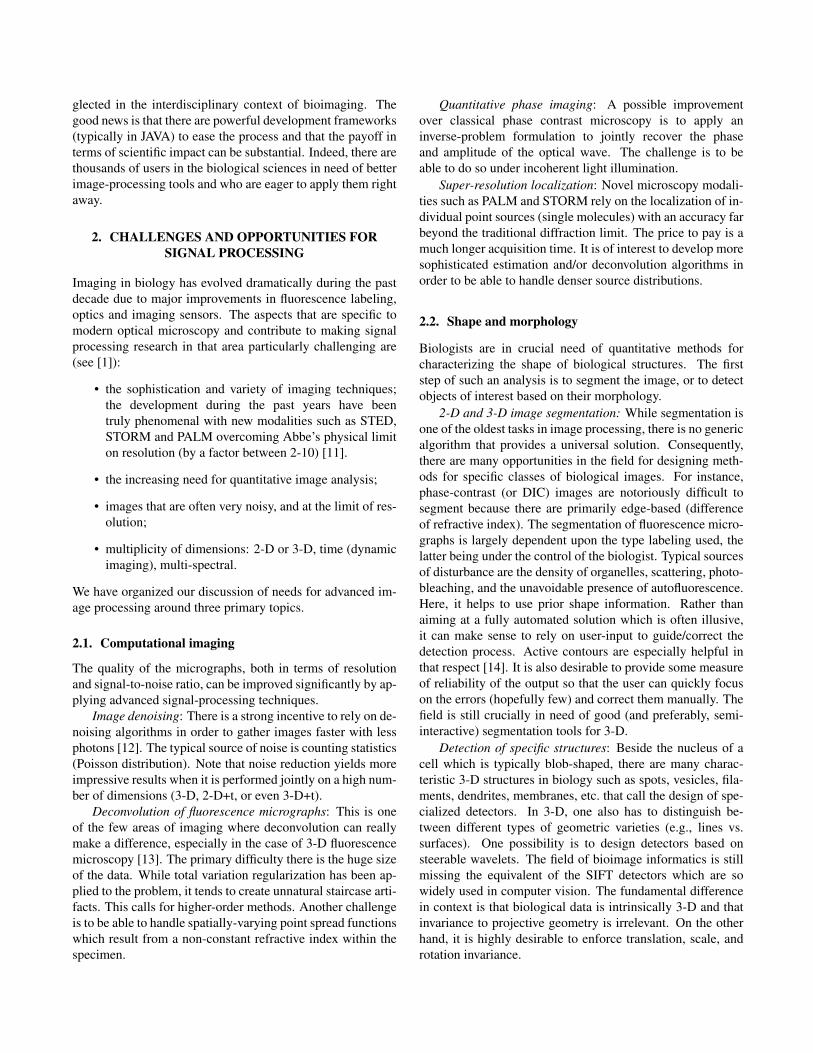
Fig. 1. Samsung Slate PC Series 7 running the open image analysissoftware Icy [10] and one of the plug-ins implementing the methodof [14]. This is the result of the efforts of the open-source communityof developers to produce an user-friendly image analysis software.
1. User-friendliness: The software should be intuitive,easy-to-use and accessible (one-click installation).Moreover, it should be accompanied with clear usermanuals and offer feedback mechanisms (e.g., forums,mailing lists, bug report systems) [17]. We show inFigure 1 an intuitive interface of an image analysissoftware running on a tablet computer.
2. Developer-friendliness: A good documentation of thestructure of the code and of its modules is crucial sinceit allows developers to understand what a program doesand how it works. Open-source software is a good ex-ample of developer-friendly software.
3. Interoperability: It is important to make software thatcommunicates using the available open standards. Inthis way, different software can easily interact withouthaving to define complementary components to trans-late the data. A successful example is the Bio-Formatsproject, a Java library for reading and writing life sci-ences image file formats [18].
4. Modularity: The implicit modularity of object-orienteddesign is key when maintaining a large piece of soft-ware. The use of modular structures with common in-terfaces allows developers to update their software withminimum effort.
5. Validation and quality control: The software should betested in ways that are relevant to the user. Moreover,for the benefit of making research reproducible, it mustbe possible to replicate the exact same computationsand quantitative results that the developers advertise. Arecent trend is to define computational challenges forsome well-defined bioimage analysis tasks such as de-convolution or particle tracking. This allows for objec-

tive performance assessment and comparison of algo-rithms and software solutions.
3.2. Open Image Analysis Platforms
In order to properly analyze an experiment and draw conclu-sions from the data provided by an image-analysis software,the biologist must be aware of what the algorithm really does.Open-source software provides the necessary transparency,giving scientists the opportunity to fully understand the com-putational methods behind their tools.
Among all open-source bioimage analysis tools, the onethat has had the most impact so far is ImageJ [7]. It was ini-tiated by Wayne Rasband at the National Institutes of Health(NIH) under the name of NIH Image. The idea was to developa low-cost image-processing platform for the Apple Macin-tosh II. This piece of software was coded in Pascal, and hadadd-on capabilities in the form of expansion slots in order toenable other developers to easily extend the software for theirown applications.
In the mid-nineties, the programming language Java wascreated by Sun Microsystems. Java applications are typicallycompiled to bytecode that can run on any machine regard-less of the architecture. This allowed developers to write theirsoftware independently of the platform. Rasband ported NIHImage to Java in the late-nineties under the name of ImageJ.As a result, the base of NIH Image users and developers wasextended to PC and Unix.
ImageJ upgraded the expansion slots of NIH Image intothe more modular concept of plug-ins. Since its creation, Im-ageJ has enjoyed a great popularity, and resulted in the devel-opment of a wide variety of plug-ins for very diverse applica-tions.
Besides the core application, another popular distributionis Fiji. It is a user-friendlier distribution of ImageJ togetherwith Java, Java 3-D and the most prominent plug-ins as wellas transparent installation and updates [19].
The largest upgrade of ImageJ since NIH Image is be-ing prepared involving several research laboratories under thename of ImageJ2. It involves a full rewrite of the source codeusing new architectures in order to overcome the limitationsof ImageJ.
Recently, other open-source related platforms are emerg-ing. Among them, we can find: µManager, a software pack-age for the control of automated microscopes [20]; CellPro-filer, a software specialized in measuring phenotypes auto-matically within images [9]; and Icy, a full integrated easy-to-use platform extensible with plug-ins [10]. We summarizeall these open-source projects in Table 1 [8].
3.3. Open Bio Image Alliance
The Open Bio Image Alliance1 (OBIA) was constituted in2012 with the aim of federating the development of the afore-mentioned image-processing packages and improving theirinteroperability. It is an international consortium that bringstogether the major developers of bioimage analysis soft-ware ranging from biologists, microscopists, computer scien-tists, to researchers involved in biomedical image processing.Given the mission of OBIA stated below, we recommend itsweb site as the primary entry point for gathering informationabout the open-source resources for bioimage informatics,both at the level of the users and developers.
The primary mission of OBIA is to
• provide biologists and researchers in the life scienceswith the highest quality public-domain software re-sources and a corresponding knowledge base to ana-lyze and quantitate their image data in a sound andreproducible fashion,
• to strengthen the interaction between biologists, imag-ing scientists and developers of bio-image analysis soft-ware and algorithms.
OBIA capitalizes on the existence of highly successfulsoftware packages such as ImageJ. [. . .] OBIA promoteslong-term availability and backward compatibility, federatesthe harmonious community-based development of interop-erable software, and promotes good software developmentpractices. OBIA will meet these challenges by implementingmechanisms and initiating actions in order to:
• facilitate the diffusion of bioimaging software andguide the choice of image analysis tools with specialattention to quality (curation), long-term availabilityand (backward) compatibility;
• federate the harmonious community-based develop-ment of interoperable software and promote good prac-tices, including the careful validation of tools;
• reinforce interactions between imaging scientists/devel-opers and create a sense of community;
• be a catalyst for new software development projects,advanced image-analysis initiatives, and interdisci-plinary collaborations in the computational and bio-logical sciences.
We can only encourage our colleagues to take part inthis alliance, or, at least, to closely follow what is goingon and available in terms of development tools and soft-ware/algorithm deployment channels and repositories.
1http://www.openbioimage.org/

Initiated Status Language LicenseNIH Image 1987 Discontinued Pascal Public domain
ImageJ 1997 Active Java Public domainµManager 2005 Active C++/Java BSD, Lesser GPLCellProfiler 2006 Active Python GNU
Fiji 2007 Active Java GNUImageJ2 2009 beta version Under development Java Simplified BSD
Icy 2011 Active Java GPL
Table 1. Summary of open-source image-processing platforms.
4. CONCLUSION
We hope to have convinced people involved in signal pro-cessing of the strategic importance of bioimage informatics.So far, the field has been mostly defined by biologists whohave become software developers by necessity. The topics areplentiful and challenging intellectually with a pressing needfor better image processing and analysis tools.
Our advice to designers of new algorithms is to thinkabout user interactions issues from the very start, to take ad-vantage of existing software frameworks such as ImageJ andIcy, and to work in close interactions with biologists. This isthe best way to maximize the impact of one’s research output.
5. REFERENCES
[1] C. Vonesch, F. Aguet, J.-L. Vonesch, and M. Unser, “The col-ored revolution of bioimaging,” IEEE Signal Processing Mag-azine, vol. 23, no. 3, pp. 20–31, May 2006.
[2] Editorial, “The quest for quantitative microscopy,” NatureMethods. Special Issue Focus on Bioimage Informatics, vol. 9,pp. 627, 2012, 2012.
[3] R. Pepperkok and J. Ellenberg, “High-throughput fluorescencemicroscopy for systems biology,” Nature Reviews MolecularCell Biology, vol. 7, no. 9, pp. 690–696, 2006.
[4] H. Peng, “Bioimage informatics: a new area of engineeringbiology,” Bioinformatics, vol. 24, no. 17, pp. 1827–1836, 2008.
[5] A. Cardona and P. Tomancak, “Current challenges in open-source bioimage informatics,” Nature Methods, vol. 9, no. 7,pp. 661–665, July 2012.
[6] A.E. Carpenter, L. Kamentsky, and K.W. Eliceiri, “A call forbioimaging software usability,” Nature Methods, vol. 6, no. 7,pp. 666–670, July 2012.
[7] T.J. Collins, “ImageJ for microscopy,” BioTechniques, vol. 43,no. 1, pp. 25–30, July 2007.
[8] C.A. Schneider, W.S. Rasband, and K.W. Eliceiri, “NIH Imageto ImageJ: 25 years of image analysis,” Nature Methods, vol.9, no. 7, pp. 671–675, July 2012.
[9] M.R. Lamprecht, D.M. Sabatini, and A.E. Carpenter, “Cellpro-filer™: Free, versatile software for automated biological imageanalysis,” BioTechniques, vol. 42, no. 1, pp. 71–75, January2007.
[10] F. De Chaumont, S. Dallongeville, N. Chenouard, N. Herve,S. Pop, T. Provoost, V. Meas-Yedid, P. Pankajakshan,T. Lecomte, Y. Le Montagner, T. Lagache, A. Dufour, and J.-C. Olivo-Marin, “Icy: An open bioimage informatics platformfor extended reproducible reresearch,” Nature Methods, vol. 9,no. 7, pp. 690–696, July 2012.
[11] L. Schermelleh, R. Heintzmann, and H. Leonhardt, “A guideto super-resolution fluorescence microscopy,” The Journal ofCell Biology, vol. 190, no. 2, pp. 165–175, 2010.
[12] F. Luisier, T. Blu, and M. Unser, “Image denoising in mixedPoisson-Gaussian noise,” IEEE Transactions on Image Pro-cessing, vol. 20, no. 3, pp. 696–708, March 2011.
[13] A. Griffa, N. Garin, and D. Sage, “Comparison of deconvo-lution software in 3D microscopy: A user point of view—Part1,” G.I.T. Imaging & Microscopy, vol. 12, no. 1, pp. 43–45,March 2010.
[14] R. Delgado-Gonzalo, P. Thevenaz, C.S. Seelamantula, andM. Unser, “Snakes with an ellipse-reproducing property,”IEEE Transactions on Image Processing, vol. 21, no. 3, pp.1258–1271, March 2012.
[15] D. Sage, M. Unser, P. Salmon, and C. Dibner, “A software so-lution for recording circadian oscillator features in time-lapselive cell microscopy,” Cell Division, vol. 5, no. 17, July 2010.
[16] E. Meijering, I. Smal, and G. Danuser, “Tracking in molecularbioimaging,” Signal Processing Magazine, IEEE, vol. 23, no.3, pp. 46–53, 2006.
[17] D. Bolchini, A. Finkelstein, V. Perrone, and S. Nagl, “Betterbioinformatics through usability analysis,” Bioinformatics, vol.25, no. 3, pp. 406–412, February 2009.
[18] M. Linkert, C.T. Rueden, C. Allan, J.-M. Burel, W. Moore,A. Patterson, B. Loranger, J. Moore, C. Neves, D. MacDonald,A. Tarkowska, C. Sticco, E. Hill, M. Rossner, K.W. Eliceiri,and J.R. Swedlow, “Metadata matters: Access to image datain the real world,” Journal of Cell Biology, vol. 189, no. 5, pp.777–782, May 2010.
[19] J. Schindelin, I. Arganda-Carreras, E. Frise, V. Kaynig,M. Longair, T. Pietzsch, S. Preibisch, C. Rueden, S. Saalfeld,B. Schmid, J.-Y. Tinevez, D.J. White, V. Hartenstein, K. Eli-ceiri, P. Tomancak, and A. Cardona, “Fiji: An open-sourceplatform for biological-image analysis,” Nature Methods, vol.9, no. 7, pp. 676–682, July 2012.
[20] A. Edelstein, N. Amodaj, K. Hoover, R. Vale, and N. Stuur-man, “Computer control of microscopes using µmanager,” inCurrent Protocols in Molecular Biology. John Wiley & Sons,Inc., 2010.