adolescent morphine exposure affects long-term microglial function and later-life relapse liability...
TRANSCRIPT
Neurobiology of Disease
Adolescent Morphine Exposure Affects Long-TermMicroglial Function and Later-Life Relapse Liability in aModel of Addiction
Jaclyn M. Schwarz1 and Staci D. Bilbo1
1Department of Psychology and Neuroscience, Duke University, Durham, North Carolina 27705
Adolescence in humans represents a unique developmental time point associated with increased risk-taking behavior and experimenta-tion with drugs of abuse. We hypothesized that exposure to drugs of abuse during adolescence may increase the risk of addiction inadulthood. To test this, rats were treated with a subchronic regimen of morphine or saline in adolescence, and their preference formorphine was examined using conditioned place preference (CPP) and drug-induced reinstatement in adulthood. The initial preferencefor morphine did not differ between groups; however, rats treated with morphine during adolescence showed robust reinstatement ofmorphine CPP after drug re-exposure in adulthood. This effect was not seen in rats pretreated with a subchronic regimen of morphine asadults, suggesting that exposure to morphine specifically during adolescence increases the risk of relapse to drug-seeking behavior inadulthood. We have previously established a role for microglia, the immune cells of the brain, and immune molecules in the risk ofdrug-induced reinstatement of morphine CPP. Thus, we examined the role of microglia within the nucleus accumbens of these rats anddetermined that rats exposed to morphine during adolescence had a significant increase in Toll-like receptor 4 (TLR4) mRNA and proteinexpression specifically on microglia. Morphine binds to TLR4 directly, and this increase in TLR4 was associated with exaggeratedmorphine-induced TLR4 signaling and microglial activation in rats previously exposed to morphine during adolescence. These datasuggest that long-term changes in microglial function, caused by adolescent morphine exposure, alter the risk of drug-induced reinstate-ment in adulthood.
IntroductionThe brain continues to undergo developmental changes through-out adolescence and early adulthood (Spear, 2000). In particular,brain regions that are necessary for critical thinking and decisionmaking remain underdeveloped in the adolescent brain relativeto the adult brain (Galvan et al., 2006). As a result, adolescents aremore likely to engage in risk-taking behavior and, in particular,increased experimentation with drugs of abuse (Baumrind, 1987;Wills et al., 1994, 1996). Similarly, exposure to drugs of abuseduring adolescence has the potential to impact the developmentof the brain and affect life-long behavior. Work from our labora-tory demonstrates that developmental events can have a lastinginfluence on microglia, the primary immune cells in the brain,including their development and function. This in turn affectsbehaviors that are directly impacted by microglial function, in-cluding addiction, and learning and memory (Bilbo, 2010; Bland
et al., 2010a; Schwarz et al., 2011; Williamson et al., 2011). Forexample, we previously determined that increased maternal careattenuates morphine-induced microglial activation within thenucleus accumbens (NAcc) in adulthood, which decreases therisk of the drug-induced reinstatement of drug-seeking behaviorin a model of conditioned place preference (CPP) (Schwarz et al.,2011). In the current study, we hypothesized that exposure todrugs of abuse during adolescence may alter the developmentand function of microglia and thus increase the risk of drug-seeking behavior into adulthood. Epidemiological evidence fromhumans suggests that adolescents who use drugs of abuse aremore likely to use drugs of abuse in adulthood, although thepotential mechanism underlying this link has not been explored(Robins and Przybeck, 1985; Simoni-Wastila and Yang, 2006).The novel results presented here demonstrate that rats exposed tomorphine during adolescence exhibit an increased risk of relapseto drug-seeking behavior in adulthood caused by long-termchanges in microglial function within the NAcc.
Materials and MethodsGeneral MethodsSubjects. Sprague Dawley rats (32–35 d or 49 –52 d) were obtained fromHarlan and housed in polypropylene cages with ad libitum access to foodand water. The colony was maintained at 22°C on a 12:12 h light-darkcycle (lights on at 0700 EST). All experiments were performed in males.
Drugs. Morphine sulfate and ibudilast were obtained from the Na-tional Institute on Drug Abuse Drug Inventory Supply. Minocyclinehydrochloride was obtained from Sigma-Aldrich. Morphine sulfate was
Received May 24, 2012; revised Nov. 20, 2012; accepted Nov. 21, 2012.Author contributions: J.M.S. and S.D.B. designed research; J.M.S. performed research; J.M.S. analyzed data;
J.M.S. and S.D.B. wrote the paper.This work was supported by National Institutes of Health Grant R01DA025978 to S.D.B. and National Institutes of
Health Grant F32DA030136 to J.M.S. We thank Ashwin Agarwal, Sonia Sabater, and Susan H. Smith for technicalassistance.
The authors declare no competing financial interests.Correspondence should be addressed to Dr. Jaclyn M. Schwarz, 572 Research Drive, Room 3017, Box 91050,
Durham, NC 27705. E-mail: [email protected]:10.1523/JNEUROSCI.2516-12.2013
Copyright © 2013 the authors 0270-6474/13/330961-11$15.00/0
The Journal of Neuroscience, January 16, 2013 • 33(3):961–971 • 961
prepared and is reported as free base concentrations. Ibudilast was dis-solved in a 35% polyethylene glycol/65% saline solution, warmed to50°C. Minocycline hydrochloride (25 mg/kg) was dissolved in water forgavage (2.5 ml/kg). Ibudilast and minocycline are both effective inhibi-tors of glial proinflammation caused by morphine or an immune chal-lenge (Schwarz et al., 2011; Williamson et al., 2011); Minocycline wasused in Experiment 1 (see Experiment 1: adolescent subchronic mor-phine treatment) because ibudilast has slightly mild sedative effects thatmake it difficult to test preference but that do not inhibit general cogni-tive function or learning (Schwarz et al., 2011). Ibudilast, which inhibitsboth microglia and astrocytes, was used in Experiment 3 (not in thecontext of CPP) based on our previous data using this drug, which indi-cates that it can effectively block the synthesis of cytokines and chemo-kines within the NAcc produced by morphine treatment (Schwarz et al.,2011).
Elevated plus maze. The maze consists of two open arms and two closedarms (50.8 � 12.7 cm). The closed arms have 45-cm-high black walls,and the maze is mounted 86 cm off the floor. Rats were placed into thecentral start area and allowed to explore the maze for 5 min. The numberof visits to each arm and the time spent in the open arms, closed arms,and central area were recorded.
Conditioned place preference. An unbiased CPP paradigm was used forthese experiments within a two-chamber shuttle box that is divided intotwo equal-sized compartments. One side of the box was white with a gridfloor, and the other side of the box was black with a rod floor (catalog no.MED-CPP2-RSAT, Med Associates). Activity was measured with auto-mated data collection using photo beam strips and a computer interfacewith Med-PC IV software. On day 1 (d1), rats were placed into thechamber, alternating the side into which the animal was introduced, andthe time spent in each side of the box was measured for 20 min (pretest).On d2 and d3, rats had two conditioning sessions: one in the morningbetween 8 A.M. and 12 noon and one in the afternoon between 1 P.M.and 5 P.M. Rats were randomly assigned to be conditioned to morphinein either the white or black box and during either the morning or after-noon, counterbalancing this parameter across treatment groups using an“unbiased assignment procedure” (Cunningham et al., 2006). Duringthe conditioning sessions, rats were injected alternately with either mor-phine (see doses for individual experiments below) or saline (1 ml/kg)and placed into one side of the conditioning chamber with the guillotinedoor shut for 45 min. The order of administration was randomizedacross treatment groups. On d4, testing occurred between 2 P.M. and 4P.M. Rats were placed into the chamber, and the time spent in each sideof the box was measured for 20 min (post-test). The CPP score wasdetermined by subtracting the time spent in the morphine-paired boxduring the post-test from the time spent in the morphine-paired boxduring the pretest.
Extinction. Rats were placed into the CPP chamber with the guillotinedoor open for 5 min sessions, three times per week. After three 5 minsessions each week, the rats received an extinction test the next day, whichconsisted of a 20 min session with the guillotine door open. Time spent inthe morphine-paired box during the pretest was subtracted from thetime spent in the morphine-paired box during each extinction test.
Microglial immunohistochemistry. Postfixed brain hemispheres weresliced through the NAcc in a 1:5 series at 30 �m (2.28 –1.08 mm frombregma, according to Paxinos and Watson (2005).
The ionized calcium-binding adaptor molecule (Iba1) protein wasused for the analysis of microglia because it is specific to microglia withinthe parenchyma, and its expression is constitutive (Imai et al., 1996).Sections were stained as previously reported (Bilbo and Tsang, 2010).
Quantification of Iba1 densitometry was achieved from digitized im-ages of tissue sections (�10) using Scion Image. Sections were capturedand digitized using a Nikon Eclipse 80i microscope and digital camera(Nikon). Signal pixels of a region of interest were defined as having a grayvalue of 3 SDs above the mean gray value of a cell-sparse area close to theregion of interest. The number of pixels and the average gray values abovethe set background were then computed for each region of interest andmultiplied, giving an integrated density measurement. One measure-ment was made for each of 6 different sections per animal for the NAcc
(including core and shell). All 6 values for each NAcc were averaged toobtain a single integrated density value for each rat.
Quantitative real-time PCR. RNA was isolated from NAcc microdis-sections using the TRIzol method and was subsequently DNase-treated.To measure Toll-like receptor 4 (TLR4), CD11b, and GFAP mRNA,cDNA was synthesized from 150 ng of isolated RNA using the Quanti-Tect Reverse Transcription Kit from QIAGEN, and gene expression wasmeasured using quantitative real-time PCR and the in-house designedprimers listed below using the QuantiTect SYBR Green PCR Kit fromQIAGEN following the manufacturer’s protocol. For the analysis of allother genes, cDNA was synthesized from 500 ng of isolated RNA usingthe RT 2 First Strand Kit (catalog no. C-03; SA Biosciences QIAGEN),and gene expression was measured using quantitative real-time PCR withstock primers designed to measure 84 genes central to TLR-mediatedsignal transduction pathways (SA Biosciences QIAGEN; catalog no.PARN-018) following the manufacturer’s protocol.
Primer specifications. GAPDH: forward, GTTTGTGATGGGTGT-GAACC; reverse, TCTTCTGAGTGGCAGTGATG; TLR4: forward, CA-GAGGAAGAACAAGAAGC; reverse, CCAGATGAACTGTAGCATTC;CD11b: forward, CTGGGAGATGTGAATGGAG; reverse, ACTGAT-GCTGGCTACTGATG; GFAP: forward, AGGGACAATCTCACA-CAGG; reverse, GACTCAACCTTCCTCTCCA.
Quantitative RT-PCR analysis. Threshold amplification cycle number(Ct) was determined for each reaction within the linear phase of theamplification plot, and relative gene expression was determined fromGAPDH using the 2 ���CT method.
Microglial isolation. Isolated NAcc and hippocampi (HP) were dicedinto small pieces using a sterile razor, transferred into a 2 ml sterile tubewith 1 ml of HBSS (without calcium and magnesium) (Invitrogen), andspun at 300 � g at room temperature for 2 min. Tissue was then broughtto a single-cell suspension using the Miltenyi Neural Tissue DissociationKit (P) (Miltenyi Biotec) according to the manufacturer’s instructions.After a final wash in HBSS containing CaCl2 and MgCl2, tissue was incu-bated with anti-myelin microbeads (Miltenyi Biotec) in MACS buffer(PBS containing 0.5% BSA and 2 mM EDTA, pH 7.2) for 15 min at 4°C.Tissue was then washed with 5 ml of MACS buffer and centrifuged at300 � g at 4°C for 10 min. The tissue was resuspended in 500 �l MACSbuffer and passed through a 70 �m nylon filter onto LD columns (Milte-nyi Biotec) exposed to a strong magnetic field. The flow through (demy-elinated cells) and subsequent washes were collected into a 5 mlpolypropylene tube and centrifuged at 300 � g at 4°C for 10 min. Thedemyelinated cells were immediately stained for flow cytometric analysisas follows.
Fluorescence staining and flow cytometry. Demyelinated cells werewashed once in 2 ml PBS supplemented with 0.5% BSA and 2 mM EDTA(wash buffer), followed by centrifugation at 350 � g for 5 min. The washbuffer was removed by aspiration and the pellet dispersed by vortexing.Cells were incubated with 5 �l rat Fc receptor block (CD32; BD Biosci-ences PharMingen) for 5 min at 4°C. Next, cells were incubated for 20min at 4°C in the dark with 100 �l of antibody mixture, includingallophycocyanin-conjugated mouse anti-rat CD11b/c (BD BiosciencesPharMingen), diluted 1:1000, and rabbit anti-TLR4 (Cell SignalingTechnology), diluted 1:100. Cells were washed in buffer before beingincubated in 100 �l secondary antibody, phycoerythrin-conjugated don-key anti-rabbit IgG (eBioscience), diluted 1:500, for 15 min at 4°C in thedark. The cells were washed and spun down at 350 � g for 5 min and fixedin 200 �l 1.5% paraformaldehyde before analysis using a FACSCanto IIflow cytometer (BD Biosciences) and FlowJo software (TreeStar). Foreach sample, 10,000 events were collected and doublets excluded fromthe analysis based on properties of size (forward scatter height and area,see Fig. 8).
Statistical analysis. A two-tailed Student’s t test (morphine vs saline, �level � 0.05) or a one-way ANOVA (morphine and ibudilast treatments)was used to analyze CPP data, as well as subsequent reinstatement data.Significant overall effects using one-way ANOVA were followed up withthe Tukey’s post hoc test (with p � 0.05) to determine group differences.Iba1 densitometry was compared using a two-tailed Student’s t test(morphine vs saline, � level � 0.05). Relative gene expression acrossgroups was compared using either a two-tailed Student’s t test (morphine
962 • J. Neurosci., January 16, 2013 • 33(3):961–971 Schwarz and Bilbo • Adolescence, Morphine, Microglia, and Reinstatement
vs saline, � level � 0.05) or a two-way ANOVA with adolescent treatment(saline or morphine) and adult treatment (saline and morphine) as fac-tors (� level � 0.01). A one-tailed t test was used to analyze the fluores-cent staining intensity of TLR4 protein and CD11b protein after flowcytometry (� level � 0.05). A two-way within-subjects ANOVA was usedto analyze data from the Elevated Plus Maze. Significant interactionsusing a two-way ANOVA were followed up with the Holm-Sidak post hoctest (with p � 0.05) to determine potential group differences. All data ingraphical form represent the mean � SEM.
Experiment 1: adolescent subchronic morphine treatmentThe goal of this experiment was to determine whether exposure to mor-phine during adolescence alters morphine CPP, extinction, or drug-induced reinstatement of morphine CPP in adulthood, and if so, whethermarkers of microglial function/innate immune signaling are significantlyaltered within the NAcc of these rats.
Treatment and CPP. Adolescent rats (�37– 40 d) were randomlyassigned to the following pre-exposure treatment groups: either sub-chronic morphine or saline twice a day (8 A.M. and 4 P.M.) for 5 d (Fig.1A). On d1 and d2, the dose of morphine was 4 mg/kg intraperitoneally(i.p.); and on d3, d4, and d5, each dose of morphine was 8 mg/kg i.p. Ratswere housed in pairs of the same treatment group until behavioral testingbegan in adulthood (60 – 63 d). CPP and extinction were performed asdescribed above (see Conditioned place preference), except that the con-ditioning dose of morphine in this experiment was 4 mg/kg (i.p.). One ratshowed a strong preference for one side of the box on d1 [the rat spentmore than two-thirds of the total time (800 s) in one side of the box] andwas eliminated from the experiments and replaced with another rat.Within 48 h after the completion of all behavioral tasks (Fig. 1A), the ratswere killed, and half of the brain was collected for mRNA analysis of
CD11b and TLR4 and the other half of the brain was submersion fixed in4% paraformaldehyde for 4 d for immunohistochemical detection ofIba1.
Intraperitoneal versus subcutaneous treatment. In Experiment 1, allmorphine and saline treatments were administered i.p. In subsequentexperiments, all morphine and saline treatments were administered sub-cutaneously (s.c.) for greater consistency with the literature. When wechanged the route of administration from i.p. to s.c., we performed a setof experiments which allowed us to determine that morphine CPP pro-duced by 8 mg/kg i.p. (149.2 � 38.2) was nearly identical to that pro-duced by 4 mg/kg s.c. (144.7 � 38.6). In addition, the two doses (viadifferent routes of administration) produce an identical effectiveness andtime course of effectiveness on the hot plate (data not shown), whichsuggests that the pharmacokinetics of the two doses is identical. Thus, inthe CPP experiments described in Experiments 2 and 3, even though wedecreased the morphine dose from 8 mg/kg (i.p.) to 4 mg/kg (s.c.), weobtained similar, if not better, CPP. Importantly, our findings (bothbehavioral and molecular) remained consistent regardless of the routeand/or dose of morphine administration.
Experiment 2: adult subchronic morphine treatmentHaving determined that exposure to morphine during adolescence in-creases reinstatement to CPP after re-exposure to morphine in adult-hood, the goal of this experiment was to determine whether pre-exposureto morphine in adulthood would similarly increase the likelihood ofdrug-induced reinstatement to morphine CPP assessed several weekslater.
Treatment and CPP. Adult rats (�54 –57 d) were randomly assigned tothe following pre-exposure treatment groups: either morphine or salinetwice a day (8 A.M. and 4 P.M.) for 5 d (Fig. 1B). On d1 and d2, the dose
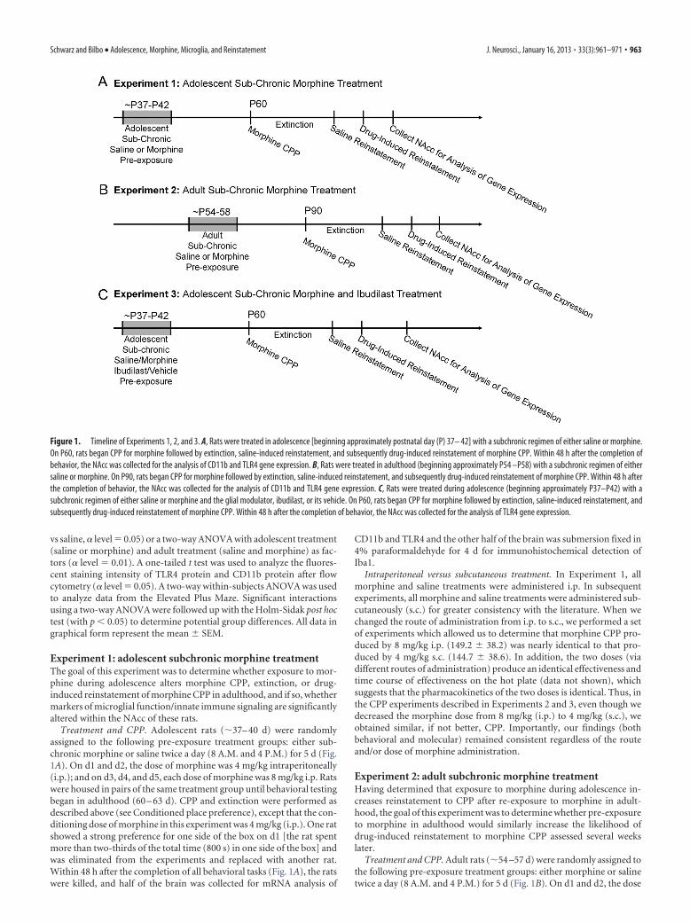
Figure 1. Timeline of Experiments 1, 2, and 3. A, Rats were treated in adolescence [beginning approximately postnatal day (P) 37– 42] with a subchronic regimen of either saline or morphine.On P60, rats began CPP for morphine followed by extinction, saline-induced reinstatement, and subsequently drug-induced reinstatement of morphine CPP. Within 48 h after the completion ofbehavior, the NAcc was collected for the analysis of CD11b and TLR4 gene expression. B, Rats were treated in adulthood (beginning approximately P54 –P58) with a subchronic regimen of eithersaline or morphine. On P90, rats began CPP for morphine followed by extinction, saline-induced reinstatement, and subsequently drug-induced reinstatement of morphine CPP. Within 48 h afterthe completion of behavior, the NAcc was collected for the analysis of CD11b and TLR4 gene expression. C, Rats were treated during adolescence (beginning approximately P37–P42) with asubchronic regimen of either saline or morphine and the glial modulator, ibudilast, or its vehicle. On P60, rats began CPP for morphine followed by extinction, saline-induced reinstatement, andsubsequently drug-induced reinstatement of morphine CPP. Within 48 h after the completion of behavior, the NAcc was collected for the analysis of TLR4 gene expression.
Schwarz and Bilbo • Adolescence, Morphine, Microglia, and Reinstatement J. Neurosci., January 16, 2013 • 33(3):961–971 • 963
of morphine was 4 mg/kg s.c.; and on d3, d4,and d5, each dose of morphine was 8 mg/kg s.c.Rats were housed in pairs of the same treat-ment group until behavioral testing began.CPP began 30 d after the subchronic treatmentbegan (84 – 87 d). CPP and extinction wereperformed as described above (see Condi-tioned place preference), except that the con-ditioning dose of morphine in this experimentwas 4 mg/kg s.c. Within 48 h after the comple-tion of all behavioral tasks, the rats were killed,and half of the brain was collected for mRNAanalysis of CD11b and TLR4 (Fig. 1B).
Experiment 3: adolescent subchronicmorphine and ibudilast treatmentThe goal of this experiment was to determinewhether exposure to morphine during adoles-cence increases the likelihood of reinstatementto morphine CPP via an impact on glia.
Treatment and CPP. Adolescent rats(�37– 40 d) were randomly assigned to the fol-lowing pre-exposure treatment groups: saline� vehicle, saline � ibudilast, morphine � ve-hicle, or morphine � ibudilast. On d1 and d2,the dose of morphine was 4 mg/kg s.c.; and ond3, d4, and d5, each dose of morphine was 8mg/kg s.c. Rats were treated with the afore-mentioned doses of morphine or saline twice aday (8 A.M. and 4 P.M.) for 5 d (Fig. 1C).Thirty minutes before the first treatment eachday, rats received a single dose of either ibudi-last (7.5 mg/kg, i.p.) or vehicle (1 ml/kg, i.p.).Rats were housed in pairs of the same treatment group until behavioraltesting began in adulthood (60 d). CPP began 30 d after the first treat-ment (60 – 63 d). CPP and CPP extinction were performed as describedabove (see Conditioned place preference), except that the conditioningdose of morphine in this experiment was 4 mg/kg s.c. Within 48 h afterthe completion of all behavioral tasks, the rats were killed, and the brainwas collected for mRNA analysis of CD11b and TLR4 (Fig. 1C). A secondgroup of adolescent rats were treated with morphine or saline as de-scribed in Experiment 1 and treated again in adulthood (P60) with asingle dose of morphine (4 mg/kg, s.c.) or saline 20 min before beingkilled to assess the acute TLR signaling and microglial response to mor-phine within the NAc.
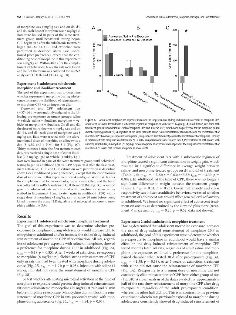
ResultsExperiment 1: adolescent subchronic morphine treatmentThe goal of this experiment was to determine whether pre-exposure to morphine during adolescence would increase CPP tomorphine in adulthood and/or increase the risk of drug-inducedreinstatement of morphine CPP after extinction. All rats, regard-less of adolescent pre-exposure with saline or morphine, showeda preference for morphine during CPP in adulthood (Fig. 2A,t(22) � �0.18; p � 0.85). After 4 weeks of extinction, re-exposureto morphine (8 mg/kg i.p.) elicited strong reinstatement of CPPonly in rats that had been treated with morphine during adoles-cence (Fig. 2B, t(22) � �1.91; p � 0.03). Injection with saline (1ml/kg, i.p.) did not cause the reinstatement of morphine CPP(Fig. 2B).
To test whether attenuating microglial activation at the time ofmorphine re-exposure could prevent drug-induced reinstatement,rats were administered minocycline (25 mg/kg) at 24 h and 30 minbefore morphine re-exposure. Minocycline did not block the rein-statement of morphine CPP in rats previously treated with mor-phine during adolescence (Fig. 2C, t(15) � �1.84; p � 0.04).
Treatment of adolescent rats with a subchronic regimen ofmorphine caused a significant attenuation in weight gain, whichresulted in a significant difference in average weight betweensaline- and morphine-treated groups on d4 and d5 of treatment(Table 1, d4: t(22) � �2.22; p � 0.03; and d5: t(22) � �3.39; p �0.002). In adulthood, at the time of CPP, there was no longer asignificant difference in weight between the treatment groups(Table 1, t(22) � 0.34; p � 0.73). Given that anxiety and stressresponsivity can influence addictive behaviors, we tested whethertreatment of adolescent rats would affect general levels of anxietyin adulthood. We found no significant effect of adolescent treat-ment on anxiety as determined by the elevated plus maze (treat-ment � maze arm: F(1,31) � 0.25; p � 0.62; data not shown).
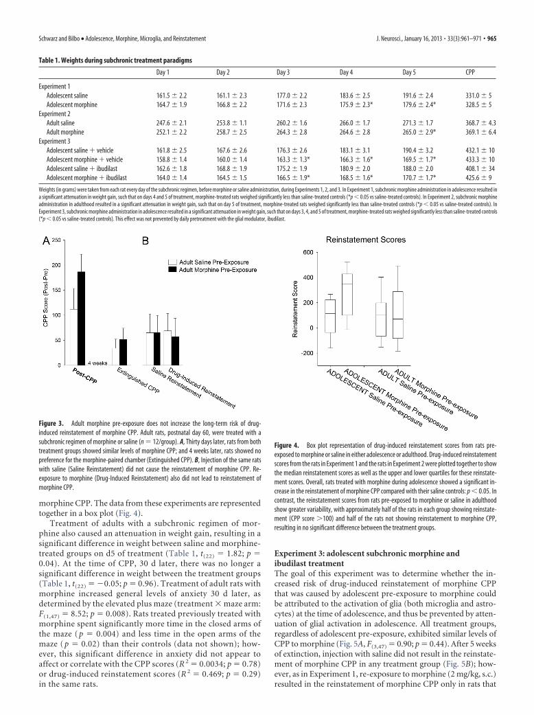
Experiment 2: adult subchronic morphine treatmentHaving determined that adolescent morphine exposure increasesthe risk of drug-induced reinstatement of morphine CPP inadulthood, the goal of this experiment was to determine whetherpre-exposure to morphine in adulthood would have a similareffect on the drug-induced reinstatement of morphine CPPtested months later. All rats, regardless of adult saline and mor-phine pre-exposure, exhibited a preference for the morphine-paired chamber when tested 30 d after pre-exposure (Fig. 3A,t(22) � �1.38; p � 0.18). After 5 weeks of extinction, treatmentwith saline did not cause the reinstatement of morphine CPP(Fig. 3A). Reexposure to a priming dose of morphine did notconsistently elicit reinstatement of CPP from either group of rats(Fig. 3B). A closer analysis of the data revealed that approximatelyhalf of the rats show reinstatement of morphine CPP after drugre-exposure, regardless of the adult pre-exposure condition,whereas the other half did not. This is in contrast to the previousexperiment wherein rats previously exposed to morphine duringadolescence consistently showed drug-induced reinstatement of
Figure 2. Adolescent morphine pre-exposure increases the long-term risk of drug-induced reinstatement of morphine CPP.Adolescent rats were treated with a subchronic regimen of morphine or saline (n � 12/group). A, In adulthood, rats from bothtreatment groups showed similar levels of morphine CPP; and 3 weeks later, rats showed no preference for the morphine-pairedchamber (Extinguished CPP). B, Injection of the same rats with saline (Saline Reinstatement) did not cause the reinstatement ofmorphine CPP; however, re-exposure to morphine (Drug-Induced Reinstatement) caused the reinstatement of morphine CPP onlyin rats treated with morphine as adolescents. *p � 0.05, compared with saline-treated rats. C, Pretreatment of both groups witha microglial inhibitor, minocycline (25 mg/kg), before morphine re-exposure did not prevent the drug-induced reinstatement ofmorphine CPP in rats that received morphine as adolescents.
964 • J. Neurosci., January 16, 2013 • 33(3):961–971 Schwarz and Bilbo • Adolescence, Morphine, Microglia, and Reinstatement
morphine CPP. The data from these experiments are representedtogether in a box plot (Fig. 4).
Treatment of adults with a subchronic regimen of mor-phine also caused an attenuation in weight gain, resulting in asignificant difference in weight between saline and morphine-treated groups on d5 of treatment (Table 1, t(22) � 1.82; p �0.04). At the time of CPP, 30 d later, there was no longer asignificant difference in weight between the treatment groups(Table 1, t(22) � �0.05; p � 0.96). Treatment of adult rats withmorphine increased general levels of anxiety 30 d later, asdetermined by the elevated plus maze (treatment � maze arm:F(1,47) � 8.52; p � 0.008). Rats treated previously treated withmorphine spent significantly more time in the closed arms ofthe maze ( p � 0.004) and less time in the open arms of themaze ( p � 0.02) than their controls (data not shown); how-ever, this significant difference in anxiety did not appear toaffect or correlate with the CPP scores (R 2 � 0.0034; p � 0.78)or drug-induced reinstatement scores (R 2 � 0.469; p � 0.29)in the same rats.
Experiment 3: adolescent subchronic morphine andibudilast treatmentThe goal of this experiment was to determine whether the in-creased risk of drug-induced reinstatement of morphine CPPthat was caused by adolescent pre-exposure to morphine couldbe attributed to the activation of glia (both microglia and astro-cytes) at the time of adolescence, and thus be prevented by atten-uation of glial activation in adolescence. All treatment groups,regardless of adolescent pre-exposure, exhibited similar levels ofCPP to morphine (Fig. 5A, F(3,47) � 0.90; p � 0.44). After 5 weeksof extinction, injection with saline did not result in the reinstate-ment of morphine CPP in any treatment group (Fig. 5B); how-ever, as in Experiment 1, re-exposure to morphine (2 mg/kg, s.c.)resulted in the reinstatement of morphine CPP only in rats that
Table 1. Weights during subchronic treatment paradigms
Day 1 Day 2 Day 3 Day 4 Day 5 CPP
Experiment 1Adolescent saline 161.5 � 2.2 161.1 � 2.3 177.0 � 2.2 183.6 � 2.5 191.6 � 2.4 331.0 � 5Adolescent morphine 164.7 � 1.9 166.8 � 2.2 171.6 � 2.3 175.9 � 2.3* 179.6 � 2.4* 328.5 � 5
Experiment 2Adult saline 247.6 � 2.1 253.8 � 1.1 260.2 � 1.6 266.0 � 1.7 271.3 � 1.7 368.7 � 4.3Adult morphine 252.1 � 2.2 258.7 � 2.5 264.3 � 2.8 264.6 � 2.8 265.0 � 2.9* 369.1 � 6.4
Experiment 3Adolescent saline � vehicle 161.8 � 2.5 167.6 � 2.6 176.3 � 2.6 183.1 � 3.1 190.4 � 3.2 432.1 � 10Adolescent morphine � vehicle 158.8 � 1.4 160.0 � 1.4 163.3 � 1.3* 166.3 � 1.6* 169.5 � 1.7* 433.3 � 10Adolescent saline � ibudilast 162.6 � 1.8 168.8 � 1.9 175.2 � 1.9 180.9 � 2.0 188.0 � 2.0 408.1 � 34Adolescent morphine � ibudilast 164.0 � 1.4 164.5 � 1.5 166.5 � 1.9* 168.5 � 1.6* 170.7 � 1.7* 425.6 � 9
Weights (in grams) were taken from each rat every day of the subchronic regimen, before morphine or saline administration, during Experiments 1, 2, and 3. In Experiment 1, subchronic morphine administration in adolescence resulted ina significant attenuation in weight gain, such that on days 4 and 5 of treatment, morphine-treated rats weighed significantly less than saline-treated controls (*p � 0.05 vs saline-treated controls). In Experiment 2, subchronic morphineadministration in adulthood resulted in a significant attenuation in weight gain, such that on day 5 of treatment, morphine-treated rats weighed significantly less than saline-treated controls (*p � 0.05 vs saline-treated controls). InExperiment 3, subchronic morphine administration in adolescence resulted in a significant attenuation in weight gain, such that on days 3, 4, and 5 of treatment, morphine-treated rats weighed significantly less than saline-treated controls(*p � 0.05 vs saline-treated controls). This effect was not prevented by daily pretreatment with the glial modulator, ibudilast.
Figure 3. Adult morphine pre-exposure does not increase the long-term risk of drug-induced reinstatement of morphine CPP. Adult rats, postnatal day 60, were treated with asubchronic regimen of morphine or saline (n � 12/group). A, Thirty days later, rats from bothtreatment groups showed similar levels of morphine CPP; and 4 weeks later, rats showed nopreference for the morphine-paired chamber (Extinguished CPP). B, Injection of the same ratswith saline (Saline Reinstatement) did not cause the reinstatement of morphine CPP. Re-exposure to morphine (Drug-Induced Reinstatement) also did not lead to reinstatement ofmorphine CPP.
Figure 4. Box plot representation of drug-induced reinstatement scores from rats pre-exposed to morphine or saline in either adolescence or adulthood. Drug-induced reinstatementscores from the rats in Experiment 1 and the rats in Experiment 2 were plotted together to showthe median reinstatement scores as well as the upper and lower quartiles for these reinstate-ment scores. Overall, rats treated with morphine during adolescence showed a significant in-crease in the reinstatement of morphine CPP compared with their saline controls: p � 0.05. Incontrast, the reinstatement scores from rats pre-exposed to morphine or saline in adulthoodshow greater variability, with approximately half of the rats in each group showing reinstate-ment (CPP score 100) and half of the rats not showing reinstatement to morphine CPP,resulting in no significant difference between the treatment groups.
Schwarz and Bilbo • Adolescence, Morphine, Microglia, and Reinstatement J. Neurosci., January 16, 2013 • 33(3):961–971 • 965
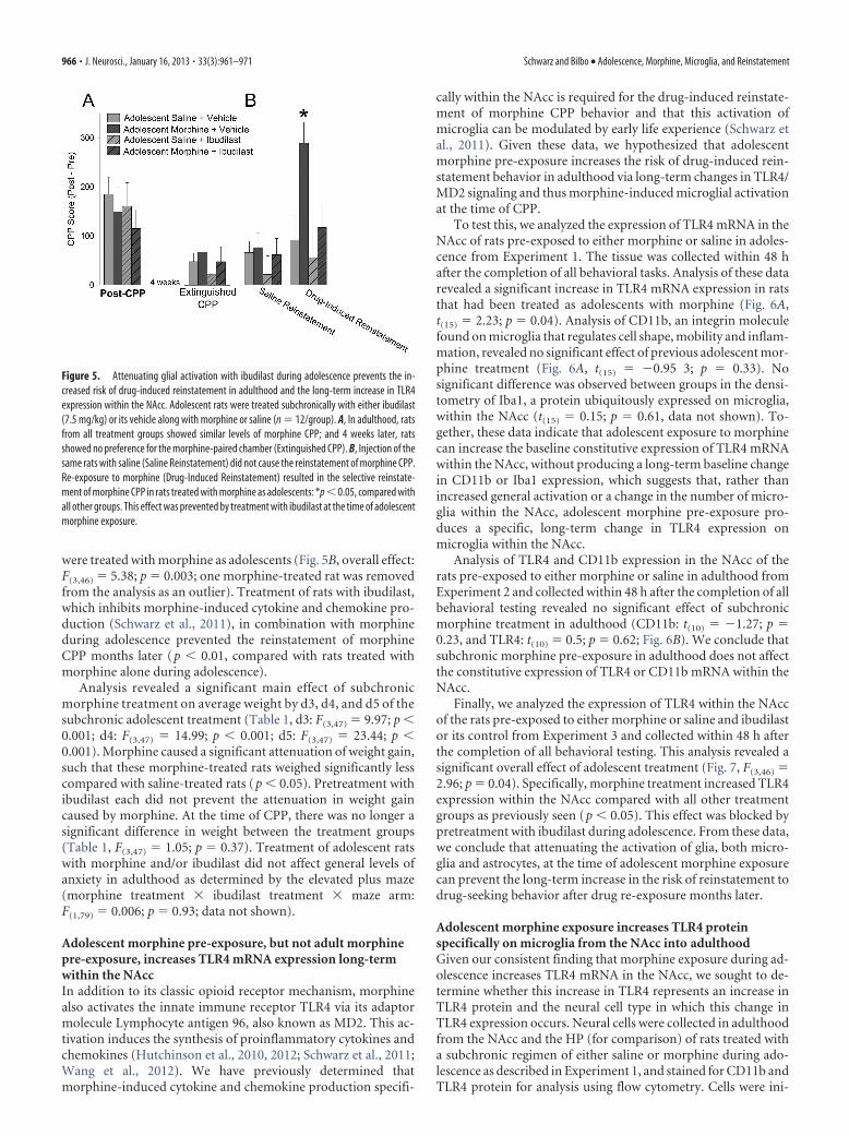
were treated with morphine as adolescents (Fig. 5B, overall effect:F(3,46) � 5.38; p � 0.003; one morphine-treated rat was removedfrom the analysis as an outlier). Treatment of rats with ibudilast,which inhibits morphine-induced cytokine and chemokine pro-duction (Schwarz et al., 2011), in combination with morphineduring adolescence prevented the reinstatement of morphineCPP months later (p � 0.01, compared with rats treated withmorphine alone during adolescence).
Analysis revealed a significant main effect of subchronicmorphine treatment on average weight by d3, d4, and d5 of thesubchronic adolescent treatment (Table 1, d3: F(3,47) � 9.97; p �0.001; d4: F(3,47) � 14.99; p � 0.001; d5: F(3,47) � 23.44; p �0.001). Morphine caused a significant attenuation of weight gain,such that these morphine-treated rats weighed significantly lesscompared with saline-treated rats (p � 0.05). Pretreatment withibudilast each did not prevent the attenuation in weight gaincaused by morphine. At the time of CPP, there was no longer asignificant difference in weight between the treatment groups(Table 1, F(3,47) � 1.05; p � 0.37). Treatment of adolescent ratswith morphine and/or ibudilast did not affect general levels ofanxiety in adulthood as determined by the elevated plus maze(morphine treatment � ibudilast treatment � maze arm:F(1,79) � 0.006; p � 0.93; data not shown).
Adolescent morphine pre-exposure, but not adult morphinepre-exposure, increases TLR4 mRNA expression long-termwithin the NAccIn addition to its classic opioid receptor mechanism, morphinealso activates the innate immune receptor TLR4 via its adaptormolecule Lymphocyte antigen 96, also known as MD2. This ac-tivation induces the synthesis of proinflammatory cytokines andchemokines (Hutchinson et al., 2010, 2012; Schwarz et al., 2011;Wang et al., 2012). We have previously determined thatmorphine-induced cytokine and chemokine production specifi-
cally within the NAcc is required for the drug-induced reinstate-ment of morphine CPP behavior and that this activation ofmicroglia can be modulated by early life experience (Schwarz etal., 2011). Given these data, we hypothesized that adolescentmorphine pre-exposure increases the risk of drug-induced rein-statement behavior in adulthood via long-term changes in TLR4/MD2 signaling and thus morphine-induced microglial activationat the time of CPP.
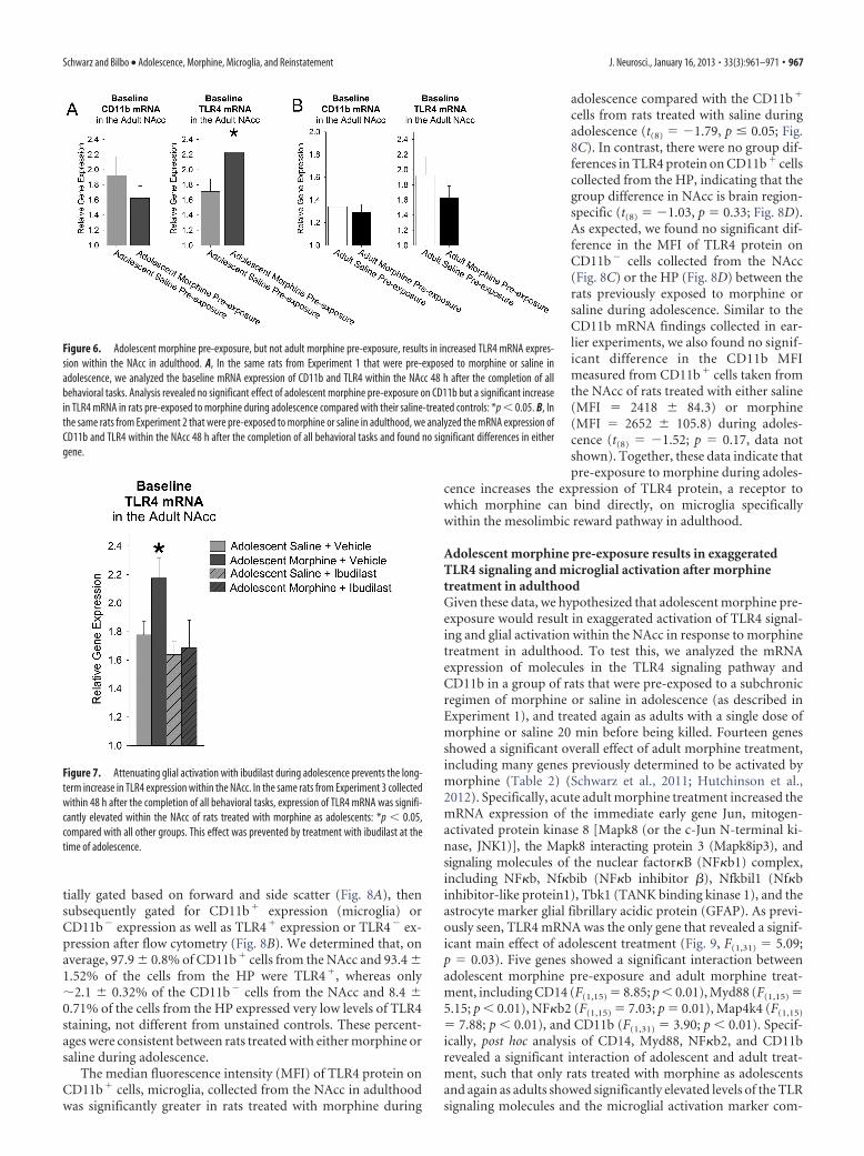
To test this, we analyzed the expression of TLR4 mRNA in theNAcc of rats pre-exposed to either morphine or saline in adoles-cence from Experiment 1. The tissue was collected within 48 hafter the completion of all behavioral tasks. Analysis of these datarevealed a significant increase in TLR4 mRNA expression in ratsthat had been treated as adolescents with morphine (Fig. 6A,t(15) � 2.23; p � 0.04). Analysis of CD11b, an integrin moleculefound on microglia that regulates cell shape, mobility and inflam-mation, revealed no significant effect of previous adolescent mor-phine treatment (Fig. 6A, t(15) � �0.95 3; p � 0.33). Nosignificant difference was observed between groups in the densi-tometry of Iba1, a protein ubiquitously expressed on microglia,within the NAcc (t(15) � 0.15; p � 0.61, data not shown). To-gether, these data indicate that adolescent exposure to morphinecan increase the baseline constitutive expression of TLR4 mRNAwithin the NAcc, without producing a long-term baseline changein CD11b or Iba1 expression, which suggests that, rather thanincreased general activation or a change in the number of micro-glia within the NAcc, adolescent morphine pre-exposure pro-duces a specific, long-term change in TLR4 expression onmicroglia within the NAcc.
Analysis of TLR4 and CD11b expression in the NAcc of therats pre-exposed to either morphine or saline in adulthood fromExperiment 2 and collected within 48 h after the completion of allbehavioral testing revealed no significant effect of subchronicmorphine treatment in adulthood (CD11b: t(10) � �1.27; p �0.23, and TLR4: t(10) � 0.5; p � 0.62; Fig. 6B). We conclude thatsubchronic morphine pre-exposure in adulthood does not affectthe constitutive expression of TLR4 or CD11b mRNA within theNAcc.
Finally, we analyzed the expression of TLR4 within the NAccof the rats pre-exposed to either morphine or saline and ibudilastor its control from Experiment 3 and collected within 48 h afterthe completion of all behavioral testing. This analysis revealed asignificant overall effect of adolescent treatment (Fig. 7, F(3,46) �2.96; p � 0.04). Specifically, morphine treatment increased TLR4expression within the NAcc compared with all other treatmentgroups as previously seen (p � 0.05). This effect was blocked bypretreatment with ibudilast during adolescence. From these data,we conclude that attenuating the activation of glia, both micro-glia and astrocytes, at the time of adolescent morphine exposurecan prevent the long-term increase in the risk of reinstatement todrug-seeking behavior after drug re-exposure months later.
Adolescent morphine exposure increases TLR4 proteinspecifically on microglia from the NAcc into adulthoodGiven our consistent finding that morphine exposure during ad-olescence increases TLR4 mRNA in the NAcc, we sought to de-termine whether this increase in TLR4 represents an increase inTLR4 protein and the neural cell type in which this change inTLR4 expression occurs. Neural cells were collected in adulthoodfrom the NAcc and the HP (for comparison) of rats treated witha subchronic regimen of either saline or morphine during ado-lescence as described in Experiment 1, and stained for CD11b andTLR4 protein for analysis using flow cytometry. Cells were ini-
Figure 5. Attenuating glial activation with ibudilast during adolescence prevents the in-creased risk of drug-induced reinstatement in adulthood and the long-term increase in TLR4expression within the NAcc. Adolescent rats were treated subchronically with either ibudilast(7.5 mg/kg) or its vehicle along with morphine or saline (n � 12/group). A, In adulthood, ratsfrom all treatment groups showed similar levels of morphine CPP; and 4 weeks later, ratsshowed no preference for the morphine-paired chamber (Extinguished CPP). B, Injection of thesame rats with saline (Saline Reinstatement) did not cause the reinstatement of morphine CPP.Re-exposure to morphine (Drug-Induced Reinstatement) resulted in the selective reinstate-ment of morphine CPP in rats treated with morphine as adolescents: *p � 0.05, compared withall other groups. This effect was prevented by treatment with ibudilast at the time of adolescentmorphine exposure.
966 • J. Neurosci., January 16, 2013 • 33(3):961–971 Schwarz and Bilbo • Adolescence, Morphine, Microglia, and Reinstatement
tially gated based on forward and side scatter (Fig. 8A), thensubsequently gated for CD11b� expression (microglia) orCD11b� expression as well as TLR4� expression or TLR4� ex-pression after flow cytometry (Fig. 8B). We determined that, onaverage, 97.9 � 0.8% of CD11b� cells from the NAcc and 93.4 �1.52% of the cells from the HP were TLR4�, whereas only�2.1 � 0.32% of the CD11b� cells from the NAcc and 8.4 �0.71% of the cells from the HP expressed very low levels of TLR4staining, not different from unstained controls. These percent-ages were consistent between rats treated with either morphine orsaline during adolescence.
The median fluorescence intensity (MFI) of TLR4 protein onCD11b� cells, microglia, collected from the NAcc in adulthoodwas significantly greater in rats treated with morphine during
adolescence compared with the CD11b�
cells from rats treated with saline duringadolescence (t(8) � �1.79, p � 0.05; Fig.8C). In contrast, there were no group dif-ferences in TLR4 protein on CD11b� cellscollected from the HP, indicating that thegroup difference in NAcc is brain region-specific (t(8) � �1.03, p � 0.33; Fig. 8D).As expected, we found no significant dif-ference in the MFI of TLR4 protein onCD11b� cells collected from the NAcc(Fig. 8C) or the HP (Fig. 8D) between therats previously exposed to morphine orsaline during adolescence. Similar to theCD11b mRNA findings collected in ear-lier experiments, we also found no signif-icant difference in the CD11b MFImeasured from CD11b� cells taken fromthe NAcc of rats treated with either saline(MFI � 2418 � 84.3) or morphine(MFI � 2652 � 105.8) during adoles-cence (t(8) � �1.52; p � 0.17, data notshown). Together, these data indicate thatpre-exposure to morphine during adoles-
cence increases the expression of TLR4 protein, a receptor towhich morphine can bind directly, on microglia specificallywithin the mesolimbic reward pathway in adulthood.
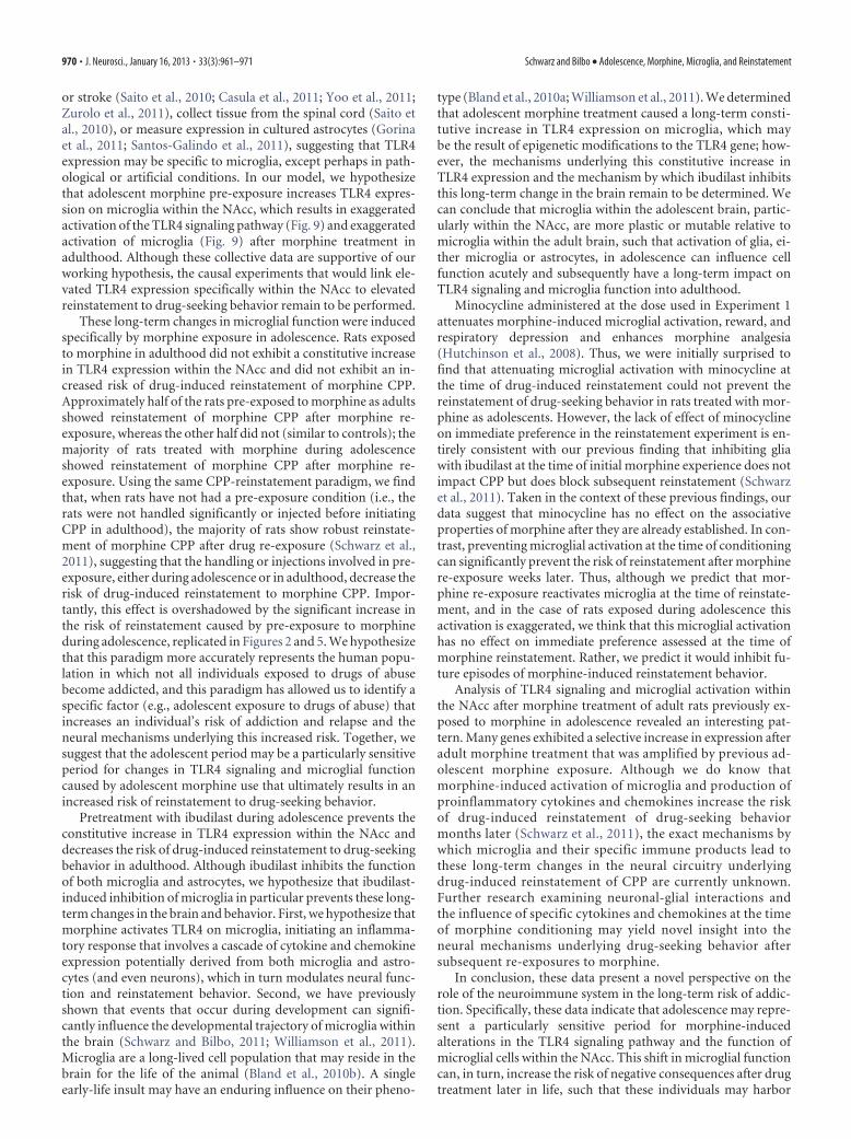
Adolescent morphine pre-exposure results in exaggeratedTLR4 signaling and microglial activation after morphinetreatment in adulthoodGiven these data, we hypothesized that adolescent morphine pre-exposure would result in exaggerated activation of TLR4 signal-ing and glial activation within the NAcc in response to morphinetreatment in adulthood. To test this, we analyzed the mRNAexpression of molecules in the TLR4 signaling pathway andCD11b in a group of rats that were pre-exposed to a subchronicregimen of morphine or saline in adolescence (as described inExperiment 1), and treated again as adults with a single dose ofmorphine or saline 20 min before being killed. Fourteen genesshowed a significant overall effect of adult morphine treatment,including many genes previously determined to be activated bymorphine (Table 2) (Schwarz et al., 2011; Hutchinson et al.,2012). Specifically, acute adult morphine treatment increased themRNA expression of the immediate early gene Jun, mitogen-activated protein kinase 8 [Mapk8 (or the c-Jun N-terminal ki-nase, JNK1)], the Mapk8 interacting protein 3 (Mapk8ip3), andsignaling molecules of the nuclear factor�B (NF�b1) complex,including NF�b, Nf�bib (NF�b inhibitor �), Nfkbil1 (Nf�binhibitor-like protein1), Tbk1 (TANK binding kinase 1), and theastrocyte marker glial fibrillary acidic protein (GFAP). As previ-ously seen, TLR4 mRNA was the only gene that revealed a signif-icant main effect of adolescent treatment (Fig. 9, F(1,31) � 5.09;p � 0.03). Five genes showed a significant interaction betweenadolescent morphine pre-exposure and adult morphine treat-ment, including CD14 (F(1,15) � 8.85; p � 0.01), Myd88 (F(1,15) �5.15; p � 0.01), NF�b2 (F(1,15) � 7.03; p � 0.01), Map4k4 (F(1,15)
� 7.88; p � 0.01), and CD11b (F(1,31) � 3.90; p � 0.01). Specif-ically, post hoc analysis of CD14, Myd88, NF�b2, and CD11brevealed a significant interaction of adolescent and adult treat-ment, such that only rats treated with morphine as adolescentsand again as adults showed significantly elevated levels of the TLRsignaling molecules and the microglial activation marker com-
Figure 6. Adolescent morphine pre-exposure, but not adult morphine pre-exposure, results in increased TLR4 mRNA expres-sion within the NAcc in adulthood. A, In the same rats from Experiment 1 that were pre-exposed to morphine or saline inadolescence, we analyzed the baseline mRNA expression of CD11b and TLR4 within the NAcc 48 h after the completion of allbehavioral tasks. Analysis revealed no significant effect of adolescent morphine pre-exposure on CD11b but a significant increasein TLR4 mRNA in rats pre-exposed to morphine during adolescence compared with their saline-treated controls: *p � 0.05. B, Inthe same rats from Experiment 2 that were pre-exposed to morphine or saline in adulthood, we analyzed the mRNA expression ofCD11b and TLR4 within the NAcc 48 h after the completion of all behavioral tasks and found no significant differences in eithergene.
Figure 7. Attenuating glial activation with ibudilast during adolescence prevents the long-term increase in TLR4 expression within the NAcc. In the same rats from Experiment 3 collectedwithin 48 h after the completion of all behavioral tasks, expression of TLR4 mRNA was signifi-cantly elevated within the NAcc of rats treated with morphine as adolescents: *p � 0.05,compared with all other groups. This effect was prevented by treatment with ibudilast at thetime of adolescence.
Schwarz and Bilbo • Adolescence, Morphine, Microglia, and Reinstatement J. Neurosci., January 16, 2013 • 33(3):961–971 • 967
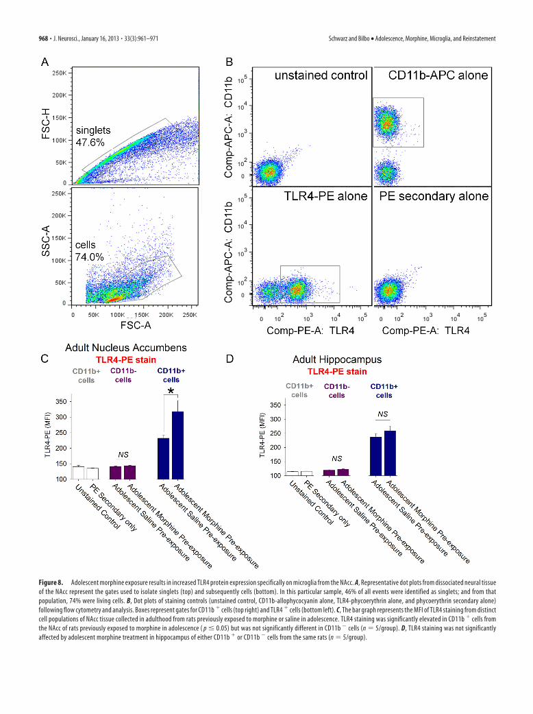
Figure 8. Adolescent morphine exposure results in increased TLR4 protein expression specifically on microglia from the NAcc. A, Representative dot plots from dissociated neural tissueof the NAcc represent the gates used to isolate singlets (top) and subsequently cells (bottom). In this particular sample, 46% of all events were identified as singlets; and from thatpopulation, 74% were living cells. B, Dot plots of staining controls (unstained control, CD11b-allophycocyanin alone, TLR4-phycoerythrin alone, and phycoerythrin secondary alone)following flow cytometry and analysis. Boxes represent gates for CD11b � cells (top right) and TLR4 � cells (bottom left). C, The bar graph represents the MFI of TLR4 staining from distinctcell populations of NAcc tissue collected in adulthood from rats previously exposed to morphine or saline in adolescence. TLR4 staining was significantly elevated in CD11b � cells fromthe NAcc of rats previously exposed to morphine in adolescence ( p � 0.05) but was not significantly different in CD11b � cells (n � 5/group). D, TLR4 staining was not significantlyaffected by adolescent morphine treatment in hippocampus of either CD11b � or CD11b � cells from the same rats (n � 5/group).
968 • J. Neurosci., January 16, 2013 • 33(3):961–971 Schwarz and Bilbo • Adolescence, Morphine, Microglia, and Reinstatement
pared with all other treatment groups (Fig. 9, p � 0.05). Analysisof Map4k4 revealed the exact opposite pattern (Fig. 9, p � 0.05).
DiscussionWe demonstrate that rats exposed to morphine during adoles-cence exhibit an increased risk of reinstatement to drug-seekingbehavior in adulthood, and treatment of rats with morphine dur-ing adolescence causes a long-lasting increase in the expression ofTLR4 on microglia specifically within the NAcc. TLR4 is an in-nate immune receptor to which morphine can bind directly(Hutchinson et al., 2012). Although the activation of TLR4 bybacteria results in a robust increase in the synthesis of manycytokines and chemokines, morphine induces the activation ofmicroglia, resulting in a robust increase in the synthesis of a selectgroup of cytokines and chemokines within the NAcc, includinginterferon-�, CCL25, CCL12, CCL21b, CCL4, and CCL17(Schwarz et al., 2011). Although the specific function of thesecytokines and chemokines within the NAcc is currently un-known, we know that their expression specifically within theNAcc at the time of morphine conditioning is necessary for thedrug-induced reinstatement of morphine CPP months later. Spe-cifically, the drug-induced reinstatement of morphine CPP can
be blocked by preventing their expression directly within theNAcc via ibudilast infusion at the time of morphine CPP(Schwarz et al., 2011). Consistent with these findings, we presentthree novel pieces of data related to the risk of drug-inducedreinstatement: (1) rats treated with morphine during adolescenceexhibit increased levels of TLR4 specifically on microglia withinthe NAcc and exhibit exaggerated TLR4 signaling and microglialactivation within the NAcc after morphine re-exposure in adult-hood; (2) rats treated with morphine during adolescence exhibitan increased risk of drug-induced reinstatement of morphineCPP in adulthood; and (3) blocking the activation of glia withibudilast during morphine treatment in adolescence prevents thelong-term increase in TLR4 expression within the NAcc and pre-vents the increased risk of drug-induced reinstatement of mor-phine CPP in adulthood.
Using flow cytometry of rapidly isolated neural tissue, we de-termined that TLR4 is selectively expressed on microglia fromboth the NAcc and the hippocampus. Others report that TLR4 isalso present on astrocytes; however, we found virtually no pro-tein expression of TLR4 on other cell types rapidly isolated fromthese two brain regions. The reports indicating TLR4 expressionon astrocytes and sometimes neurons use either models of disease
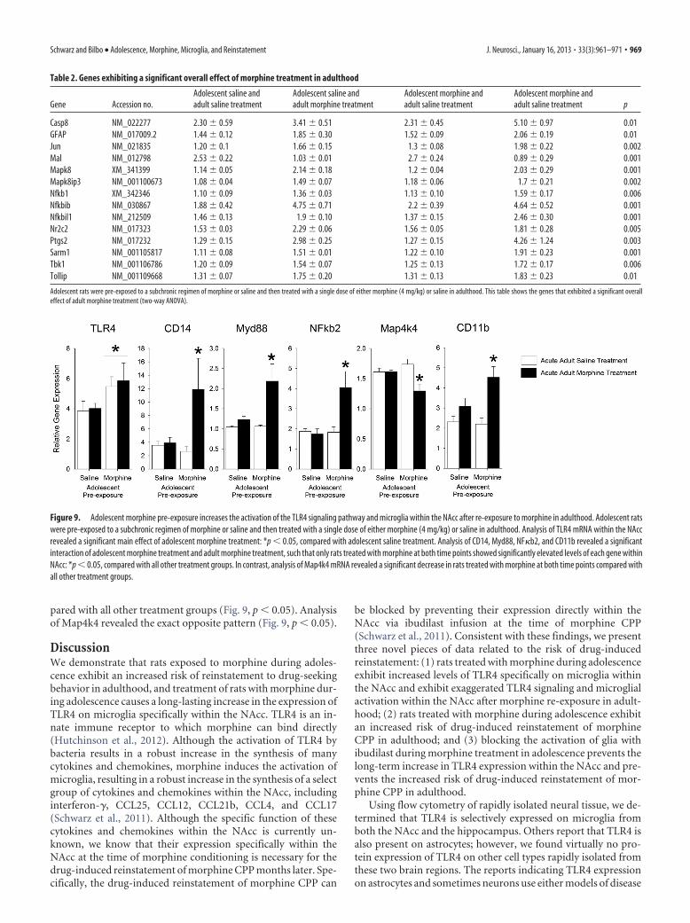
Table 2. Genes exhibiting a significant overall effect of morphine treatment in adulthood
Gene Accession no.Adolescent saline andadult saline treatment
Adolescent saline andadult morphine treatment
Adolescent morphine andadult saline treatment
Adolescent morphine andadult saline treatment p
Casp8 NM_022277 2.30 � 0.59 3.41 � 0.51 2.31 � 0.45 5.10 � 0.97 0.01GFAP NM_017009.2 1.44 � 0.12 1.85 � 0.30 1.52 � 0.09 2.06 � 0.19 0.01Jun NM_021835 1.20 � 0.1 1.66 � 0.15 1.3 � 0.08 1.98 � 0.22 0.002Mal NM_012798 2.53 � 0.22 1.03 � 0.01 2.7 � 0.24 0.89 � 0.29 0.001Mapk8 XM_341399 1.14 � 0.05 2.14 � 0.18 1.2 � 0.04 2.03 � 0.29 0.001Mapk8ip3 NM_001100673 1.08 � 0.04 1.49 � 0.07 1.18 � 0.06 1.7 � 0.21 0.002Nfkb1 XM_342346 1.10 � 0.09 1.36 � 0.03 1.13 � 0.10 1.59 � 0.17 0.006Nfkbib NM_030867 1.88 � 0.42 4.75 � 0.71 2.2 � 0.39 4.64 � 0.52 0.001Nfkbil1 NM_212509 1.46 � 0.13 1.9 � 0.10 1.37 � 0.15 2.46 � 0.30 0.001Nr2c2 NM_017323 1.53 � 0.03 2.29 � 0.06 1.56 � 0.05 1.81 � 0.28 0.005Ptgs2 NM_017232 1.29 � 0.15 2.98 � 0.25 1.27 � 0.15 4.26 � 1.24 0.003Sarm1 NM_001105817 1.11 � 0.08 1.51 � 0.01 1.22 � 0.10 1.91 � 0.23 0.001Tbk1 NM_001106786 1.20 � 0.09 1.54 � 0.07 1.25 � 0.13 1.72 � 0.17 0.006Tollip NM_001109668 1.31 � 0.07 1.75 � 0.20 1.31 � 0.13 1.83 � 0.23 0.01
Adolescent rats were pre-exposed to a subchronic regimen of morphine or saline and then treated with a single dose of either morphine (4 mg/kg) or saline in adulthood. This table shows the genes that exhibited a significant overalleffect of adult morphine treatment (two-way ANOVA).
Figure 9. Adolescent morphine pre-exposure increases the activation of the TLR4 signaling pathway and microglia within the NAcc after re-exposure to morphine in adulthood. Adolescent ratswere pre-exposed to a subchronic regimen of morphine or saline and then treated with a single dose of either morphine (4 mg/kg) or saline in adulthood. Analysis of TLR4 mRNA within the NAccrevealed a significant main effect of adolescent morphine treatment: *p � 0.05, compared with adolescent saline treatment. Analysis of CD14, Myd88, NF�b2, and CD11b revealed a significantinteraction of adolescent morphine treatment and adult morphine treatment, such that only rats treated with morphine at both time points showed significantly elevated levels of each gene withinNAcc: *p � 0.05, compared with all other treatment groups. In contrast, analysis of Map4k4 mRNA revealed a significant decrease in rats treated with morphine at both time points compared withall other treatment groups.
Schwarz and Bilbo • Adolescence, Morphine, Microglia, and Reinstatement J. Neurosci., January 16, 2013 • 33(3):961–971 • 969
or stroke (Saito et al., 2010; Casula et al., 2011; Yoo et al., 2011;Zurolo et al., 2011), collect tissue from the spinal cord (Saito etal., 2010), or measure expression in cultured astrocytes (Gorinaet al., 2011; Santos-Galindo et al., 2011), suggesting that TLR4expression may be specific to microglia, except perhaps in path-ological or artificial conditions. In our model, we hypothesizethat adolescent morphine pre-exposure increases TLR4 expres-sion on microglia within the NAcc, which results in exaggeratedactivation of the TLR4 signaling pathway (Fig. 9) and exaggeratedactivation of microglia (Fig. 9) after morphine treatment inadulthood. Although these collective data are supportive of ourworking hypothesis, the causal experiments that would link ele-vated TLR4 expression specifically within the NAcc to elevatedreinstatement to drug-seeking behavior remain to be performed.
These long-term changes in microglial function were inducedspecifically by morphine exposure in adolescence. Rats exposedto morphine in adulthood did not exhibit a constitutive increasein TLR4 expression within the NAcc and did not exhibit an in-creased risk of drug-induced reinstatement of morphine CPP.Approximately half of the rats pre-exposed to morphine as adultsshowed reinstatement of morphine CPP after morphine re-exposure, whereas the other half did not (similar to controls); themajority of rats treated with morphine during adolescenceshowed reinstatement of morphine CPP after morphine re-exposure. Using the same CPP-reinstatement paradigm, we findthat, when rats have not had a pre-exposure condition (i.e., therats were not handled significantly or injected before initiatingCPP in adulthood), the majority of rats show robust reinstate-ment of morphine CPP after drug re-exposure (Schwarz et al.,2011), suggesting that the handling or injections involved in pre-exposure, either during adolescence or in adulthood, decrease therisk of drug-induced reinstatement to morphine CPP. Impor-tantly, this effect is overshadowed by the significant increase inthe risk of reinstatement caused by pre-exposure to morphineduring adolescence, replicated in Figures 2 and 5. We hypothesizethat this paradigm more accurately represents the human popu-lation in which not all individuals exposed to drugs of abusebecome addicted, and this paradigm has allowed us to identify aspecific factor (e.g., adolescent exposure to drugs of abuse) thatincreases an individual’s risk of addiction and relapse and theneural mechanisms underlying this increased risk. Together, wesuggest that the adolescent period may be a particularly sensitiveperiod for changes in TLR4 signaling and microglial functioncaused by adolescent morphine use that ultimately results in anincreased risk of reinstatement to drug-seeking behavior.
Pretreatment with ibudilast during adolescence prevents theconstitutive increase in TLR4 expression within the NAcc anddecreases the risk of drug-induced reinstatement to drug-seekingbehavior in adulthood. Although ibudilast inhibits the functionof both microglia and astrocytes, we hypothesize that ibudilast-induced inhibition of microglia in particular prevents these long-term changes in the brain and behavior. First, we hypothesize thatmorphine activates TLR4 on microglia, initiating an inflamma-tory response that involves a cascade of cytokine and chemokineexpression potentially derived from both microglia and astro-cytes (and even neurons), which in turn modulates neural func-tion and reinstatement behavior. Second, we have previouslyshown that events that occur during development can signifi-cantly influence the developmental trajectory of microglia withinthe brain (Schwarz and Bilbo, 2011; Williamson et al., 2011).Microglia are a long-lived cell population that may reside in thebrain for the life of the animal (Bland et al., 2010b). A singleearly-life insult may have an enduring influence on their pheno-
type (Bland et al., 2010a; Williamson et al., 2011). We determinedthat adolescent morphine treatment caused a long-term consti-tutive increase in TLR4 expression on microglia, which maybe the result of epigenetic modifications to the TLR4 gene; how-ever, the mechanisms underlying this constitutive increase inTLR4 expression and the mechanism by which ibudilast inhibitsthis long-term change in the brain remain to be determined. Wecan conclude that microglia within the adolescent brain, partic-ularly within the NAcc, are more plastic or mutable relative tomicroglia within the adult brain, such that activation of glia, ei-ther microglia or astrocytes, in adolescence can influence cellfunction acutely and subsequently have a long-term impact onTLR4 signaling and microglia function into adulthood.
Minocycline administered at the dose used in Experiment 1attenuates morphine-induced microglial activation, reward, andrespiratory depression and enhances morphine analgesia(Hutchinson et al., 2008). Thus, we were initially surprised tofind that attenuating microglial activation with minocycline atthe time of drug-induced reinstatement could not prevent thereinstatement of drug-seeking behavior in rats treated with mor-phine as adolescents. However, the lack of effect of minocyclineon immediate preference in the reinstatement experiment is en-tirely consistent with our previous finding that inhibiting gliawith ibudilast at the time of initial morphine experience does notimpact CPP but does block subsequent reinstatement (Schwarzet al., 2011). Taken in the context of these previous findings, ourdata suggest that minocycline has no effect on the associativeproperties of morphine after they are already established. In con-trast, preventing microglial activation at the time of conditioningcan significantly prevent the risk of reinstatement after morphinere-exposure weeks later. Thus, although we predict that mor-phine re-exposure reactivates microglia at the time of reinstate-ment, and in the case of rats exposed during adolescence thisactivation is exaggerated, we think that this microglial activationhas no effect on immediate preference assessed at the time ofmorphine reinstatement. Rather, we predict it would inhibit fu-ture episodes of morphine-induced reinstatement behavior.
Analysis of TLR4 signaling and microglial activation withinthe NAcc after morphine treatment of adult rats previously ex-posed to morphine in adolescence revealed an interesting pat-tern. Many genes exhibited a selective increase in expression afteradult morphine treatment that was amplified by previous ad-olescent morphine exposure. Although we do know thatmorphine-induced activation of microglia and production ofproinflammatory cytokines and chemokines increase the riskof drug-induced reinstatement of drug-seeking behaviormonths later (Schwarz et al., 2011), the exact mechanisms bywhich microglia and their specific immune products lead tothese long-term changes in the neural circuitry underlyingdrug-induced reinstatement of CPP are currently unknown.Further research examining neuronal-glial interactions andthe influence of specific cytokines and chemokines at the timeof morphine conditioning may yield novel insight into theneural mechanisms underlying drug-seeking behavior aftersubsequent re-exposures to morphine.
In conclusion, these data present a novel perspective on therole of the neuroimmune system in the long-term risk of addic-tion. Specifically, these data indicate that adolescence may repre-sent a particularly sensitive period for morphine-inducedalterations in the TLR4 signaling pathway and the function ofmicroglial cells within the NAcc. This shift in microglial functioncan, in turn, increase the risk of negative consequences after drugtreatment later in life, such that these individuals may harbor
970 • J. Neurosci., January 16, 2013 • 33(3):961–971 Schwarz and Bilbo • Adolescence, Morphine, Microglia, and Reinstatement
long-term changes in the neural circuitry that promote the cycleof morphine addiction and relapse.
ReferencesBaumrind D (1987) A developmental perspective on adolescent risk taking
in contemporary America. New Dir Child Dev 37:93–125. MedlineBilbo SD (2010) Early-life infection is a vulnerability factor for aging-related
glial alterations and cognitive decline. Neurobiol Learn Mem 94:57– 64.CrossRef Medline
Bilbo SD, Tsang V (2010) Enduring consequences of maternal obesity forbrain inflammation and behavior of offspring. FASEB J 24:2104 –2115.CrossRef Medline
Bland ST, Beckley JT, Watkins LR, Maier SF, Bilbo SD (2010a) NeonatalEscherichia coli infection alters glial, cytokine, and neuronal gene expres-sion in response to acute amphetamine in adolescent rats. Neurosci Lett474:52–57. CrossRef Medline
Bland ST, Beckley JT, Young S, Tsang V, Watkins LR, Maier SF, Bilbo SD(2010b) Enduring consequences of early-life infection on glial and neuralcell genesis within cognitive regions of the brain. Brain Behav Immun24:329 –338. CrossRef Medline
Casula M, Iyer AM, Spliet WG, Anink JJ, Steentjes K, Sta M, Troost D,Aronica E (2011) Toll-like receptor signaling in amyotrophic lateralsclerosis spinal cord tissue. Neuroscience 179:233–243. CrossRef Medline
Cunningham CL, Gremel CM, Groblewski PA (2006) Drug-induced condi-tioned place preference and aversion in mice. Nat Protoc 1:1662–1670.CrossRef Medline
Galvan A, Hare TA, Parra CE, Penn J, Voss H, Glover G, Casey BJ (2006)Earlier development of the accumbens relative to orbitofrontal cortexmight underlie risk-taking behavior in adolescents. J Neurosci 26:6885– 6892. CrossRef Medline
Gorina R, Font-Nieves M, Marquez-Kisinousky L, Santalucia T, Planas AM(2011) Astrocyte TLR4 activation induces a proinflammatory environ-ment through the interplay between MyD88-dependent NFkappaB sig-naling, MAPK, and Jak1/Stat1 pathways. Glia 59:242–255. CrossRefMedline
Hutchinson MR, Northcutt AL, Chao LW, Kearney JJ, Zhang Y, Berkelham-mer DL, Loram LC, Rozeske RR, Bland ST, Maier SF, Gleeson TT, Wat-kins LR (2008) Minocycline suppresses morphine-induced respiratorydepression, suppresses morphine-induced reward, and enhances sys-temic morphine-induced analgesia. Brain Behav Immun 22:1248 –1256.CrossRef Medline
Hutchinson MR, Zhang Y, Shridhar M, Evans JH, Buchanan MM, Zhao TX,Slivka PF, Coats BD, Rezvani N, Wieseler J, Hughes TS, Landgraf KE,Chan S, Fong S, Phipps S, Falke JJ, Leinwand LA, Maier SF, Yin H, RiceKC, Watkins LR (2010) Evidence that opioids may have Toll-like recep-tor 4 and MD-2 effects. Brain Behav Immun 24:83–95. CrossRef Medline
Hutchinson MR, Northcutt AL, Hiranita T, Wang X, Lewis SS, Thomas J, vanSteeg K, Kopajtic TA, Loram LC, Sfregola C, Galer E, Miles NE, Bland ST,Amat J, Rozeske RR, Maslanik T, Chapman TR, Strand KA, Fleshner M,Bachtell RK, Somogyi AA, Yin H, Katz JL, Rice KC, Maier SF, Watkins LR.
(2012) Opioid activation of Toll-like receptor 4 contributes to drug re-inforcement. J Neurosci 32:11187–11200. CrossRef Medline
Imai Y, Ibata I, Ito D, Ohsawa K, Kohsaka S (1996) A novel gene iba1 in themajor histocompatibility complex class III region encoding an EF handprotein expressed in a monocytic lineage. Biochem Biophys Res Commun224:855– 862. CrossRef Medline
Paxinos G, Watson C (2005) The rat brain in stereotaxic coordinates, Ed 5.New York: Academic.
Robins LN, Przybeck TR (1985) Age of onset of drug use as a factor in drugand other disorders. NIDA Res Monogr 56:178 –192. Medline
Saito O, Svensson CI, Buczynski MW, Wegner K, Hua XY, Codeluppi S,Schaloske RH, Deems RA, Dennis EA, Yaksh TL (2010) Spinal glialTLR4-mediated nociception and production of prostaglandin E(2) andTNF. Br J Pharmacol 160:1754 –1764. CrossRef Medline
Santos-Galindo M, Acaz-Fonseca E, Bellini MJ, Garcia-Segura LM (2011)Sex differences in the inflammatory response of primary astrocytes tolipopolysaccharide. Biol Sex Differ 2:7. CrossRef Medline
Schwarz JM, Bilbo SD (2011) LPS elicits a much larger and broader inflam-matory response than Escherichia coli infection within the hippocampusof neonatal rats. Neurosci Lett 497:110 –115. CrossRef Medline
Schwarz JM, Hutchinson MR, Bilbo SD (2011) Early-life experience de-creases drug-induced reinstatement of morphine CPP in adulthood viamicroglial-specific epigenetic programming of anti-inflammatory IL-10expression. J Neurosci 31:17835–17847. CrossRef Medline
Simoni-Wastila L, Yang HK (2006) Psychoactive drug abuse in older adults.Am J Geriatr Pharmacother 4:380 –394. CrossRef Medline
Spear LP (2000) The adolescent brain and age-related behavioral manifes-tations. Neurosci Biobehav Rev 24:417– 463. CrossRef Medline
Wang X, Loram LC, Ramos K, de Jesus AJ, Thomas J, Cheng K, Reddy A,Somogyi AA, Hutchinson MR, Watkins LR, Yin H (2012) Morphineactivates neuroinflammation in a manner parallel to endotoxin. Proc NatlAcad Sci U S A 109:6325– 6330. CrossRef Medline
Williamson LL, Sholar PW, Mistry RM, Smith SH, Bilbo SD (2011) Mi-croglia and memory: modulation by early-life infection. J Neurosci31:15511–15521. CrossRef Medline
Wills TA, Vaccaro D, McNamara G (1994) Novelty seeking, risk taking, andrelated constructs as predictors of adolescent substance use: an applica-tion of Cloninger’s theory. J Subst Abuse 6:1–20. CrossRef Medline
Wills TA, Vaccaro D, McNamara G, Hirky AE (1996) Escalated substanceuse: a longitudinal grouping analysis from early to middle adolescence.J Abnorm Psychol 105:166 –180. CrossRef Medline
Yoo KY, Yoo DY, Hwang IK, Park JH, Lee CH, Choi JH, Kwon SH, Her S, LeeYL, Won MH (2011) Time-course alterations of Toll-like receptor 4 andNF-kappaB p65, and their coexpression in the gerbil hippocampal CA1region after transient cerebral ischemia. Neurochem Res 36:2417–2426.CrossRef Medline
Zurolo E, Iyer A, Maroso M, Carbonell C, Anink JJ, Ravizza T, Fluiter K, SplietWG, van Rijen PC, Vezzani A, Aronica E (2011) Activation of Toll-likereceptor, RAGE and HMGB1 signalling in malformations of cortical de-velopment. Brain 134:1015–1032. CrossRef Medline
Schwarz and Bilbo • Adolescence, Morphine, Microglia, and Reinstatement J. Neurosci., January 16, 2013 • 33(3):961–971 • 971