adenosine-binding motif mimicry and cellular effects of a ... · london wc2a 3ly, u.k., ‡cancer...
TRANSCRIPT

Biochem. J. (2013) 451, 329–342 (Printed in Great Britain) doi:10.1042/BJ20121871 329
Adenosine-binding motif mimicry and cellular effects of athieno[2,3-d]pyrimidine-based chemical inhibitor of atypical proteinkinase C isoenzymesSvend KJÆR*1, Mark LINCH†1, Andrew PURKISS*, Brenda KOSTELECKY*†, Phillip P. KNOWLES*, Carine ROSSE†, PhilippeRIOU†, Christelle SOUDY‡, Sarah KAYE‡, Bhavisha PATEL‡, Erika SORIANO*, Judith MURRAY-RUST*, Caroline BARTON‡,Christian DILLON‡, Jon ROFFEY‡, Peter J. PARKER†§2 and Neil Q. MCDONALD*‖2
*Structural Biology, Cancer Research UK, 44 Lincoln’s Inn Fields, London WC2A 3LY, U.K., †Protein Phosphorylation Laboratories, Cancer Research UK, 44 Lincoln’s Inn Fields,London WC2A 3LY, U.K., ‡Cancer Research Technology Discovery Laboratories, Wolfson Institute for Biomedical Research, University College London, Gower Street, London WC1E6BT, U.K., §Division of Cancer Studies, King’s College London, New Hunts House, Guy’s Campus, London SE1 1UL, U.K., and ‖Institute of Structural and Molecular Biology,Department of Biological Sciences, Birkbeck College, University of London, Malet Street, London WC1E 7HX, U.K.
The aPKC [atypical PKC (protein kinase C)] isoforms ι and ζ playcrucial roles in the formation and maintenance of cell polarity andrepresent attractive anti-oncogenic drug targets in Ras-dependenttumours. To date, few isoform-specific chemical biology tools areavailable to inhibit aPKC catalytic activity. In the present paper,we describe the identification and functional characterizationof potent and selective thieno[2,3-d]pyrimidine-based chemicalinhibitors of aPKCs. A crystal structure of human PKCι kinasedomain bound to a representative compound, CRT0066854,reveals the basis for potent and selective chemical inhibition.Furthermore, CRT0066854 displaces a crucial Asn-Phe-Aspmotif that is part of the adenosine-binding pocket and engages
an acidic patch used by arginine-rich PKC substrates. We showthat CRT0066854 inhibits the LLGL2 (lethal giant larvae 2) phos-phorylation in cell lines and exhibits phenotypic effects in a rangeof cell-based assays. We conclude that this compound can be usedas a chemical tool to modulate aPKC activity in vitro and in vivoand may guide the search for further aPKC-selective inhibitors.
Key words: ATP pocket, atypical protein kinase C, cell-basedassay, cell migration, cell polarity, chemical inhibitor,crystallography, Madin–Darby canine kidney (MDCK) cell,protein kinase A/protein kinase G/protein kinase C family kinase(AGC kinase).
INTRODUCTION
PKC (protein kinase C) isoenzymes are an important familyof serine/threonine protein kinases that contribute to manydiverse cellular and tissue functions, as well as human diseasepathologies including cancer development and progression[1,2]. The molecular architecture of PKC family members isconserved throughout the cPKC (classical PKC; cPKCα, cPKCβand cPKCγ ), nPKC (novel PKC; nPKCδ, nPKCε, nPKCη andnPKCθ ) and aPKC (atypical PKC; aPKCζ and aPKCι) isoforms.They all comprise a C-terminal serine/threonine protein kinasedomain [belonging to the AGC kinase (protein kinase A/proteinkinase G/PKC family kinase) superfamily] and an N-terminalregulatory domain [3]. The kinase domain has a C-terminalextension unique to AGC kinases that contain essential ‘priming’phosphorylation sites required for catalytic activation [4]; inaPKCs, the most C-terminal of these is replaced by a phospho-mimetic glutamate residue. The regulatory domain within allPKC isoforms has an inhibitory region (pseudo-substrate motif)and distinctive arrangements of C1, C2 and/or PB1 (Phox/Bem1)domains (the last being exclusive to the aPKCs) that together arecrucial for conferring isoform-specific functions [3].
The aPKC isoforms (PKCζ and PKCι) diverge from other PKCfamily members as their regulatory domains are unresponsiveto diacylglycerol, phosphatidylserine and Ca2 + . Instead theyare regulated by protein interactions, for example with polarityproteins Par-6, Par-3 and the Rho family GTPase cdc42 [5]. Thesecomponents assemble into an evolutionarily conserved Par-6–aPKC–Par-3 complex present in both invertebrates and vertebratesthat controls apical–basal polarity in epithelial cells and anterior–posterior positioning in asymmetric cell division [6,7]. In additionto an established role in cell polarity, aPKC is required for normalcell proliferation, mitotic spindle orientation [8,9] and migration[10,11].
There has been considerable interest in aPKCs as drug targetsdue to their role in cancer development and progression [12]. Incancer-specific cell models, PKCζ is required for EGF (epidermalgrowth factor)-induced migration of human breast and lung cancercells and CSF-1 (colony-stimulating factor 1) chemotaxis ofmacrophages and acute monocytic leukaemic cells [13–15]. PKCιpromotes nicotine-induced migration and invasion of lung cancercells via phosphorylation of m- and μ-calpains [16] and is requiredfor non-adherent cell growth in a Rac-1-dependent manner [17].The potential importance of aPKC in Her2-driven malignancies
Abbreviations used: Ade, adenosine-binding; AGC kinase, protein kinase A/protein kinase G/protein kinase C family kinase; aPKC, atypical proteinkinase C; BIM-1, bisindolylmaleimide-1; CCD, charge-coupled-device; cPKC, classical protein kinase C; 2D, two-dimensional; 3D, three-dimensional;DCM, dichloromethane; DMEM, Dulbecco’s modified Eagle’s medium; DTT, dithiothreitol; F-actin, filamentous actin; FBS, fetal bovine serum; GFP, greenfluorescent protein; GST, glutathione transferase; HEK, human embryonic kidney; HRP, horseradish peroxidase; HTS, high-throughput screen; IMAP,immobilized metal ion-affinity fluorescence polarization; LC, liquid chromatography; LLGL, lethal giant larvae; MDCK, Madin–Darby canine kidney; MI,mass ion; nPKC, novel protein kinase C; NRK, normal rat kidney cell; PKC, protein kinase C; PSAL, predominantly single apical lumen; RMSD, root meansquare deviation; RNAi, RNA interference; RT, retention time; SPR, surface plasmon resonance; TLS, translation libration screw-motion.
1 These authors contributed equally to this work.2 Correspondence may be addressed to either of these authors (email [email protected] or [email protected]).The structural co-ordinates for PKCιk bound to CRT0066854 will appear in the PDB under the accession number 3ZH8.
c© The Authors Journal compilation c© 2013 Biochemical Society

330 S. Kjær and others
was demonstrated when the aberrant polarity induced by forcedHer2 dimerization in human mammary epithelial MCF-10A cellsembedded in MatrigelTM was partially reversed following geneticdisruption of aPKC binding to Par-6 [18]. In model organisms,aPKC has been shown to co-operate with bona fide oncogenes,including Ras and Notch in Drosophila melanogaster, to leadto invasive and metastatic tumours [19,20]. Furthermore, intransgenic mice K-Ras-induced lung cancer is reduced followingdepletion of PKCι or its adaptor protein p62 [21,22].
In human malignancy, one of the most frequent amplicons is3q26, the location of the PKCι gene (HGNC symbol PRKCI).PKCι gene amplification leads to high protein levels and is cor-related with poor clinical outcomes in squamous cell carcinomasof the lung [17] and oesophagus [23]. High PKCι expression hasbeen correlated with poor prognosis and/or worse stage in an in-creasing number of human cancers including breast [24], pancreas[25], stomach [26], bile duct [27] and brain [28]. In addition to thecompelling evidence for a role of PKCι in human malignancy, theinterest in PKCι as a therapeutic target was further enhanced bythe finding that elevated PKCι protein is associated with chemo-therapy resistance [29] and that depletion/inhibition of PKCι issynergistic with some conventional chemotherapies [30–32].
Like many protein kinases, aPKC isoenzymes are amenableto small-molecule chemical inhibition either through their ATP-binding clefts or allosteric pockets [33]. However, there arefew selective chemical biology tools to inhibit aPKC catalyticactivity. Available tools include the non-selective and relativelyimpotent BIM-1 (bisindolylmaleimide 1), Go6983 and Go6976compounds or a PKCζ pseudo-substrate peptide containing amembrane-targeting myristoylation site [34]. The developmentof effective ATP-competitive inhibitors against aPKCs, similarto other protein kinases, has been complicated by challengesin both potency and selectivity [33]. In general, such inhibitorsacquire selectivity by associating with both the ATP-binding siteand with adjacent residues, which are less well conserved. Otherapproaches include screening for allosteric inhibitors of PKCζthat target the PIF pocket [35] or blocking PKCι interaction withPar-6, for example with ATM (aurothiomalate), a quite differentclass of PKCι inhibitor which exhibits potent antitumour activityin lung cancer [36].
In the present study, we report the identification of an aPKCinhibitor CRT0066854 which is able to mimic both ATP anda key side chain that forms part of the aPKC nucleotide-binding cleft. We define the compound’s mechanism of actionby crystallographic and biochemical methods and demonstrate itsutility through the use of phenotypic cell-based assays. Together,our data indicate that CRT0066854 is a potent and selectivechemical tool to modulate aPKC activity both in vitro and incells.
EXPERIMENTAL
Chemical synthesis of CRT0066854 and CRT0066390
Synthesis of (S)-N*1*-(2-pyridin-4-yl-5,6,7,8-tetrahydro-benzo[4,5]thieno[2,3-d]pyrimidin-4-yl)-butane-1,2-diamine(CRT0066854)
Synthesis of 2-pyridin-4-yl-5,6,7,8-tetrahydro-benzo[4,5]thieno[2,3-d]pyrimidin-4-ol (A-1)
To a solution of 4-cyanopyridine (1.25 g, 12 mmol) indry dioxane (10 ml) was added 2-amino-4,5,6,7-tetrahydro-benzo[b]thiophene-3-carboxylic acid ethyl ester (2.25 g,10 mmol) followed by potassium-tert-pentylate 1.7 M in toluene(12 ml, 20 mmol). The reaction mixture was stirred at roomtemperature (20 ◦C) overnight. After completion the precipitateformed was filtered and washed with diethyl ether. The residuewas used without any further purification in the next step.LC (liquid chromatography)–MS method: RT (retention time):3.54 min, MI (mass ion): 284 [M + 1]. 1H NMR (300 MHz,DMSO): 8.56 (d, 2 H), 8.12 (d, 2 H), 2.90 (m, 2 H), 2.67(m, 2 H), 1.76 (m, 4 H).
Synthesis of 2,4,6-tri-isopropyl-benzenesulfonic acid 2-pyridin-4-yl-5,6,7,8-tetrahydro-benzo[4,5]thieno[2,3-d]pyrimidin-4-yl es-ter (A-2)
To a solution of 2-pyridin-4-yl-5,6,7,8-tetrahydro-benzo[4,5]thieno[2,3-d]pyrimidin-4-ol (1 g, 3.5 mmol) (A-1) inDCM (dichloromethane, 10 ml) was added 2,4,6-tri-isopropylbenzenesulfonyl chloride (1.3 g, 4.2 mmol),triethylamine (Et3N) (1.5 ml, 10.5 mmol) and DMAP (4-dimethylaminopyridine) (6 mg, 0.05 mmol). The mixture wasstirred at room temperature for 1 h. After completion the mixturewas diluted with water and the product was extracted into DCM(2×2 ml). The combined extracts were dried with magnesiumsulfate, filtered and evaporated under reduced pressure to yieldthe title compound as a brown solid, which was used withoutfurther purification in the next step. LC–MS method: RT:6.23 min, MI: 550 [M + 1].
Synthesis of (S)-3-phenyl-propane-1,2-diamine (A-3)
To a suspension of (S)-2-amino-3-phenyl-propionamide hy-drochloride (540 mg, 2.7 mmol) in DCM (5 ml) was addedtriethylamine (Et3N) (380 μl, 2.7 mmol). The suspension wasstirred for 2 h at room temperature, the resulting solid was filteredand the filtrate was concentrated under reduced pressure to yield awhite solid, to which was added dropwise a 1 M solution of BH3 inTHF (tetrahydrofuran, 20 ml, 20 mmol), the solution was stirredovernight at reflux. After cooling the solution was hydrolysed byslow addition of an excess of 10% acetic acid/methanol (30 ml)and refluxed for a further 2 h. The solvent was removed underreduced pressure, and the residue was dissolved in methanol andpassed through a SCX-2 cartridge and washed with methanol.The product was released from the cartridge using a solution of2 M ammonia/methanol. The solvent was evaporated to providethe title compound as a white solid which was used in the nextstep without further purification. LC–MS method: RT: 0.36 min,MI: 151 [M + 1].
c© The Authors Journal compilation c© 2013 Biochemical Society

A novel small molecule aPKC inhibitor 331
Synthesis of (S)-N*1*-(2-pyridin-4-yl-5,6,7,8-tetrahydro-benzo[4,5]thieno[2,3-d]pyrimidin-4-yl)-butane-1,2-diamine(Compound A, CRT0066854)
To a solution of 2,4,6-tri-isopropyl-benzenesulfonic acid2-pyridin-4-yl-5,6,7,8-tetrahydro-benzo[4,5]thieno[2,3-d]pyrimidin-4-yl ester (A-2) (100 mg, 0.180 mmol) in DMA(dimethylacetamide, 2 ml) was added (S)-3-phenyl-propane-1,2-diamine (A-3) (30 mg, 0.180 mmol) followed by triethylamine(Et3N) (50 μl, 0.36 mmol), the mixture was stirred at roomtemperature for 2 h. After completion the mixture was loadedon to a SCX-2 cartridge and washed with methanol. Theproduct was released from the cartridge using a solution of2 M ammonia/methanol. The ammonia/methanol eluent wasconcentrated under reduced pressure and the crude productwas purified by preparative HPLC to yield the title compound.LC–MS method: 4, RT: 2.51 min, MI: 416 [M + 1]. 1H NMR(300 MHz, DMSO): 8.64 (d, 2 H), 7.95 (d, 2 H), 7.36 (m, 5 H),3.92 (m, 2 H), 3.46 (m, 2 H), 2.92 (m, 1 H), 2.91 (m, 2 H), 2.79(m, 2 H), 1.83 (m, 4 H).
Synthesis of (R)-N*1*-(2-pyridin-4-yl-5,6,7,8-tetrahydro-benzo[4,5]thieno[2,3-d]pyrimidin-4-yl)-butane-1,2-diamine(Compound B, CRT0066390)
(R)-N*1*-(2-pyridin-4-yl-5,6,7,8-tetrahydro-benzo[4,5]thieno[2,3-d]pyrimidin-4-yl)-butane-1,2-diamine(Compound B, CRT0066390) was prepared from (R)-3-phenyl-propane-1,2-diamine (A-4) and 2,4,6-tri-isopropyl-benzenesulfonic acid 2-pyridin-4-yl-5,6,7,8-tetrahydro-benzo[4,5]thieno[2,3-d]pyrimidin-4-yl ester (A-2) to yieldthe title compound. LC–MS method: RT: 2.54 min, MI: 416[M + 1]. 1H NMR (300 MHz, DMSO): 8.74 (d, 2 H), 7.15 (d, 2H), 7.34 (m, 5 H), 3.90 (m, 2 H), 3.52 (m, 2 H), 2.95 (m, 1 H),2.94 (m, 2 H), 2.79 (m, 2 H), 1.83 (m, 4 H).
The tricyclic thienopyrimidine compound ASN2993118(2-pyridin-4-yl-5,6,7,8-tetrahydro-benzo[4,5]thieno[2,3-d]pyrimidin-4-ylamine) was purchased directly from Asinex.
Plasmids
For mammalian studies, human PKCι cDNA (GenBank® acces-sion number NM_002740.5), a gift from T. Biden (Garvan Insti-tute of Medical Research, Australia), was subcloned into pEGFP-
C1 vector (Clontech) using 5′-SalI and 3′-BamHI restriction sites.The cDNA contains two start codons at bp 1–3 and bp 28–30 withthe second methionine residue denoted as the first amino acidof the protein. LLGL2 (lethal giant larvae 2) cDNA (GenBank®
accession number NM_004524.2), a gift from T. Pawson (SamuelLunenfeld Research Institute, ON, Canada), was subcloned intopEGFP-C1 with and without a 5′ FLAG-tag sequence using5′-BamH1 and 3′-Sal1 restriction sites. For biochemical andstructural studies, the kinase domain of PKCι (residues 239–587) was inserted into a pBacPAK-His3 vector (Clontech) usingBamHI and XhoI restriction sites. The vector was modified tohave a GST (glutathione transferase) followed by a 3C proteasecleavage site upstream of the kinase domain. A C586S mutationwas also introduced into the kinase domain using QuikChange®
(Stratagene) according to the manufacturer’s instructions in orderto avoid oxidative cross-linking during crystallization.
PKCιk and PKCι protein expression and purification
Recombinant baculoviruses used for infection were obtainedusing standard protocols (Oxford Expression Technologies). HighFive cells were grown in shaker flasks in SFIII medium (LifeTechnologies) and 10 μg/ml gentamycin (Life Technologies).High Five cells were infected with recombinant baculoviruses at amultiplicity of infection of 2. To increase the phosphorylation stateof PKCι to the fully active form, High Five cells were co-infectedwith PDK-1 baculovirus at a multiplicity of infection of 1. HighFive cells were harvested 72 h post-infection and resuspendedin lysis buffer {20 mM Hepes (pH 7.4) (Sigma), 150 mM NaCl(Sigma), 10 mM benzamidine (Sigma), 0.2 mM AEBSF [4-(2-aminoethyl)benzenesulfonyl fluoride] (Melford Laboratories),1 mM EDTA (Sigma) and 1 mM DTT (dithiothreitol; MelfordLaboratories)}. All purification steps were carried out at4 ◦C or on ice. Cells were lysed by sonication and spundown at 30000 g for 30 min. PKCιk or PKCι were purifiedusing glutathione–Sepharose 4B beads (Amersham Biosciences),followed by removal of the GST affinity tag with GST-3C protease(PreScission Protease, Amersham Bioscience) and ion-exchangechromatography (Hi-Trap Q column, GE Healthcare). The proteinwas dialysed into the final buffer [25 mM Tris (pH 8.0), 150 mMNaCl, 1 mM EDTA and 1 mM DTT] and concentrated before use.
High-throughput screening and selectivity screens
We performed a small molecule HTS (high-throughput screen)against recombinant PKCιk with confirmed hits profiled againstrecombinant PKCζ k to verify pan-aPKC inhibition. Wescreened a 56000 CRT (Cancer Research Technology) chemicallibrary collection at a single concentration of 30 μM [IMAP(immobilized metal ion-affinity fluorescence polarization) format,Molecular Devices]. The 56000 compounds comprises 46000from a diverse collection and 10000 from a kinase-focusedcollection (including in-house compounds). The HTS assayused 10 pM PKCι and 100 nM 5FAM (fluorescein-amidite)-ERMRPRKRQGSVRRRV-NH2 peptide and 30 μM ATP in abuffer consisting of 20 mM Hepes (pH 7.5), 10 mM MgCl2,1 mM DTT and 0.25 mM EGTA. The PKCιk Km for ATP wasdetermined to be 30 μM using this IMAP assay. We used anisoquinoline sulfonamide H-series compound H-9 as the assaystandard (IC50 = 2 μM). All final Z’-factors were greater than0.5 and most were above 0.6. The H-9 assay standard inhibitionat 2 μM gave 46.7 +− 4.8% inhibition of PKCιk activity. Hitsfrom the HTS screen gave IC50 values from 0.2 to 30 μM. Theinitial hit rate was unusually low for a kinase HTS at 0.2%with no confirmed hits arising from the kinase-focused subset
c© The Authors Journal compilation c© 2013 Biochemical Society

332 S. Kjær and others
of our library. As the HTS used a truncated form of PKCι,we profiled our hits against the full-length human PKCι. Thetricyclic thienopyrimidine ASN2993118 demonstrated activityagainst both full-length and the kinase domain of PKCι (as wellas the kinase domain of PKCζ ). Further validation of PKCιinhibitory activity was undertaken in orthogonal assay formatswith ASN2993118, and mechanistically it was shown to havean ATP-competitive mode of inhibition. These studies confirmedASN2993118 as a bona fide hit, and it was progressed into leadidentification. Lead identification involved assaying modificationsto the C4 position of the central pyrimidine ring of ASN2993118.The Millipore KinaseProfilerTM service (Millipore) was used todetermine the selectivity of CRT0066854 against a panel ofkinases. This service used a radiometric assay and 1 μM single-point concentrations of CRT0066854. An ATP concentration of30 μM (equivalent to the Km of PKCιk) was used.
In vitro kinetic assays for PKCι and IC50 determinations
The ADP Quest kit (DiscoveRx) was used to determine the kcat andKm values for fully primed phospho-species of PKCιk, PKCζ k andthe nPKC isoform PKCεk as a positive control (SupplementaryTable S1 at http://www.biochemj.org/bj/451/bj4510329add.htm).This assay uses a coupled reaction to convert ADP into aproduct that has a fluorescence excitation at 530 nm and emissionat 590 nm. Two synthetic peptides, a model substrate peptide(ERMRPRKQGSVRRRV) derived from PKCε and humanLLGL2 substrate peptide (LKKSLRQSFRRMRR). For the kcat
(app) and Km (app) reactions, the ATP concentration ranged from0 to 200 μM while the synthetic peptide substrates were keptconstant at 200 μM to avoid product inhibition. PKC isoenzymeconcentrations were 5 nM for all three isoenzymes. The reactionswere measured every 2 min for 30 min in a 384-well plateusing a Safire2 plate reader (Tecan). For determination of IC50
values, reactions were prepared using 20 μM ATP and 20 μMmodel substrate peptide. Protein concentrations were the same asindicated in the previous paragraph. Serial dilutions of inhibitorswith concentrations ranging from 1 nM to 100 μM were addedand fluorescence intensity was measured in end point assay mode.
SPR (surface plasmon resonance) measurements for CRT0066854binding to PKCιk
To conduct SPR measurements to obtain affinity and on and offrates for the inhibitor, we immobilized an anti-GST antibody(GEHC GST-capture kit) by amine coupling using standardprotocols in flow cells 1 and 2. We then captured GST-taggedPKCιk in flow cell 2 using 25 μg/ml in running buffer, 180 s,5 μl/min. The running buffer consisted of 50 mM Tris, 150 mMNaCl, 10 mM MgCl2, 1 mM MnCl2, 1 mM DTT and 0.05% P20at pH 7.5. A GST control was captured in flow cell 1 using25 μg/ml in the same running buffer (180 s, 5 μl/min). Thenwe ran a concentration series of inhibitor CRT0066854 over thecaptured PKCιk. Concentrations used were 0, 16.125, 32.25, 62.5,125, 250, 500, 1000, 1500 and 2000 nM.
Crystallography and structure determination
Crystals of PKCιk bound to CRT0066854 were obtained at 20 ◦Cusing 200 nl drops formed by 100 nl of protein solution in 20 mMTris (pH 7.5), 150 mM NaCl and 2 mM DTT buffer (containinga 5-fold molar excess of CRT0066854), mixed with 100 nlof precipitant solution containing 0.2 M ammonium iodide and20% (v/v) PEG3350 [poly(ethylene glycol) 3350]. Crystals werecryoprotected using 20% ethylene glycol. Diffraction data
were collected at Diamond [beamline IO3, at a wavelength of 0.92Å (1 Å = 0.1 nm)] and processed with the CCP4 (collaborativecomputational project 4) software [37]. Molecular replacementwas performed using the program Phaser [38], using the proteinstructure of PKCιk bound to BIM-1 (PDB code 1ZRZ) as asearch model. Refinement was carried out with Phenix [39], usingtorsion angle NCS (non-crystallographic symmetry) and TLS(translation libration screw-motion; with three TLS groups perprotein chain) parameters. For a summary of the crystallographicdata, see Table 3. There are three molecules within the asymmetricunit, denoted A, B and C, with RMSDs (root mean squaredeviations) between Cα atoms A–B 0.13 Å for 321 pairs, A–C 0.13 Å for 312 pairs and B–C 0.19 Å for 314 pairs. Missingregions in the final model due to poor or non-existent electrondensity included residues 448–455 and 543–551. These residuesbelong to an aPKC-specific insert motif (residues 448–455) andthe Ade (adenosine-binding) motif respectively. Selected well-defined water molecules were included together with two iodideions. The position of the iodide ions was confirmed by inspectionof an anomalous difference Fourier electron density map.
Cell culture
HEK (human embryonic kidney)-293, A549 and MDCK(Madin–Darby canine kidney) cells were obtained from CellProduction, Cancer Research UK (CRUK) and cultivated inDMEM (Dulbecco’s modified Eagle’s medium), 10 % FBS (fetalbovine serum) and penicillin–streptomycin (Invitrogen). HCT-116 cells were obtained from Cell Production, CRUK, andgrown in McCoy’s 5A medium with 10% FBS and penicillin–streptomycin. NRK (normal rat kidney)-49F cells were a gift fromDr J. Camonis (Institut Curie, Paris, France) and have previouslybeen characterized [10]. The cells were cultivated in DMEM and10% FBS under 5% CO2 conditions.
Antibodies
The following antibodies were used for immunoblotting: mousemonoclonal anti-PKCλ/ι antibody used at a 1:500 dilution(BD Biosciences), mouse monoclonal anti-α-tubulin antibody ata 1:10000 dilution (Sigma), rabbit polyclonal anti-pLLGL1/2antibody at a 1:500 dilution (Abnova), rabbit polyclonalanti-phospho-serine PKC substrate antibody used at a 1:500dilution (Cell Signaling Technology; broad phospho-serine motifrecognition for AGC kinase substrates) and rabbit polyclonalanti-GFP (green fluorescent protein) antibody used at a 1:1000dilution (Santa Cruz Biotechnology). In ELISA, the anti-pLLGL1/2 antibody (Abnova) was used at a 1:1000 dilution andHRP (horseradish peroxidase)-conjugated goat anti-(rabbit IgG)antibody (Perbio Science) was used at a 1:2000 dilution.
Western blotting
For immunoblotting, lysates or immunoprecipitates were resolvedusing precast NuPAGE 4–12% Bis-Tris gels (Invitrogen)and transferred on to nitrocellulose membranes (PROTRAN,Whatman). Immunoblots were blocked in 3% BSA–TBST(Tris-buffered saline containing 0.1% Tween 20) andprobed with primary antibodies as indicated. Followingincubation with species-specific HRP-conjugated secondaryantibodies, bands were visualized radiographically.
Immunoprecipitation
After 36 h, transfected HCT-116 cells were lysed in lysis buffer[1% Triton X-100, 20 mM Tris/HCl (pH 8), 130 mM NaCl, 1 mM
c© The Authors Journal compilation c© 2013 Biochemical Society

A novel small molecule aPKC inhibitor 333
DTT, 10 mM sodium fluoride, complete EDTA-free proteaseinhibitor cocktail (Roche) and phosphatase inhibitor cocktails(set II + IV; Calbiochem)]. After centrifugation (13000 g, 4 ◦C,10 min), soluble proteins were precleared then incubated withanti-GFP magnetic beads (GFP–TRAP; Chromotek) for 90 minat 4 ◦C. Beads were washed five times with lysis buffer(containing 260 mM NaCl), then the bound proteins were elutedin Laemmli sample buffer, resolved by SDS/PAGE and analysedby immunoblotting. Where indicated, cells were treated for2 h before lysis with DMSO (as a control), 10 μM BIM-1(Calbiochem) or 5 μM Go6983 (Calbiochem) or CRT0066854.
Transfection
Transient reverse transfections of cDNA for HEK-293 cellswere performed on poly-L-lysine precoated plates usingLipofectamineTM 2000 as per the manufacturer’s instructions(Invitrogen). Co-transfections of PKCι and LLGL2 were at a 4:1DNA ratio. HCT-116 cells were transfected 24 h after platingusing FuGENE® HD as per the manufacturer’s instructions(Roche). The cells were then grown in normal medium for36 h before further manipulations. In ELISA, HEK-293 cellswere transfected with cDNA using JetPEI reagent as per themanufacturer’s instructions (Autogen Bioclear).
ELISA
HEK-293 cells were transfected in a 10-cm dish as per themanufacturer’s instructions (Corning). After 16 h, the cellswere trypsinized and seeded into a 96-well plate at 1.5×10⁴
cells/well and medium was replenished. After a further 24 h,the medium was replaced by new medium and a range ofCRT0066854 inhibitor concentrations. After 1 h of inhibitortreatment, lysates were prepared using ice-cold Tris lysis buffer[150 mM NaCl, 20 mM Tris (pH 7.5), 1 mM EGTA, 1 mM EDTAand 1% Triton X-100]. Lysates were transferred on to an anti-FLAG-coated ELISA plate (Sigma) and incubated for 2 h withgentle shaking, followed by an automated wash step (Tecanplate washer) with wash buffer [50 mM Tris (pH 7.5), 0.15 MNaCl and 0.02% Tween 20]. The immunocomplexed proteinwas incubated with anti-pLLGL1/2 primary antibody overnightat 4 ◦C, followed by an automated wash and then additionof HRP-conjugated secondary antibody. After a further wash,3,3′,5,5′-tetramethylbenzidine (Sigma) was added according tothe manufacturer’s instructions and attenuance was read at 450 nmusing an Ascent plate reader (Thermo Labsystems).
Cell viability
CellTitre-Glo® (Promega) cell viability assay was carriedout as per the manufacturer’s instructions. The luminescentmeasurement of ATP was performed using the EnVision platereader (PerkinElmer).
Soft agar assay
Single-cell suspension of HeLa cells were resuspended in 0.35 %agarose in RPMI 1640 medium and 10% FBS at a densityof 1500 cells/ml. The agarose–cell mixture (3 ml) was addedto each 35-mm well that had been precoated with 1.5 ml of0.5% agarose. Plates were incubated at 4 ◦C for 20 min to allowthe agarose to set before being placed in the 37 ◦C incubatorfor 10–14 days. Colonies were stained with 0.005 % CrystalViolet for 1 h, after which images were taken using a CCD(charge-coupled-device) camera and colony number was countedusing ImageJ software version 1.40g.
3D (three-dimensional) cystogenesis assay
The 3D culture of MDCK cells in MatrigelTM was performedas follows: MDCK cells in exponential-phase growth weretrypsinized and resuspended in standard medium, supplementedwith 2% low growth factor MatrigelTM (BD Biosciences) at2×104 cells/ml. Each well of an eight-well chamber slide (BDBiosciences) was precoated with 30 μl of 100% MatrigelTM
to which 400 μl of the cell suspension was added. Inhibitorswere added on day 1 and the medium of 2 % MatrigelTM in thepresence or absence of inhibitor was changed on alternate days for5 days.
Wound migration assay
Wounds were inflicted by scratching the cell surface witha plastic pipette tip. Images were recorded using a NikonEclipse Ti inverted videomicroscope and Coolsnap HQ2 camera(Photometrics). All inhibitor treatments were performed with 1 hpre-incubation. The migration speed was monitored by measuringthe change in the area surrounding the wound over time, usingMetamorph software. Statistical analysis (ANOVA 2 factors) wereperformed using Prism Software.
Immunofluorescence microscopy
MDCK cells grown in MatrigelTM on chamber slides werefixed with 2% formaldehyde in PBS, washed in PBS and thenpermeablized with 0.5% Triton X-100 in PBS. Following afurther wash, cells were directly stained with Phalloidin AlexaFluor® 546 (1:200 dilution; Invitrogen) and Hoechst dyes (1:5000dilution; Invitrogen). After a quick rinse with PBS, the cultureswere mounted with Prolong Gold hard-set mounting medium(Invitrogen).
MDCK cysts that had their F-actin (filamentous actin)stained with Phalloidin Alexa Fluor® 546 were visualizedwith a confocal microscope (Zeiss 510) and an apical lumenassessment was carried out. The middle of the cyst inthe z-plane was identified and the following criteria wereobserved to determine whether the cyst had a ‘predominantlysingle lumen’: (i) a continuous F-actin-defined lumen; (ii)the luminal actin staining being more intense than thebasal (outside) staining; (iii) a uni-dimensional measurementof the lumen being at least one-third of the cyst diameter; and(iv) the uni-dimensional measurement of the lumen being twicethe size of any other luminal structure.
Statistical analysis
Enzyme kinetic constants for PKCιk were determined by fittingthe data to the Michaelis–Menten equation using the graphicsprogram Prism (GraphPad Prism, version 5.0d, GraphPadSoftware). Differences between the treatment groups wereassessed by ANOVA with a repeated measurement module usingPrism statistical software. P values of <0.05 were regardedas significant. Dose–response curves and the calculation ofthe Geometric Mean IC50 values were performed in SARviewsoftware (IDPS, version 6.2).
RESULTS
Discovery of potent and selective chemical inhibitors of aPKCs
To identify molecules that inhibit PKCι, a 56000-compounddiverse chemical library assembled by the CRT Discovery
c© The Authors Journal compilation c© 2013 Biochemical Society

334 S. Kjær and others
Table 1 IC50 values for selected aPKC inhibitors
CRT0066854, CRT0066390 and ASN2993118 were compared against full-length (FL) aPKCι.PKCζ and ROCK2 kinase domain (KD) were used for CRT0066854, and CRT0066390 was usedas a control. IC50 values are geometric mean values determined using five times the kinase K m
for ATP.
aPKC inhibitors CRT0066854 (μM) CRT0066390 (μM) ASN2993118 (μM)
PKCι FL 0.132 2.5 26PKCζ FL 0.623 7.1 –ROCK2 KD 0.620 30 –
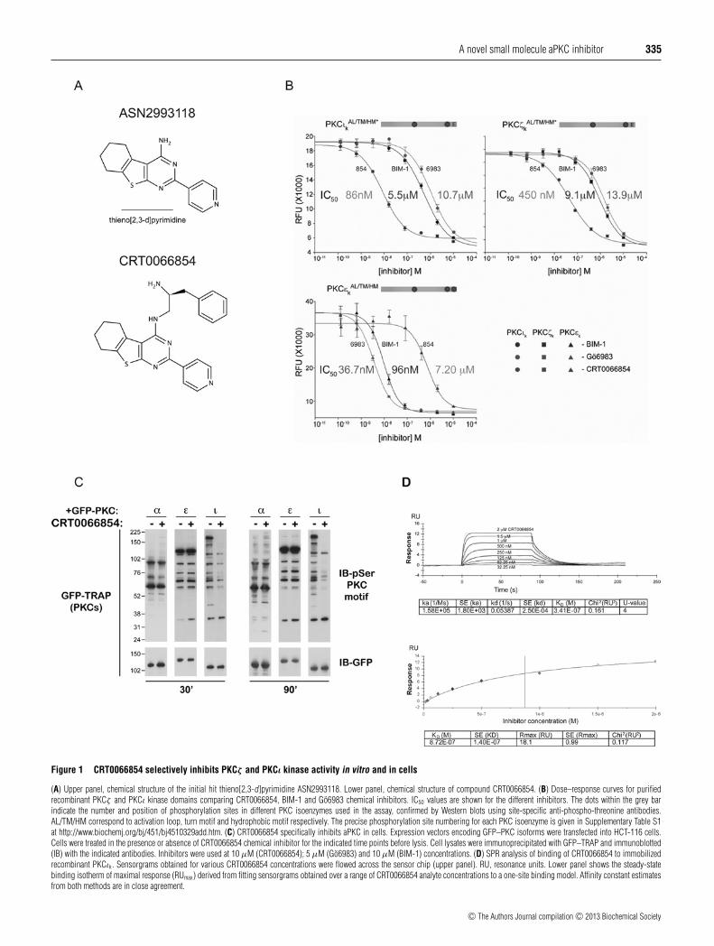
Laboratories was screened at 30 μM against a 10 pMbiphosphorylated (i.e. fully primed) form of the PKCι kinasedomain (residues 239–587, referred to hereafter as PKCιk). ThePKCι Km for ATP was determined to be 30 μM (SupplementaryTable S1). The screen employed an IMAP (Molecular Devices)assay using a fluorescein-amidite (FAM)-tagged PKCε-derivedpseudo-substrate sequence containing an alanine-to-serine mutant(PSS; pseudo-substrate substrate) that we found to be an excellentPKCι substrate. The screen identified several compounds withpotent anti-PKCιk activity including a tricyclic thieno[2,3-d]pyrimidine ASN2993118 (Figure 1A, upper panel). This hithad an IC50 of 11 μM against PKCιk and 26.3 μM againstfull-length PKCι. Counter screening against PKCζ k showedthe compound inhibited with an IC50 of 36.5 μM. The low-molecular mass and calculated lipophilicity of ASN2993118made it an attractive starting point for optimization. Explorationof the activity landscape around the central pyrimidine ringof ASN2993118 led to the identification of compounds with adiamine side chain at the pyrimidine C4 position that exhibitedincreased inhibitory activity. This included CRT0066854 in whichthe pyrimidine C4 position was substituted with a S-3-phenyl-propane-1,2-diamine moiety (Figure 1A, lower panel).
To characterize CRT0066854 further, we used an ADP QuestTM
assay (Discoverx) to generate dose–response curves for fullyprimed phosphospecies of PKCζ k, PKCιk and the nPKC isoformPKCεk as a control. The kcat and Km kinetic constants weredetermined for PKCζ k, PKCιk and the nPKC isoform PKCεk usingthe ADP QuestTM assay (Supplementary Table S1). CRT0066854was used at 10 μM. We used the structurally unrelated PKCinhibitors BIM-1 (10 μM) and Go6983 (5 μM) from commercialsources as positive controls. IC50 values for CRT0066854against PKCζ k and PKCιk were 450 nM and 86 nM respectively(Figure 1B). Comparing the IC50 values measured for BIM-1(PKCζ k IC50 = 9.1 μM and PKCιk IC50 = 5.5 μM), indicates thatCRT0066854 is approximately 20-fold more potent for PKCζ k
and over 60-fold more potent for PKCιk than BIM-1. A similarincrease in potency is observed for CRT0066854 compared withGo6983 (PKCζ k IC50 = 13.9 μM and PKCιk IC50 = 10.7 μM)(Figure 1B). Inhibition of the nPKCεk by CRT0066854 issignificantly weaker, with a 16-fold higher IC50 (IC50 = 7.21 μM)compared with PKCζ k and more than 80-fold greater than the IC50
for PKCιk. We then measured IC50 values for full-length PKCζ andPKCι, which gave 639 nM and 132 nM respectively, comparablewith those obtained from isolated aPKC kinase domains.Since CRT0066854 has a chiral centre, we prepared its enantiomerCRT0066390 and measured its IC50 values against full-lengthPKCι and PKCζ . This showed only low levels of inhibition ofaPKC isoenzymes by CRT0066390 (Table 1).
To examine the selectivity profile of CRT0066854 in vitro,the compound was tested at Millipore using the KinaseProfilerTM
screening service (Millipore) against 106 protein kinases at
a 1 μM inhibitor concentration, screened at the Km for ATP(Table 2). This is a radiometric assay format used with each kinase.The selectivity score S(80) is the number of kinases inhibited bygreater than 80% at 1 μM compound/total number of kinasestested. For CRT0066854, the S(80) score is close to 0.02, whichindicates a good kinase selectivity [40]. Moreover, the compoundshowed striking selectivity for aPKC over other PKC isoenzymes(Table 2).
To test whether there was selectivity of the tool compoundbetween the PKC subclasses in cells, GFP-tagged cPKCα,nPKCε and aPKCι proteins were expressed in HCT-116 cellsand the cultures were treated with CRT0066854 for 30 or90 min. As a control we used BIM-1 and Go6983 to potentlyinhibit full-length novel and cPKC isoenzymes expressed inthis cell line (Supplementary Figure S1 at http://www.biochemj.org/bj/451/bj4510329add.htm). Immuno-complexes wereimmune-isolated through the GFP-tag and immunoblotted withan anti-phospho-serine-PKC substrate antibody. In CRT0066854-treated cells, the number of phosphorylated substrate bandsapparent was significantly diminished for cells overexpressingPKCι, but not PKCα or PKCε, demonstrating aPKC specificityin cells (Figure 1C). Together, these data suggested thatCRT0066854 would be a potent and selective chemical tool tomodulate aPKC activity both in vitro and in cells.
Kinetics of CRT0066854 inhibitor binding to PKCιk
To characterize the interaction of CRT0066854 in solution withpurified recombinant PKCιk protein, we used SPR on a BIACORET200 to measure the kinetics and affinity of this inhibitor. GST-tagged PKCιk was captured using anti-GST monoclonal antibodyimmobilized via amine coupling. Sensorgrams were fitted to a 1:1Langmuir binding model and kinetics constants were obtainedand an affinity constant of 340 nM was calculated from kd/ka
(Figure 1D, upper panel). A binding isotherm was also fitted toa 1:1 steady-state binding model and an affinity of 870 nM wasobtained in good agreement with the value derived from the rateconstants (Figure 1D, lower panel).
Structural basis for CRT0066854 selectivity for the PKCι kinasedomain
To understand both the selectivity and binding mode ofCRT0066854 we determined the crystal structure of CRT0066854bound to PKCιk (Table 3). Crystals of PKCιk bound toCRT0066854 contain three very similar PKCιk molecules in theasymmetric unit that each have a single inhibitor bound withinthe ATP cleft. The overall PKCιk conformation bears many of thehallmarks of an active AGC kinase conformation (SupplementaryFigure S2 at http://www.biochemj.org/bj/451/bj4510329add.htm)except for the lack of bound magnesium and the absence of anordered Ade motif as discussed below [41]. The CRT0066854compound makes predominantly hydrophobic interactions withinthe PKCι nucleotide-binding cleft, as well as five hydrogenbonds, including a water-mediated hydrogen bond (Figures 2Aand 2B). The fused tricyclic scaffold (cyclohexa[4,5]thieno[2,3-d]pyrimidine) makes hydrophobic contacts with the PKCιk
glycine loop, stabilizing a ‘closed’ conformation. This tricyclicring system deviates significantly from planarity and is curvedwith a puckered cyclohexane ring (Figure 2A). The thieno-1nitrogen of the pyrimidine ring makes a crucial hydrogen bondto the Thr386 side chain. The thieno[2,3-d]pyrimidine ring systemalso has two important substituents: (i) a pyridine substituentat position 2 and (ii) a (2-amino-3-phenylpropyl)(methyl)amine
c© The Authors Journal compilation c© 2013 Biochemical Society
A novel small molecule aPKC inhibitor 335
Figure 1 CRT0066854 selectively inhibits PKCζ and PKCι kinase activity in vitro and in cells
(A) Upper panel, chemical structure of the initial hit thieno[2,3-d]pyrimidine ASN2993118. Lower panel, chemical structure of compound CRT0066854. (B) Dose–response curves for purifiedrecombinant PKCζ and PKCι kinase domains comparing CRT0066854, BIM-1 and Go6983 chemical inhibitors. IC50 values are shown for the different inhibitors. The dots within the grey barindicate the number and position of phosphorylation sites in different PKC isoenzymes used in the assay, confirmed by Western blots using site-specific anti-phospho-threonine antibodies.AL/TM/HM correspond to activation loop, turn motif and hydrophobic motif respectively. The precise phosphorylation site numbering for each PKC isoenzyme is given in Supplementary Table S1at http://www.biochemj.org/bj/451/bj4510329add.htm. (C) CRT0066854 specifically inhibits aPKC in cells. Expression vectors encoding GFP–PKC isoforms were transfected into HCT-116 cells.Cells were treated in the presence or absence of CRT0066854 chemical inhibitor for the indicated time points before lysis. Cell lysates were immunoprecipitated with GFP–TRAP and immunoblotted(IB) with the indicated antibodies. Inhibitors were used at 10 μM (CRT0066854); 5 μM (Go6983) and 10 μM (BIM-1) concentrations. (D) SPR analysis of binding of CRT0066854 to immobilizedrecombinant PKCιk. Sensorgrams obtained for various CRT0066854 concentrations were flowed across the sensor chip (upper panel). RU, resonance units. Lower panel shows the steady-statebinding isotherm of maximal response (RUmax) derived from fitting sensorgrams obtained over a range of CRT0066854 analyte concentrations to a one-site binding model. Affinity constant estimatesfrom both methods are in close agreement.
c© The Authors Journal compilation c© 2013 Biochemical Society

336 S. Kjær and others
Table 2 Effect of 1 μM CRT0066854 on the activity of 106 protein kinases
A radiometric assay was carried out by the Millipore Kinase Profiling Service (Millipore). Theassay determined the selectivity of 1 μM single-point concentrations of CRT0066854 againsta panel of 106 protein kinases and 30 μM ATP (K m of PKCιk). The selectivity score S(80) isthe number of kinases inhibited by more than 80 % at 1 μM compound/total number of kinasestested. h, human; m, murine.
Percentage of activity1 μM CRT0066854
Percentage of inhibition1 μM CRT0066854
Abl(h) 93 11 1Abl (M351T)(h) 97 3 2ALK(h) 95 5 3ARK5(h) 91 9 4ASK1(h) 104 −4 5Aurora-A(h) 96 −8 6Axl(h) 106 −6 7Blk(m) 98 2 8Bmx(h) 107 −7 9BRK(h) 112 −12 10CDK1/cyclinB(h) 86 14 11CDK2/cyclinA(h) 76 30 12CDK5/p35(h) 32 68 13CHK1(h) 100 0 14CHK2(h) 90 10 15CK1δ(h) 100 0 16CK1γ 1(h) 101 −1 17CK1γ 2(h) 120 −20 18CK1γ 3(h) 110 −10 19cKit(h) 99 1 20cKit(D816H)(h) 80 20 21cKit(V560G)(h) 90 10 22CSK(h) 103 −3 23c-RAF(h) 94 3 24cSRC(h) 89 8 25DAPK1(h) 94 −3.5 26DDR2(h) 108 −8 27DYRK2(h) 94 6 28EGFR(h) 102 −4 29EGFR(L858R)(h) 78 22 30EGFR(L861Q)(h) 87 13 31EGFR(T790M)(h) 61 39 32EGFR(T790M, L858R)(h) 69 31 33EphA2(h) 102 −2 34EphA7(h) 101 −1 35EphB4(h) 106 −6 36ErbB4(h) 102 −2 37FAK(h) 99 1 38Fer(h) 88 12 39Fes(h) 91 9 40FGFR1(h) 109 −9 41FGFR2(h) 102 −2 42FGFR3(h) 96 −11 43FGFR4(h) 84 16 44Flt1(h) 96 4 45Flt3(D835Y)(h) 33 67 46Flt3(h) 97 3 47Flt4(h) 98 2 48Fms(h) 96 4 49GSK3α(h) 94 6 50Hck(h) 92 8 51HIPK1(h) 98 2 52HIPK2(h) 93 7 53HIPK3(h) 103 −3 54IGF-1R(h) 88 12 55IKKα(h) 29 65 56IR(h) 85 15 57IRAK4(h) 117 −17 58JAK3(h) 94 6 59JNK1α1(h) 102 −2 60KDR(h) 96 4 61LIMK1(h) 42 58 62LKB1(h) 95 5 63
Table 2 Continued
Percentage of activity1 μM CRT0066854
Percentage of inhibition1 μM CRT0066854
MAPKAP-K2(h) 101 −1 64MAPK2(h) 99 1 65MEK1(h) 100 −0.5 66MELK(h) 77 23 67Mer(h) 89 11 68Met(h) 100 0 69MSK1(h) 106 −6 70MST1(h) 108 −8 71MST3(h) 90 10 72NEK2(h) 100 0 73p70S6K(h) 88 12 74PAK4(h) 103 −2 75PDGFRα(h) 105 −5 76PDGFRα(D842V)(h) 80 20 77PDK1(h) 89 15 78Pim-1(h) 89 11 79PKBα(h) 96 −3.5 80PKCα(h) 97 0 81PKCβ I(h) 107 −7 82PKCγ (h) 102 −2 83PKCδ(h) 89 11 84PKCε(h) 99 −1 85PKCη(h) 109 −5 86PKCι(h) 13 83.5 87PKCμ(h) 101 0 88PKCθ (h) 108 −1 89PKCζ (h) 23 77 90PKD2(h) 96 4 91Plk3(h) 83 17 92PRK2(h) 16 84 93Ret(h) 106 −6 94ROCK-I(h) 109 −9 95ROCK-II(h) 37 63 96Ron(h) 114 −14 97Ros(h) 94 6 98SAPK2a(h) 102 −2 99Snk(h) 102 −2 100TAK1(h) 100 0 101Tie2(h) 114 −14 102TrkA(h) 114 −14 103Yes(h) 102 −2 104ZAP-70(h) 91 9 105ZIPK(h) 86 14 106
S(80)0.019
substituent including a benzyl group, a chiral carbon and a primaryamine. The pyridine substituent acts as a kinase domain hinge-binding moiety by forming a hydrogen bond to the main-chainamide of Val326, approximating to the position of the adenine ringof ATP. The primary amine of the 3-phenyl-propane-1,2-diaminoheadpiece of CRT0066854 makes a crucial charged interactionwith both the backbone carbonyl of Asp373 and the side chain ofAsp330. Finally, the benzyl group in the inhibitor headpiece bindsa small hydrophobic pocket formed by the kinase domain hinge,usually occupied by Phe543 from the Ade motif [41] (Figure 2C;compare left-hand and middle panel). The benzyl group alsomakes an orthogonal π-stacking interaction with the pyridinering hinge-binder, thus locking the inhibitor into a distinctly non-planar and U-shape conformation (Figure 2A).
Superimpositions of CRT0066854 and ATP-bound structuresof PKCιk indicate the general equivalence of the benzyl moietyand Phe543 (Figure 2D). An important consequence of inhibitorbinding is that the entire Ade motif, including Phe543 fromthe Phe-Xaa-Xaa-Phe motif, becomes disordered, whereas it isan integral part of the ATP-binding cleft in the ATP–PKCιk
c© The Authors Journal compilation c© 2013 Biochemical Society

A novel small molecule aPKC inhibitor 337
Table 3 X-ray data collection and refinement statistics
Parameter Values
Data collectionSpace group P31
Cell dimensionsa, b, c (A) 113.6, 113.6, 82.4α, β , γ (◦) 90, 90, 120
Resolution (A)* 49.2–2.74 (2.89–2.74)Rmerge 11.0 % (71.4 %)Rpim 5.8 % (38.4 %)I/σ 6.3 (1.1)Completeness 100 % (100 %)Multiplicity 5.6 (5.4)
RefinementResolution (A) 2.74Number of unique reflections 31 307 (4570)Rwork/R free 21.5 % / 25.6%Atoms
Protein 7468Ligand/ion 180Water 66
B-factorsProtein 49.8Ligand/ion 42.6Water 31.9
RMSDBond lengths (A) 0.003Bond angles (◦) 0.726
*Numbers in parentheses are values in the highest resolution shell.
complex (PDB code 3A8W) (Figure 2C). Recent data haveshown for PKCβ2, an equivalent phenyl side chain to Phe543
that has been implicated in allosteric activation [42]. Thisresidue is held out of the ATP cleft by the C1 domainpreventing full enzyme activation until diacylglycerol binds,providing an additional layer of PKC regulation. It seemslikely that CRT0066854 targets PKCιk for inhibition not onlyby competitive binding to the nucleotide-binding cleft, butalso by preventing a catalytically competent conformation viadisplacement of the Ade motif by the benzyl inhibitor substituent.Both features combine to contribute towards the high specificityof CRT0066854 for aPKCs. A detailed comparison of inhibitorcontents compared with other AGC kinases is given in theSupplementary Online Data and Supplementary Table S2 (athttp://www.biochemj.org/bj/451/bj4510329add.htm).
CRT0066854 inhibition of LLGL2 phosphorylation in cells
LLGL is a recognized substrate of aPKC in D. melanogaster[43] and in immunocomplexes from HEK-293 cells expressingexogenous aPKC [44]. As LLGL2 is expressed at low levelsin HEK-293 cells and due to the lack of a sensitive phospho-specific antibody against LLGL1/2, we used a co-expressionsystem of aPKC and LLGL2 to evaluate the cellular IC50 ofCRT0066854 in HEK-293 cells. Treatment of HEK-293 cells,co-transfected with PKCι wild-type and FLAG-tagged LLGL2,with CRT0066854 (0.125–8 μM) for 1 h led to a reproducibledose-dependent reduction in LLGL2 phosphorylation as seen byWestern blotting (Figure 3A). In order to test this robustly ina high-throughput format, the phosphorylation signal windowwas optimized using the co-transfection of constitutively activePKCι-A120E [45] and FLAG-tagged LLGL2 and an ELISA wasestablished. This demonstrated that CRT006854 has a cellularphospho-LLGL2 IC50 of 0.87 μM comparable with the IC50
measured in vitro against PKCι (Figure 3B).
Effect of CRT0066854 on A549 lung carcinoma cell viability
A549 cells are K-Ras mutant cells of lung origin, known tobe dependent on PKCι for survival [17]. The A549 cells weretreated with CRT0066854 for 48 h at various concentrations(0.01–20 μM), and cell viability determined by the CellTitre-Gloluminescent assay. An IC50 of 3.47 μM was obtained from threeindependent determinations (Figure 3C).
Decreased colony formation due to CRT0066854
Although a reduction in adherent cell growth has been reportedfollowing the depletion of PKCι in selected cell lines [46],the anti-proliferative effects appear more pronounced for non-adherent cell growth [17,46]. As such, we cultured HeLa cells,which reliably form colonies within 12 days when embedded insoft agar, and replenished inhibitor (0.1–10 μM) on alternatedays. Two separate inhibitors were used for these experiments,the aPKC-specific CRT0066854 and its enantiomeric equivalentCRT0066390, which is essentially inactive towards PKCι in vitro(Table 1). At 1 μM the CRT0066854 inhibitor led to a 65 %decrease in colony formation and, as expected, the same dose ofCRT0066390 resulted in considerably less suppression of colonyformation (25% decrease) (Figures 3D and 3E). This is consistentwith CRT0066854 having a specific cellular effect via PKCιinhibition.
Inhibition of polarized epithelial morphogenesis by CRT0066854
Depletion of PKCι in cells using RNAi (RNA interference) leadsto impaired luminal morphogenesis in epithelial cell 3D cysts asa consequence of impaired spindle formation and apical–basalpolarity [9,47]. We therefore tested whether chemical inhibitioncould induce the same phenotype in MDCK cells embeddedin MatrigelTM. MDCK cells are widely used as models forstudying epithelia, as they have clear apical–basolateral polarityand polarize in 2D (two-dimensional) and 3D cell cultures. InMatrigelTM, MDCK cells are known to form cysts with a singlelumen. We determined the number of MDCK cysts that had aPSAL (predominantly single apical lumen) after growing in thepresence of 0, 0.33 or 1 μM concentrations of CRT0066854.Surprisingly, inhibitor addition caused significant disruption tolumen formation, resulting in a multiple luminal morphology(Figure 4A). This disruption in lumen formation was dose-dependent and quantification of this effect indicated that at 1 μMconcentrations of CRT0066854 there was a 50% reduction inPSALs compared with a DMSO control (Figure 4B).
CRT0066854 impedes directed migration of NRK cells
Depletion of aPKC using RNAi decreases the rate of healingin an NRK cell scratch-wound assay due to an impairmentof the polarized delivery of the exocyst to the cell’s leadingedge [10]. We used this model to test the effect of aPKCinhibition by CRT0066854 on directional migration. Scratchwounds were generated in wells of confluent NRK cells afterwhich DMSO, or 3, 6 or 12 μM CRT0066854 was added tothe wells and time-lapse imaging recorded the wound healingover 10 h (Supplementary Movie S1 at http://www.biochemj.org/bj/451/bj4510329add.htm). Both 6 μM and 12 μM CRT0066854led to a significant reduction in wound healing, with 12 μMhalving the normalized migration speed (Figures 4C and 4D).Note that optimum knockdown of aPKCs in this model reducesmigration by 60% [10].
c© The Authors Journal compilation c© 2013 Biochemical Society

338 S. Kjær and others
Figure 2 Structural basis for CRT0066854 inhibition of PKCιk
(A) Close-up of the CRT0066854-binding site within the PKCι nucleotide-binding cleft and kinase domain hinge, highlighting the five hydrogen-bond contacts and the lack of planarity of CRT0066854.Selected protein side chains and main chains are shown for clarity. A bound water molecule (red sphere) and an iodide ion (purple sphere) are also shown. (B) A 2D representation of CRT0066854inhibitor–PKCι contacts produced by LIGPLOT + [51]. (C) Comparison of the nucleotide pockets between CRT0066854 and ATP complexes of PKCι. The left-hand panel shows the electrostaticsurface of PKCι, including the two acidic residues that interact with the inhibitor (yellow sticks). Phe543 from the Asn-Phe-Asp motif of PKCι (middle panel, green sticks and salmon-pink cartoon)contacts ATP in a similar manner to Phe327 of protein kinase A (right-hand panel, green cartoon and sticks). Inhibitor binding leads to (i) displacement of the Asn-Phe-Asp motif (left-hand panel),(ii) changes in the glycine loop, and (iii) changes in the Tyr256 (glycine loop)–Trp289 (Cα-helix) interaction. (D) Superimposition of the nucleotide-binding pockets of PKCι bound to ATP and toCRT0066854. This shows that the phenyl group of CRT0066854 (yellow sticks) mimics the placement of Phe543 (green sticks) from the Ade motif observed in the ATP complex.
DISCUSSION
In the present study, we report the identification andcharacterization of a selective inhibitor of the aPKC isoenzymes.Selectivity and potency issues have proved particularlychallenging in the search for inhibitors of individual PKCisoforms, particularly aPKCζ and aPKCι, where all commercially
available inhibitors have greater efficacy for the cPKCs andnPKCs [34]. The discovery of CRT0066854 and a knowledgeof the detailed contacts it makes within the aPKC kinase ATP-binding cleft demonstrates that isoform specificity can be obtainedusing the residues surrounding the nucleotide-binding site, andprovides a useful starting point for rational design of even morepotent and specific inhibitors.
c© The Authors Journal compilation c© 2013 Biochemical Society

A novel small molecule aPKC inhibitor 339
Figure 3 CRT0066854 inhibits LLGL2 phosphorylation and cell viability
(A) HEK-293 cells were co-transfected with LLGL2 and PKCι-WT (wild-type). After 35 h, the cells were treated with CRT0066854 (0.125–8 μM) for 1 h, lysates were generated separated by gelelectrophoresis and immunoblotted with pLLGL1/2. (B) HEK-293 cells co-transfected with FLAG–LLGL2 and PKCι-A120E (constitutively active). After 40 h, cells were treated with CRT0066854(0.001–12 μM) for 1 h and an ELISA was performed. (C) A549 cells in log-phase growth as a monolayer were treated with CRT0066854 at the indicated concentrations for 48 h and the cell viabilitywas estimated using CellTitreGlo. (D) HeLa cells from a single cell suspension were cultured in soft agar for 14 days, with the medium and inhibitor being replenished on alternate days. Colonieswere stained with Crystal Violet and images of the 35-mm well taken using a mounted CCD camera. (E) The effect of aPKC inhibitors on HeLa colony growth was quantified and the means +− S.E.M.is presented for at least three separate experiments.
Our crystallographic analysis has revealed that CRT0066854has a bifunctional inhibitory effect. First, it is a competitive ATPinhibitor, binding within the nucleotide-binding pocket of aPKC.Secondly, it uses a benzyl group to displace the Ade motif froma peripheral hinge pocket adjacent to the nucleotide cleft andprevents it forming a functional ATP-binding pocket. These twofeatures ensure that CRT0066854 is highly selective for aPKC.Some strain/induced fit is evident within the compound in viewof its lack of planarity when bound to PKCιk. Inhibitory effectson other kinases such as ROCK2 and PRK2 may be due to thepresence of a similar phenyl group equivalent to Phe543, suggestingthat related scaffolds may permit a more generic approach towardsinhibiting AGC kinases bearing an Asn-Phe-Asp motif. Bindingisotherms show that the inhibitor has a low off-rate, generallydeemed to be an advantageous feature for a kinase inhibitor.
Recent studies have shown differential effects between PKCζand PKCι for compounds targeting other aPKC-specific pocketsoutside of the ATP cleft [35,48]. The selectivity of CRT0066854shown in the present study for both aPKCζ and aPKCιcould prove useful in both clinical and research settings. Weprovide compelling evidence for both in vitro and phenotypiceffects of CRT0066854 using a range of cellular assaysrelevant to cancer therapy. Non-adherent cell growth, migrationand aberrant polarity are all processes exploited by cancer andwere more affected by CRT0066854 than adherent cellgrowth. This differential dependence on aPKC for 3D and 2Dcellular phenotypes has been demonstrated previously by geneticdepletion of aPKC [17], and together with the work described inthe present paper, supports the role of CRT0066854 as a selectiveaPKC inhibitor in cells. PKCι overexpression has been linked to
c© The Authors Journal compilation c© 2013 Biochemical Society

340 S. Kjær and others
Figure 4 Phenotypic effects of CRT0066854 on cell polarity and directed cell migration
(A) MDCK cells were cultured in MatrigelTM for 6 days. The cultures were treated with 0, 0.33 or 1 μM of the aPKCι inhibitor CRT0066854 which were replenished, along with growth medium,on alternate days. Cultures were fixed and stained with Phalloidin (F-actin) and counterstained with Hoechst (DNA). Representative single mid-section confocal images are shown. The scale barsrepresent 50 μM. (B) Quantification of the number of PSALs was made. At least 100 cysts were counted per condition in each experiment and the means +− S.E.M. are presented for at least threeseparate experiments. (C) Confluent monolayers of NRK cells were wounded, treated with CRT0066854 at the indicated concentrations and healing was followed over time. The panels shown arerepresentative of at least three independent experiments. (D) The normalized mean migration speed is plotted for different CRT0066854 concentrations. The error bars represent the S.E.M. andone-way ANOVA was used to determine statistical significance of difference: *P < 0.05, ***P < 0.001.
several types of cancer, including non-small-cell lung cancer andovarian cancer [17,49,50] and further optimization of inhibitors onthe basis of the CRT0066854 structure could yield more specificcancer therapeutics. CRT0066854 and its derivative compoundswill be useful tools to further dissect PKCζ and PKCι roles inestablishing cell polarity and growth-factor-stimulated signallingpathways.
AUTHOR CONTRIBUTION
Svend Kjær purified, assembled and crystallized the CRT0066854 inhibitor–PKCιk
complex; Andrew Purkiss collected the data, and determined and refined the CRT0066854-bound structure; Brenda Kostelecky and Erika Soriano carried out kinetic assays withinhibitors; Phillip Knowles carried out the SPR experiments; Philippe Riou assessed
inhibitor efficacy against multiple PKC isoforms in cells; Christian Dillon and Jon Roffeyled the chemistry project team that discovered CRT0066854; Caroline Barton and BhavishaPatel conducted the LLGL2 phosphorylation assays and cell viability assay; ChristelleSoudy synthesized the inhibitors used in these studies; Carine Rosse conducted thedirected cell migration assay; Sarah Kaye carried out IC50 assays on full-length aPKCisoenzymes; Mark Linch carried out the LLGL2 phosphorylation assay, cell polarity andcolony formation assay; Peter Parker and Neil McDonald planned the project and designedexperiments; and Neil McDonald, Judith Murray-Rust and Mark Linch prepared the Figuresand wrote the paper.
ACKNOWLEDGEMENTS
We thank members of the N.Q.M. and P.J.P. laboratories for helpful discussions andcomments on the paper before submission. We gratefully acknowledge the expertassistance of Tamar Telem-Shafir (GE Healthcare) in performing and analysing the SPRdata.
c© The Authors Journal compilation c© 2013 Biochemical Society

A novel small molecule aPKC inhibitor 341
FUNDING
S. Kjær was funded by a Cancer Research UK fellowship and by a Marie Curie Fellowship[grant number MEIF-CT-2005-010656]; M.L. was funded by a Cancer Research UKClinical Research Fellowship. Research in the N.Q.M. and P.J.P. laboratories is supportedby CRUK core funding to the London Research Institute.
REFERENCES
1 Bosco, R., Melloni, E., Celeghini, C., Rimondi, E., Vaccarezza, M. and Zauli, G. (2011)Fine tuning of protein kinase C (PKC) isoforms in cancer: shortening the distance fromthe laboratory to the bedside. Mini-Rev. Med. Chem. 11, 185–199
2 Rosse, C., Linch, M., Kermorgant, S., Cameron, A. J., Boeckeler, K. and Parker, P. J.(2010) PKC and the control of localized signal dynamics. Nat. Rev. Mol. Cell Biol. 11,103–112
3 Parker, P. J. and Murray-Rust, J. (2004) PKC at a glance. J. Cell Sci. 117, 131–1324 Pearce, L. R., Komander, D. and Alessi, D. R. (2010) The nuts and bolts of AGC protein
kinases. Nat. Rev. Mol. Cell Biol. 11, 9–225 Suzuki, A., Akimoto, K. and Ohno, S. (2003) Protein kinase C λ/ι (PKCλ/ι): a PKC
isotype essential for the development of multicellular organisms. J. Biochem. 133, 9–166 Knoblich, J. A. (2010) Asymmetric cell division: recent developments and their
implications for tumour biology. Nat. Rev. Mol. Cell Biol. 11, 849–8607 Suzuki, A. and Ohno, S. (2006) The PAR–aPKC system: lessons in polarity. J. Cell Sci.
119, 979–9878 Hao, Y., Du, Q., Chen, X., Zheng, Z., Balsbaugh, J. L., Maitra, S., Shabanowitz, J., Hunt,
D. F. and Macara, I. G. (2010) Par3 controls epithelial spindle orientation byaPKC-mediated phosphorylation of apical Pins. Curr. Biol. 20, 1809–1818
9 Durgan, J., Kaji, N., Jin, D. and Hall, A. (2011) Par6B and atypical PKC regulate mitoticspindle orientation during epithelial morphogenesis. J. Biol. Chem. 286, 12461–12474
10 Rosse, C., Formstecher, E., Boeckeler, K., Zhao, Y., Kremerskothen, J., White, M. D.,Camonis, J. H. and Parker, P. J. (2009) An aPKC–exocyst complex controls paxillinphosphorylation and migration through localised JNK1 activation. PLoS Biol. 7,e1000235
11 Etienne-Manneville, S. and Hall, A. (2003) Cell polarity: Par6, aPKC and cytoskeletalcrosstalk. Curr. Opin. Cell Biol. 15, 67–72
12 Fields, A. P. and Regala, R. P. (2007) Protein kinase C iota: human oncogene, prognosticmarker and therapeutic target. Pharmacol. Res. 55, 487–497
13 Guo, H., Gu, F., Li, W., Zhang, B., Niu, R., Fu, L., Zhang, N. and Ma, Y. (2009) Reductionof protein kinase C ζ inhibits migration and invasion of human glioblastoma cells. J.Neurochem. 109, 203–213
14 Liu, Y., Wang, B., Wang, J., Wan, W., Sun, R., Zhao, Y. and Zhang, N. (2009)Down-regulation of PKCζ expression inhibits chemotaxis signal transduction in humanlung cancer cells. Lung Cancer 63, 210–218
15 Sun, R., Gao, P., Chen, L., Ma, D., Wang, J., Oppenheim, J. J. and Zhang, N. (2005)Protein kinase C ζ is required for epidermal growth factor-induced chemotaxis of humanbreast cancer cells. Cancer Res. 65, 1433–1441
16 Xu, L. and Deng, X. (2006) Protein kinase Cι promotes nicotine-induced migration andinvasion of cancer cells via phosphorylation of μ- and m-calpains. J. Biol. Chem. 281,4457–4466
17 Regala, R. P., Weems, C., Jamieson, L., Copland, J. A., Thompson, E. A. and Fields, A. P.(2005) Atypical protein kinase Cι plays a critical role in human lung cancer cell growthand tumorigenicity. J. Biol. Chem. 280, 31109–31115
18 Aranda, V., Haire, T., Nolan, M. E., Calarco, J. P., Rosenberg, A. Z., Fawcett, J. P., Pawson,T. and Muthuswamy, S. K. (2006) Par6-aPKC uncouples ErbB2 induced disruption ofpolarized epithelial organization from proliferation control. Nat. Cell Biol. 8, 1235–1245
19 Brumby, A. M. and Richardson, H. E. (2003) scribble mutants cooperate with oncogenicRas or Notch to cause neoplastic overgrowth in Drosophila. EMBO J. 22, 5769–5779
20 Pagliarini, R. A. and Xu, T. (2003) A genetic screen in Drosophila for metastatic behavior.Science 302, 1227–1231
21 Duran, A., Linares, J. F., Galvez, A. S., Wikenheiser, K., Flores, J. M., Diaz-Meco, M. T.and Moscat, J. (2008) The signaling adaptor p62 is an important NF-κB mediator intumorigenesis. Cancer Cell 13, 343–354
22 Regala, R. P., Davis, R. K., Kunz, A., Khoor, A., Leitges, M. and Fields, A. P. (2009)Atypical protein kinase C ι is required for bronchioalveolar stem cell expansion and lungtumorigenesis. Cancer Res. 69, 7603–7611
23 Yang, Y. L., Chu, J. Y., Luo, M. L., Wu, Y. P., Zhang, Y., Feng, Y. B., Shi, Z. Z., Xu, X., Han,Y. L., Cai, Y. et al. (2008) Amplification of PRKCI, located in 3q26, is associated withlymph node metastasis in esophageal squamous cell carcinoma. Genes, ChromosomesCancer 47, 127–136
24 Kojima, Y., Akimoto, K., Nagashima, Y., Ishiguro, H., Shirai, S., Chishima, T., Ichikawa, Y.,Ishikawa, T., Sasaki, T., Kubota, Y. et al. (2008) The overexpression and alteredlocalization of the atypical protein kinase C λ/ι in breast cancer correlates with thepathologic type of these tumors. Hum. Pathol. 39, 824–831
25 Scotti, M. L., Bamlet, W. R., Smyrk, T. C., Fields, A. P. and Murray, N. R. (2010) Proteinkinase Cι is required for pancreatic cancer cell transformed growth and tumorigenesis.Cancer Res. 70, 2064–2074
26 Takagawa, R., Akimoto, K., Ichikawa, Y., Akiyama, H., Kojima, Y., Ishiguro, H., Inayama, Y.,Aoki, I., Kunisaki, C., Endo, I. et al. (2010) High expression of atypical protein kinase Cλ/ι in gastric cancer as a prognostic factor for recurrence. Ann. Surg. Oncol. 17, 81–88
27 Li, Q., Wang, J. M., Liu, C., Xiao, B. L., Lu, J. X. and Zou, S. Q. (2008) Correlation ofaPKC-ι and E-cadherin expression with invasion and prognosis of cholangiocarcinoma.Hepatobiliary Pancreatic Dis. Int. 7, 70–75
28 Patel, R., Win, H., Desai, S., Patel, K., Matthews, J. A. and Acevedo-Duncan, M. (2008)Involvement of PKC-ι in glioma proliferation. Cell Proliferation 41, 122–135
29 Murray, N. R. and Fields, A. P. (1997) Atypical protein kinase C ι protects humanleukemia cells against drug-induced apoptosis. J. Biol. Chem. 272, 27521–27524
30 Baldwin, R. M., Garratt-Lalonde, M., Parolin, D. A., Krzyzanowski, P. M., Andrade, M. A.and Lorimer, I. A. (2006) Protection of glioblastoma cells from cisplatin cytotoxicity viaprotein kinase Cι -mediated attenuation of p38 MAP kinase signaling. Oncogene 25,2909–2919
31 Jin, Y. T., Ying, X. X., Hu, Y. H., Zou, Q., Wang, H. Y. and Xu, Y. H. (2008) aPKC inhibitorsmight be the sensitizer of chemotherapy and adoptive immunotherapy in the treatment ofhASIPa-overexpressed breast cancer. Oncol. Res. 17, 59–68
32 Kikuchi, K., Soundararajan, A., Zarzabal, L. A., Weems, C. R., Nelon, L. D., Hampton, S.T., Michalek, J. E., Rubin, B. P., Fields, A. P. and Keller, C. (2012) Protein kinase C ι as atherapeutic target in alveolar rhabdomyosarcoma. Oncogene 32, 286–95
33 Knight, Z. A. and Shokat, K. M. (2005) Features of selective kinase inhibitors. Chem. Biol.12, 621–637
34 Roffey, J., Rosse, C., Linch, M., Hibbert, A., McDonald, N. Q. and Parker, P. J. (2009)Protein kinase C intervention: the state of play. Curr. Opin. Cell Biol. 21, 268–279
35 Frohner, W., Lopez-Garcia, L. A., Neimanis, S., Weber, N., Navratil, J., Maurer, F., Stroba,A., Zhang, H., Biondi, R. M. and Engel, M. (2011) 4-Benzimidazolyl-3-phenylbutanoicacids as novel PIF-pocket-targeting allosteric inhibitors of protein kinase PKCζ . J. Med.Chem. 54, 6714–6723
36 Erdogan, E., Lamark, T., Stallings-Mann, M., Lee, J., Pellecchia, M., Thompson, E. A.,Johansen, T. and Fields, A. P. (2006) Aurothiomalate inhibits transformed growth bytargeting the PB1 domain of protein kinase Cι. J. Biol. Chem. 281, 28450–28459
37 Collaborative Computational Project-Number 4 (1994) The CCP-4 suite: programs forprotein crystallography. Acta Crystallogr., Sect. D: Biol. Crystallogr. 50, 760–763
38 McCoy, A. J., Grosse-Kunstleve, R. W., Adams, P. D., Winn, M. D., Storoni, L. C. andRead, R. J. (2007) Phaser crystallographic software. J. Appl. Crystallogr. 40, 658–674
39 Adams, P. D., Grosse-Kunstleve, R. W., Hung, L. W., Ioerger, T. R., McCoy, A. J., Moriarty,N. W., Read, R. J., Sacchettini, J. C., Sauter, N. K. and Terwilliger, T. C. (2002) PHENIX:building new software for automated crystallographic structure determination. ActaCrystallogr., Sect. D: Biol. Crystallogr. 58, 1948–1954
40 Bain, J., Plater, L., Elliott, M., Shpiro, N., Hastie, C. J., McLauchlan, H., Klevernic, I.,Arthur, J. S., Alessi, D. R. and Cohen, P. (2007) The selectivity of protein kinaseinhibitors: a further update. Biochem. J. 408, 297–315
41 Romano, R. A., Kannan, N., Kornev, A. P., Allison, C. J. and Taylor, S. S. (2009) Achimeric mechanism for polyvalent trans-phosphorylation of PKA by PDK1. Protein Sci.18, 1486–1497
42 Leonard, T. A., Rozycki, B., Saidi, L. F., Hummer, G. and Hurley, J. H. (2011) Crystalstructure and allosteric activation of protein kinase C β II. Cell 144, 55–66
43 Betschinger, J., Mechtler, K. and Knoblich, J. A. (2003) The Par complex directsasymmetric cell division by phosphorylating the cytoskeletal protein Lgl. Nature 422,326–330
44 Plant, P. J., Fawcett, J. P., Lin, D. C., Holdorf, A. D., Binns, K., Kulkarni, S. and Pawson, T.(2003) A polarity complex of mPar-6 and atypical PKC binds, phosphorylates andregulates mammalian Lgl. Nat. Cell Biol. 5, 301–308
45 Pears, C. J., Kour, G., House, C., Kemp, B. E. and Parker, P. J. (1990) Mutagenesis of thepseudosubstrate site of protein kinase C leads to activation. Eur. J. Biochem. 194, 89–94
46 Ishiguro, H., Akimoto, K., Nagashima, Y., Kojima, Y., Sasaki, T., Ishiguro-Imagawa, Y.,Nakaigawa, N., Ohno, S., Kubota, Y. and Uemura, H. (2009) aPKCλ/ι promotes growth ofprostate cancer cells in an autocrine manner through transcriptional activation ofinterleukin-6. Proc. Natl. Acad. Sci. U.S.A. 106, 16369–16374
47 Horikoshi, Y., Suzuki, A., Yamanaka, T., Sasaki, K., Mizuno, K., Sawada, H., Yonemura, S.and Ohno, S. (2009) Interaction between PAR-3 and the aPKC–PAR-6 complex isindispensable for apical domain development of epithelial cells. J. Cell Sci. 122,1595–1606
c© The Authors Journal compilation c© 2013 Biochemical Society

342 S. Kjær and others
48 Pillai, P., Desai, S., Patel, R., Sajan, M., Farese, R., Ostrov, D. and Acevedo-Duncan, M.(2011) A novel PKC-ι inhibitor abrogates cell proliferation and induces apoptosis inneuroblastoma. Int. J. Biochem. Cell Biol. 43, 784–794
49 Eder, A. M., Sui, X., Rosen, D. G., Nolden, L. K., Cheng, K. W., Lahad, J. P., Kango-Singh,M., Lu, K. H., Warneke, C. L., Atkinson, E. N. et al. (2005) Atypical PKCι contributes topoor prognosis through loss of apical-basal polarity and cyclin E overexpression inovarian cancer. Proc. Natl. Acad. Sci. U.S.A. 102, 12519–12524
50 Zhang, L., Huang, J., Yang, N., Liang, S., Barchetti, A., Giannakakis, A., Cadungog,M. G., O’Brien-Jenkins, A., Massobrio, M., Roby, K. F. et al. (2006) Integrativegenomic analysis of protein kinase C (PKC) family identifies PKCι as a biomarkerand potential oncogene in ovarian carcinoma. Cancer Res. 66, 4627–4635
51 Laskowski, R. A. and Swindells, M. B. (2011) LigPlot + : multiple ligand–proteininteraction diagrams for drug discovery. J. Chem. Inf. Model. 51,2778–2786
Received 18 December 2012/12 February 2013; accepted 18 February 2013Published as BJ Immediate Publication 18 February 2013, doi:10.1042/BJ20121871
c© The Authors Journal compilation c© 2013 Biochemical Society
Biochem. J. (2013) 451, 329–342 (Printed in Great Britain) doi:10.1042/BJ20121871
SUPPLEMENTARY ONLINE DATAAdenosine-binding motif mimicry and cellular effects of athieno[2,3-d]pyrimidine-based chemical inhibitor of atypical proteinkinase C isoenzymesSvend KJÆR*1, Mark LINCH†1, Andrew PURKISS*, Brenda KOSTELECKY*†, Phillip P. KNOWLES*, Carine ROSSE†, PhilippeRIOU†, Christelle SOUDY‡, Sarah KAYE‡, Bhavisha PATEL‡, Erika SORIANO*, Judith MURRAY-RUST*, Caroline BARTON‡,Christian DILLON‡, Jon ROFFEY‡, Peter J. PARKER†§2 and Neil Q. MCDONALD*‖2
*Structural Biology, Cancer Research UK, 44 Lincoln’s Inn Fields, London WC2A 3LY, U.K., †Protein Phosphorylation Laboratories, Cancer Research UK, 44 Lincoln’s Inn Fields,London WC2A 3LY, U.K., ‡Cancer Research Technology Discovery Laboratories, Wolfson Institute for Biomedical Research, University College London, Gower Street, London WC1E6BT, U.K., §Division of Cancer Studies, King’s College London, New Hunts House, Guy’s Campus, London SE1 1UL, U.K., and ‖Institute of Structural and Molecular Biology,Department of Biological Sciences, Birkbeck College, University of London, Malet Street, London WC1E 7HX, U.K.
CRT0066854-bound PKCιk adopts an active kinase conformation
Both PKCιk structures bound to ATP (PDB code 3A8W) orbound to CRT0066854, have similar relative N-lobe and C-lobeorientations consistent with a closed ‘active’ kinase conformation.Active kinase conformational elements include (i) an occupiednucleotide-binding pocket, (ii) an ordered activation loop and (iii)a salt bridge between the Cα-helix Glu293 and N-lobe Lys274 thatco-ordinates ATP phosphate moieties adjacent to the predicteddivalent metal site. The glycine-rich loop of PKCιk bound toCRT0066854 adopts a ‘closed’ conformation making multiplevan der Waals contacts with the inhibitor (Figure 2 in the maintext). The threonine residues of the activation loop (Thr403)and the turn motif (Thr555) are phosphorylated in both PKCιk
structures. Comparison with other PKC kinase domain structuresshows considerable variation in the precise position of the turnmotif, indicating some plasticity within this poorly conservedregion. The hydrophobic motif (residues 570–578) is well orderedand are bound to the hydrophobic cleft of the N-lobe asexpected.
Structural comparisons of CRT0066854 and BIM-1 bound to thePKCι kinase domain
We compared contact residues from the CRT0066854 and BIM-1 complexes to further characterize the selectivity of the aPKC-selective inhibitor. This analysis does not consider the contributionof bound solvent between inhibitor and protein as few orderedsolvent molecules are observed for these structures given theirmodest resolution. Supplementary Table S2 shows the contactresidues together with their structural equivalents in other classesof PKC isoenzymes, the AGC kinases PKA (protein kinase A),PKB (protein kinase B) as well as PRK2 and ROCK (bothinhibited by CRT0066854, Table 2 in the main text). Of the PKCιside chains involved in CRT0066854 binding, BIM-1 bindingor both, five residues stand out: Ile251, Val307, Ile323, Leu376 andThr386 (Figure 2B of the main text). Thr386 is a good candidatefor conferring CRT0066854 sensitivity on aPKCs owing to itsside-chain hydrogen bond to a nitrogen atom of the thieno[2,3-
d]pyrimidine scaffold of CRT0066854. The equivalent residue inmost other PKC isoforms is alanine, which is unable to form sucha side-chain hydrogen bond. ROCK2 and PRK2 also have alanineat this position. The exception is PKCγ , which also containsthreonine at this position, as do PKB and PKA. However, PKBand PKCγ are unresponsive to CRT0066854 at 1 μM inhibitorconcentration in in vitro assays, compared with 77% inhibitionfor PKCζ (Table 2 in the main text).
Both aPKCs and nPKCs have a leucine residue at position376, whereas cPKCs have methionine. The higher potencyof BIM-1 towards cPKC isoenzymes may be influenced by amethionine residue at this position as has been suggested forPKB [1]. However, the presence of Leu376 alone cannot accountfor CRT0066854 potency towards aPKC over nPKCs. Val307 ofaPKCs aligns with threonine in the cPKC and nPKC isoformsas well as in PKB. The side-chain oxygen of this threonineresidue is involved in binding BIM-1, whereas such an equivalentinteraction is not possible for the aPKC isoenzymes. Val307 maythus select against BIM-1 binding [2]. The equivalent to thegatekeeper residue Ile323 is methionine in all cPKC and nPKCisoforms as well as in PKB, PKA, PRK2 and ROCK2. The Ile323
side chain forms hydrophobic contacts with CRT0066854 and,although the hydrophobic methionine would add some additionalbulk, its presence in PRK2 and ROCK2 suggests that it does notperturb CRT0066854 binding. Ile251 may also play a role in drugselectivity. Isoleucine at this position is unique to the aPKCs,and ROCK2 with the equivalent residue being leucine in all otherPKCs and many AGC kinases. The slight change induced bythe isoleucine to leucine switch may also help to determine thedifference in BIM-1 sensitivities for the respective isoforms. TheAGC kinases PRK2 and ROCK2 are both inhibited to a significantextent by CRT0066854. Each has an Asn-Phe-Asp motif andtherefore may provide a positive selection for these kinases tobind CRT0066854 in a similar manner to PKCι. We concludethat rather than depending on a single individual side chain, acombination of several residues is responsible for the isoformspecificity of CRT0066854 for aPKCs by both selecting for thisinhibitor, as well as selecting against CRT0066854 in other AGCkinases.
1 These authors contributed equally to this work.2 Correspondence may be addressed to either of these authors (email [email protected] or [email protected]).The structural co-ordinates for PKCιk bound to CRT0066854 will appear in the PDB under the accession number 3ZH8.
c© The Authors Journal compilation c© 2013 Biochemical Society
S. Kjær and others
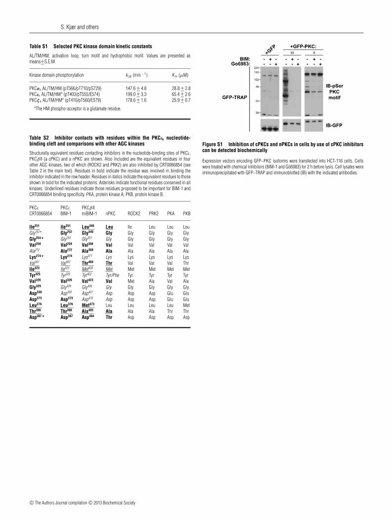
Table S1 Selected PKC kinase domain kinetic constants
AL/TM/HM, activation loop, turn motif and hydrophobic motif. Values are presented asmeans+−S.E.M.
Kinase domain phosphorylation k cat (min − 1) K m (μM)
PKCεk AL/TM/HM (pT566/pT710/pS729) 147.6 +− 4.8 28.8 +− 2.8PKCιk AL/TM/HM* (pT403/pT555/E574) 199.0 +− 3.3 65.4 +− 2.6PKCζ k AL/TM/HM* (pT410/pT560/E579) 178.6 +− 1.6 25.9 +− 0.7
*The HM phospho-acceptor is a glutamate residue.
Table S2 Inhibitor contacts with residues within the PKCιk nucleotide-binding cleft and comparisons with other AGC kinases
Structurally equivalent residues contacting inhibitors in the nucleotide-binding sites of PKCι,PKCβ II (a cPKC) and a nPKC are shown. Also included are the equivalent residues in fourother AGC kinases, two of which (ROCK2 and PRK2) are also inhibited by CRT0066854 (seeTable 2 in the main text). Residues in bold indicate the residue was involved in binding theinhibitor indicated in the row header. Residues in italics indicate the equivalent residues to thoseshown in bold for the indicated proteins. Asterisks indicate functional residues conserved in allkinases. Underlined residues indicate those residues proposed to be important for BIM-1 andCRT0066854 binding specificity. PKA, protein kinase A; PKB, protein kinase B.
PKCι PKCι PKCβ IICRT0066854 BIM-1 miBIM-1 nPKC ROCK2 PRK2 PKA PKB
Ile251 Ile251 Leu348 Leu Ile Leu Leu LeuGly252* Gly252 Gly349 Gly Gly Gly Gly GlyGly254* Gly254 Gly351 Gly Gly Gly Gly GlyVal259 Val259 Val356 Val Val Val Val ValAla272 Ala272 Ala369 Ala Ala Ala Ala AlaLys274* Lys274 Lys371 Lys Lys Lys Lys LysVal307 Val307 Thr404 Thr Val Val Val ThrIle323 Ile323 Met420 Met Met Met Met MetTyr325 Tyr325 Tyr422 Tyr/Phe Tyr Tyr Tyr TyrVal326 Val326 Val423 Val Met Ala Val AlaGly329 Gly329 Gly426 Gly Gly Gly Gly GlyAsp330 Asp330 Asp427 Asp Asp Asp Glu GluAsp373 Asp373 Asp470 Asp Asp Asp Glu GluLeu376 Leu376 Met473 Leu Leu Leu Leu MetThr386 Thr386 Ala483 Ala Ala Ala Thr ThrAsp387* Asp387 Asp484 Thr Asp Asp Asp Asp
Figure S1 Inhibition of cPKCs and nPKCs in cells by use of cPKC inhibitorscan be detected biochemically
Expression vectors encoding GFP–PKC isoforms were transfected into HCT-116 cells. Cellswere treated with chemical inhibitors (BIM-1 and Go6983) for 2 h before lysis. Cell lysates wereimmunoprecipitated with GFP–TRAP and immunoblotted (IB) with the indicated antibodies.
c© The Authors Journal compilation c© 2013 Biochemical Society

A novel small molecule aPKC inhibitor
Figure S2 Structure of PKCι bound to CRT0066854 compared with PKCι bound to ATP and PKA bound to ATP
Overall fold of PKCιk in complex with CRT0066854 (left-hand panel, cyan cartoon and yellow sticks), and ATP (middle panel, PDB code 3A8W, salmon-pink cartoon and sticks), with the PKA complexof ATP (right-hand panel, PDB code 1ATP, green cartoon and sticks) for comparison.
REFERENCES
1 Gassel, M., Breitenlechner, C. B., Konig, N., Huber, R., Engh, R. A. and Bossemeyer, D.(2004) The protein kinase C inhibitor bisindolyl maleimide 2 binds with reversedorientations to different conformations of protein kinase A. J. Biol. Chem. 279,23679–23690
2 Messerschmidt, A., Macieira, S., Velarde, M., Badeker, M., Benda, C., Jestel, A.,Brandstetter, H., Neuefeind, T. and Blaesse, M. (2005) Crystal structure of the catalyticdomain of human atypical protein kinase C-ι reveals interaction mode of phosphorylationsite in turn motif. J. Mol. Biol. 352, 918–931
Received 18 December 2012/12 February 2013; accepted 18 February 2013Published as BJ Immediate Publication 18 February 2013, doi:10.1042/BJ20121871
c© The Authors Journal compilation c© 2013 Biochemical Society