adenoid cystic carcinomas of the breast and salivary...
TRANSCRIPT

Adenoid cystic carcinomas of the breast and salivaryglands (or ‘The strange case of Dr Jekyll and Mr Hyde’of exocrine gland carcinomas)
Caterina Marchio,1 Britta Weigelt,2 Jorge S Reis-Filho3
ABSTRACTAdenoid cystic carcinoma (AdCC) is a tumour withmyoepithelial differentiation and characterised by thepresence of a dual population of basaloid and luminalcells arranged in specific growth patterns. Thesetumours, regardless of the anatomical site, arecharacterised by expression of the proto-oncogene andtherapeutic target c-KIT, and seem to harbour a specificchromosomal translocation t(6;9) leading to the fusiongene MYB-NFIB and overexpression of the oncogeneMYB. However, the clinical behaviour of salivary glandand breast AdCC differs; while salivary gland lesions havea relatively high proclivity to metastasise, patients withbreast AdCCs have an excellent outcome. Here theclinical, morphological and molecular features, andpotential therapeutic targets of salivary gland and breastAdCCs are reviewed.
INTRODUCTIONAdenoid cystic carcinomas (AdCCs) are among themost common salivary gland malignancies but alsoaffect other exocrine tubulo-acinar glands1e7 suchas the breast.8 Despite the long recognisedmorphological similarities of tumours arising inthese two glandular structures and the fact thatthese tumours harbour a recurrent chromosomaltranslocation,9 AdCCs of the salivary glands and thebreast differ in incidence and clinical behaviour.Here, we review the clinical, morphological andmolecular features, and potential therapeutictargets of salivary gland and breast AdCCs.
DEFINITION, CLASSIFICATION AND OVERVIEW OFADENOID CYSTIC CARCINOMASThe term cylindroma, the pathological entity latercalled ‘adenoid cystic carcinoma’, was coined byBillroth10 to describe a salivary gland tumourcomposed of entwined cylinders of hyaline stromaand epithelial cells (indeed the illustrations fromBillroth’s study10 depict the typical architecturalpattern of cylindromas/AdCCs, and the cribriformstructures with pseudoglands found in AdCCs. Theterm adenoid cystic carcinoma (carcinoma adenoidescysticum) of the salivary glands was first used byEwing (1919)11 and applied by Geschickter in 1945 totumours of the breast.12 Since then, AdCCs have alsobeen described in several other organs such aslacrimal glands, auditory canal, upper respiratorytract and lung, digestive tract, skin, prostate andlower female genital tract.1e7
AdCC belongs to the subgroup of tumours of themyoepithelial lineage and is defined as a tumour
where both epithelial (luminal) and myoepithelial(basaloid) cells are neoplastic (figure 1). Multiplearchitectural patterns have been reported (eg, crib-riform, tubular, trabecular and solid) in bothmammary and salivary glands, and generallya mixture of different growth patterns are found inAdCCs.1 14 The cribriform growth pattern is themost characteristic and features variably sized andusually smoothly contoured islands of neoplasticcells arranged to compose pseudolumens (which arenot true glandular lumens but represent stromalinvaginations) and true glandular spaces (formed bythe epithelial cells) giving rise to a ‘sieve-like’appearance (figure 1).1 14 These spaces are filled witheosinophilic hyaline material (Periodic acid-Schiff(PAS) positive, diastase resistant), and/or lightlybasophilic myxoid substance (Alcian blue positive):these materials have been demonstrated to repre-sent duplicated basal lamina and glycosaminogly-cans by ultrastructural studies (see Cheuk andChan1 and Bennett et al15 and references therein).Within the cribriform islands, there are occasionaltrue narrow glands lined by cuboidal cells witheosinophilic cystoplasm1 (see figure 1). Histologicalvariants are the glandular (or tubular), reticular (ortrabecular) and solid growth patterns (figure 1). Thetubular variant is characterised by glandular spacesof elongated tubules lined by epithelial cells andsurrounded by single or multiple layers of basaloidcells; the glandular lumens are either empty orcontain secretion. In the trabecular variant cells arearranged to form small nests,14 whereas the solidvariant is composed of island and sheets of closelypacked basaloid cells, with very few or even nopseudocystic spaces; few true glandular spaces canbe found.1
The two distinct cells types of AdCCs, basaloidand luminal, are best appreciated by immunohis-tochemical staining (table 1). The basaloid cellsexpress basal cytokeratins (Cks) such as Ck14 andCk17, vimentin, S-100 protein, actin, calponin andp63. In addition, strong nuclear and cytoplasmicimmunoreactivity of maspin, a mammary inhibi-tory serine protease, has been described in themyoepithelial component of a series of AdCCs ofthe breast.16 The epithelial cells show strong posi-tivity for luminal cytokeratins such as Ck7, forCEA, EMA and CD117 (c-KIT).1 8 17
The stromal hyaline material is best highlightedby staining for collagen IV and laminin. In addi-tion, PAS and Alcian blue staining may helpdifferentiate the material present in true glandularlumens and pseudolumen, respectively (seeabove).1 8
1Department of BiomedicalSciences and Human Oncology,University of Turin, Turin, Italy2Cancer Research UK, LondonResearch Institute, London, UK3Molecular Pathology Team, TheBreakthrough Breast CancerResearch Centre, Institute ofCancer Research, London, UK
Correspondence toJorge S Reis-Filho, TheBreakthrough Breast CancerResearch Centre, Institute ofCancer Research, 237 FulhamRoad, London SW3 6JB, UK;[email protected]
Accepted 27 November 2009
220 J Clin Pathol 2010;63:220e228. doi:10.1136/jcp.2009.073908
Review
on 14 July 2018 by guest. Protected by copyright.
http://jcp.bmj.com
/J C
lin Pathol: first published as 10.1136/jcp.2009.073908 on 4 M
arch 2010. Dow
nloaded from

ADENOID CYSTIC CARCINOMAS OF THE SALIVARY GLANDSClinical and morphological featuresAdCCs are part of the malignant epithelial tumours of the salivaryglands and have been recognised as a specific variant of adenocar-cinoma of the salivary and mucous glands since 1853.18 19 AdCCaffects patients preferentially in the fourth to sixth decade of life,with a slight female predominance (about 3:2).1 17 The parotidgland, submandibular gland and the palate are most commonlyinvolved,1 17 however minor salivary glands in the oesophagus canalso be affected.20
AdCCs account for about 10e15% of all parotid malignan-cies,21 usually present as a slow-growing swelling, and, because ofthe propensity for perineural invasion, pain may be present(table 2).1 24 In advanced cases, fixation to skin or deeper tissuescan occur.1 Owing to its growth pattern, it should be also notedthat at time of presentation these tumours have often invadedbeyond the clinically apparent borders.25e33 AdCC of the salivarygland is a slow-growing but aggressive cancer which is reflectedby the good short-term but very poor long-term outcome ofpatients with this disease.1 34 The 5-year survival is about60e75%, while the 10-year survival drops to 30e54%; mostpatients eventually die of disease after multiple local recurrencesand development of distant metastases (distant metastases aremore common than regional lymph-node involvement).32 35 36
Histologically, AdCC of the salivary glands presents variablecombinations of the three main growth patterns (ie, cribriform,tubular and solid) in each individual case. The cribriform is themost characteristic and is almost invariably found, at leastfocally.1 Cytologically, the basaloid cells constitute the major cellpopulation, showing mild nuclear pleomorphism and few or nomitoses; in the solid variants these cells usually show a morepleomorphic appearance and mitoses are more commonlyfound.1
Salivary gland AdCCs are graded using a specific three-tiergrading system, originally proposed by Szanto et al18 in 1984,which is solely based on the main type of growth pattern present
Figure 1 Representative micrographsof the different variants of adenoid cysticcarcinoma (AdCC). Breast AdCC showinga mixture of cribriform (A), tubular (B),trabecular (C) and solid (D) growthpatterns. Examples of grade 1 (I), grade 2(II) and grade 3 (III) AdCCs, according tothe grading system proposed byRo et al.13
Table 1 Detailed immunophenotypical characterisationof the two distinct cell types of adenoid cystic carcinoma
MarkerLuminal/epithelial
Basal/myoepithelial
Cytokeratin 7 +++ e
Cytokeratin 14 � +++
Cytokeratin 17 � +++
Vimentin � +++
p63 � +++
Maspin � ++
Laminin � ++
Fibronectin � ++
c-KIT(CD117) +/+++ �CyclinD1 +/+++ �b-Catenin nuclear <5% <5%
J Clin Pathol 2010;63:220e228. doi:10.1136/jcp.2009.073908 221
Review
on 14 July 2018 by guest. Protected by copyright.
http://jcp.bmj.com
/J C
lin Pathol: first published as 10.1136/jcp.2009.073908 on 4 M
arch 2010. Dow
nloaded from
in the tumour. Grade 1 AdCCs are well differentiated andcomposed of tubular and cribriform patterns without solidcomponents; grade 2 AdCCs are characterised by a pure cribri-form pattern or mixed with less than 30% of solid areas; andgrade 3 AdCCs are tumours with marked predominance of thesolid pattern.18 This histological grading system has been shownto be associated with prognosis in retrospective studies.1 18 37 38
Recent studies have reported that some low-grade salivarygland carcinomas, including AdCC, can undergo ‘dedif-ferentiation’.39e41 This histological variant would show twocomponents, a conventional low-grade AdCC and a high-grade‘dedifferentiated’ carcinoma, which can be either undifferenti-ated carcinoma or poorly differentiated adenocarcinoma. Dedif-ferentiation is a well recognised phenomenon in bone and softtissue tumour pathology42 and is generally associated with a poorprognosis. In AdCCs it is an extremely rare event (11 casesreported to date), whose clinical behaviour and molecular char-acteristics are still not entirely understood,43 even though recentdata seem to support a more aggressive clinical behaviour ofdedifferentiated AdCCs.43
Molecular featuresThe defining molecular feature of AdCCs of the salivary glandsand breast appears to be the presence of a recurrent chromo-somal translocation t(6;9)(q22e23;p23e24), which generatesa fusion transcript involving the genes MYB and NFIB.9 Thistranslocation is described in detail below.
AdCCs of the salivary glands have a stable genome.44 TP53mutations45 or p53 nuclear expression have been reported,46e48
but in small studies and with discrepant results (see table 3).45 46
The presence of aneuploidy appears to be less common in thecribriform variant (16%) than in the solid lesions (67%, table 3).53
Interestingly, a TP53 point mutation and p53 expression havebeen demonstrated in the dedifferentiated component of twodedifferentiated AdCCs, together with HER2 overexpression anda high proliferation index.43
Loss of heterozygosis (LOH), comparative genomic hybrid-isation (CGH) and microarray-based CGH studies showconflicting results. Cytogenetic analysis of AdCCs of salivaryglands showed deletions of 6q and 12q as well as rearrangementsinvolving chromosomes 6q and 9p.65 Recently, high-resolutionCGH analysis showed that bronchial and salivary AdCCsharbour low levels of genetic instability with few copy number
alterations or high-level amplifications. Recurrent gains included7p15.2, 17q21e25 and 22q11e13, and recurrent losses included1p35, 6q22e25, 8q12e13, 9p21, 12q12e13 and 17p11e13.61 Inaddition, Rao et al have recently shown that deletion of1p32ep36 holds prognostic significance in these lesions.64 Giventhe rather pervasive presence of recurrent regions of deletions,some have studied the possible involvement of tumoursuppressor genes: the minimal region of deletions on chromo-some 9p contains CDKN2A and CDKN2B; other possible candi-date tumour suppressor genes map to 6q75 and 12q.76
Gene amplifications have been investigated, however studieshave yielded conflicting results. The majority of studies wheregene amplifications were investigated using fluorescence in situhybridisation (FISH) or CGH have reported a low prevalence ofgene amplifications. The only report that identified frequentamplification of ERBB1, CCND1 and PIK3CA (67%, 46% and38%, respectively) in AdCCs relied on a PCR-based method,62
which is known to be prone to artefacts when formalin fixedparaffin embedded (FFPE) samples are analysed. Bernheim et al61
and Greer et al63 have recently identified amplifications ofMDM2(12q15), CCND1 (11q13.3) and CTTN (11q13.3) using FISH(table 3); it should be noted, however, that these amplificationsaffected less than 5% of cases. On the other hand, CCND1overexpression seems to be more pervasive than CCND1 ampli-fication, as it is found in up to 90% of cases.63
Apart from the recurrent chromosomal translocationdescribed above, additional translocations have been found insingle cases of AdCCs (see table 3)55 57 59; however neither thegenes involved in these translocations nor their recurrencefrequency have been determined.Two studies have analysed the gene expression profiles of
AdCC in salivary glands as compared to normal salivary glandtissues and an AdCC cell line (ACC3): unsupervised hierarchicalcluster analysis showed AdCCs separated in three differentgroups, and interestingly, these groups were not correlated withhistological grade.67 Overexpression of several genes encodingtranscription factors was demonstrated,67 and among them,SOX4was found to be up-regulated in both studies.66 67 Patel et alshowed that genes associated with morphogenesis, neurogenesis,proliferation, apoptosis, a group of genes encoding extracellularmatrix proteins and basement membrane components seem tocharacterise AdCCs. In addition, Frierson et al67 found epsilon andfrizzled-7, both members of the Wnt/b-catenin signalling
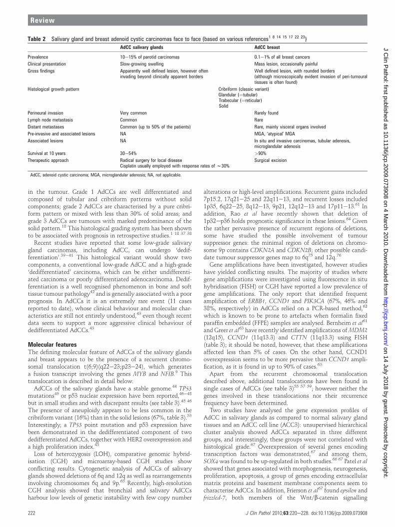
Table 2 Salivary gland and breast adenoid cystic carcinomas face to face (based on various references1 8 14 15 17 22 23)
AdCC salivary glands AdCC breast
Prevalence 10e15% of parotid carcinomas 0.1e1% of all breast cancers
Clinical presentation Slow-growing swelling Mass lesion, occasionally painful
Gross findings Apparently well defined lesion, however ofteninvading beyond clinically apparent borders
Well defined lesion, with rounded borders(although microscopically evident invasion of peri-tumouraltissues is often found)
Histological growth pattern Cribriform (classic variant)Glandular (¼tubular)Trabecular (¼reticular)Solid
Perineural invasion Very common Rarely found
Lymph node metastasis Common Rare
Distant metastases Common (up to 50% of the patients) Rare, mainly visceral organs involved
Pre-invasive and associated lesions NA MGA, ‘atypical’ MGA
Associated lesions NA In situ and invasive carcinomas, tubular adenosis,microglandular adenosis
Survival at 10 years 30e54% >90%
Therapeutic approach Radical surgery for local diseaseCisplatin usually employed with response rates of w30%
Surgical excision
AdCC, adenoid cystic carcinoma; MGA, microglandular adenosis; NA, not applicable.
222 J Clin Pathol 2010;63:220e228. doi:10.1136/jcp.2009.073908
Review
on 14 July 2018 by guest. Protected by copyright.
http://jcp.bmj.com
/J C
lin Pathol: first published as 10.1136/jcp.2009.073908 on 4 M
arch 2010. Dow
nloaded from

pathway, to be up-regulated in AdCCs compared to 175 othercarcinomas from 10 anatomical sites.
ADENOID CYSTIC CARCINOMAS OF THE BREASTClinical, morphological and immunohistochemical featuresAfter Geschickter ’s description of ‘adenocystic basal cell carci-noma’ of the breast,8 three adenocystic basal cell carcinomaswere reported by Foote and Stewart in 1946,3 but it was only twodecades afterwards that Galloway77 at the Mayo clinic describedthe first series of mammary adenoid cystic carcinomas.8 Sincethen several groups have described AdCCs of the breast; howeverdue to the relative rarity of this tumour type, most studies are inthe form of case reports or small cohorts.
The existence of AdCC in the breast has been questioned,given its morphological similarity with cribriform carcinoma22 78;however, immunohistochemical and ultrastructural studies haveeventually proven the existence of true AdCC in the breast (seebelow), which is now recognised as one of 17 histological specialtypes of breast cancer by theWorld HealthOrganization.15 21e23 79
AdCCs account for 0.1e1% of all breast cancers8 15 23; they areusually diagnosed in adult female patients as unilateral painfulmasses.8 15 23 This associated pain has been suggested to be due tothe contractile myoepithelial component of these tumours, asperineural invasion is not commonly seen in these lesions(table 2).8 15 All quadrants seem to be affected, with a particulartrend for the peri-areolar region.8 The most striking feature ofAdCCs of the breast, which is in stark contrast with AdCCs ofsalivary glands, is the excellent long-term prognosis (table 2):a 90e100% 10-year survival rate is reported, and lymph-nodemetastases are rare, as well as distant metastases, which affectmainly visceral organs.15 54
AdCC can be found in conjunction with other breast lesions,including ipsilateral and controlateral in situ and invasivecarcinomas.15 In addition, recent studies have noted an associa-tion between AdCC and microglandular adenosis (MGA),80e83
and some authors have suggested that AdCC may develop in
a background of and in continuity with MGA. Following thishypothesis there would be a spectrum of lesions with a trend ofprogression, encompassing MGA, ‘atypical’MGA (also known as‘in situ AdCC’) and invasive AdCC.80 An association betweenAdCC and tubular adenosis (TA) has also been reported in onestudy, but the molecular analysis performed on these two lesionsfailed to provide evidence of molecular evolution from TA toAdCC.14
The variety of growth patterns described for AdCCs of thesalivary glands is also found when this tumour arises in thebreast: the classic cribriform pattern, as well as the glandular,trabecular and solid variants.8 A sebaceous differentiation isfound in up to 14% of cases, and foci of adenosquamous differ-entiation may also be encountered.8 84
In a way akin to salivary gland AdCC, breast AdCCs aregraded according to the proportion of solid growth: cases witheither cribriform or glandular pattern are considered low grade/G1 tumours, cases with <30% of solid elements are labelled asG2, whereas cases showing>30% of solid growth are classified ashigh grade/G3 tumours (figure 1).8 13 Ro et al13 reported thisgrading system to be clinically meaningful, as in their series,tumours with solid components (G2 and G3 cases) were morelikely to develop recurrences; in addition, in the same cohort, theonly patient who experienced metastases was affected by a high-grade tumour. It should be noted however that histological gradedefined by this system was not associated with outcome in twoother studies.85 86
Other authors72 have suggested the proliferative indices to beof some relevance as they showed greater values in high-gradewhen compared with low-grade lesions; however proliferativeactivity was not found to be significantly related to prognosis.85
Phenotypically, AdCCs are described as hormone receptornegative carcinomas (table 4). In the series of 18 cases analysedby Azoulay et al,73 neither oestrogen receptor (ER) nor proges-terone receptor (PR) expression was identified. Similar findingshave been reported in other independent series (table 4).49 50 74
Table 3 Summary of genomic features reported in adenoid cystic carcinomas of breast and salivary glands
AdCC salivary gland AdCC breast
TP53 mutation 40e100% (higher prevalence in solid variants)45 * NA
p53 nuclear expression Reported between 17%46e48 and 87%48 50% (049e100%50 y)Other mutations Contradictory information about KIT mutations. Most studies failed
to identify mutations, while one study using FFPE tissues reportedmultiple mutations51
PTEN and PIK3CA (case report)52
Aneuploidy 16% (cribriform variant) to 67% (solid variant)53 <10%54
Recurrent translocation and cloned fusion transcript t(6;9)(q22e23;p23e24)9 55 56 MYB-NFIB9 t(6;9)(q22e23;p23e24) MYB-NFIB9
Translocations in single cases t(6;12)(p21;q13)z 57
t(6;14)(q22;q11)58
t(6;9)(q23eq25;p22ep24)55 59
t(6;15)(q25;q15)60
t(1;9)(q21;p21e22)55
der(9)i(9)(q10)inv(9)(q12q13)55
der(X)t(X;9)(p21;p22e23)55
NA
Gains 7p15.2, 17q21e2561; 8p, 9q, 14q, 15q, 16q, 20p, 20qx 6p, 8q, 14q, 16q, 20p, 20qxAmplifications ERBB1, CCND1 and PIK3CA and their co-amplification (by PCR
method)62; CCND1, MDM2, CTTN (by FISH)61 63Not reported
Losses/deletions 1p32ep3664; 6q, 9p, 12q65; 1p35, 6q22e25, 8q12e13, 9p21,12q12e13 and 17p11e1361
2q, 6q, 15p, 16q, 19q**
Transcriptome Up-regulation of genes related to morphogenesis, neurogenesis,proliferation, apoptosis, extracellular matrix proteins and basementmembrane,66 transcription factors (SOX-4)66 67
Basal-like subtype49
Oncogenes overexpressed{ c-KIT (CD117),68e70 CCND1, Cortactin,63 MDM261 and pAKT61 71 c-KIT (CD117)49 51 72e74
*Performed with nucleic acids extracted from FFPE tissues.yp53 immunohistochemical analysis using pressure cooking antigen retrieval.zTranslocation described in one case of AdCC and one case of polymorphous low-grade adenocarcinomas.xFulford, Reis-Filho and Lakhani (unpublished observations).{As defined by immunohistochemistry.AdCC, adenoid cystic carcinoma; IHC, immunohistochemistry; NA, not applicable.
J Clin Pathol 2010;63:220e228. doi:10.1136/jcp.2009.073908 223
Review
on 14 July 2018 by guest. Protected by copyright.
http://jcp.bmj.com
/J C
lin Pathol: first published as 10.1136/jcp.2009.073908 on 4 M
arch 2010. Dow
nloaded from

One study72 reported that 15% and 10% of cases were positivefor ER and PR, respectively (table 4). In contrast, Arpino et al54
demonstrated the presence of ER and PR expression in up to 46%(13/28) and 36% (10/28) of cases, respectively (table 4).Although this cohort represents one of the largest series ofAdCCs reported to date (n¼28), it should be noted that the caseswere collected from different institutions and did not undergoa central review; hence, it cannot be formally ruled out thata substantial number of cases included in that study were crib-riform carcinomas, which are usually ER and PR positive. Inaddition, it should be noted that dextran-coated charcoal assay,87
instead of immunohistochemistry, was used to assess positivityfor ER and PR and only 85% agreement between the two tech-niques is reported.88 The use of the dextran-coated charcoalassay for ER and PR assessment of AdCCs is particularly prob-lematic, given that normal breast lobules and ducts are oftenentrapped within the bulk of AdCCs, which may lead to falsepositive results.
Molecular featuresIn a way akin to salivary gland AdCCs, breast AdCCs consis-tently display the recurrent chromosomal translocation t(6;9)(q22e23;p23e24), which generates fusion transcripts involvingthe genes MYB and NFIB.9 This translocation is described indetail below.
Although microarray-based gene expression profiling has beenextensively applied to the study of breast cancer, most of theseanalyses ignored the histological special types of breast cancer(reviewed in Weigelt et al89 and Weigelt and Reis-Filho90), andthere is paucity on the transcriptomic features of AdCCs. Themolecular subtypes of breast cancer (ie, basal-like, HER2, luminalA, luminal B and normal breast-like), which provide a widelyused working model for a breast cancer molecular taxonomy,have been identified by microarray analysis of only the mostcommon types, invasive ductal and invasive lobularcarcinomas.91e93 This molecular classification was shown to beof prognostic significance, with tumours of luminal A subtypebeing associated with the best outcome, and tumours of basal-like or HER2 subtype with the worst outcome.91 94
To date, only one study has formally investigated the tran-scriptome of breast AdCCs. Using microarray-based geneexpression profiling, Weigelt et al49 analysed a series of 113tumours from 11 special histological types of breast cancer,including four AdCCs. Unsupervised hierarchical cluster anal-ysis showed that AdCCs clustered together with metaplasticand medullary carcinomas, whose similar gene expressionpatterns were also reflected at the immunohistochemical level(triple negative phenotype; ie, lack of ER, PR and HER2; lowlevels of Ck19, AR and Ck8/18; high levels of CD117, vimentin,S100, Ck14 and Ck5/6 expression). In addition, molecularsubtype analysis using a single sample predictor (ie, centroids)
showed that two AdCCs were of basal-like and two AdCCs ofnormal breast-like phenotype, a molecular subtype which iscurrently considered to be an artefact of sample representation(ie, high content of normal tissue contamination).95 Indeed,molecular subtype assignment by hierarchical clusteringshowed that AdCCs consistently displayed a basal-like pheno-type, as did medullary and metaplastic carcinomas. Theseresults support earlier immunohistochemical observations byAzoulay et al,73 that breast AdCCs lack expression of ER, PRand HER2 (ie, triple negative phenotype) and express basalcytokeratins (Ck5/6). These observations illustrate the hetero-geneity of basal-like breast cancers and emphasise that triplenegative and basal-like breast cancer is not a single entity, butrather a spectrum of lesions. Although the majority of triplenegative and basal-like breast cancers are high grade cancers (ie,medullary carcinomas, grade 3 invasive ductal carcinomas of nospecial type (IDC-NSTs), metaplastic carcinomas and apocrinecarcinomas49 73 96e99), there is a subgroup of low grade tumourswith indolent clinical behaviour that also display a triplenegative and basal-like phenotype (ie, AdCCs and secretorycarcinomas). Contrary to high grade triple negative and basal-like breast cancers,100 which have been shown to harbour TP53mutations in >80% of cases,101 two studies have reported thatp53 nuclear expression is either absent49 or seen in 25% ofAdCCs102; on the other hand, one study reported p53 expres-sion in 6/6 AdCCs analysed.50 Furthermore, AdCCs do notappear to be more prevalent in BRCA1 germ-line mutationcarriers.There is not only a paucity of data on transcriptomic but also
on the genomic features of AdCC of the breast. Cytogeneticanalyses of a few cases showed 46,XX,t(4;4)(q21;q35),t(5;11)(q13;q21), 46,XX,+1,der(1;16)(q10;p10)103 and 46,XX,inv(9).65
Aneuploidy is reported in less than 10% of cases.54
In a recent case report of a 76-year-old woman affected bya breast AdCC with a kidney metastasis, molecular geneticanalysis of the primary and metastatic tumour revealed botha PTEN and a PIK3CAmutation.52 These findings led the authorsto speculate that PTEN and PIK3CA mutations may be respon-sible for the unusually aggressive course of this particular AdCCand suggested identified novel molecular targets for therapeuticintervention. Further studies on larger cohorts are needed toconfirm the prevalence of these mutations and their biologicalrole in AdCC.The limited molecular features of breast AdCC available
suggest that these tumours have a relatively stable genome andare characterised by a recurrent chromosomal translocationinvolving MYB and NFIB. Although AdCCs are of triple nega-tive and basal-like phenotype, a diagnosis of breast AdCCshould not necessarily prompt the use of systemic chemo-therapy, given the excellent prognosis of patients with thistumour type.
Table 4 Overview of data reported on the expression of prognostic and predictive factors, p63, basal cytokeratins and c-KIT in adenoid cysticcarcinoma of the breast
Trendell-Smith et al,199950 (N[6)
Arpino et al, 200254
(N[28)Mastropasquaet al, 200572 (N[20)
Crisi et al, 200574
(N[6)Azoulay et al,200573 (N[18)
Weigelt et al,200849 (N[4)
ER 0% 46% 15% 0% 0% 0%
PR 0% 36% 10% 0% 0% 0%
HER2 0% NA 0% 0% 0% 0%
p63 NA NA 85% NA 100% 50%
Cytokeratin 5/6 NA NA NA NA 100% 75%
c-KIT (CD117) NA NA 95% 100% 100% 100%
ER, oestrogen receptor; NA, not available; PR, progesterone receptor.
224 J Clin Pathol 2010;63:220e228. doi:10.1136/jcp.2009.073908
Review
on 14 July 2018 by guest. Protected by copyright.
http://jcp.bmj.com
/J C
lin Pathol: first published as 10.1136/jcp.2009.073908 on 4 M
arch 2010. Dow
nloaded from
MOLECULAR COMPARATIVE PATHOLOGY AND THERAPEUTICINSIGHTS FOR ADCC: FACTS AND ARTEFACTSc-KIT: therapeutic target or unjustified hope?The gene KIT maps to 4q11eq12 and encodes c-KIT (CD117),a type III transmembrane tyrosine kinase receptor, which isactivated by mutations in gastrointestinal stromal tumours andmelanomas. Tumours harbouring KIT mutation but not over-expression of wild-type c-KIT can be targeted by the smallmolecule inhibitor imatinib mesylate (Gleevec, Novartis) andsorafenib (Nexavar, Bayer).
c-KIT has been shown to be expressed in up to 100% of AdCCsof the breast49 51 72e74 and in 80e100% of AdCCs of the salivaryglands arising from the head and neck.68e70 These observationshave prompted several groups to investigate whether imatinibwould be a valid therapeutic option for those lesions arising insalivary glands. Bold claims were made in 2005 in a study byFaivre et al,104 showing a remarkable response to imatinib treat-ment in a patient affected by AdCC of salivary glands. These datacould, however, never be confirmed by subsequent clinical studieswhich failed to identify objective clinical responses in patientswith AdCC treated with imatinib.69 105 106 In contrast withFaivre et al,104 there are descriptions of progression of metastaticAdCC under treatment with imatinib (table 5).105 These disap-pointing results should not come as a surprise, given that acti-vating KIT mutations, the determinant for response to imatinibtreatment,109 have been repeatedly reported to be vanishingly rarein these cancers.69 70 104 105 Last year, however, Vila et al51
demonstrated the presence of multiple KIT mutations in AdCCsand even multiple mutations in single cases. Owing to the smallnumber of tumours analysed (extraction of DNA was successfulin only 8/14 cases) and, most importantly, to the type of samplesubjected to mutation analysis (ie, FFPE samples), these findingsneeds to be confirmed in larger cohorts using optimally processedsamples (ie, fresh/frozen samples).
Taken together, the balance of evidence available to datesuggest that c-KIT may not constitute a potential novel thera-peutic target for AdCC.
A translocation in commonPersson et al9 have recently shown that breast, salivary, lachrymaland ceruminal gland AdCCs harbour a recurrent specific trans-location t(6;9)(q22e23;p23e24). This genetic alteration hadalready been described more than 10 years ago, by the samegroup, as characteristic of salivary glands. However, the fusiongene partners in this translocation have only now been identified:the oncogene MYB on chromosome 6q22eq23 and the tran-scription factor NFIB on chromosome 9p23ep24.9 MYB maps tochromosome 6q22eq23 and encodes a transcription factor withan N-terminal DNA binding domain, a centrally locatedtranscription activation domain, and a C-terminal negativeregulatory domain, which plays a pivotal role in the control ofcell proliferation, apoptosis and differentiation. It is highlyexpressed in immature, proliferating cells, fetal salivary gland;however it is down-regulated as cells become more differenti-ated.9 In the t(6;9)(q22eq23;p23ep24), the exon 14 of MYB isfused to the last coding exons of NFIB, most often due tobreakpoints in MYB intron 14 and in NFIB intron 8. Thistranslocation was shown to produce distinct types of fusiontranscripts due to splice variations of the MYB gene. Thecommon denominator of these genetic aberrations, however, wasa deletion of exon 15 of MYB and its 39-UTR, which containsseveral highly conserved target sites for miR-15a/16 and miR-150microRNAs. In normal and cancer cells, these miRNAs have beenshown to down-regulate the expression ofMYB. Consistent withthis hypothesis, transfection of leukaemic cells with premiR-15a/16 and premiR-150 resulted in a significant down-regulation ofMYB mRNA expression, whereas transfection of ACC cells didnot alter the expression levels of MYB significantly.9
Despite the limited number of samples analysed so far,9 this isthe first fusion transcript identified in AdCCs. Although the useof split apart probes for the identification of this translocation areunlikely to be used in diagnostic practice, due to the character-istic histological features of AdCC, one can envisage that theseprobes may help reclassify some tumours with features over-lapping with those of AdCC or mixed salivary gland tumours.
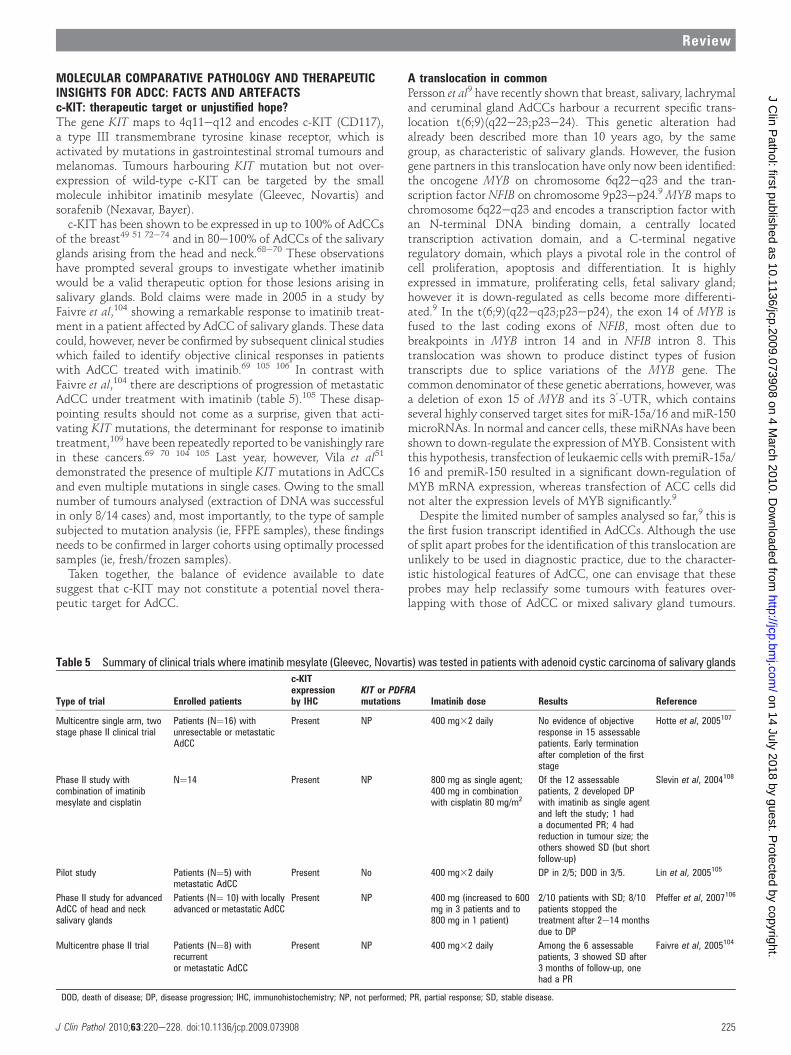
Table 5 Summary of clinical trials where imatinib mesylate (Gleevec, Novartis) was tested in patients with adenoid cystic carcinoma of salivary glands
Type of trial Enrolled patients
c-KITexpressionby IHC
KIT or PDFRAmutations Imatinib dose Results Reference
Multicentre single arm, twostage phase II clinical trial
Patients (N¼16) withunresectable or metastaticAdCC
Present NP 400 mg32 daily No evidence of objectiveresponse in 15 assessablepatients. Early terminationafter completion of the firststage
Hotte et al, 2005107
Phase II study withcombination of imatinibmesylate and cisplatin
N¼14 Present NP 800 mg as single agent;400 mg in combinationwith cisplatin 80 mg/m2
Of the 12 assessablepatients, 2 developed DPwith imatinib as single agentand left the study; 1 hada documented PR; 4 hadreduction in tumour size; theothers showed SD (but shortfollow-up)
Slevin et al, 2004108
Pilot study Patients (N¼5) withmetastatic AdCC
Present No 400 mg32 daily DP in 2/5; DOD in 3/5. Lin et al, 2005105
Phase II study for advancedAdCC of head and necksalivary glands
Patients (N¼ 10) with locallyadvanced or metastatic AdCC
Present NP 400 mg (increased to 600mg in 3 patients and to800 mg in 1 patient)
2/10 patients with SD; 8/10patients stopped thetreatment after 2e14 monthsdue to DP
Pfeffer et al, 2007106
Multicentre phase II trial Patients (N¼8) withrecurrentor metastatic AdCC
Present NP 400 mg32 daily Among the 6 assessablepatients, 3 showed SD after3 months of follow-up, onehad a PR
Faivre et al, 2005104
DOD, death of disease; DP, disease progression; IHC, immunohistochemistry; NP, not performed; PR, partial response; SD, stable disease.
J Clin Pathol 2010;63:220e228. doi:10.1136/jcp.2009.073908 225
Review
on 14 July 2018 by guest. Protected by copyright.
http://jcp.bmj.com
/J C
lin Pathol: first published as 10.1136/jcp.2009.073908 on 4 M
arch 2010. Dow
nloaded from

This fusion gene may provide new therapeutic avenues for themanagement of advanced AdCC; however, one must rememberthat targeting transcription factors is by no means a trivial task.Further functional studies investigating in greater depth thebiological consequences of the MYB gain of function due to theMYB-NFIB fusion are eagerly awaited. Importantly, genesilencing experiments to demonstrate that MYB expression isselectively required for the survival of cancer cells with geneti-cally activated MYB have yet to be performed.
CONCLUSIONSAs salivary and mammary glands are exocrine glands bothshowing a tubulo-acinar architecture, the remarkable similarmorphological features observed in tumours arising in these twoglandular structures should perhaps not come as a surprise.110
A good example is offered by salivary duct carcinoma, a lesionthat, at the morphological level, is remarkably similar to invasiveductal carcinomas of the breast,92 and that can be categorised inthe samemolecular subtypes as those identified in invasive ductalbreast carcinomas, namely luminal, HER2 and basal subtypes.111
AdCCs constitute an even better example of the similaritiesbetween the tumours affecting these sites. AdCCs of breast andsalivary glands share a common pattern of growth, harbour anidentical recurrent fusion gene (MYB-NFIB), and similar patternsof numerical chromosomal alterations. Surprisingly, however,despite these similarities, the behaviour of salivary gland andbreast AdCCs differs: while salivary gland lesions have anaggressive behaviour and often metastasise, breast lesions havea much more indolent clinical course.
Although great strides have been made in the characterisationof these tumours, several questions remain unanswered. DoAdCCs constitute a tumour type with pluripotent patterns ofevolution? And if so, what are the determinants of the distinctevolution/clinical behaviour? If overexpression of MYB and itstarget genes is the common denominator in the genesis of AdCCsregardless of the site of origin, what then makes their behaviourdiffer in salivary gland and breast? It would be plausible thatdifferent tumour-microenvironment interactions may accountfor the different metastatic proclivity of breast and salivary glandAdCCs; alternatively, it could be that the MYB-NFIB trans-location may have similar impacts on the morphology of thetumour in different sites, but different effects on the metastaticability dependent on the target epithelial cell (ie, cell of origin).With the advent of massively parallel sequencing and theopportunity to characterise the genome and transcriptome fromthese cancers, new opportunities to define the molecular deter-minants of the behaviour of these fascinating lesions willemerge.112 113
Acknowledgements The authors are grateful to Dr Riccardo Arisio (Sant’AnnaHospital, Turin) for the courtesy of the breast AdCC case illustrated in (figure 1).
Funding This study was funded by Breakthrough Breast Cancer and Ricerca SanitariaFinalizzata Regione Piemonte 2009. BW is funded by a Cancer Research UK fellowship.NHS funding to the NIHR Biomedical Research Centre is also acknowledged.
Competing interests None.
Provenance and peer review Commissioned; not externally peer reviewed.
REFERENCES1. Cheuk W, Chan J. Salivary gland tumors. In: Fletcher C, ed. Diagnostic
histopathology of tumors. New York: Churchill Livingstone Elsevier, 2007: 280e4.2. Gray HR, Helwig EB. Epithelioma adenoides cysticum and solitary trichoepithelioma.
Arch Dermatol 1963;87:102e14.3. Hajdu SI, Huvos AG, Goodner JT, et al. Carcinoma of the trachea. Clinicopathologic
study of 41 cases. Cancer 1970;25:1448e56.4. Huvos AG, Strong EW. Epithelial tumors of the lacrimal gland. Lab Invest
1973;28:386.5. Lassaletta L, Patron M, Oloriz J, et al. Avoiding misdiagnosis in ceruminous gland
tumours. Auris Nasus Larynx 2003;30:287e90.6. Nelms DC, Luna MA. Primary adenocystic carcinoma (cylindromatous carcinoma) of
the esophagus. Cancer 1972;29:440e3.7. Weltzer S. Adenoid cystic carcinoma of the breast. Am Surg 1970;36:271.8. Rosen PP. Adenoid cystic carcinoma. In: Rosen PP, ed. Rosen’s breast pathology.
Philadelphia: Lippincott Williams and Wilkins, 2009:590e604.9. Persson M, Andren Y, Mark J, et al. Recurrent fusion of MYB and NFIB transcription
factor genes in carcinomas of the breast and head and neck. Proc Natl Acad SciU S A 2009;106:18740e4.
10. Billroth T. Beobachtungen uber Geschwulste der Speicheldrusen. Virchows ArchPath Anat 1859;17:357e75.
11. Ewing J. Epithelial tumors of the salivary gland, in neoplastic diseases. Philadelphia:WB Saunders, 1919: 780.
12. Geschickter CK. Diseases of the breast, in diagnosis, pathology, treatment.Philadelphia: JB Lippincott, 1945: 824.
13. Ro JY, Silva EG, Gallager HS. Adenoid cystic carcinoma of the breast. Hum Pathol1987;18:1276e81.
14. Da Silva L, Buck L, Simpson PT, et al. Molecular and morphological analysis ofadenoid cystic carcinoma of the breast with synchronous tubular adenosis. VirchowsArch 2009;454:107e14.
15. Bennett AK, Mills SE, Wick MR. Salivary-type neoplasms of the breast and lung.Semin Diagn Pathol 2003;20:279e304.
16. Reis-Filho JS, Milanezi F, Silva P, et al. Maspin expression in myoepithelial tumorsof the breast. Pathol Res Pract 2001;197:817e21.
17. Ellis GL, Auclair PL. Adenoid cystic carcinoma, in atlas of tumor pathologydtumorsof the salivary glands. Washington, DC: Armed Forces Institute of Pathology, 1996:203e15.
18. Szanto PA, Luna MA, Tortoledo ME, et al. Histologic grading of adenoid cysticcarcinoma of the salivary glands. Cancer 1984;54:1062e9.
19. Tauxe WN,Mc DJ, Devine KD. A century of cylindromas. short review and report of27 adenoid cystic carcinomas arising in the upper respiratory passages. ArchOtolaryngol 1962;75:364e76.
20. De Dosso S, Mazzucchelli L, Ghielmini M, et al. Response to oxaliplatin withcetuximab in minor salivary gland adenoid cystic carcinoma. Tumori2009;95:378e81.
21. Barnes L, Eveson J, Reichart P, et al. Pathology and genetics of head and necktumours. In: Kleihues R, Sobin LH, eds. World Health Organization classification oftumours. Lyon: IARC, 2005:221e2.
22. Ellis IO, Pinder S, Lee AHS. Tumors of the breastdadenoid cystic carcinoma.In: Fletcher C, ed. Diagnosti histopathology of tumors. Churchill Livingstone Elsevier,2007: 953e4.
23. Ellis IO, Schnitt SJ, Sastre-Garau X, et al. Invasive Breast Carcinoma. In: TavassoliFA, Devilee P, eds. World health organization classification of tumoursdpathologyand genetics, tumours of the breast and female genital organs. Lyon: IARC Press,2003: 44e5.
24. Friedrich RE, Bleckmann V. Adenoid cystic carcinoma of salivary and lacrimal glandorigin: localization, classification, clinical pathological correlation, treatment resultsand long-term follow-up control in 84 patients. Anticancer Res 2003;23:931e40.
25. Chomette G, Auriol M, Tranbaloc P, et al. Adenoid cystic carcinoma of minor salivaryglands. Analysis of 86 cases. Clinico-pathological, histoenzymological andultrastructural studies. Virchows Arch A Pathol Anat Histol 1982;395:289e301.
26. Conley J, Dingman DL. Adenoid cystic carcinoma in the head and neck (cylindroma).Arch Otolaryngol 1974;100:81e90.
27. Eby LS, Johnson DS, Baker HW. Adenoid cystic carcinoma of the head and neck.Cancer 1972;29:1160e8.
28. Eneroth CM, Zajicek J. Aspiration biopsy of salivary gland tumors. IV. Morphologicstudies on smears and histologic sections from 45 cases of adenoid cysticcarcinoma. Acta Cytol 1969;13:59e63.
29. Leafstedt SW, Gaeta JF, Sako K, et al. Adenoid cystic carcinoma of major andminor salivary glands. Am J Surg 1971;122:756e62.
30. Matsuba HM, Simpson JR, Mauney M, et al. Adenoid cystic salivary glandcarcinoma: a clinicopathologic correlation. Head Neck Surg 1986;8:200e4.
Take-home messages
< Adenoid cystic carcinomas (AdCCs) in salivary glandsrepresent a very aggressive type of lesion, whereas breastadenoid cystic carcinoma shows a very good clinicalbehaviour, with an excellent 10-year prognosis.
< Breast AdCCs have a triple negative and basal-like phenotype;however they have a good long-term outcome.
< Breast and salivary gland AdCCs harbour the t(6;9)(q22eq24;p21ep23) recurrent translocation, involving theMYB and NFIBgenes.
< At the present time, no specific targeted therapy is availablefor AdCC of either breast or salivary glands.
226 J Clin Pathol 2010;63:220e228. doi:10.1136/jcp.2009.073908
Review
on 14 July 2018 by guest. Protected by copyright.
http://jcp.bmj.com
/J C
lin Pathol: first published as 10.1136/jcp.2009.073908 on 4 M
arch 2010. Dow
nloaded from

31. Nascimento AG, Amaral AL, Prado LA, et al. Adenoid cystic carcinoma of salivary glands.A study of 61 cases with clinicopathologic correlation. Cancer 1986;57:312e19.
32. Perzin KH, Gullane P, Clairmont AC. Adenoid cystic carcinomas arising in salivaryglands: a correlation of histologic features and clinical course. Cancer1978;42:265e82.
33. Spiro RH, Huvos AG, Strong EW. Adenoid cystic carcinoma of salivary origina clinicopathologic study of 242 cases. Am J Surg 1974;128:512e20.
34. Wahlberg P, Anderson H, Biorklund A, et al. Carcinoma of the parotid and submandibularglandsea study of survival in 2465 patients. Oral Oncol 2002;38:706e13.
35. Hamper K, Lazar F, Dietel M, et al. Prognostic factors for adenoid cystic carcinomaof the head and neck: a retrospective evaluation of 96 cases. J Oral Pathol Med1990;19:101e7.
36. Chilla R, Schroth R, Eysholdt U, et al. Adenoid cystic carcinoma of the head andneck. Controllable and uncontrollable factors in treatment and prognosis. ORL JOtorhinolaryngol Relat Spec 1980;42:346e67.
37. Batsakis JG, el-Naggar AK. Myoepithelium in salivary and mammary neoplasms ishost-friendly. Adv Anat Pathol 1999;6:218e26.
38. Batsakis JG, Luna MA, el-Naggar A. Histopathologic grading of salivary glandneoplasms: III. Adenoid cystic carcinomas. Ann Otol Rhinol Laryngol 1990;99:1007e9.
39. Chau Y, Hongyo T, Aozasa K, et al. Dedifferentiation of adenoid cystic carcinoma:report of a case implicating p53 gene mutation. Hum Pathol 2001;32:1403e7.
40. Cheuk W, Chan JK, Ngan RK. Dedifferentiation in adenoid cystic carcinoma ofsalivary gland: an uncommon complication associated with an accelerated clinicalcourse. Am J Surg Pathol 1999;23:465e72.
41. MolesMA, Avila IR, Archilla AR. Dedifferentiation occurring in adenoid cystic carcinoma ofthe tongue. Oral Surg Oral Med Oral Pathol Oral Radiol Endod 1999;88:177e80.
42. Meis JM. "Dedifferentiation" in bone and soft-tissue tumors. A histological indicatorof tumor progression. Pathol Annu 1991;26(Pt 1):37e62.
43. Nagao T, Gaffey TA, Serizawa H, et al. Dedifferentiated adenoid cystic carcinoma:a clinicopathologic study of 6 cases. Mod Pathol 2003;16:1265e72.
44. Yu Y, Baras AS, Shirasuna K, et al. Concurrent loss of heterozygosity and copynumber analysis in adenoid cystic carcinoma by SNP genotyping arrays. Lab Invest2007;87:430e9.
45. Yamamoto Y, Wistuba II, Kishimoto Y, et al. DNA analysis at p53 locus in adenoidcystic carcinoma: comparison of molecular study and p53 immunostaining. Pathol Int1998;48:273e80.
46. Kiyoshima T, Shima K, Kobayashi I, et al. Expression of p53 tumor suppressor genein adenoid cystic and mucoepidermoid carcinomas of the salivary glands. Oral Oncol2001;37:315e22.
47. Preisegger KH, Beham A, Kopp S, et al. Prognostic impact of molecular analyses inadenoid cystic carcinomas of the salivary gland. Onkologie 2001;24:273e7.
48. Zhu QR, White FH, Tipoe GL. p53 oncoprotein accumulation in adenoid cysticcarcinoma of parotid and palatine salivary glands. Pathology 1997;29:154e8.
49. Weigelt B, Horlings HM, Kreike B, et al. Refinement of breast cancer classificationby molecular characterization of histological special types. J Pathol2008;216:141e50.
50. Trendell-Smith NJ, Peston D, Shousha S. Adenoid cystic carcinoma of the breast:a tumour commonly devoid of oestrogen receptors and related proteins.Histopathology 1999;35:241e8.
51. Vila L, Liu H, Al-Quran SZ, et al. Identification of c-kit gene mutations in primaryadenoid cystic carcinoma of the salivary gland. Mod Pathol 2009;22:1296e302.
52. Vranic S, Bilalovic N, Lee LM, et al. PIK3CA and PTEN mutations in adenoid cysticcarcinoma of the breast metastatic to kidney. Hum Pathol 2007;38:1425e31.
53. Enamorado I, Lakhani R, Korkmaz H, et al. Correlation of histopathological variants,cellular DNA content, and clinical outcome in adenoid cystic carcinoma of the salivaryglands. Otolaryngol Head Neck Surg 2004;131:646e50.
54. Arpino G, Clark GM, Mohsin S, et al. Adenoid cystic carcinoma of the breast:molecular markers, treatment, and clinical outcome. Cancer 2002;94:2119e27.
55. Nordkvist A, Mark J, Gustafsson H, et al. Non-random chromosomerearrangements in adenoid cystic carcinoma of the salivary glands. GenesChromosomes Cancer 1994;10:115e21.
56. Jin Y, Mertens F, Limon J, et al. Characteristic karyotypic features in lacrimal andsalivary gland carcinomas. Br J Cancer 1994;70:42e7.
57. Martins C, Fonseca I, Roque L, et al. Cytogenetic similarities between two types ofsalivary gland carcinomas: adenoid cystic carcinoma and polymorphous low-gradeadenocarcinoma. Cancer Genet Cytogenet 2001;128:130e6.
58. Bell D, Zhao Y, Rao HP, et al. Translocation t(6;14) as the sole chromosomalabnormality in adenoid cystic carcinoma of the base of tongue. Head Neck Pathol2007;1:165e8.
59. Stenman G, Sandros J, Dahlenfors R, et al. 6q- and loss of the Y chromosomeetwocommon deviations in malignant human salivary gland tumors. Cancer GenetCytogenet 1986;22:283e93.
60. el-Naggar AK, Lovell M, Callender DL, et al. Limited nonrandom chromosomalaberrations in a recurrent adenoid cystic carcinoma of the parotid gland. CancerGenet Cytogenet 1999;109:66e9.
61. Bernheim A, Toujani S, Saulnier P, et al. High-resolution array comparative genomichybridization analysis of human bronchial and salivary adenoid cystic carcinoma.Lab Invest 2008;88:464e73.
62. Sequeiros-Santiago G, Garcia-Carracedo D, Fresno MF, et al. Oncogeneamplification pattern in adenoid cystic carcinoma of the salivary glands. Oncol Rep2009;21:1215e22.
63. Greer RO Jr, Said S, Shroyer KR, et al. Overexpression of cyclin D1 and cortactin isprimarily independent of gene amplification in salivary gland adenoid cysticcarcinoma. Oral Oncol 2007;43:735e41.
64. Rao PH, Roberts D, Zhao YJ, et al. Deletion of 1p32-p36 is the most frequent geneticchange and poor prognostic marker in adenoid cystic carcinoma of the salivaryglands. Clin Cancer Res 2008;14:5181e7.
65. Mark HF, Hanna I, Gnepp DR. Cytogenetic analysis of salivary gland type tumors.Oral Surg Oral Med Oral Pathol Oral Radiol Endod 1996;82:187e92.
66. Patel KJ, Pambuccian SE, Ondrey FG, et al. Genes associated with earlydevelopment, apoptosis and cell cycle regulation define a gene expression profile ofadenoid cystic carcinoma. Oral Oncol 2006;42:994e1004.
67. Frierson HF Jr, El-Naggar AK, Welsh JB, et al. Large scale molecular analysisidentifies genes with altered expression in salivary adenoid cystic carcinoma. Am JPathol 2002;161:1315e23.
68. Edwards PC, Bhuiya T, Kelsch RD. C-kit expression in the salivary gland neoplasmsadenoid cystic carcinoma, polymorphous low-grade adenocarcinoma, and monomorphicadenoma. Oral Surg Oral Med Oral Pathol Oral Radiol Endod 2003;95:586e93.
69. Holst VA, Marshall CE, Moskaluk CA, et al. KIT protein expression and analysis ofc-kit gene mutation in adenoid cystic carcinoma. Mod Pathol 1999;12:956e60.
70. Jeng YM, Lin CY, Hsu HC. Expression of the c-kit protein is associated with certainsubtypes of salivary gland carcinoma. Cancer Lett 2000;154:107e11.
71. Volker HU, Scheich M, Berndt A, et al. Expression of p-AKT characterizes adenoidcystic carcinomas of head and neck with a higher risk for tumor relapses. DiagnPathol 2009;4:18.
72. Mastropasqua MG,Maiorano E, Pruneri G, et al. Immunoreactivity for c-kit and p63as an adjunct in the diagnosis of adenoid cystic carcinoma of the breast. Mod Pathol2005;18:1277e82.
73. Azoulay S, Lae M, Freneaux P, et al. KIT is highly expressed in adenoid cysticcarcinoma of the breast, a basal-like carcinoma associated with a favorableoutcome. Mod Pathol 2005;18:1623e31.
74. Crisi GM, Marconi SA, Makari-Judson G, et al. Expression of c-kit in adenoid cysticcarcinoma of the breast. Am J Clin Pathol 2005;124:733e9.
75. Rutherford S, Yu Y, Rumpel CA, et al. Chromosome 6 deletion and candidate tumorsuppressor genes in adenoid cystic carcinoma. Cancer Lett 2006;236:309e17.
76. Rutherford S, Hampton GM, Frierson HF, et al. Mapping of candidate tumorsuppressor genes on chromosome 12 in adenoid cystic carcinoma. Lab Invest2005;85:1076e85.
77. Galloway JR,Woolner LB, Clagett OT. Adenoid cystic carcinoma of the breast. SurgGynecol Obstet 1966;122:1289e94.
78. Azzopardi JE. Problems in breast pathology. London: WB Saunders, 1979.79. Wells CA, Nicoll S, Ferguson DJ. Adenoid cystic carcinoma of the breast: a case
with axillary lymph node metastasis. Histopathology 1986;10:415e24.80. Acs G, Simpson JF, Bleiweiss IJ, et al. Microglandular adenosis with transition into
adenoid cystic carcinoma of the breast. Am J Surg Pathol 2003;27:1052e60.81. James BA, Cranor ML, Rosen PP. Carcinoma of the breast arising in microglandular
adenosis. Am J Clin Pathol 1993;100:507e13.82. Khalifeh IM, Albarracin C, Diaz LK, et al. Clinical, histopathologic, and
immunohistochemical features of microglandular adenosis and transition into in situand invasive carcinoma. Am J Surg Pathol 2008;32:544e52.
83. Koenig C, Dadmanesh F, Bratthauer GL, et al. carcinoma arising in microglandularadenosis: an immunohistochemical analysis of 20 intraepithelial and invasiveneoplasms. Int J Surg Pathol 2000;8:303e15.
84. Tavassoli FA, Norris HJ. Mammary adenoid cystic carcinoma with sebaceousdifferentiation. A morphologic study of the cell types. Arch Pathol Lab Med1986;110:1045e53.
85. Kleer CG, Oberman HA. Adenoid cystic carcinoma of the breast: value of histologicgrading and proliferative activity. Am J Surg Pathol 1998;22:569e75.
86. Lamovec J, Us-Krasovec M, Zidar A, et al. Adenoid cystic carcinoma of the breast:a histologic, cytologic, and immunohistochemical study. Semin Diagn Pathol1989;6:153e64.
87. McGuire WL, De La Garza M, Chamness GC. Evaluation of estrogen receptor assaysin human breast cancer tissue. Cancer Res 1977;37:637e9.
88. Hayat MA. Microscopy, immunohistochemistry and antigen retrieval methods forlight and electron microscopy. New York: Kluwer Academic/Plenum Publisher, 2002.
89. Weigelt B, Baehner FL, Reis-Filho JS. The contribution of gene expression profilingto breast cancer classification, prognostication and prediction: a retrospective of thelast decade. J Pathol 2010;220:263e80.
90. Weigelt B, Reis-Filho JS. Histological and molecular types of breast cancer: is therea unifying taxonomy. Nat Rev Clin Oncol 2009;6:718e30.
91. Hu Z, Fan C, Oh DS, et al. The molecular portraits of breast tumors are conservedacross microarray platforms. BMC Genomics 2006;7:96.
92. Perou CM, Sorlie T, Eisen MB, et al. Molecular portraits of human breast tumours.Nature 2000;406:747e52.
93. Sorlie T, Tibshirani R, Parker J, et al. Repeated observation of breast tumor subtypesin independent gene expression data sets. Proc Natl Acad Sci U S A2003;100:8418e23.
94. Sorlie T, Perou CM, Tibshirani R, et al. Gene expression patterns of breastcarcinomas distinguish tumor subclasses with clinical implications. Proc Natl AcadSci U S A 2001;98:10869e74.
95. Peppercorn J, Perou CM, Carey LA. Molecular subtypes in breast cancer evaluationand management: divide and conquer. Cancer Invest 2008;26:1e10.
J Clin Pathol 2010;63:220e228. doi:10.1136/jcp.2009.073908 227
Review
on 14 July 2018 by guest. Protected by copyright.
http://jcp.bmj.com
/J C
lin Pathol: first published as 10.1136/jcp.2009.073908 on 4 M
arch 2010. Dow
nloaded from

96. Reis-Filho JS, Milanezi F, Steele D, et al. Metaplastic breast carcinomas are basal-like tumours. Histopathology 2006;49:10e21.
97. Lae M, Freneaux P, Sastre-Garau X, et al. Secretory breast carcinomas with ETV6-NTRK3 fusion gene belong to the basal-like carcinoma spectrum. Mod Pathol2009;22:291e8.
98. Rakha EA, Reis-Filho JS, Ellis IO. Basal-like breast cancer: a critical review. J ClinOncol 2008;26:2568e81.
99. Weigelt B, Kreike B, Reis-Filho JS. Metaplastic breast carcinomas are basal-like breastcancers: a genomic profiling analysis. Breast Cancer Res Treat 2009;117:273e80.
100. Reis-Filho JS, Tutt AN. Triple negative tumours: a critical review. Histopathology2008;52:108e18.
101. Manie E, Vincent-Salomon A, Lehmann-Che J, et al. High frequency of TP53mutation in BRCA1 and sporadic basal-like carcinomas but not in BRCA1 luminalbreast tumors. Cancer Res 2009;69:663e71.
102. Pastolero G, Hanna W, Zbieranowski I, et al. Proliferative activity and p53expression in adenoid cystic carcinoma of the breast. Mod Pathol 1996;9:215e19.
103. Pandis N, Teixeira MR, Gerdes AM, et al. Chromosome abnormalities in bilateralbreast carcinomas. cytogenetic evaluation of the clonal origin of multiple primarytumors. Cancer 1995;76:250e8.
104. Faivre S, Raymond E, Casiraghi O, et al. Imatinib mesylate can induce objectiveresponse in progressing, highly expressing KIT adenoid cystic carcinoma of thesalivary glands. J Clin Oncol 2005;23:6271e3; author reply 6273e4.
105. Lin CH, Yen RF, Jeng YM, et al. Unexpected rapid progression of metastatic adenoidcystic carcinoma during treatment with imatinib mesylate. Head Neck2005;27:1022e7.
106. Pfeffer MR, Talmi Y, Catane R, et al. A phase II study of Imatinib for advancedadenoid cystic carcinoma of head and neck salivary glands. Oral Oncol2007;43:33e6.
107. Hotte SJ,Winquist EW, Lamont E, et al. Imatinib mesylate in patients with adenoidcystic cancers of the salivary glands expressing c-kit: a Princess Margaret HospitalPhase II Consortium Study. J Clin Oncol 2005;23:585e90.
108. Slevin NJ, Mais KL, Bruce I, et al. Imatinib with cisplatin in recurrent and/ormetastatic salivary adenoidcystic carcinomadresponse assessed by FDG-PETscanning [abstract]. J Clin Oncol 2004;22:5604.
109. Hornick JL, Fletcher CD. The role of KIT in the management of patients withgastrointestinal stromal tumors. Hum Pathol 2007;38:679e87.
110. Foschini MP, Reis-Filho JS, Eusebi V, et al. Salivary gland-like tumours of thebreast: surgical and molecular pathology. J Clin Pathol 2003;56:497e506.
111. Di Palma S, Skalova A, Ungari M, et al. Pure salivary duct carcinomas can beclassified into luminal, Her2 and basal-like phenotypes. Abstract 1072. Mod Pathol2008;21(Suppl 1).
112. Aparicio SA, Huntsman DG. Does massively parallel DNA resequencing signify theend of histopathology as we know it? J Pathol 2010;220:307e15.
113. Reis-Filho JS. Next generation sequencing. Breast Cancer Res 2009;11(Suppl 3):S12.
228 J Clin Pathol 2010;63:220e228. doi:10.1136/jcp.2009.073908
Review
on 14 July 2018 by guest. Protected by copyright.
http://jcp.bmj.com
/J C
lin Pathol: first published as 10.1136/jcp.2009.073908 on 4 M
arch 2010. Dow
nloaded from