adeno-associated virus site-specific integration is regulated by trp
TRANSCRIPT

1
Adeno-associated virus site-specific integration is regulated by TRP-185
Running title: TRP-185 regulate AAV site-specific integration
Noriaki Yamamoto1, Masato Suzuki1, Masa-aki Kawano1, Takamasa Inoue1, Ryou-u
Takahashi1, Hiroko Tsukamoto1, Teruya Enomoto1, Yuki Yamaguchi1, Tadashi Wada2,
and Hiroshi Handa1, *
Graduate School of Bioscience and Biotechnology1, and Integrated Research Institute2,
Tokyo Institute of Technology, Yokohama 226-8501, Japan
* Corresponding Author: Hiroshi Handa
Corresponding Author. Mailing address: Graduate school of Bioscience and
Biotechnology, Tokyo Institute of Technology, 4259 Nagatsuta-cho, Midori-ku,
Yokohama, Kanagawa 226-8501, Japan.
Phone: 81-45-924-5872. Fax: 81-45-924-5145. E-mail: [email protected].
Word count: Abstract: 168
Text: 5,390
ACCEPTED
Copyright © 2006, American Society for Microbiology and/or the Listed Authors/Institutions. All Rights Reserved.J. Virol. doi:10.1128/JVI.02014-06 JVI Accepts, published online ahead of print on 6 December 2006
on April 5, 2018 by guest
http://jvi.asm.org/
Dow
nloaded from

2
Abstract 1
Adeno-associated virus (AAV) site-specifically integrates into the AAVS1 locus on 2
human chromosome 19. Although recruitment of the AAV nonstructural protein 3
Rep78/68 to the Rep binding site (RBS) on AAVS1 is thought to be an essential step, the 4
mechanism of site-specific integration remains largely unknown, in particular, how the 5
site of integration is determined. Here we describe the identification and characterization 6
of a new cellular regulator of AAV site-specific integration. TAR-RNA loop binding 7
protein (TRP) 185, previously reported to associate with human immunodeficiency virus 8
(HIV)-1 TAR RNA, binds to AAVS1 DNA. Our data suggest that TRP-185 suppresses 9
AAV integration at the AAVS1 RBS, and enhances integration into a region downstream 10
of the RBS. TRP-185 bound to Rep68 directly, changing the Rep68 DNA binding 11
property, and stimulating Rep68 helicase activity. We present a model in which TRP-185 12
changes the specificity of the AAV integration site from the RBS to a downstream region 13
by acting as a molecular chaperone that promotes Rep68 complex formation competent 14
for 3’-->5’ DNA helicase activity. 15 ACCEPTED
on April 5, 2018 by guest
http://jvi.asm.org/
Dow
nloaded from

3
Introduction 1
AAV is a nonpathogenic human parvovirus that contains a linear single-stranded DNA 2
genome of approximately 4.7 kb, carrying palindromic inverted terminal repeats (ITRs) at 3
both ends that serve as the viral origin of replication. The AAV genome consists of two major 4
open reading frames, rep and cap. The cap gene encodes three structural proteins, VP1, VP2, 5
and VP3. The rep gene encodes four nonstructural proteins, Rep78, Rep68, Rep52, and 6
Rep40. The nonstructural proteins have multiple activities, such as DNA binding, ATPase, 7
helicase, and endonuclease activities, and play pivotal roles in various stages of the viral life 8
cycle, including integration, replication, and regulation of viral gene expression. For efficient 9
AAV replication and gene expression, co-infection of a helper virus, such as adenovirus or 10
herpesvirus, is required. In its absence, AAV infection results in stable integration of the viral 11
DNA genome into a specific locus on chromosome 19, called AAVS1(17, 25). This property 12
of AAV is unique among eukaryotic DNA viruses. 13
In the current model of AAV site-specific integration, following infection, the 14
single-stranded AAV genome is converted to duplex DNA. Next, the viral p5 promoter is 15
activated and directs the synthesis of Rep78 and Rep68. These proteins bind to the 16-bp 16
RBS present in the AAV ITRs and the p5 promoter. Rep78/68 also binds to an RBS in the 17
AAVS1 region of chromosome 19, and recruits the AAV genome to AAVS1 (8, 31). 18
Rep78/68 then creates a nick at the 6-bp terminal resolution site (trs) flanking the RBS on 19
AAVS1, and its helicase activity unwinds the duplex trs in the 3’-->5’ direction to facilitate 20
DNA replication (8, 29). Subsequently, the AAV genome is integrated into the AAVS1 site 21
within ca. 1 kb downstream of the RBS (8, 21). Currently, there are two models of formation 22
of the AAV-AAVS1 junction. One model assumes that there is Rep78/68-mediated strand 23
ACCEPTED
on April 5, 2018 by guest
http://jvi.asm.org/
Dow
nloaded from

4
switching during DNA replication (21); the other model assumes the involvement of Rep in 1
the ligation of AAV and AAVS1 sequences (27). 2
AAV site-specific integration requires three components: (1) the RBS on the ITRs 3
and the p5 promoter of the AAV genome, (2) a 33-bp sequence containing the trs and the 4
RBS in AAVS1 of chromosome 19, and (3) Rep78 or Rep68 (12, 24). Rep78 and Rep68 5
function in the same way in AAV site-specific integration, and either protein is sufficient for 6
integration in vitro. However, as the levels of Rep proteins are extremely low in latently 7
infected cells (36), additional cellular factors that help Rep function may exist and contribute 8
to AAV site-specific integration. Indeed, it has been reported that the high mobility group 9
protein 1 (HMG-1) binds to and promotes the DNA binding and endonuclease activities of 10
Rep68, thereby stimulating junction formation at the RBS in vitro (3, 6). Nevertheless, the 11
complicated mechanism of AAV site-specific integration has not yet been fully elucidated, in 12
particular, the mechanism of integration of the AAV genome within ca. 1 kb downstream of 13
the RBS in AAVS1 in vivo (8, 21). Although Rep helicase activity is thought to be involved 14
in AAV integration into a region downstream of the RBS (21), it is unclear how the helicase 15
activity is regulated during latent infection of AAV. 16
We have been interested in identifying cellular regulators of AAV site-specific 17
integration, which we assume interact with central components of the integration machinery, 18
such as Rep68 and AAVS1 DNA. In a previous paper (9), we searched for Rep68-binding 19
proteins and identified two members of the 14-3-3 protein family. Subsequent analysis 20
suggested that 14-3-3 is involved in AAV DNA replication but not in integration (9 and data 21
not shown). In the current study, we focused on AAVS1, and searched for proteins that bind 22
to the core elements of AAVS1. Towards this end, we carried out affinity chromatography 23
using latex beads to which one can conjugate various biologically active components, such as 24
ACCEPTED
on April 5, 2018 by guest
http://jvi.asm.org/
Dow
nloaded from

5
chemical compounds (26), nucleic acids (30), and proteins (9). In a one-step affinity 1
chromatography procedure, we purified TRP-185, a 185-kDa protein previously implicated in 2
the activation of HIV-1 gene expression (32), from HeLa cell nuclear extracts (NE). We 3
present several lines of evidence suggesting that TRP-185 controls selection of the AAV 4
integration site through its interaction with the AAVS1 RBS and Rep68.5
ACCEPTED
on April 5, 2018 by guest
http://jvi.asm.org/
Dow
nloaded from

6
Materials and Methods 1
Plasmids 2
An approximately 1.6-kb fragment of AAVS1 was excised from pUC18 (HindIII) AAVS1 (a 3
gift from Dr. Tadahito Kanda) and cloned into EcoRI and BamHI sites of pBluescript SK+ to 4
create pBSAAVS1 .The EcoRI and BamHI sites of pBSAAVS1 were defined as nucleotide 5
numbers 0 and 1612, respectively (see Fig. 2A). The RBS of pBSAAVS1 6
(GCTCGCTCGCTCGCTG) was mutated to CCTCCCTCCCTCCCTG by the PCR-based 7
overlap extension method. Mutated nucleotides in the above sequence are underlined. To 8
construct pFASTRep68, a DNA fragment containing Rep68 was excised from pBS-Rep68 9
(9) and cloned into pFastBac1 (Invitrogen). Rep68 K340H mutant was generated by 10
PCR-based mutagenesis. The TRP-185 cDNA, modified to express a C-terminal His or Flag 11
tag, was cloned into pFastBac1 and pcDNA3.1(+) (Invitrogen), respectively. Thus generating 12
pFASTTRP-185-His and pcTRP-185-Flag. For the expression of a shRNA that targeted 13
nucleotides 1414-1434 of the TRP-185 mRNA, the oligonucleotides 14
5’-ACTCAGTATATAGCGGAAAGTTCAAGAGACTTTCCGCTATACTGAGTCTTTTT-15
3’ and 16
5’-GATCAAAAAGACTCAGTATAGCGGAAAGTCTCTTGAACTTTCCGCTATACTGA17
GTCA-3’ were annealed and inserted into pBS-U6 (35), which contains the mouse U6 18
promoter. Next, a DNA fragment containing the U6 promoter and the oligonucleotides was 19
inserted into the BanIII and SacII sites of pLenti6/V5-GW/lacZ (Invitrogen). 20
21
Preparation of recombinant proteins 22
Recombinant (r) hemagglutinin (HA)- and Flag-tagged Rep68 (rRep68) and His-tagged 23
TRP-185 (rTRP-185) proteins were expressed in insect cells (sf9) as described (9). rRep68 24
ACCEPTED
on April 5, 2018 by guest
http://jvi.asm.org/
Dow
nloaded from

7
was purified by affinity chromatography using anti-Flag M2 agarose (Sigma), followed by 1
elution with a Flag peptide. rTRP-185 was purified by Ni-NTA affinity chromatography 2
according to the manufacturer’s instructions (Qiagen). 3
4
Affinity purification by DNA immobilized latex particles 5
AAVS1 DNA-binding proteins were affinity purified from HeLa cell nuclear extract (NE) 6
that had been prepared by the Dignam’s method (4). The following oligonucleotides were 7
annealed and covalently conjugated to latex particles as described (14): 8
5’-AATTCGGCGGTTGGGGCTCGGC(GCTC)3GCTGGGCGGGCGG-3’ and 9
5’-GATCCCGCCCGCCCAGC(GAGC)3GCCGAGCCCCAACCGCCG-3’ for wild-type 10
mnAAVS1, 5’-AATTCGGCGGTTGGGGCTCGGC(CCTC)3CCTGGGCGGGCGG-3’ and 11
5’-GATCCCGCCCGCC CAGG(GAGG)3GCCGAGCCCCAACCGCCG-3’ for mutant 12
mnAAVS1. Mutated nucleotides in the above sequences are underlined. Latex particles (0.43 13
mg) carrying 1 µg of DNA were equilibrated with HGEDN (10 mM Hepes [pH 7.9], 10% 14
glycerol, 1 mM EDTA, 1 mM DTT, 0.1% NP40) containing 0.1 M KCl (0.1 HGEDN; the 15
number preceding HGEDN denotes the molar concentration of KCl) and incubated with 16
HeLa NE (800 µg), salmon sperm DNA (50 µg), and poly (dI-dC) (2 µg) at 4˚C for 1 h. The 17
latex particles were washed three times with 0.2 HGEDN, and bound proteins were eluted 18
with 0.3 HGEDN. 19
20
Formaldehyde cross-linking and chromatin immunoprecipitation (ChIP)21
HeLa cells (7x10⁶ cells on a 15-cm dish) were transfected with 6 µg of pcTRP-185-Flag 22
using Lipofectamine 2000 (Invitrogen). Two days post transfection, the cells were harvested 23
ACCEPTED
on April 5, 2018 by guest
http://jvi.asm.org/
Dow
nloaded from

8
for ChIP analysis as described (7). Genomic DNA fragments in the input and 1
immunoprecipitated samples were purified and subjected to real-time PCR analysis using iQ 2
SYBR Green Supermix (Bio-Rad). The following primers were used: 3
5’-ATCCGTGACGTCAGCAAGC-3’ and 5’-CATCCTCTCCGGACATCG-3’ for the 4
AAVS1 RBS region, 5’-GCCTTAAGGTTTATACCAAAATCA-3’ and 5
5’-GGAAGGCACTGTTAAAGTTGAG-3’ for a chromosome 2q34 region. Amplification 6
conditions consisted of 95˚C for 3 min, followed by 41 cycles of 95˚C for 15 s, 56˚C for 15 s, 7
and 72˚C for 15 s. 8
9
In vitro junction formation 10
In vitro junction formation assays were performed as described previously (6) except that the 11
reactions were carried out in a 15-µl solution containing 40 mM Hepes (pH 7.9), 7 mM 12
MgCl2, 4 mM ATP, 2 mM DTT, 15 fmol of the AAV genome substrate (19), 30 fmol of 13
pBSAAVS1 wild-type or mutant, and various amounts of rRep68. HeLa NE, rTRP-185, and 14
BSA were included in the reaction where indicated. DNA was purified and subjected to 15
real-time PCR analysis using iQ SYBR Green Supermix and the following primers: 16
V-4526/PITR (5’-TTAACTACAAGGAACCCCTA-3’, AAV 4526-4545), PITR left/V-149 17
(5’- CTCCAGGAACCCCTAGT-3’, AAV 133-149), S1-447/H4d1 18
(5’-GGCAAGCTTCCATCCTCTCCGGACATCGCAC-3’, pBSAAVS1 426-447), S1-1562 19
(5’- GCAACACAGCAGAGAGCAAG-3’, pBSAAVS1 1543-1562), S1-2513 20
(5’-GCCTACATACCTCGCTCTGC-3’, pBSAAVS1 2494-2513), and S1-3693 21
(5’-TTTGCCTTCCTGTTTTTGCT-3’, pBSAAVS1 3674-3693) (6, 10, 11). Amplification 22
conditions consisted of 95˚C for 3 min, followed by 30 cycles of 95˚C for 15 s, 56˚C for 15 s, 23
ACCEPTED
on April 5, 2018 by guest
http://jvi.asm.org/
Dow
nloaded from
9
and 72˚C for 1 min. For each primer set, 500-bp linear DNA containing primer-binding sites 1
at both ends was made by PCR and used to generate a standard curve. PCR products were 2
analyzed by 2% agarose gel electrophoresis or cloned into pGEM-T Easy (Promega) for 3
sequencing. 4
5
Immunodepletion of TRP-185 6
Immunodepletion of TRP-185 from HeLa NE was carried out essentially as described 7
previously (7). NE (100 µl) was incubated three times for 2 h each at 4ºC with protein 8
A-Sepharose onto which 160 µg of anti-TRP-185 antiserum (raised against TRP-185 amino 9
acids 1411-1621) or preimmune serum was absorbed. 10
11
TRP-185 knockdown 12
For knockdown experiments, a recombinant lentivirus expressing shRNA against TRP-185 13
was prepared using the ViraPower Lentiviral Expression System (Invitrogen) and 14
pLentisiTRP-185. Quantification of the TRP-185 mRNA level was carried out using 15
QuantiTect SYBR Green RT-PCR master mix (Qiagen) and TRP-185 specific primers, 16
5’-CAGGTGACTGGTCTCAGCAA-3’ and 5’-CTGAAGCCCCAAATACCTCA-3’. 17
18
In vivo AAV integration assay 19
AAV preparation and infection were performed as previously described (11). Sixteen hours 20
post infection, genomic DNA was prepared using DNeasy Tissue Kit (Qiagen). PCR was 21
carried out using iQ Supermix (Bio-Rad), 200 ng of genomic DNA, and the following 22
primers: V-3569 (5’-ACGCAGTCAAGGCTTCAGTT-3’, AAV 3569-3588) and 23
ACCEPTED
on April 5, 2018 by guest
http://jvi.asm.org/
Dow
nloaded from

10
S1-447/H4d1 for the RBS, V-3569 and S1-1615 (5’-ATCCGCTCAGAGGACATCAC-3’, 1
AAVS1 1596-1615) for the RBS downstream region, V-4526/PITR and the Alu-element 2
primer (5’-GCCTCCCAAAGTGCTGGGATTACAG-3’) for flanking regions of 3
Alu-elements. Amplification conditions consisted of 95˚C for 3 min, followed by 35 cycles of 4
95˚C for 1 min, 56˚C for 1 min, and 72˚C for 3 min. Amplified products were analyzed by 5
Southern hybridization using a 32P-labeled AAV-specific probe (AAV 4526-4679) or cloned 6
into pGEM-T Easy (Promega) for sequencing.7
8
Helicase assays 9
Circular M13 substrate was prepared as described previously (13). Linear AAVS1 substrates 10
were prepared by annealing complementary oligonucleotides 11
5’-TGGGGCTCGGCGCTCGCTCGCTCGCTGGG-3’ and 12
5’-GCCCGCCCAGCGAGCGAGCGAGCGCCGAGCCCCAACCGCCGCCACCACCCGC13
CCGCCCGC-3’(for wild-type AAVS1) or 14
5’-TGGGGCTCGGCcCTCcCTCcCTCcCTGGG-3’ and 15
5’-GCCCGCCCAGgCAGgCAGgGAGgGCCGAGCCCCAACCGCCGCCACCACCCGCC16
CGCCCGC-3’ (for mutant AAVS1), followed by labeling of the 3' ends of the sense strands 17
with Klenow fragment and [α-32P] dCTP. The RBS sequence in each oligonucleotide is 18
underlined, and mutated positions are shown in lowercase. Wild-type or mutant AAVS1 19
substrate (12.5 fmol) or M13 substrate (10 fmol) was incubated with the indicated amounts of 20
rRep68 and rTRP-185 in a 20-µl reaction containing 25 mM Hepes (pH 7.9), 0.5 mM ATP, 5 21
mM MgCl2, 1 mM DTT, and 200 ng of BSA for 30 min at 37˚C, and the reactions were 22
terminated by the addition of 10 µl of stop buffer (0.5% SDS, 50 mM EDTA). The mixtures 23
ACCEPTED
on April 5, 2018 by guest
http://jvi.asm.org/
Dow
nloaded from

11
were then electrophoresed on nondenaturing 10% (AAVS1) or 6% (M13) polyacrylamide 1
gels and visualized by autoradiography. 2
3
Gel-shift assays 4
Gel-shift probe was made by end-labeling of double-stranded wild-type mnAAVS1 5
oligonucleotides (the same as the one used for affinity purification) with Klenow fragment. 6
Reactions (10 µl) containing 5 µg of BSA, 10 ng of poly(dI-dC), 0.01 pmol of probe, and 7
various amounts of rRep68 and rTRP-185 in 0.1 HGEDN were incubated on ice for 30 min 8
and separated by 4% polyacrylamide gel electrophoresis in 0.5×TBE at 4˚C. 9
10
Gel filtration 11
Reaction mixtures (50 µl) containing BSA (25 µg), rTRP-185 (7.5 pmol), rRep68 (0.75 12
pmol), and mnAAVS1 DNA (2.5 pmol) in 0.1 HGEDN were incubated on ice for 30 min and 13
then applied to a Superose 6 gel filtration column (Amersham Pharmacia) equilibrated with 14
0.1 HGEDN. The fractions were analyzed by immunoblotting for rRep68 and rTRP-185 or 15
Southern blotting for mnAAVS1 DNA. 16
17
His-tag pull-down assays 18
The Ni-NTA resin was equilibrated with 0.1 HGEDN containing 20 mM imidazole and 19
incubated with 1.2 pmol of rTRP-185, 3.5 pmol of rRep68, and 50 µg of BSA in the presence 20
or absence of 2.5 pmol of mnAAVS1 DNA wild-type or mutant in a 100-µl reaction for 30 21
min at 4˚C. The resin was washed three times with the imidazole-containing buffer. Bound 22
ACCEPTED
on April 5, 2018 by guest
http://jvi.asm.org/
Dow
nloaded from

12
proteins were eluted with SDS sample buffer (30), separated by 7.5% SDS-PAGE, and 1
immunoblotted.2
ACCEPTED
on April 5, 2018 by guest
http://jvi.asm.org/
Dow
nloaded from

13
Results 1
Identification of TRP-185 as an AAVS1-binding factor 2
To purify cellular factors that interacted with AAVS1, we performed affinity 3
chromatography using an approximately 50-basepair (bp) minimal AAVS1 sequence 4
(mnAAVS1 wild-type), containing the trs and the RBS, or its mutant immobilized on 5
latex beads (Fig. 1A). The latex beads were incubated with HeLa NE, and proteins that 6
bound to the DNA-conjugated beads were eluted with high salt buffer and analyzed by 7
SDS polyacrylamide gel electrophoresis (PAGE). As shown in Fig. 1B, a protein of 8
approximately 185 kDa in size was specifically eluted from the mnAAVS1-conjugated 9
beads (compare lanes 3 and 4). On the silver-stained gel, proteins of 80, 60, and 50 kDa 10
(indicated by dots) also seemed to be recovered selectively from the wild-type beads; 11
however, the observation was not reproducible (data not shown). The 185 kDa protein 12
band was therefore subjected to in-gel digestion with lysyl-C endopeptidase, and the 13
resulting peptides were subjected to sequence analysis by tandem mass spectrometry. We 14
obtained four sequences, GRPAGGPDPSLQP, LLPVLVQCGGAALR, LLDKDELVSK, 15
and QQLLSHGDTKP, that were identical to the protein sequence of TRP-185. We 16
confirmed the identity of the purified protein using immunoblot analysis. As shown in 17
Fig. 1B, lower panel, the 185 kDa protein was recognized by anti-TRP-185 monoclonal 18
antibody.19
To assess whether TRP-185 interacted with AAVS1 in cells, we performed ChIP 20
analysis of HeLa cells expressing or not expressing Flag-tagged TRP-185. After 21
formaldehyde cross-linking, immunoprecipitation was carried out with anti-Flag 22
antibody, and coprecipitated DNA and input DNA were subjected to real-time PCR 23
ACCEPTED
on April 5, 2018 by guest
http://jvi.asm.org/
Dow
nloaded from

14
analysis with primers that specifically amplified the AAVS1 RBS region, or a control 1
region on chromosome 2q34. As shown in Fig. 1C, TRP-185-Flag associated with the 2
AAVS1 RBS region, whereas its association with the control region on chromosome 3
2q34 was comparable to background levels. These results together indicated that 4
TRP-185 binds to the AAVS1 RBS region in vitro and in vivo.5
6
Rep68 mediates AAV-AAVS1 junction formation specifically at the RBS in vitro7
Prior to further characterization of TRP-185, we carried out reevaluation of previously 8
developed in vitro junction formation assays. In a previous study (6), it was shown that 9
junctions between AAV and AAVS1 were formed almost precisely at the RBS of AAV 10
and AAVS1 in the presence of Rep68 and ATP, and that the junction formation was 11
enhanced by components of HeLa cell extracts. In our hands, however, integration 12
products were obtained in amounts sufficient for reliable quantification in the absence of 13
crude cell extracts (Fig. 2) and in fact, HeLa cell NE showed an inhibitory, rather than 14
stimulatory, effect on integration (see Fig. 3 and Discussion). Hence, our standard 15
reactions contained two DNA substrates (Fig. 2A), ATP, and purified recombinant (r) 16
Rep68 (Fig. 2B) as the only source of protein factor. Since the previous study used only 17
PCR primers that would detect AAV-AAVS1 junctions at or near the RBS (6), we first 18
used various AAVS1 primers at different distances away from the RBS in combination 19
with AAV primers at both ends of the AAV genome in order to detect various potential 20
integration products (Fig.2A). As a result, integration events were efficiently observed 21
only at the RBS region of AAVS1 in the presence of rRep68, although some weak signals 22
were also seen in other regions in a Rep68-independent manner (Fig. 2C). In addition, 23
ACCEPTED
on April 5, 2018 by guest
http://jvi.asm.org/
Dow
nloaded from

15
integration at the AAVS1 RBS was dependent on intact RBS and concentrations of 1
rRep68 (Fig. 2D). Furthermore, sequence analysis of eight cloned products revealed that 2
all the junctions were formed precisely at the RBS of AAV and AAVS1 (Fig. 2E). These 3
results lead us to conclude that our in vitro integration assay is specific to the AAVS1 4
RBS, Rep68-dependent, and orientation-independent, which is consistent with the 5
previous report (6). In a subsequent study, we therefore used only primer set 1. 6
7
TRP-185 inhibits Rep68-mediated AAV-AAVS1 junction formation at the RBS in 8
vitro9
To determine whether TRP-185 plays a role in AAV-AAVS1 junction formation, we 10
performed in vitro junction formation assay using recombinant (r) histidine-tagged 11
TRP-185 that was expressed in insect cells and affinity-purified by nickel column 12
chromatography (Fig. 3A). Addition of increasing amounts of rTRP-185 to the junction 13
formation reaction resulted in clear inhibition of junction formation in a dose-dependent 14
manner (Fig. 3B, lanes 3-5), whereas heat-denatured rTRP-185 or BSA had no 15
appreciable effect (Fig. 3B, lanes 6 and 7). 16
To further confirm the involvement of TRP-185 in junction formation, TRP-185 17
was immunodepleted from HeLa NE using anti-TRP-185 antibody. HeLa NEs were 18
passed three times through an anti-TRP-185 antibody-immobilized column or a control 19
column, and the presence of TRP-185 was monitored by immunoblotting (Fig. 3C). After 20
three rounds of immunodepletion, more than 90% of TRP-185 was removed from the NE, 21
whereas little change in the level of a control protein, HMG-1, was observed. Addition of 22
control NE to the junction formation assay strongly inhibited junction formation (Fig. 3D, 23
ACCEPTED
on April 5, 2018 by guest
http://jvi.asm.org/
Dow
nloaded from

16
lanes 2 and 3). In contrast, the addition of TRP-185-immunodepleted NE had only a weak 1
inhibitory effect (lane 4), and simultaneous addition of rTRP-185 restored strong 2
inhibition of junction formation (lanes 5 and 6). These results demonstrated that TRP-185 3
inhibits Rep68-dependent AAV-AAVS1 junction formation at the RBS in vitro. 4
5
TRP-185 alters AAV integration sites from the RBS to a downstream region in vivo6
A previous report showed that in vivo AAV integration sites on AAVS1 are scattered 7
within ca. 1 kb downstream of the RBS (21). This prompted us to examine whether 8
TRP-185 affected determination of the AAV integration site. To this end, we generated 9
TRP-185-knockdown HeLa cells using a lentiviral expression vector encoding a short 10
hairpin RNA (shRNA) targeting TRP-185. At seven days post lentivirus infection, the 11
protein level of TRP-185 in knockdown cells was reduced to approximately 20% of the 12
control level (Fig. 4A). The level of TRP-185 mRNA was also reduced to approximately 13
7% of the control level, as determined by real-time RT-PCR analysis (Fig. 4B). 14
Downregulation of TRP-185 to this level was maintained for at least four weeks post 15
infection without any significant effect on cell proliferation (data not shown). 16
Control and knockdown cells were infected with AAV at a multiplicity of 17
infection of 500. Since it was reported that AAV site-specific integration becomes 18
detectable between 8 and 16 h post infection (11), total DNA was prepared 16 h post 19
infection and subjected to PCR and Southern blot analysis. For PCR, three different 20
primer sets were used to analyze integration at the AAVS1 RBS, at a region downstream 21
of the RBS, and near Alu repeat regions. Since Alu repeats are distributed throughout the 22
human genome (1), we considered that AAV-Alu signals would represent nonspecific 23
ACCEPTED
on April 5, 2018 by guest
http://jvi.asm.org/
Dow
nloaded from
17
AAV integration into the genome outside AAVS1, which is known to occur occasionally 1
in AAV-infected cells (22). Using these primer sets, PCR products of various lengths 2
should be generated due to heterogeneity of junction points, especially in the case of the 3
Alu primer set. In control cells, junction formation occurred efficiently at a region 4
approximately 1 kb downstream of the RBS (Fig. 4C), consistent with previous studies 5
(11, 21). Strikingly, however, in TRP-185-depleted cells, there was a reduction in 6
junction formation at the RBS downstream region and a concomitant increase in junction 7
formation at the RBS. By contrast, nonspecific integration around Alu repeats was 8
unaffected in the TRP-185 knockdown cells. These results were consistent with the 9
results of our in vitro junction formation assays and suggested that TRP-185 positively or 10
negatively affects AAV integration around the RBS in cells. 11
Sequence analysis of cloned products revealed that in TRP185-depleted cells, 12
junctions were formed precisely at the RBS of AAV and AAVS1, as observed in our in 13
vitro assay. In control cells, however, junction points were distributed over a 50-bp 14
region spanning the AAV RBS and a 600-bp region downstream of the AAVS1 RBS, and 15
there were overlapping sequences of 2 to 11 bp between AAV and AAVS1. 16
17
TRP-185 promotes Rep68 helicase activity in an RBS-dependent manner 18
According to a model presented by Linden et al. (37), the 3’-->5’-helicase activity of Rep 19
is involved in junction formation at the RBS downstream region. To determine whether 20
TRP-185 affects Rep68 helicase activity, we carried out helicase assay using a partially 21
double-stranded linear DNA substrate containing the AAVS1 RBS. As previously 22
described (37), rRep68 unwound the AAVS1 substrate in a dose-dependent manner (Fig. 23
ACCEPTED
on April 5, 2018 by guest
http://jvi.asm.org/
Dow
nloaded from

18
5B), while the K340H mutant (Fig. 5A), which lacks ATPase/helicase activity (18), had 1
no appreciable effect (Fig. 5B), demonstrating that the observed helicase activity is due to 2
Rep68. In the presence of 45 fmol of rRep68, which had modest helicase activity, further 3
addition of rTRP-185 increased DNA unwinding in a dose-dependent manner (Fig. 5C). 4
No such effect was observed in the absence of rRep68 (Fig. 5C) or when the RBS was 5
mutated (Fig. 5D), suggesting that TRP-185 stimulates Rep68 helicase activity. 6
It remained to be determined whether the stimulatory function of TRP-185 7
requires intact RBS, because with the mutant RBS substrate, Rep68 helicase activity was 8
too low to be quantified (Fig. 5D). We therefore used circular M13 DNA substrate, which 9
was efficiently unwound by rRep68 in an RBS-independent manner (Fig. 5E). With this 10
substrate, rTRP-185 did not promote Rep68-mediated DNA unwinding appreciably (Fig. 11
5F). These results indicated that TRP-185 enhances Rep68 helicase activity in an 12
RBS-dependent manner. 13
14
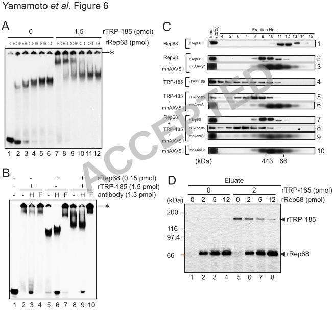
Rep68 and TRP-185 do not bind to AAVS1 DNA simultaneously 15
Since both TRP-185 and Rep68 bind to AAVS1 in a sequence-specific manner, we 16
examined whether these proteins bind to AAVS1 simultaneously by gel-shift assays (Fig. 17
6A). rRep68 alone formed protein-DNA complexes, sizes of which increased in a 18
concentration-dependent manner (lanes 2-6). rTRP-185 alone formed a slower migrating 19
protein-DNA complex (lane 7), and simultaneous addition of rRep68 and rTRP-185 20
resulted in the generation of a novel mobility species (lanes 8-12). 21
We next examined the components of the protein-DNA complexes using 22
anti-His and anti-Flag antibodies, which were specific for rTRP-185 and rRep68, 23
ACCEPTED
on April 5, 2018 by guest
http://jvi.asm.org/
Dow
nloaded from

19
respectively. Anti-Flag antibody caused a supershift of the Rep68-DNA complex and the 1
newly appeared complex (Fig. 6B, lanes 5, 7, 8, and 10). On the other hand, anti-His 2
antibody seemed to dissociate the slow-migrating TRP-185-DNA complex but had no 3
appreciable effect on the newly appeared complex (lanes 2, 3, 8, and 9). These results 4
indicated that at least Rep68 is contained in the newly appeared complex. TRP-185 may 5
also be incorporated into the complex in such a way that the His tag is not recognized by 6
the antibody. Alternatively, TRP-185 may not be incorporated into the complex but 7
instead affect the Rep68-DNA complex structure to cause its mobility shift, by transiently 8
interacting with the Rep68-DNA complex. 9
To discriminate these possibilities, we performed gel filtration analysis (Fig. 6C). 10
In the presence of mnAAVS1 DNA, the rRep68 peak was shifted from fractions 11 and 11
12 to fractions 9 and 10, which most likely reflects DNA binding of rRep68 (panels 1-3). 12
As for rTRP-185, the protein was fractionated broadly in high molecular weight fractions 13
in the absence of DNA but with a peak at fractions 8 and 9 in the presence of mnAAVS1 14
DNA, probably reflecting DNA binding of rTRP-185 (panels 4-6). Remarkably, in the 15
presence of rRep68, rTRP-185, and mnAAVS1 DNA, formation of the rTRP-185-DNA 16
complex seemed to be abrogated, while the rRep68-DNA complex remained intact 17
(panels 7-9). These results are consistent with the idea that Rep68 binds to AAVS1 with 18
higher affinity than TRP-185, and that Rep68 and TRP-185 do not bind to AAVS1 DNA 19
simultaneously. 20
The same issue was examined in a different way, i.e., by DNA pull-down assays 21
using AAVS1-immobilized latex beads. As described above, rTRP-185 bound to AAVS1 22
DNA in the absence of rRep68 (Fig. 6D, lane 5). When increasing amounts of rRep68 23
ACCEPTED
on April 5, 2018 by guest
http://jvi.asm.org/
Dow
nloaded from
20
were added together with rTRP-185 (Fig. 6D, lanes 6-8), rRep68 bound to the 1
AAVS1-immobilized beads and concomitantly, rTRP-185 was released from the beads in 2
an rRep68-dose-dependent manner. These results supported the above conclusion that 3
Rep68 and TRP-185 do not bind to AAVS1 DNA simultaneously and, together with the 4
results of gel-shift assays, suggested that TRP-185 affects the Rep68-DNA complex 5
structure, possibly by transiently interacting with the Rep68-DNA complex. 6
7
TRP-185 directly interacts with Rep68 8
The results obtained thus far indicated that TRP-185 and Rep68 physically interact with 9
each other. To test this idea, we carried out an in vitro binding assay using His-tagged 10
rTRP-185. Ni-NTA beads loaded with rTRP-185 were incubated with purified rRep68 11
and washed extensively, and bound proteins were subjected to immunoblot analysis. As 12
expected, rRep68 bound to the Ni-NTA beads in an rTRP-185-dependent manner (Fig. 7, 13
lanes 2 and 3), indicating that TRP-185 directly associates with Rep68. 14
We then examined whether AAVS1 DNA has any effect on the interaction 15
between TRP-185 and Rep68. As expected, the TRP-185-Rep68 interaction was 16
significantly reduced by the presence of mnAAVS1 wild-type DNA, but not by its mutant 17
DNA (Fig. 7, lanes 4 and 5). These results were fully consistent with the findings in Fig. 18
6 and suggested that the direct Rep68-TRP185 interaction is abrogated by the presence of 19
AAVS1 DNA. 20
ACCEPTED
on April 5, 2018 by guest
http://jvi.asm.org/
Dow
nloaded from

21
Discussion 1
The mechanism of AAV site-specific integration has not yet been fully elucidated, in 2
particular, the mechanism of integration site selection and the proteins that regulate this 3
complicated process. In the present study, we identified TRP-185 as an AAVS1-binding 4
protein in HeLa NEs. Immunodepletion experiments in vitro and RNAi-mediated 5
knockdown analysis in vivo suggested that TRP-185 alters selection of the AAV 6
integration site from the RBS to a downstream region. We also provided evidence that 7
TRP-185 binds to Rep68 directly, changes the Rep68 DNA binding property, and 8
stimulates Rep68 helicase activity on AAVS1 DNA. 9
10
Identification of TRP-185 as an AAVS1-binding protein 11
Previous studies (32, 34) have shown that TRP-185 stimulates HIV-1 gene expression 12
from the long terminal repeat by binding to TAR RNA. Although TRP-185 is expressed 13
in a variety of human tissues, its function in other processes remains largely unknown. In 14
this study, we showed that TRP-185 binds to AAVS1 in vitro and in vivo (Fig. 1). 15
Whereas TRP-185 binding to TAR RNA is critically dependent on a group of cellular 16
cofactors (32, 33), we showed using a highly purified recombinant protein that TRP-185 17
directly binds to AAVS1 DNA (Fig. 6). TRP-185 does not contain a classical nucleic 18
acid-binding domain. However, it contains multiple basic amino acid residues and a 19
leucine zipper motif in its N-terminus, which may be involved in mediating binding to 20
AAVS1 DNA. 21
22
Rep68 alone is sufficient for mediating AAV-AAVS1 junction formation at the RBS 23
ACCEPTED
on April 5, 2018 by guest
http://jvi.asm.org/
Dow
nloaded from

22
We showed that Rep68 alone is sufficient for the AAV-AAVS1 junction formation at the 1
RBS (Fig. 2). Although previous studies have shown that crude cell extract or DNA 2
replication promotes low-level junction formation by Rep68 (6, 28), in our in vitro 3
integration assay, Rep68 alone was sufficient to support junction formation at the RBS, 4
and cellular proteins in HeLa cell NE had no appreciable effect on junction formation 5
other than TRP-185 (Fig. 3). The apparent discrepancy between their and our studies may 6
arise from differences in the methods used to prepare cell extracts. It may be that cellular 7
factors promoting AAV integration were not enriched in our extract. Junction formation 8
at the RBS by Rep68 is possible in the absence of any additional protein factors because 9
Rep68 has all the activities expected to be required for the process, i.e., Rep68 is capable 10
of DNA binding sequence-specifically, mediating interactions between AAV and 11
AAVS1, creating a nick, and unwinding and ligating DNA (13, 23, 27, 31). 12
13
TRP-185 may regulate AAV integration site determination through its 14
chaperone-like activity for Rep68 15
We propose that TRP-185 acts as a molecular chaperone that facilitates the formation of 16
helicase-competent, oligomerized Rep68-AAVS1 complexes through its transient 17
interactions with Rep68 and AAVS1 DNA. Our data showed that whereas a high 18
concentration of Rep is necessary for its helicase activity in the absence of TRP-185, a 19
low concentration of Rep is sufficient for its helicase activity in the presence of TRP-185 20
(Fig. 5). We also showed that the presence of TRP-185 and a high concentration of 21
Rep68 both contribute to the formation of slow-migrating Rep68-DNA complexes (Fig. 22
6). Previous biochemical studies have shown that Rep78/68 can form oligomerized 23
ACCEPTED
on April 5, 2018 by guest
http://jvi.asm.org/
Dow
nloaded from

23
complexes on DNA and that this oligomerization enhances its nicking and helicase 1
activities (15, 20). It is therefore likely that TRP-185 enhances Rep68 helicase activity by 2
promoting its oligomerization. 3
We provided evidence that TRP-185 regulates AAV integration site 4
determination through regulation of Rep DNA binding property and sequence-specific 5
helicase activity. We showed that TRP-185, as well as a high concentration of Rep68, 6
inhibits Rep-dependent integration at the RBS in vitro (Fig. 2 and 3). This inhibition 7
probably reflects a shift in the integration site from the RBS to a downstream region, 8
caused by Rep68 oligomerization and activation of its helicase activity. Since the 9
expression of Rep78/68 is strictly regulated to a low level during latent infection of AAV 10
(36), we assume that this TRP-185-dependent mechanism operates during the viral life 11
cycle. This assumption is supported by the results of our in vivo assays (Fig. 1C and 4). 12
Here we present a model as to how TRP-185 affects AAV integration site 13
determination. Following AAV infection, Rep78/68 proteins are synthesized and 14
associate with the AAVS1 RBS, which is already occupied by TRP-185. Through 15
protein-protein and protein-DNA interactions, TRP-185 affects the DNA binding 16
property of Rep78/68, activating Rep helicase activity, and then dissociates from the 17
DNA. Subsequently, Rep introduces a nick at the trs, proceeds downstream, unwinding 18
the DNA through its helicase activity, and induces AAV integration at the downstream 19
region. In the absence of TRP-185, Rep helicase activity is not activated, and therefore, 20
after introducing a nick at the trs, Rep remains bound to the AAVS1 RBS and facilitates 21
AAV integration at the RBS. 22
23
ACCEPTED
on April 5, 2018 by guest
http://jvi.asm.org/
Dow
nloaded from

24
Possible interplay of TRP-185 and other cellular proteins during AAV integration 1
It has been shown that the high mobility group protein HMG-1 promotes the formation of 2
Rep-DNA complexes (3). Unlike TRP-185, HMG-1 enhances AAV-AAVS1 junction 3
formation at the RBS (6). HMG-1 and TRP-185 seem to exert differential effects on 4
Rep68, as HMG-1, but not TRP-185, promotes its endonuclease activity (3). 5
In a previous study, the zinc finger protein ZF5 was identified as a cellular 6
protein that binds to the RBS of the AAV ITR (2). A transient overexpression study 7
indicated that ZF5 inhibits transcription, replication, and production of AAV, possibly by 8
competing with Rep for binding to the ITR (2). It seems possible that ZF5 also regulates, 9
either positively or negatively, site-specific integration through its binding to the RBS on 10
the AAV genome and/or AAVS1, although this issue has not been examined. Thus, it will 11
be interesting to investigate the possible functional interplay of TRP-185, HMG-1, and 12
ZF5 on the AAV integration process in future studies. 13
14
The biological significance of TRP-185 in AAV life cycle 15
What is the physiological significance of the role of TRP-185 in AAV integration site 16
determination? From an evolutionary perspective, there is no evidence for AAV 17
site-specific integration in mice. Interestingly, whereas an ortholog of human AAVS1 18
was recently discovered in the mouse genome (5), the homologue of the gene encoding 19
TRP-185 does not exist in the mouse genome (32). These points suggest that TRP-185 20
plays a role in AAV site-specific integration specifically in humans. Second, abnormal 21
integration at the AAVS1 RBS may be harmful to host cells and may be prevented by 22
TRP-185. In fact, however, infection and abnormal integration of AAV had no 23
ACCEPTED
on April 5, 2018 by guest
http://jvi.asm.org/
Dow
nloaded from
25
appreciable effect on the proliferation of deltaTRP-185 cells (data not shown). This may 1
be due to the partial downregulation of TRP-185, and a more significant effect of 2
abnormal integration would be evident upon complete downregulation. Unfortunately, 3
this hypothesis could not be tested, because greater than 90% downregulation of TRP-185 4
using a different shRNA construct caused cell death (data not shown), indicating that 5
TRP-185 is essential for viability in humans. Third, AAV integration at the RBS 6
downstream region may be important for the rescue of AAV from host cells. It remains 7
our future challenge to address the significance of TRP-185 function for both the host cell 8
and AAV. 9
10
Finally, it is important to note AAV is a prospective vector for gene therapy because of 11
its property of integrating into a specific locus. Further analyses of AAV site-specific 12
integration will make it possible to control site-specific integration of a desired gene and 13
will lead to significant developments in gene therapy. 14 ACCEPTED
on April 5, 2018 by guest
http://jvi.asm.org/
Dow
nloaded from

26
Acknowledgements 1
We thank Richard B. Gaynor for providing anti-TRP-185 monoclonal antibodies, 2
Tadahito Kanda for providing pUC18 (HindIII) AAVS1 plasmid DNA, Yasunori Tsuboi 3
for technical support, and Masasuke Yoshida and Akira Nakanishi for discussions and 4
comments on the manuscript. This work was supported in part by Grant-in-Aid for 5
Scientific Research on Priority Areas and by the Grant of the 21st Century COE Program 6
from the Ministry of Education, Culture, Sports, Science, and Technology of Japan. 7
8
ACCEPTED
on April 5, 2018 by guest
http://jvi.asm.org/
Dow
nloaded from

27
References 1
1. Bouyac-Bertoia, M., J. D. Dvorin, R. A. Fouchier, Y. Jenkins, B. E. Meyer, L. 2
I. Wu, M. Emerman, and M. H. Malim. 2001. HIV-1 infection requires a 3
functional integrase NLS. Mol Cell 7:1025-35. 4
2. Cathomen, T., T. H. Stracker, L. B. Gilbert, and M. D. Weitzman. 2001. A 5
genetic screen identifies a cellular regulator of adeno-associated virus. Proc Natl 6
Acad Sci U S A 98:14991-6. 7
3. Costello, E., P. Saudan, E. Winocour, L. Pizer, and P. Beard. 1997. High 8
mobility group chromosomal protein 1 binds to the adeno-associated virus 9
replication protein (Rep) and promotes Rep-mediated site-specific cleavage of 10
DNA, ATPase activity and transcriptional repression. Embo J 16:5943-54. 11
4. Dignam, J. D., R. M. Lebovitz, and R. G. Roeder. 1983. Accurate transcription 12
initiation by RNA polymerase II in a soluble extract from isolated mammalian 13
nuclei. Nucleic Acids Res 11:1475-89. 14
5. Dutheil, N., M. Yoon-Robarts, P. Ward, E. Henckaerts, L. Skrabanek, K. I. 15
Berns, F. Campagne, and R. M. Linden. 2004. Characterization of the mouse 16
adeno-associated virus AAVS1 ortholog. J Virol 78:8917-21. 17
6. Dyall, J., P. Szabo, and K. I. Berns. 1999. Adeno-associated virus (AAV) 18
site-specific integration: formation of AAV-AAVS1 junctions in an in vitro 19
system. Proc Natl Acad Sci U S A 96:12849-54. 20
7. Endoh, M., W. Zhu, J. Hasegawa, H. Watanabe, D. K. Kim, M. Aida, N. 21
Inukai, T. Narita, T. Yamada, A. Furuya, H. Sato, Y. Yamaguchi, S. S. 22
Mandal, D. Reinberg, T. Wada, and H. Handa. 2004. Human Spt6 stimulates 23
ACCEPTED
on April 5, 2018 by guest
http://jvi.asm.org/
Dow
nloaded from
28
transcription elongation by RNA polymerase II in vitro. Mol Cell Biol 1
24:3324-36. 2
8. Hamilton, H., J. Gomos, K. I. Berns, and E. Falck-Pedersen. 2004. 3
Adeno-associated virus site-specific integration and AAVS1 disruption. J Virol 4
78:7874-82. 5
9. Han, S. I., M. A. Kawano, K. Ishizu, H. Watanabe, M. Hasegawa, S. N. 6
Kanesashi, Y. S. Kim, A. Nakanishi, K. Kataoka, and H. Handa. 2004. Rep68 7
protein of adeno-associated virus type 2 interacts with 14-3-3 proteins depending 8
on phosphorylation at serine 535. Virology 320:144-55. 9
10. Huser, D., and R. Heilbronn. 2003. Adeno-associated virus integrates 10
site-specifically into human chromosome 19 in either orientation and with equal 11
kinetics and frequency. J Gen Virol 84:133-7. 12
11. Huser, D., S. Weger, and R. Heilbronn. 2002. Kinetics and frequency of 13
adeno-associated virus site-specific integration into human chromosome 19 14
monitored by quantitative real-time PCR. J Virol 76:7554-9. 15
12. Huser, D., S. Weger, and R. Heilbronn. 2003. Packaging of human 16
chromosome 19-specific adeno-associated virus (AAV) integration sites in AAV 17
virions during AAV wild-type and recombinant AAV vector production. J Virol 18
77:4881-7. 19
13. Im, D. S., and N. Muzyczka. 1990. The AAV origin binding protein Rep68 is an 20
ATP-dependent site-specific endonuclease with DNA helicase activity. Cell 21
61:447-57. 22
ACCEPTED
on April 5, 2018 by guest
http://jvi.asm.org/
Dow
nloaded from

29
14. Inomata, Y., T. Wada, H. Handa, K. Fujimoto, and H. Kawaguchi. 1994. 1
Preparation of DNA-carrying affinity latex and purification of transcription 2
factors with the latex. J Biomater Sci Polym Ed 5:293-302. 3
15. James, J. A., C. R. Escalante, M. Yoon-Robarts, T. A. Edwards, R. M. 4
Linden, and A. K. Aggarwal. 2003. Crystal structure of the SF3 helicase from 5
adeno-associated virus type 2. Structure 11:1025-35. 6
16. Kotin, R. M., R. M. Linden, and K. I. Berns. 1992. Characterization of a 7
preferred site on human chromosome 19q for integration of adeno-associated 8
virus DNA by non-homologous recombination. Embo J 11:5071-8. 9
17. Kotin, R. M., M. Siniscalco, R. J. Samulski, X. D. Zhu, L. Hunter, C. A. 10
Laughlin, S. McLaughlin, N. Muzyczka, M. Rocchi, and K. I. Berns. 1990. 11
Site-specific integration by adeno-associated virus. Proc Natl Acad Sci U S A 12
87:2211-5. 13
18. Kyostio, S. R., and R. A. Owens. 1996. Identification of mutant 14
adeno-associated virus Rep proteins which are dominant-negative for DNA 15
helicase activity. Biochem Biophys Res Commun 220:294-9. 16
19. Laughlin, C. A., J. D. Tratschin, H. Coon, and B. J. Carter. 1983. Cloning of 17
infectious adeno-associated virus genomes in bacterial plasmids. Gene 23:65-73. 18
20. Li, Z., J. R. Brister, D. S. Im, and N. Muzyczka. 2003. Characterization of the 19
adenoassociated virus Rep protein complex formed on the viral origin of DNA 20
replication. Virology 313:364-76. 21
ACCEPTED
on April 5, 2018 by guest
http://jvi.asm.org/
Dow
nloaded from
30
21. Linden, R. M., P. Ward, C. Giraud, E. Winocour, and K. I. Berns. 1996. 1
Site-specific integration by adeno-associated virus. Proc Natl Acad Sci U S A 2
93:11288-94. 3
22. Muzyczka, N. 1992. Use of adeno-associated virus as a general transduction 4
vector for mammalian cells. Curr Top Microbiol Immunol 158:97-129. 5
23. Owens, R. A., M. D. Weitzman, S. R. Kyostio, and B. J. Carter. 1993. 6
Identification of a DNA-binding domain in the amino terminus of 7
adeno-associated virus Rep proteins. J Virol 67:997-1005. 8
24. Philpott, N. J., J. Gomos, K. I. Berns, and E. Falck-Pedersen. 2002. A p5 9
integration efficiency element mediates Rep-dependent integration into AAVS1 at 10
chromosome 19. Proc Natl Acad Sci U S A 99:12381-5. 11
25. Samulski, R. J., X. Zhu, X. Xiao, J. D. Brook, D. E. Housman, N. Epstein, 12
and L. A. Hunter. 1991. Targeted integration of adeno-associated virus (AAV) 13
into human chromosome 19. Embo J 10:3941-50. 14
26. Shimizu, N., K. Sugimoto, J. Tang, T. Nishi, I. Sato, M. Hiramoto, S. Aizawa, 15
M. Hatakeyama, R. Ohba, H. Hatori, T. Yoshikawa, F. Suzuki, A. Oomori, 16
H. Tanaka, H. Kawaguchi, H. Watanabe, and H. Handa. 2000. 17
High-performance affinity beads for identifying drug receptors. Nat Biotechnol 18
18:877-81. 19
27. Smith, R. H., and R. M. Kotin. 2000. An adeno-associated virus (AAV) initiator 20
protein, Rep78, catalyzes the cleavage and ligation of single-stranded AAV ori 21
DNA. J Virol 74:3122-9. 22
ACCEPTED
on April 5, 2018 by guest
http://jvi.asm.org/
Dow
nloaded from
31
28. Song, S., Y. Lu, Y. K. Choi, Y. Han, Q. Tang, G. Zhao, K. I. Berns, and T. R. 1
Flotte. 2004. DNA-dependent PK inhibits adeno-associated virus DNA 2
integration. Proc Natl Acad Sci U S A 101:2112-6. 3
29. Urcelay, E., P. Ward, S. M. Wiener, B. Safer, and R. M. Kotin. 1995. 4
Asymmetric replication in vitro from a human sequence element is dependent on 5
adeno-associated virus Rep protein. J Virol 69:2038-46. 6
30. Wada, T., T. Takagi, Y. Yamaguchi, H. Kawase, M. Hiramoto, A. Ferdous, 7
M. Takayama, K. A. Lee, H. C. Hurst, and H. Handa. 1996. Copurification of 8
casein kinase II with transcription factor ATF/E4TF3. Nucleic Acids Res 9
24:876-84. 10
31. Weitzman, M. D., S. R. Kyostio, R. M. Kotin, and R. A. Owens. 1994. 11
Adeno-associated virus (AAV) Rep proteins mediate complex formation between 12
AAV DNA and its integration site in human DNA. Proc Natl Acad Sci U S A 13
91:5808-12. 14
32. Wu-Baer, F., W. S. Lane, and R. B. Gaynor. 1995. The cellular factor TRP-185 15
regulates RNA polymerase II binding to HIV-1 TAR RNA. Embo J 16
14:5995-6009. 17
33. Wu-Baer, F., W. S. Lane, and R. B. Gaynor. 1996. Identification of a group of 18
cellular cofactors that stimulate the binding of RNA polymerase II and TRP-185 19
to human immunodeficiency virus 1 TAR RNA. J Biol Chem 271:4201-8. 20
34. Wu, F., J. Garcia, D. Sigman, and R. Gaynor. 1991. tat regulates binding of the 21
human immunodeficiency virus trans-activating region RNA loop-binding protein 22
TRP-185. Genes Dev 5:2128-40. 23
ACCEPTED
on April 5, 2018 by guest
http://jvi.asm.org/
Dow
nloaded from

32
35. Yamada, T., Y. Yamaguchi, N. Inukai, S. Okamoto, T. Mura, and H. Handa. 1
2006. P-TEFb-mediated phosphorylation of hSpt5 C-terminal repeats is critical 2
for processive transcription elongation. Mol Cell 21:227-37. 3
36. Young, S. M., Jr., D. M. McCarty, N. Degtyareva, and R. J. Samulski. 2000. 4
Roles of adeno-associated virus Rep protein and human chromosome 19 in 5
site-specific recombination. J Virol 74:3953-66. 6
37. Zhou, X., I. Zolotukhin, D. S. Im, and N. Muzyczka. 1999. Biochemical 7
characterization of adeno-associated virus rep68 DNA helicase and ATPase 8
activities. J Virol 73:1580-90. 9
10
11
12
ACCEPTED
on April 5, 2018 by guest
http://jvi.asm.org/
Dow
nloaded from
33
Figure Legends 1
Figure 1. Identification of TRP-185 as an AAVS1-binding factor. (A) mnAAVS1 2
wild-type and mutant DNA sequence and schematic representation of DNA-immobilized 3
latex beads. (B) Input NE (lane 1), eluate from control beads (lane 2), and eluate from 4
mnAAVS1 wild-type (lane 3) or mutant-immobilized beads (lane 4) were separated on a 5
5-20% SDS polyacrylamide gel, and proteins were visualized by silver staining. The 6
protein bands that were differentially purified by mnAAVS1-wild-type-conjugated beads 7
are marked by dots. In the lower panel, the same samples were immunoblotted using 8
anti-TRP-185 monoclonal antibody (NK5.18, a gift of Dr. Richard B. Gaynor). The 9
positions of molecular weight markers are shown on the left. (C) HeLa cells were 10
transiently transfected with pcTRP-185-Flag or pcDNA3.1 (+) (mock) and then subjected 11
to ChIP analysis using anti-Flag antibody. Input and immunoprecipitated (Ppt) DNAs 12
were subjected to real-time PCR analysis using primers that specifically amplified the 13
AAVS1 RBS region or a control region on chromosome 2q34. PCR products were 14
analyzed by 2% agarose gel electrophoresis. Data represent the means ± standard error of 15
the mean (SEM) from six independent experiments, and statistical significance was 16
determined by Student’s t-test (P<0.05). 17
18
Figure 2. Rep68 mediates AAV-AAVS1 junction formation specifically at the RBS in 19
vitro. (A) Schematic diagrams of the AAV genome and the plasmid pBSAAVS1 20
containing a 1.6-kb AAVS1 sequence (left) and primer sets (right) used for in vitro 21
junction formation assay. The positions of the primers used are indicated in the diagrams. 22
(B) rRep68 used in this study was visualized by silver staining. The positions of 23
ACCEPTED
on April 5, 2018 by guest
http://jvi.asm.org/
Dow
nloaded from

34
molecular weight markers are shown on the right. (C) In vitro junction formation assays 1
were performed with or without 1.5 pmol of rRep68. The recombination products and the 2
indicated amounts of PCR standards were subjected to PCR analysis with various primer 3
sets as shown in (A). The PCR products were analyzed by 2% agarose gel electrophoresis 4
and stained with ethidium bromide. The positions of size markers are shown on the right. 5
(D) Junction formation assays were performed with the indicated amounts of rRep68 and 6
pBSAAVS1 wild-type or mutant. The recombination products were examined by PCR 7
with primer set 1. The positions of size markers are shown on the right. Quantitation of 8
the results from three independent experiments is shown below. (E) AAV-AAVS1 9
junction sequences from eight clones. The RBS-flanking regions of the two substrates are 10
shown above. In the junction sequences, overlapping sequences between AAV and 11
AAVS1 are indicated by open boxes, and respective junction points are indicated by 12
numbers according to the published numbering system (16). The RBS sequences are 13
indicated by letters. 14
15
Figure 3. TRP-185 inhibits Rep68-mediated AAV-AAVS1 junction formation at the RBS 16
in vitro. (A) rTRP-185 used in this study was visualized by silver staining. The positions 17
of molecular weight markers are shown on the right. (B) Junction formation assays were 18
performed with the indicated amounts of rRep68, rTRP-185, heat-denatured (h.d.) 19
rTRP-185, and BSA. The positions of size markers are shown on the right. Quantitation 20
of the results from three independent experiments is shown below. (C) Immunoblot 21
analysis of HeLa NE depleted of TRP-185. HeLa NE was repeatedly (one to three times) 22
passed over protein A-Sepharose to which either anti-TRP-185 or preimmune serum (ctrl) 23
ACCEPTED
on April 5, 2018 by guest
http://jvi.asm.org/
Dow
nloaded from

35
was absorbed. The supernatants were analyzed for the presence of TRP-185 and HMG-1 1
by immunoblotting. Quantitation of each protein level is shown below. (D) AAV-AAVS1 2
junction formation reactions were performed in the presence or absence of control (ctrl) 3
or TRP-185-depleted (deltaTRP) NE (8 µg) and the indicated amounts of rRep68 and 4
rTRP-185. The positions of size markers are shown on the right. Quantitation of the 5
results from three independent experiments is shown below. 6
7
Figure 4. TRP-185 alters AAV integration sites from the RBS to a downstream region in 8
vivo. (A, B) HeLa cells were infected with a lentiviral expression vector encoding an 9
shRNA that targeted TRP-185 (deltaTRP) or a control vector (ctrl). Seven days post 10
infection, whole cell extracts were prepared and immunoblotted with anti-TRP-185 and 11
anti-β-tubulin antibodies (A). Alternatively, total RNA was prepared, and the mRNA 12
level of TRP-185 was quantified by real-time RT-PCR (B). (C) In vivo integration assays 13
were performed in ctrl and deltaTRP HeLa cells. Following AAV or mock infection, 14
AAV integration into the RBS, a region downstream of the RBS, and Alu repeat regions 15
was analyzed by PCR using specific primer sets for each region, followed by Southern 16
blot analysis. AAV-infected samples were analyzed in duplicate. The positions of DNA 17
size markers are shown on the right. (D) Sequence analysis of AAV-AAVS1 junctions at 18
the RBS (in deltaTRP cells) and downstream of the RBS (in ctrl cells). In the top panel, 19
the positions of the PCR primers and Southern probe used are indicated, and integration 20
events are denoted by arrows. In the RBS junction sequences, the RBS sequences are 21
indicated by letters, and overlapping sequences between AAV and AAVS1 are indicated 22
ACCEPTED
on April 5, 2018 by guest
http://jvi.asm.org/
Dow
nloaded from

36
by open boxes. In the downstream junction sequences, overlapping sequences between 1
AAV and AAVS1 are indicated by letters. 2
3
Figure 5. TRP-185 promotes Rep68 helicase activity in an RBS-dependent manner. (A) 4
rRep68 wild-type and K340H mutant were visualized by Coomassie staining. The 5
positions of molecular weight markers are shown on the left. (B-F) Helicase assays were 6
performed using 12.5 fmol of 32P-labeled linear AAVS1 substrate containing a wild-type 7
(B, C) or mutant RBS (D) or 10 fmol of M13 circular substrate (E, F) and the indicated 8
amounts of rRep68 and rTRP-185. The products were then electrophoresed on a 9
nondenaturing polyacrylamide gel. "Boil" samples were heated to 98˚C for 5 min 10
immediately before electrophoresis. Quantitation of the unwound products is shown 11
below each panel. 12
13
Figure 6. Rep68 and TRP-185 do not bind to AAVS1 DNA simultaneously. (A, B) 14
Gel-shift assays were performed with 10 fmol of 32P-labeled mnAAVS1 wild-type probe 15
and the indicated amounts of rRep68 and rTRP-185. In (B), anti-His (H) and anti-Flag (F) 16
antibodies were included in the binding reaction, as indicated. The reaction mixtures were 17
electrophoresed on a 4% nondenaturing polyacrylamide gel. The asterisks indicate the 18
positions of the wells. (C) rTRP-185, rRep68, and mnAAVS1 DNA were incubated either 19
individually or in combination and then subjected to gel filtration analysis. Fractionated 20
samples and input materials were analyzed by immunoblotting using anti-Flag and 21
anti-His antibodies for rRep68 and rTRP-185, respectively, or by Southern blotting using 22
32P-labeled mnAAVS1-specific probe. The positions of molecular weight markers are 23
ACCEPTED
on April 5, 2018 by guest
http://jvi.asm.org/
Dow
nloaded from

37
shown below. (D) The indicated amounts of rTRP-185 and rRep68 were examined, either 1
individually or in combination, for AAVS1 binding using latex beads onto which 2
mnAAVS1 wild-type DNA was immobilized. Eluted proteins were visualized by silver 3
staining. The positions of molecular weight markers are shown on the left. 4
5
Figure 7. TRP-185 directly interacts with Rep68. rTRP-185 (1.2 pmol) was coupled to 6
Ni-NTA beads and incubated with 3.75 pmol of rRep68 in the absence or presence of 2.5 7
pmol of mnAAVS1 wild-type (w) or mutant (mt) DNA. Eluted samples and input 8
material were subjected to immunoblot analysis using anti-Flag and anti-His antibodies 9
for rRep68 and rTRP-185, respectively. 10
11
ACCEPTED
on April 5, 2018 by guest
http://jvi.asm.org/
Dow
nloaded from

A
C
CGGC GGTTGG GGCTCGGC GCTCGCTC GCTCGCTG GGCGGGCGG 3'
GCCG CCAACC CCGAGCCG CGAGCGAGCGAGCGAC CCGCCCGCC
5' AATT
CTAG 5'
RBS
OH
NH2
mnAAVS1 DNA
latex
beads DNA
trs
3'
Input
Ppt
control
Input
Ppt
1 2 3 4 5 6 7 8
Input
Ppt
AAVS1 (RBS)
Input
Ppt
Yamamoto et al. Figure 1
B
Input Eluate
(-) mt mnAAVS1w
66
45
200
116
97.4
(kD)
31
TRP-185
TRP-185
CGGC GGTTGG GGCTCGGC CCTCCCTC CCTCCCTG GGCGGGCGG 3'
GCCG CCAACC CCGAGCCG GGAGGGAGGGAGGGAC CCGCCCGCC
5' AATT
CTAG 5'
mRBStrs
3'
wild-type (w)
mutant (mt)
1 2 3 4
mock TRP-185 mock TRP-185
0
0.5
1
1.5
2
2.5mockTRP-185
control AAVS1 (RBS)
% input
ACCEPTED
on April 5, 2018 by guest
http://jvi.asm.org/
Dow
nloaded from

C
E
Yamamoto et al. Figure 2
0 0.15 0.45 1.5 4.5 15 rRep68
(pmol)
mt w mt w mt
0.1
0.3
w mt w mt w mtw
arb
itra
ry u
nits
0
100
(kb)
AAVS1
B
rRep68
66
45
200
116
97.4
(kD)
31
21.5
D
rRep68
- +
Standard(copy number)
0.5 k
0.1k
0.5 k
0.1k
0.5 k
0.5 k
0.5 k
0.5 k
0.5 k
0.5 k
103
104
105
1
2
3
4
5
6
7
8
A AAV primer
V-4526/PITR (R)
V-149/PITR left (L)
V-4526/PITR (R)
V-149/PITR left (L)
V-4526/PITR (R)
V-149/PITR left (L)
V-4526/PITR (R)
V-149/PITR left (L)
AAVS1 primer
S1-447/H4d1(A)
S1-447/H4d1(A)
S1-1562 (B)
S1-1562 (B)
S1-2513 (C)
S1-2513 (C)
S1-3693 (D)
S1-3693 (D)
primer set
1
2
3
4
5
6
7
8
dsAAV DNAITR ITR
RBS
R
L
0 4.7 (kb)
pBS-
AAVS1
A
B
C
D
RBS
1.6
(kb) 4.5/0
(kb)
rRep68
- +
Standard(copy number)
103
104
105 (kb)
4576
410
gcgc gctcgctcgctc gctg
4576
410
gcgc gctcgctc gctg
AAV AAVS1
RBS
RBS
junction
sequences
Clone 1,2,3,4,5,6
Clone 7,8
gcgc gctcgctcgctc
right end AAV
RBS
398
gctcgctcgctc gctg
AAVS1
RBS
4573 4588nt (base) nt (base)R
A
413
ACCEPTED
on April 5, 2018 by guest
http://jvi.asm.org/
Dow
nloaded from

C
Input N
Ect
rlanti-
TR
P
x 1
TRP-185
HMG-1
1 2 3 4 5 6 7
ctrl
anti-
TR
P
x 2
ctrl
anti-
TR
P
x 3
TR
P-1
85
le
ve
l (%
)
ctrl
anti-TRP
ctrl
anti-TRP
HM
G-1
le
ve
l (%
)
numbers of immunodepletion
1 2 30
1 2 30
rTR
P-1
85
66
45
200
116
97.4
(kD)
31
21.5
B
0.3
0.1
1.5
-
-
-
1.5
+
-
-
1.5
-
+
-
rRep68 (pmol)
x 3 ctrl NE
x 3 deltaTRP NE
rTRP-185 (pmol)
-
-
-
-
1.5
-
+
0.4
1.5
-
+
0.13
0
0.20.4
0.60.8
11.2
1 2 3 4 5 61 2 3 4 5 6
100
0
(kb)
arb
itra
ry u
nits
0.1
0.3
arb
itra
ry u
nits100
(kb)
rRep68 (pmol)
rTRP-185 (pmol)
h.d. rTRP-185 (pmol)
BSA (pmol)
1.5
-
-
-
1.5
0.13
-
-
-
-
-
-
1.5
-
-
4
1.5
-
1.3
-
1 2 3 4 5 60
7
1.5
0.4
-
-
1.5
1.3
-
-
Yamamoto et al. Figure 3
A
DACCEPTED
on April 5, 2018 by guest
http://jvi.asm.org/
Dow
nloaded from

00.20.40.60.8
11.2
1 2
TRP-185
A C
2
deltaT
RP
1
b-tubulin
ctr
l 1
0T
RP
-18
5
m
RN
A
deltaT
RP
ctr
l
B
RB
SA
lu
ctr
lm
ock
AAV
in
fect
ed
ctr
l
de
lta
TR
P
1
1
0.1
0.5
0.5
0.5
1
1 2 3 4 5 6
do
wn
str
ea
m
de
lta
TR
P
(kb)
Yamamoto et al. Figure 4
4532
1557
caagga
4561
1494
ct
4584
1311
ct
4565
1153
ct
4567
1535
tct
4567
1350
tctgcgcgc
4550
1571
ggag
4558
1523
ccactccctct
4563
1552
cc
RBS junction sequences downstream junction sequences
Clones 1,2,3,4,5,6,7
Clones 8,9,10
Clone 1
Clone 2
Clone 3
Clone 4
Clones 5,6
Clone 7
Clone 8
Clone 9
Clone 10
D
0 500 1000nt (base) 1615
AAVS1
RBS
V-35694573 4679
S1-447/H4d1 S1-1615
(base)
4576
410
gcgc gctcgctcgctc gctg
4576
410
gcgc gctcgctc gctg
RBS
RBS
AAV
probe
trs RBS
1500ACCEPTED
on April 5, 2018 by guest
http://jvi.asm.org/
Dow
nloaded from

0
20
40
60
80
100
0 100 200 300 400 500
0
20
40
60
80
100
0 100 200 300 400 500
0
10
20
30
40
50
60
0 50 100 150 200 250
0
10
20
30
40
50
60
0 50 100 150 200 250
0
10
2030
40
50
60
0 50 100 150 200 250
Yamamoto et al. Figure 5
A B
Rep(-)
Rep(+)
Rep(-)
Rep(+)
un
wo
un
d p
rod
uct
(%)
un
wo
un
d p
rod
uct
(%)
TRP-185 (fmol) TRP-185 (fmol)
CrRep68
rTRP-185 (fmol)
RBS
60 mer
34 mer
5' 3'
5' 3'
Boil
0 45
rTRP-185 (fmol)
RBS mutant
66
45
200
11697.4
31wild-type
K340H
RBS wild-type
60 mer
34 mer
5'
5' 3'
Boil
0 23 70 230
0 45
RBS wild-type
Boilwild-type K340H
rRep68 (fmol) 15 45 150 450
(-)w
ild-t
ype
K340H
D
un
wo
un
d p
rod
uct
(%)
Rep68 (fmol)
(kDa)
Boil
rTRP-185 (fmol)
M13
Boil
wild-type K340HrRep68 (fmol)
1.5 4.5 15 45 150 450 450 (-)
M13
M13mp18
27 mer
E F
un
wo
un
d p
rod
uct
(%)
TRP-185 (fmol)
un
wo
un
d p
rod
uct
(%)
Rep68 (fmol)
Rep 0 fmol
Rep 4.5 fmol
0 23 70 230 0 23 70 230 0 23 70 230
0 4.5
0 23 70 230
15
15 45 150 450
Rep 15 fmol
RBS wild-type
0 23 70 230 0 23 70 230
wild-type(fmol)rRep68wild-type(fmol)
rRep68wild-type(fmol)ACCEPTED
on April 5, 2018 by guest
http://jvi.asm.org/
Dow
nloaded from
A
Yamamoto et al. Figure 6
1 2 3 4 5 6 7 8 9 10 1112
rTRP-185 (pmol)
rRep68 (pmol) 0
0 1.5
B
1.50.450.150.0450.015
rTRP-185 (1.5 pmol)antibody (1.3 pmol)- H F
rRep68 (0.15 pmol)+
+
- H F
+
-
- H F
-
+-
-
-
1 2 3 4 5 6 7 8 9 10
0 1.50.450.150.0450.015
rRep68 (pmol)
rTRP-185 (pmol)
Eluate
(kDa)
200
116
97.4
66 rRep68
rTRP-185
2
0 2 5 12
0
0 2 5 12
1 2 3 4 5 6 7 8
D
Inp
ut
(20
%)
4 5 6 7 8 9 10 11 12 13 14 15
mnAAVS1
Rep68
+
TRP-185
+
mnAAVS1
C
Rep68
Rep68
+
mnAAVS1
TRP-185
+
mnAAVS1
TRP-185
Fraction No.
rTRP-185
rRep68
mnAAVS1
rRep68
rRep68
rTRP-185
rTRP-185
mnAAVS1
mnAAVS1
mnAAVS1
1
2
3
4
5
6
7
8
9
10
(kDa) 443 66
ACCEPTED
on April 5, 2018 by guest
http://jvi.asm.org/
Dow
nloaded from

Yamamoto et al. Figure 7
Eluate
+
+
wInput (8
%)
rTRP-185
rRep68
rTRP-185
mnAAVS1 DNA
1 2 3 4 5
rRep68+
-
-
+
+
-
+
+
mt
ACCEPTED
on April 5, 2018 by guest
http://jvi.asm.org/
Dow
nloaded from