adcy10 frameshift variant leading to severe recessive ......of phenol red (c19h14o5s), 0.2485 g of...
TRANSCRIPT
© The Author(s) 2019. Published by Oxford University Press on behalf of the European Society of Human Reproduction and Embryology. All rights reserved.For permissions, please e-mail: [email protected].
Human Reproduction, Vol.34, No.6, pp. 1155–1164, 2019Advance Access Publication on May 23, 2019 doi:10.1093/humrep/dez048
ORIGINAL ARTICLE Reproductive genetics
ADCY10 frameshift variant leading tosevere recessive asthenozoospermiaand segregating with absorptivehypercalciuriaArvand Akbari1,2, Giovanni Battista Pipitone3, Zahra Anvar4,5,Mojtaba Jaafarinia1,2, Maurizio Ferrari3,6,7, Paola Carrera3,6,*, andMehdi Totonchi8,9,*1Department of Biology, Faculty of Science, Fars Science and Research Branch, Islamic Azad University, Marvdasht, Iran 2Department ofBiology, Faculty of Science, Marvdasht Branch, Islamic Azad University, Marvdasht, Iran 3Laboratory of Clinical Molecular Biology andCytogenetics, IRCCS San Raffaele Hospital, Milan, Italy 4Infertility Research Center, Shiraz University of Medical Sciences, Shiraz, Iran5Department of Obstetrics & Gynecology, School of Medicine, Shiraz University of Medical Sciences, Shiraz, Iran 6Genomic Unit for theDiagnosis of Human Disorders, Division of Genetics and Cell Biology, IRCCS San Raffaele Hospital, Milan, Italy 7Vita-Salute San RaffaeleUniversity, Milan, Italy 8Department of Genetics, Reproductive Biomedicine Research Center, Royan Institute for ReproductiveBiomedicine, ACECR, Tehran, Iran 9Department of Stem Cells and Developmental Biology, Cell Science Research Center, Royan Institutefor Stem Cell Biology and Technology, ACECR, Tehran, Iran
*Correspondence address. Genetics and Stem Cell departments, Royan Institute, Tehran, Iran. E-mail: [email protected];Genomic Unit for the Diagnosis of Human Disorders, IRCCS San Raffaele Hospital, Milan, Italy. E-mail: [email protected]
Submitted on January 19, 2019; resubmitted on February 18, 2019; editorial decision on March 13, 2019
STUDY QUESTION: Can whole exome sequencing (WES) reveal a novel pathogenic variant in asthenozoospermia in a multiplex familyincluding multiple patients?
SUMMARY ANSWER: Patients were discovered to be homozygous for a rare 2-bp deletion in the ADCY10 coding region (c.1205 1206del,rs779944215).
WHAT IS KNOWN ALREADY: ADCY10 encodes for soluble adenylyl cyclase (sAC), which is the predominant adenylate cyclase in sperm.It is already established that proper sAC activity and a constant supply of cAMP are crucial to sperm motility regulation, and knockoutmouse models have been reported as severely asthenozoospermic. ADCY10 is a susceptibility gene for dominant absorptive hypercalciuria(OMIM#143870); however, no ADCY10 variations have been confirmed to cause human asthenozoospermia to date.
STUDY DESIGN, SIZE, DURATION: This was a retrospective genetics study of a highly consanguineous pedigree of asthenozoospermia.The subject family was recruited in Iran in 2016.
PARTICIPANTS/MATERIALS, SETTING, METHODS: The two patients were diagnosed as asthenozoospermic through careful clinicalinvestigations. Both patients, respective parents, and an unaffected brother were subjected to WES. The discovered variant was validated bySanger sequencing and segregated with the phenotype. To confirm the pathogenicity of the variant, sperm samples from both patients, 10normozoospermic men and 10 asthenozoospermic patients not representing the variation, were treated with a cAMP analogue dissolved inhuman tubal fluid medium, followed by computer-assisted sperm analysis and statistical analyses.
MAIN RESULTS AND THE ROLE OF CHANCE: The discovered homozygous variant occurs at 10 amino acids upstream of theADCY10 nucleotide binding site leading to a premature termination (p.His402Argfs∗41). Treatment of the patients’ sperm samples with acell-permeable cAMP analogue resulted in a significant increase in sperm motility, indicating the pathogenic role of the variant. Moreover,absorptive hypercalciuria, segregating within the family, was also associated with the same variant following a dominant inheritance.
LIMITATIONS, REASONS FOR CAUTION: Though nonsense-mediated decay is highly likely to occur in the mutated transcripts, wewere not able to confirm this due to low RNA levels in mature sperm.
WIDER IMPLICATIONS OF THE FINDINGS: Our finding enlarges the phenotypic spectrum associated with the ADCY10 gene, previouslydescribed as a susceptibility gene for dominant absorptive hypercalciuria.
Dow
nloaded from https://academ
ic.oup.com/hum
rep/article-abstract/34/6/1155/5492390 by Promedica H
ealth System user on 20 July 2019

1156 Akbari et al.
STUDY FUNDING/COMPETING INTEREST(S): This study was supported by grants from the Royan Institute, Tehran, Iran, and SanRaffaele Hospital, Milan, Italy. The authors have no conflict of interest.
TRIAL REGISTRATION NUMBER: N/A
Key words: male infertility / asthenozoospermia / absorptive hypercalciuria / familial exome sequencing / ADCY10
IntroductionThe World Health Organization (WHO) has described infertility as areproductive system disease defined by the failure to achieve a clinicalpregnancy after 12 months or more of regular unprotected sexualintercourse (Zegers-Hochschild et al., 2009). It affects 15% of couplesworldwide and it is estimated that in 50% of cases, the cause is the malefactor (Nieschlag et al., 2010). However, a more recent study revealedthat male factor contributes to 60–70% of cases in the Middle East,which is higher than average (Agarwal et al., 2015). It has been reportedthat 30–50% of male infertility cases have an underlying genetic cause(Zorrilla and Yatsenko, 2013). Despite considerable efforts towarddiscovery of these genetic factors, 30–40% of such cases still remainidiopathic (Nieschlag et al., 2010).
Asthenozoospermia, which is the most prevalent infertility pheno-type (Curi et al., 2003), is a condition in which less than 32% of totalspermatozoa are progressively motile (WHO, 2010). It might resultfrom structural or metabolic defects. Normal flagellar ultrastructure isof utmost importance and its malfunction and/or structural defects invarious protein components of the axoneme result in asthenozoosper-mia (Coutton et al., 2015). Metabolic defects can lead to deficientenergy metabolism, oxidative stress, or inhibition of different pathwayssuch as malfunction of ion channels involved in the regulation of spermmotility. These pathways are interrelated and abnormalities usuallyhave adverse effects on various sperm functions.
Apart from being an energy source, ATP is also involved in theregulation of sperm motility where it is used as a precursor by adeny-late cyclase enzymes to produce cAMP (Visconti, 2012). It has beenshown that oxidative phosphorylation process inhibition by a chemicalcompound or by knocking out Gapdhs, which encodes for an integralsperm-specific component of the glycolytic pathway, both lead tosperm immobility (Miki et al., 2004; Goodson et al., 2012).
cAMP/PKA and calcium pathways are the most important signalingpathways of sperm motility regulation (Turner, 2006). Soluble adenylylcyclase (sAC) is the predominant adenylate cyclase in the principalpiece of the sperm tail (Buffone et al., 2014). This enzyme is activated
by Ca2+ and bicarbonate (HCO3−) to turn ATP into cAMP, which
is consumed by PKA to phosphorylate serine/threonine residues ofa number of downstream proteins. These proteins target a spe-cific subset of flagellar proteins for tyrosine phosphorylation includingaxonemal dynein, which is an essential step in the initiation of sperm
motility (Turner, 2006). Interestingly, sAC knockout mice were infertiledue to complete loss of progressive motility in sperms (Espositoet al., 2004). Furthermore, targeted deletion of the catalytic subunitof the sperm-specific isoform of PKA was also reported to causemale infertility in mice as a consequence of decreased sperm motility(Skålhegg et al., 2002).
ADCY10 is the only human gene that encodes for sAC with a highlybiased expression in testis (www.ncbi.nlm.nih.gov/gene/55811). Thisenzyme synthesizes cAMP, which is the required substrate for the
.
.
.
.
.
.
.
.
.
.
.
.
.
.
.
.
.
.
.
.
.
.
.
.
.
.
.
.
.
.
.
.
.
.
.
.
.
.
.
.
.
.
.
.
.
.
.
.
.
.
.
.
.
.
.
.
.
.
.
.
.
.
.
.
.
.
.
.
.
.
.
.
.
.
.
.
.
.
.
.
.
.
.
.
.
.
.
.
.
.
.
.
.
.
.
.
.
.
.
.
.
.
.
.
.
.
.
.
activation of PKA. Through its phosphorylation of downstreamproteins, it is assumed that PKA activity is critically essential formotility, capacitation, and fertilization competence of spermatozoon(Hess et al., 2005).
In a more severe form, asthenozoospermia may exist as mul-tiple morphological abnormalities of the sperm flagella (MMAF)representing absolute loss of sperm motility occasionally. On thewhole, asthenozoospermia may manifest itself as an isolated disorder,accompanied by other sperm abnormalities or as part of a syndromein primary ciliary dyskinesia (PCD) (Zuccarello et al., 2008). To thispoint, the following genes have been reported to cause isolatedasthenozoospermia: KLHL10 (Yatsenko et al., 2006), PLA2G6 (Visseret al., 2011), SEPT12 (Kuo et al., 2012), SLC26A8 (Dirami et al., 2013),GALNTL5 (Takasaki et al., 2014), NSUN7 (Khosronezhad et al.,2015), CATSPER1–4 (Sun et al., 2017), and SPAG17 (Xu et al., 2018).Ray et al. (2017) have extensively reviewed the genes involved inMMAF and PCD.
Herein, we provide the first report of a homozygous frameshiftvariation (c.1205 1206del; dbSNP rs779944215) in the exonic regionof ADCY10 associated with recessive asthenozoospermia, in aninbred family. Noticeably, all relatives who were heterozygous orhomozygous for the variant had a history of developing calcium kidneystones, confirming the association with absorptive hypercalciuria(OMIM#143870). This is an original finding showing the samepathogenic variant associated with distinct phenotypes, dependingon recessive or dominant inheritance.
Materials and Methods
Human subject recruitmentA 34-year-old male from the Fars province, Iran, with a chief com-plaint of infertility was referred to the infertility clinic at Royan Insti-tute. His parents were first cousins and genetic counseling revealedone infertile first cousin once removed who had first-cousin par-ents as well (Fig. 1). Considering the level of consanguinity in thefamily, they were chosen as candidates for whole exome sequenc-ing (WES) in case the cause of infertility could not be determinedby routine clinical procedures. A written consent form was signedby each participant after receiving information about the study pro-cedure and aims. This study was approved by the Royan Instituteethics committee and conducted in accordance with the declarationof Helsinki.
Conventional karyotypingKaryotype analysis was performed on trypsin-banded metaphase chro-mosomes. The analysis was performed by a standard protocol togenerate a resolution of 550 bands per haploid set, from a singlecell of the corresponding parent. Normally, 30 random metaphasespreads per sample were targeted. Results were reported based on
Dow
nloaded from https://academ
ic.oup.com/hum
rep/article-abstract/34/6/1155/5492390 by Promedica H
ealth System user on 20 July 2019
sAC deficiency leads to severe asthenozoospermia 1157
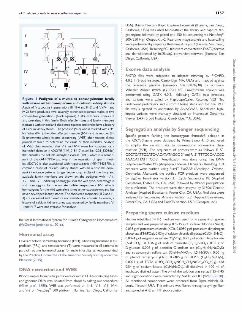
Figure 1 Pedigree of a multiplex consanguineous familywith severe asthenozoospermia and calcium kidney stones.A pair of first cousins in generations III (III-4 and III-5) and IV (IV-1 andIV-2) have produced two severely asthenozoospermic males in twoconsecutive generations (black squares). Calcium kidney stones arealso prevalent in this family. Both infertile males and family membersindicated with striped and checkered squares and circles have a historyof calcium kidney stones. The proband (V-2) who is marked with a ‘P’,his father (IV-1), the other affected member (IV-4) and his mother (III-5) underwent whole exome sequencing (WES) after routine clinicalprocedure failed to determine the cause of their infertility. Analysisof WES data revealed that V-2 and IV-4 were homozygous for aframeshift deletion in ADCY10 (NM 018417:exon11:c.1205 1206del)that encodes the soluble adenylate cyclase (sAC) which is a compo-nent of the cAMP/PKA pathway in the regulation of sperm motil-ity. ADCY10 is also associated with hypercalciuria (MIM#143870), acommon cause of calcium kidney stones with an autosomal domi-nant inheritance pattern. Sanger Sequencing results of the living andavailable family members are shown on the pedigree with +/+,+/− and −/− indicating homozygous for the wild-type, heterozygousand homozygous for the mutated allele, respectively. IV-3 who ishomozygous for the wild type allele is not asthenozoospermic and hasnever developed kidney stones. The checkered members (III-2 and III-4) are deceased and therefore not available for analysis. However, ahistory of calcium kidney stones was reported by family members. V-1 and IV-7 were not available for analysis.
the latest International System for Human Cytogenetic Nomenclature(McGowan-Jordan et al., 2016).
Hormonal assayLevels of follicle-stimulating hormone (FSH), luteinizing hormone (LH),prolactin (PRL), and testosterone (T) were measured in all patients aspart of routine hormonal assay for male infertility as recommendedby the Practice Committee of the American Society for ReproductiveMedicine (2015).
DNA extraction and WESBlood samples from participants were drawn in EDTA-containing tubesand genomic DNA was isolated from blood by salting-out procedure(Miller et al., 1988). WES was performed on III-5, IV-1, IV-3, IV-4,and V-2 on NextSeq® 500 platform (Illumina, San Diego, California,
.
.
.
.
.
.
.
.
.
.
.
.
.
.
.
.
.
.
.
.
.
.
.
.
.
.
.
.
.
.
.
.
.
.
.
.
.
.
.
.
.
.
.
.
.
.
.
.
.
.
.
.
.
.
.
.
.
.
.
.
.
.
.
.
.
.
.
.
.
.
.
.
.
.
.
.
.
.
.
.
.
.
.
.
.
.
.
.
.
.
.
.
.
.
.
.
.
.
.
.
.
.
.
.
.
.
.
.
.
.
.
.
.
.
.
.
.
.
.
.
.
.
.
.
USA). Briefly, Nextera Rapid Capture Exome kit (Illumina, San Diego,California, USA) was used to construct the library and capture tar-get regions followed by paired-end 150-bp sequencing via NextSeq®
500/550 High Output Kit v2. Real-time image analysis and base callingwere performed by sequence Real-time Analysis 2 (Illumina, San Diego,California, USA). Resulting BCL files were converted to FASTQ formatand demultiplexed by bcl2fastq2 conversion software (Illumina, SanDiego, California, USA).
Exome data analysisFASTQ files were subjected to adapter trimming by PICARD4.0.2.1 (Broad Institute, Cambridge, MA, USA) and mapped againstthe reference genome (assembly GRCh38/hg38) by Burrows-Wheeler Aligner (BWA 0.7.17-r1188). Downstream analysis wasperformed using GATK 4.0.2.1 following GATK best practicesand variants were called by HaplotypeCaller. Resulting VCF filesunderwent preliminary and custom filtering steps and the final VCFfile was subjected to annotation by ANNOVAR. Shortlisted high-impact variants were manually visualized by Interactive GenomicsViewer 2.4.4 (Broad Institute, Cambridge, MA, USA).
Segregation analysis by Sanger sequencingSpecific primers flanking the homozygous frameshift deletion inthe ADCY10 gene were designed by Primer3web 4.1.0 and usedto amplify the variation site by conventional polymerase chainreaction (PCR). The sequences of primers were as follows: F: 5′-CCCTCATTCCATCAACATATAGCC-3′ and R: 5′-TTTCCCAACCCAGACATTATTTCC-3′. Amplification was done using Taq DNAPolymerase Master Mix (Ampliqon, Odense, Denmark). Resulting PCRproducts were purified using PureIT ExoZAP (Ampliqon, Odense,Denmark). Afterward, the purified PCR products were sequencedby BigDye Terminator version 3.1 Cycle Sequencing Kit (AppliedBiosystems, Foster City, CA, USA) followed by ethanol precipitationfor purification. The products were then assayed by 3130xl GeneticAnalyzer (Applied Biosystems, Foster City, CA, USA). Final data wereanalyzed by Sequencing Analysis version 5.2 (Applied Biosystems,Foster City, CA, USA) and FinchTV version 1.5.0 (Geospiza Inc.)
Preparing sperm culture mediumHuman tubal fluid (HTF) medium was used for treatment of spermsamples and was prepared using 0.5935 g of sodium chloride (NaCl),0.035 g of potassium chloride (KCl), 0.0050 g of potassium dihydrogenphosphate (KH2PO4), 0.03 g of calcium chloride dihydrate (CaCl2.2H2O),0.0024 g of magnesium sulfate (MgSO4), 0.21 g of sodium bicarbonate(NaHCO3), 0.0036 g of sodium pyruvate (C3H3NaO3), 0.05 g ofD-glucose, 0.006 g of penicillin G sodium salt (C16H17N2NaO4S)and streptomycin sulfate salt (C21H39N7O12. 1.5 H2SO4), 0.001 gof phenol red (C19H14O5S), 0.2485 g of HEPES (C8H18N2O4S),0.0021 g of EDTA ((HO2CCH2)2NCH2CH2N(CH2CO2H)2), and0.54 g of sodium lactate (C3H5NaO3), all dissolved in 100 ml ofincubated distilled water. The pH of the solution was set at 7.35–7.45and slight deviations were corrected by NaOH or HCl (WHO, 2010).All mentioned components were procured from Sigma-Aldrich, St.Louis, Missouri, USA. This mixture was filtered through a syringe filterand stored at 4◦C as HTF stock solution.
Dow
nloaded from https://academ
ic.oup.com/hum
rep/article-abstract/34/6/1155/5492390 by Promedica H
ealth System user on 20 July 2019

1158 Akbari et al.
Density gradient centrifugation for spermisolationSperm samples were subjected to a simple wash with HTF at 2500 rpmfor 8 min (Beydola et al., 2014). Next, density gradient centrifugation(DGC) was used to effectively remove seminal plasma and achieveliquefaction by Allgrad® (Lifeglobal, CT, USA) according to manufac-turer’s instructions.
Sperm sample treatment with cAMPanalogueDibutyryl cAMP sodium salt (bucladesine sodium salt) was used asthe cAMP analogue as it can easily permeate the cell through sim-ple diffusion and activate cAMP-related pathways (Sigma-Aldrich, St.Louis, Missouri, USA). Also, 0.00983 g of dibutyryl cAMP sodium salt(MW = 491.37 g) was dissolved in 1 ml of HTF to prepare a stocksolution of 20 mM.
HTF was mixed with human serum albumin (Vitrolife, Sweden)and incubated in a CO2 incubator for 3 h at 37◦C prior to use. Asimple wash was performed on sperm samples (Edwards, 1969, EarlyStages of Fertilization in vitro of Human Oocytes Matured in vitro).Briefly, 2 ml of sperm sample was mixed with 2 ml of incubated HTF,pipetted gently and centrifuged at 2000 rpm for 15 min. The pellet wasresuspended in 2 ml of HTF and distributed among treatment tubes.Then, 5 and 20 μl of dibutyryl cAMP sodium salt stock solution, equalto 0.05 mg and 0.2 mg, respectively, of the Bucladesine sodium saltpowder, was added to 500 μl of treatment media.
Sperm motility assessmentThe effect of treatment on sperm motility was assessed by SpermClass Analyzer® computer-assisted sperm analysis (CASA) system(MICROPTIC, Barcelona, Spain) with lower reference limits set to theWHO guidelines.
Accession numberThe variant has been submitted to the ClinVar database underSCV000746242.
.
.
.
.
.
.
.
.
.
.
.
.
.
.
.
.
.
.
.
.
.
.
.
.
.
.
.
.
.
.
.
.
.
.
.
.
.
.
.
.
.
.
.
.
.
.
.
.
.
.
.
.
.
.
.
.
.
.
.
.
.
.
.
.
.
.
.
.
.
.
.
.
.
.
.
.
.
.
.
.
Results
Clinical description of patients and familymembersThe proband V-2 who was a product of a consanguineous marriagebetween first cousins (IV-1 and IV-2) was referred with a chief com-plaint of primary infertility. He reported having regular intercourse (2–3times a week) during 12 years of marriage with no success in pregnancywhile exhibiting normal erection and ejaculation. Physical examina-tion by a urologist indicated normal development of male externalgenitalia, normal bilateral testis and testis size, and normally palpablevas deferens with no varicocele. He had no history of exposure tochemotoxic materials or engagement in unhealthy activities affectingfertility, except for smoking. There was no history of systemic orrespiratory diseases. The noteworthy point in his medical record washis and his parents’ history of kidney stones for which he regularly tookmedication. Further genetic counseling revealed one first cousin onceremoved (IV-4) with infertility who was the son of consanguineousparents as well. This cousin, his late father (III-4), his mother (III-5), andtwo of his brothers (IV-5 and IV-6) also had a history of developingkidney stones. After being clinically examined, the cousin’s medicalhistory was obtained (Table I). Both patients (V-2; IV-4) underwenthormonal assay for LH, FSH, PRL, and T and all hormone levels werewithin the normal range (Table II). The patients were also tested bykaryotyping and the results complied with apparent normal males(46,XY). Routine semen analysis showed the absence of spermatozoawith rapid progressive motility (Class A) in both patients and verylow percentages of spermatozoa with medium progressive motility(Class B) (Table II).
Detecting frameshift variation in ADCY10WES was performed on III-5, IV-1, IV-3, IV-4, and V-2 and an averagecoverage of 150X was reached. Of the total reads, 91% passed thequality filter (Q ≥ 30). Given the similarity in phenotypes of the twopatients and high level of consanguinity in the pedigree, we hypothe-sized that the pathogenic variant was possibly homozygous and shared
Table I Medical history of the infertile patients.
IV-4 V-2........................................................................................................................................................................................38 34 Age
10 y 12 y History of I infertility
Once ectopic NO Abortion/pregnancy
Smoker Smoker Smoking/drinking
A first cousin once removed A first cousin once removed Family history
Bilateral normal Bilateral normal Testes
NO NO Varicocele Physical examination
Palpable Palpable Vas deferens
Asthenteratozoospermic Asthenozoospermic Semen analysis
NO 7 y ago Varicocelectomy surgery
NO NO History of systemic diseases
NO NO Exposure to gonadotoxins and/or chemical toxins
Information obtained from genetic counseling, semen analysis, physical examination and historical investigation by urologist.
Dow
nloaded from https://academ
ic.oup.com/hum
rep/article-abstract/34/6/1155/5492390 by Promedica H
ealth System user on 20 July 2019

sAC deficiency leads to severe asthenozoospermia 1159
Table II Semen analysis and hormonal assay results of the infertile.
IV-4 V-2 Normal range (WHO, 2010).......................................................................................................................................................................................Volume (ml) 4.4 4.2 2–7
PH 7.8 7.8 >7.2
Sperm count (∗106/ml) 100 37 >15
Class A 0% 0% >32%
Class B 2% 3%
Class C 39% 54%
Class D 59% 43% <60%
Normal morphology 2% 5% >4%
Vitality test 95% 90% >58%
Hormonal assay
Test Result Normal range
PRL (mlU/lit) 95.4 130.8 72–459
LH (mlU/ml) 8.32 5.63 1.0–10
FSH(mlU/ml)
2.4 10.6 1.5–12.4
T (ng/ml) 6.2 7.0 2.0–8.0
Class A: percentage of rapidly progressive sperm; Class B: percentage of medium progressive sperm; Class C: percentage of non-progressive sperm; Class D: percentage of immotilesperm. PRL, prolactin; LH, luteinizing hormone; FSH, follicle-stimulating hormone; T, testosterone
among the affected. Thus, an autosomal recessive inheritance modewas assumed. Subsequently, we started the analysis by investigatingvariants that were homozygous in the proband V-2 and heterozygous inhis father (IV-1); 16 900 variants fitted into this category. Variants withminor allele frequency >0.01 in ExAC and gnomAD were excluded.The remaining variants were categorized based on ACMG guidelinefor the interpretation of sequence variants (Richards et al., 2015). Weexcluded synonymous Single Nucleotide Variation (SNVs), intronic andintergenic variants that resulted in a list of 114 remaining variants.The effect of non-synonymous SNVs of this list was predicted usingvarious prediction tools such as SIFT, PolyPhen2, FATHMM, CADD,DANN, MetaLR, MetaSVM, Mutation taster, Mutation assessor, andPROVEAN. For assessing conservation of the variation sites, GERP++, PhyLoP, and PhastCons were used. Variants with predicted highimpacts that were mapped to genes involved in spermatogenesis,sperm motility, maturation, and function or had restricted expressionto testis were prioritized for segregation analysis with the infertilityphenotype in the pedigree. The best candidate was the homozygousframeshift variant c.1205 1206del, p.(His402Argfs∗41) in ADCY10 thatwas reported in ExAC only once in a heterozygous state (dbSNP:rs779944215).
Variant confirmation and segregationanalysisADCY10 has been linked with hypercalciuria (MIM#143870), which is aknown cause of calcium kidney stones (Reed et al., 2002). Interestingly,the proband (V-2) had kidney stones and was under medical care sincestone removal (Fig. S1). Both of the proband’s parents (IV-1 and IV-2)and three first cousins once removed including the infertile one (IV-4,IV-5, and IV-6) had kidney stones. We observed that the presence of
.
.
.
.
.
.
.
.
.
.
.
.
.
.
.
.
.
.
.
.
.
.
.
.
.
.
.
.
.
.
.
.
.
.
.
.
.
.
.
.
.
.
.
.
.
.
.
.
.
.
.
.
.
.
.
.
.
.
.
.
.
.
.
.
.
.
.
.
.
.
kidney stones in this pedigree followed a dominant inheritance pattern(Fig. 1).
The candidate variation for asthenozoospermia, a two base pairdeletion causing a frameshift producing a putative stop codon 41codons after His402, is located 10 amino acids upstream of thenucleotide binding site of the enzyme (Fig. 2a). This juncture is highlyconserved across mammals and the discovered truncating variantcan theoretically render the expression of the allele completely null(Fig. 2b). Both the affected subjects were confirmed to be homozygousfor ADCY10:NM 018417:c.1205 1206del:p.(His402Argfs∗41) bySanger sequencing (Fig. 2c). All of the family members genotypeddisplayed a perfect correspondence with phenotype; only thehomozygous mutated subjects segregated with the asthenozoospermiaphenotype while the heterozygous and the wild-type subjects were notassociated with the affected phenotype.
Sperm motility rescue with cAMP analogueSperm samples were collected from 10 normozoospermic men, 10asthenozoospermic men, and the two patients. Sanger sequencingconfirmed that all sample donors except for the two patients werehomozygous for the wild-type allele at the site of ADCY10 frameshiftvariation. After DGC and a simple wash, 0.5 ml of sperm sample wasmixed with 0.5 ml of HTF in two three-microtube sets. Treatment wasperformed with 0 (non-treated), 0.05, and 0.2 mg of dibutyryl cAMPsodium salt, meaning that respectively 0, 5, and 20 μl of the analoguewas added from the 20-mM stock solution to the final medium volumeof 1 ml. Samples were incubated at 37◦C in a CO2 incubator at intervalsof 30 and 60 min.
The percentage of spermatozoa with rapid progressive motility(Class A), medium progressive motility (Class B) (World HealthOrganization, 1999), progressive motility (A + B) (WHO, 2010), and
Dow
nloaded from https://academ
ic.oup.com/hum
rep/article-abstract/34/6/1155/5492390 by Promedica H
ealth System user on 20 July 2019

1160 Akbari et al.
Tab
leII
IR
esul
tsof
sper
msa
mpl
estr
eatm
ent
wit
hcA
MP
anal
ogue
.
....
....
....
....
....
....
....
....
....
....
....
....
....
....
....
....
....
....
....
....
....
....
....
....
....
....
....
....
....
....
....
....
....
....
....
....
....
....
....
....
....
....
....
....
....
....
....
....
....
....
....
....
....
....
....
....
....
....
....
....
..G
roup
sIn
terv
al(m
in)
cAM
Pan
alog
ue(m
g)C
lass
A(%
)C
lass
B(%
)P
R(%
)V
SL
(μm
/s)
pva
lue
(vs.
non-
trea
ted)
Nor
moz
oosp
erm
ic
(Ave
rage
of10
)
300
21.0
622
.36
43.4
218
.52
_
0.05
22.5
322
.54
45.0
720
.77
0.02
496∗
0.2
24.2
224
.11
48.3
321
.07
0.00
961∗
∗60
018
.13
23.1
541
.28
15.6
9_
0.05
17.7
220
.58
38.3
017
.01
0.83
49
0.2
16.4
622
.35
38.8
115
.42
0.96
27A
sthe
nozo
ospe
rmic
(Ave
rage
of10
)
300
5.05
6.17
11.2
213
.89
_
0.05
5.24
6.66
11.9
014
.49
0.00
9852
∗∗0.
25.
766.
5912
.35
14.2
90.
0149
4∗60
010
.26
7.85
18.1
121
.76
_
0.05
8.26
9.70
17.9
621
.04
0.61
44
0.2
7.08
8.10
15.1
817
.92
0.96
18IV
-430
00.
313.
383.
697.
68_
0.05
8.28
19.6
727
.95
19.6
20.
0113
5∗0.
21.
687.
629.
309.
200.
0276
5∗60
00.
111.
982.
097.
16_
0.05
7.14
9.8
16.9
424
.83
0.01
012∗
0.2
3.13
9.39
12.5
316
.30
0.00
9449
∗∗V
-230
00.
873.
214.
088.
67_
0.05
6.97
13.1
520
.12
16.7
30.
0092
8∗∗
0.2
8.48
14.6
523
.14
16.8
70.
0108
9∗60
00.
443.
113.
568.
55_
0.05
3.56
9.25
12.8
116
.08
0.00
7611
∗∗0.
21.
2810
.46
11.7
38.
110.
0843
7
Four
grou
psin
clud
ing
10as
then
ozoo
sper
mic
and
10no
rmoz
oosp
erm
icsa
mpl
es(w
how
ere
confi
rmed
asho
moz
ygou
sfo
rth
ew
ildty
peal
lele
)an
dth
etw
opa
tient
sun
derw
ent
trea
tmen
tw
ithcA
MP
anal
ogue
.Per
cent
ages
ofsp
erm
with
prog
ress
ive
mot
ility
,spe
rms
with
clas
sA
mot
ility
,spe
rmw
ithcl
ass
Bm
otili
tyan
dst
raig
htlin
eve
loci
ty(V
SL)
was
com
pare
dbe
twee
nth
eno
n-tr
eate
dsa
mpl
esan
dtr
eate
dsa
mpl
esw
ith0.
05m
gan
d0.
2m
gof
cAM
Pan
alog
uein
each
grou
pvi
ain
depe
nden
tt-
test
with
the
alte
rnat
ive
hypo
thes
isse
tto
‘less
’.P
valu
esle
ssth
an0.
05w
ere
cons
ider
edst
atis
tical
lysi
gnifi
cant
(P<
0.05
∗,P<
0.01
∗∗).
Nor
moz
oosp
erm
ican
das
then
ozoo
sper
mic
patie
nts
show
eda
sign
ifica
ntin
crea
sein
the
men
tione
dfo
urpa
ram
eter
saf
ter
30m
inut
esof
trea
tmen
tbu
tno
sign
ifica
ntin
crea
sew
asde
tect
edaf
ter
60m
inut
es.B
oth
patie
nts
show
edsi
gnifi
cant
incr
ease
sin
sper
mpa
ram
eter
saf
ter
30an
d60
min
utes
oftr
eatm
ent.
The
only
exce
ptio
nw
asV
2,at
0.2
mg
ofan
alog
ueaf
ter
60m
inut
es,w
hich
retu
rned
aP
valu
eof
0.08
437.
Dow
nloaded from https://academ
ic.oup.com/hum
rep/article-abstract/34/6/1155/5492390 by Promedica H
ealth System user on 20 July 2019

sAC deficiency leads to severe asthenozoospermia 1161
Figure 2 Site of the variation within the C2 domain of the sAC. a) ADCY10 is located on reverse strand so 5′ to 3′ is shown from rightto left. The C1 and C2 catalytic domains are encoded by exons 2 to 7 and 9 to 12 respectively. A GT deletion in exon 11 causes a frameshift atHis402 which is located ten residues upstream of Asn412 of the NXXXR catalytic motif. The catalytic site of the enzyme is located at the interfaceof the C1/C2 dimer. This variation potentially causes a disruption in exon 12 and prevents C2 from forming a dimer with C1 rendering the enzymedysfunctional. b) The amino acid sequence of the interval between the His402 and the NXXR catalytic motif is highly conserved across mammals. Thelast sequence belongs to Atlantic Salmon. Indeed, the whole sequences of C1 and C2 domains are highly conserved across the species. The Leu413and Ile413 residues are both conserved and effective within the catalytic motif although they might cause some differences in the efficiency of thecatalytic activity. c) Sanger Sequencing confirmed the variation in patients and close family members (Homo: Homozygous; Hetero: Heterozygous;WT: Wild-Type; MU: Mutant).
Dow
nloaded from https://academ
ic.oup.com/hum
rep/article-abstract/34/6/1155/5492390 by Promedica H
ealth System user on 20 July 2019

1162 Akbari et al.
straight line velocity (VSL) parameters was compared statisticallybefore and after treatment by paired t-test with the alternativehypothesis set to ‘less’, i.e. a decrease is expected in the second datagroup in comparison with the non-treated group. A P value <0.05 wasconsidered statistically significant.
In the normozoospermic group, treatment with 0.05 and 0.2 mg ofcAMP analogue for 30 min resulted in a significant increase in mea-sured parameters with P values of 0.02496 and 0.00961, respectively.However, in the 60-min interval, no significant increase was detected.The asthenozoospermic group followed the same pattern, meaningthat treatment with both amounts of the cAMP analogue for 30 minshowed a significant increase in sperm parameters with P values of0.009852 and 0.01494 for 0.05 and 0.2 mg, respectively, while nosignificant increase was observed after treatment for 60 min. However,for the patients, treatment with both volumes of analogue at bothintervals yielded significant increases. The only exception was treat-ment of the V-2 sample with 0.2 mg of analogue at 60-min interval(Table III). Graphical comparison of the four measured parametersin all groups (Supp. Figs 2–5) along with video files of non-treated(Supp. Movies 1, 2, 5, and 6) and treated (Supp. Movies 3, 4, 7,and 8) V-2 and IV-4 sperm samples are available in supplementalfiles.
DiscussionsAC is the predominant adenylate cyclase in sperm (Hess et al., 2005).It is encoded solely by the ADCY10 gene. The longest transcript, cor-responding to NM 018417, has 33 exons and translates into a 1610-amino-acid long polypeptide with a mass of ∼187 kDa. The C1 domainspans six exons in exons 2 to 7 and the C2 domain is encoded by fourexons in exons 9 to 12 (Chen et al., 2014) (Fig. 2a). The discoveredframeshift candidate pathogenic variation for asthenozoospermia isdescribed as ADCY10:NM 018417:exon11:c.1205 1206del:p.H402fsin which a GT deletion causes a frameshift at the His402 residue,altering the translation downstream of the deletion and causing apremature termination after 41 residues in the new reading frame.Since this region is highly conserved in mammals, we hypothesized thatthis variation severely affects the function of the protein as it disruptsthe C2 domain (Fig. 2a), particularly because the variation occurs 10residues upstream of the NXXXR catalytic motif residing two highlyconserved residues in Asn412 and Arg416 (Fig. 2b). Alternatively, itmay be that the mutant transcript is subjected to nonsense-mediatedmRNA decay, leading to a low/very low concentration of transcript.Mammalian ACs possess two structurally similar catalytic domains(C1 and C2) located on one polypeptide chain. The active enzymeincludes a dimer formed by either homo- or heterodimerization ofthese catalytic sites and the catalytic unit of the enzyme is located atthe interface of the dimer (Kamenetsky et al., 2006). As C1 and C2contribute different residues to the catalytic centers, the two resultingactive sites are not identical and only one of them possesses all thefeatures required for catalysis. The other one, which is degenerate,
is the designated binding site for enzyme specific activators, that isforskolin for tmACs and bicarbonate for sAC. Hence, although C1
and C2 have the same overall structures, they serve different purposes(Tesmer et al., 1997; Zhang et al., 1997; Kamenetsky et al., 2006). Thecrystal structure of the human sAC in complex with calcium ions andan ATP analogue confirms a single ATP binding site. ATP is inserted
.
.
.
.
.
.
.
.
.
.
.
.
.
.
.
.
.
.
.
.
.
.
.
.
.
.
.
.
.
.
.
.
.
.
.
.
.
.
.
.
.
.
.
.
.
.
.
.
.
.
.
.
.
.
.
.
.
.
.
.
.
.
.
.
.
.
.
.
.
.
.
.
.
.
.
.
.
.
.
.
.
.
.
.
.
.
.
.
.
.
.
.
.
.
.
.
.
.
.
.
.
.
.
.
.
.
.
.
.
.
.
.
.
.
.
.
.
.
.
.
.
.
.
.
.
.
into a hydrophobic cleft formed by the side chains of Ala97, Phe296,Leu345, Phe336, and Val411 (Saalau-Bethell et al., 2014). Adenine iscoordinated by the carboxyl oxygen of Val406 to be recognized by thetwo ATP recognizing residues in Lys334 and Thr405 through hydrogenbonds between the adenine ring N1 and Lys334 and between the6-amino group and Thr405 (Tesmer et al., 1997; Kamenetsky et al.,2006; Kleinboelting et al., 2014). Further along the cleft, the highlyconserved Asn412 and Arg416 of the NXXXR cyclase catalytic motifsit on opposite sides of the ribose ring and the α-phosphate. Both ofthese residues are assumed to be essential for the stabilization of thetransition state (Yan et al., 1997). Accordingly, a frameshift variation atHis402 may completely render the enzyme inactive as it affects variousfunctionally indispensable residues.
In 2004, Esposito et al. produced a knockout mouse model of sACby homologous recombination. The generated male mice were viableand developed normal testes and epididymis but were sterile due todrastically reduced total and almost non-existent progressive motilityof spermatozoa. Subsequently, the sAC-deficient spermatozoa weretreated with cAMP-acetoxymethyl, which resulted in restoration ofprogressive motility (Esposito et al., 2004). Based on these findings,Visser et al. shortlisted ADCY10 as candidates for screening in astheno-zoospermic men. They reported two ADCY10 missense variations(c.1020-40A > C and c.3542A > G) that were detected in patientsonly. However, both variations were heterozygous and the functionalassay performed to test the pathogenic capacity of c.3542A > Gdid not confirm any significant alteration in the enzyme activity(Visser et al., 2011).
In the present study, the patients’ sperm samples displayed completeabsence of rapid and a conspicuous reduction of medium progressivemotility, features that were similar to the phenotype observed in thesAC-knockout mice (Esposito et al., 2004). Treatment of the patients’sperm samples with a cAMP analogue was performed to confirm thevariant’s pathogenicity. Class A, class B, progressive motility, and VSLof sperm in both patients were measured and compared with thoseof their own non-treated samples at two intervals of 30 and 60 min.The results showed a statistically significant increase in all measuredparameters. Barring the normozoospermic and asthenozoospermicsamples that did not represent the variant in question, the patients’samples treatment results were in agreement with the findings ofEsposito et al. (2004), indicating a restoration of sperm motility thusconfirming deficiency of the sAC, as the cause of the phenotype in thestudied family (Table III and Figs S2–5).
On another note, OMIM listed ADCY10 as a disease gene forfamilial idiopathic hypercalciuria, which is a common cause of kidneystones (MIM#143870) (Reed et al., 2002). Interestingly, in the presentpedigree, both patients and all other relatives who were heterozygousfor the frameshift variant had a history of calcium kidney stones,confirmed by medical radiography of kidneys (Fig. S1). Only IV-3 who ishomozygous for the wild-type allele never experienced kidney stones.On this account, the association between absorptive hypercalciuria andmutations in ADCY10 was confirmed. Importantly, our finding enlargesthe phenotypic spectrum associated with this gene, as also causative ofa recessive form of asthenozoospermia.
Supplementary dataSupplementary data are available at Human Reproduction online.
Dow
nloaded from https://academ
ic.oup.com/hum
rep/article-abstract/34/6/1155/5492390 by Promedica H
ealth System user on 20 July 2019
sAC deficiency leads to severe asthenozoospermia 1163
AcknowledgementsWe would like to express our sincere gratitude to the personnelof Royan Department of Genetics and the Genomic Unit for theDiagnosis of Human Disorders at San Raffaele Hospital.
Authors’ rolesA.A. collected samples, arranged for clinical and performed thevalidation and functional tests, analyzed the data, and wrote themanuscript; G.B.P. performed the WES; Z.A. recruited the familyand revised the manuscript; M.J. revised the manuscript; M.F. revisedthe manuscript and provided the funding; P.C. and M.T. designedand supervised the study, provided the funding, and revised themanuscript.
FundingRoyan Institute in Tehran, Iran; San Raffaele Hospital in Milan, Italy.
Conflict of interestNone declared.
ReferencesAgarwal A, Mulgund A, Hamada A, Chyatte MR. A unique view on
male infertility around the globe. Reprod Biol Endocrinol 2015;13:37.Beydola T, Sharma Rakesh K, Agarwal A. Chapter-29 sperm prepa-
ration and selection techniques. Medical and Surgical Managementof Male Infertility. 1/e. New Delhi, India: Jaypee Brothers MedicalPublishers (P) Ltd, 2014, 244–251.
Buffone MG, Wertheimer EV, Visconti PE, Krapf D. Central role ofsoluble adenylyl cyclase and cAMP in sperm physiology. BiochimBiophys Acta 2014;1842:2610–2620.
Chen X, Baumlin N, Buck J, Levin LR, Fregien N, Salathe M. Asoluble adenylyl cyclase form targets to axonemes and rescues beatregulation in soluble adenylyl cyclase knockout mice. Am J Respir CellMol Biol 2014;51:750–760.
Coutton C, Escoffier J, Martinez G, Arnoult C, Ray PF. Teratozoosper-mia: spotlight on the main genetic actors in the human. Hum ReprodUpdate 2015;21:455–485.
Curi SM, Ariagno JI, Chenlo PH, Mendeluk GR, Pugliese MN, SardiSegovia LM, Repetto HE, Blanco AM. Asthenozoospermia: analysisof a large population. Arch Androl 2003;49:343–349.
Dirami T, Rode B, Jollivet M, Da Silva N, Escalier D, Gaitch N, NorezC, Tuffery P, Wolf JP, Becq F et al. Missense mutations in SLC26A8,encoding a sperm-specific activator of CFTR, are associated withhuman asthenozoospermia. Am J Hum Genet 2013;92:760–766.
Edwards, R, Bavister, B., Steptoe, P.. (1969) Early stages of fertilizationin vitro of human oocytes matured in vitro. Nat., 221; 632–635.
Esposito G, Jaiswal BS, Xie F, Robben TJ, Strik AM, Kuil C, Philipsen RL,van Duin M, Conti M, Gossen JA. Mice deficient for soluble adenylylcyclase are infertile because of a severe sperm-motility defect. ProcNatl Acad Sci U S A 2004;101:2993–2998.
Goodson SG, Qiu Y, Sutton KA, Xie G, Jia W, O’Brien DA. Metabolicsubstrates exhibit differential effects on functional parameters of
.
.
.
.
.
.
.
.
.
.
.
.
.
.
.
.
.
.
.
.
.
.
.
.
.
.
.
.
.
.
.
.
.
.
.
.
.
.
.
.
.
.
.
.
.
.
.
.
.
.
.
.
.
.
.
.
.
.
.
.
.
.
.
.
.
.
.
.
.
.
.
.
.
.
.
.
.
.
.
.
.
.
.
.
.
.
.
.
.
.
.
.
.
.
.
.
.
.
.
.
.
.
.
.
.
.
.
.
.
.
.
.
.
.
.
.
.
.
.
.
.
.
.
.
mouse sperm capacitation. Biol Reprod 2012;87, DOI: 10.1095/bi-olreprod. 112.102673.
Hess KC, Jones BH, Marquez B, Chen Y, Ord TS, Kamenetsky M,Miyamoto C, Zippin JH, Kopf GS, Suarez SS. The “soluble” adenylylcyclase in sperm mediates multiple signaling events required forfertilization. Dev Cell 2005;9:249–259.
Kamenetsky M, Middelhaufe S, Bank EM, Levin LR, Buck J, SteegbornC. Molecular details of cAMP generation in mammalian cells: a taleof two systems. J Mol Biol 2006;362:623–639.
Khosronezhad N, Colagar AH, Jorsarayi SGA. T26248G-transversionmutation in exon7 of the putative methyltransferase Nsun7 genecauses a change in protein folding associated with reduced spermmotility in asthenospermic men. Reprod Fertil Dev 2015;27:471–480.
Kleinboelting S, Diaz A, Moniot S, van den Heuvel J, Weyand M,Levin LR, Buck J, Steegborn C. Crystal structures of human sol-uble adenylyl cyclase reveal mechanisms of catalysis and of itsactivation through bicarbonate. Proc Natl Acad Sci U S A 2014;111:3727–3732.
Kuo YC, Lin YH, Chen HI, Wang YY, Chiou YW, Lin HH, Pan HA, WuCM, Su SM, Hsu CC. SEPT12 mutations cause male infertility withdefective sperm annulus. Hum Mutat 2012;33:710–719.
McGowan-Jordan J, Simons A, Schmid M. ISCN: An International Systemfor Human Cytogenomic Nomenclature (2016). Basel, Switzerland:Karger, 2016.
Miki K, Qu W, Goulding EH, Willis WD, Bunch DO, Strader LF,Perreault SD, Eddy EM, O’Brien DA. Glyceraldehyde 3-phosphatedehydrogenase-S, a sperm-specific glycolytic enzyme, is requiredfor sperm motility and male fertility. Proc Natl Acad Sci U S A2004;101:16501–16506.
Miller SA, Dykes DD, Polesky HF. A simple salting out procedurefor extracting DNA from human nucleated cells. Nucleic Acids Res1988;16:1215.
Nieschlag E, Behre H, Nieschlag S. Andrology: Male Reproductive Healthand Dysfunction—Google Books. New York City, United States:Springer, 2010.
Practice Committee of the American Society for ReproductiveMedicine. Diagnostic evaluation of the infertile male: a committeeopinion. Fertil Steril 2015;103:e18–e25.
Ray PF, Toure A, Metzler-Guillemain C, Mitchell MJ, Arnoult C, Cout-ton C. Genetic abnormalities leading to qualitative defects of spermmorphology or function. Clin Genet 2017;91:217–232.
Reed BY, Gitomer WL, Heller HJ, Hsu MC, Lemke M, Padalino P,Pak CY. Identification and characterization of a gene with basesubstitutions associated with the absorptive hypercalciuria pheno-type and low spinal bone density. J Clin Endocrinol Metab 2002;87:1476–1485.
Richards S, Aziz N, Bale S, Bick D, Das S, Gastier-Foster J, Grody WW,Hegde M, Lyon E, Spector E et al. Standards and guidelines for theinterpretation of sequence variants: a joint consensus recommen-dation of the American College of Medical Genetics and Genomicsand the Association for Molecular Pathology. Genet Med 2015, DOI:10.1038/gim.2015.30.
Saalau-Bethell SM, Berdini V, Cleasby A, Congreve M, Coyle JE, LockV, Murray CW, O’Brien MA, Rich SJ, Sambrook T et al. Crystalstructure of human soluble adenylate cyclase reveals a distinct,highly flexible allosteric bicarbonate binding pocket. ChemMedChem2014;9:823–832.
Dow
nloaded from https://academ
ic.oup.com/hum
rep/article-abstract/34/6/1155/5492390 by Promedica H
ealth System user on 20 July 2019

1164 Akbari et al.
Skålhegg BS, Huang Y, Su T, Idzerda RL, McKnight GS, Bur-ton KA. Mutation of the Cα subunit of PKA leads to growthretardation and sperm dysfunction. Mol Endocrinol 2002;16:630–639.
Sun XH, Zhu YY, Wang L, Liu HL, Ling Y, Li ZL, Sun LB. The Catsperchannel and its roles in male fertility: a systematic review. Reprod BiolEndocrinol 2017;15:1–12.
Takasaki N, Tachibana K, Ogasawara S, Matsuzaki H, Hagiuda J,Ishikawa H, Mochida K, Inoue K, Ogonuki N, Ogura A et al.A heterozygous mutation of GALNTL5 affects male infertilitywith impairment of sperm motility. Proc Natl Acad Sci U S A2014;111:1120–1125.
Tesmer JJ, Sunahara RK, Gilman AG, Sprang SR. Crystal structureof the catalytic domains of adenylyl cyclase in a complex withGsalpha.GTPgammaS. Science 1997;278:1907–1916.
Turner RM. Moving to the beat: a review of mammalian sperm motilityregulation. Reprod Fertil Dev 2006;18:25–38.
Visconti PE. Sperm bioenergetics in a nutshell. Biol Reprod 2012;87,DOI: 10.1095/biolreprod.112.104109.
Visser L, Westerveld GH, Xie F, van Daalen SK, van der Veen F,Lombardi MP, Repping S. A comprehensive gene mutation screenin men with asthenozoospermia. Fertil Steril 2011;95:1020–1024.e9.
WHO. Examination and Processing of Human Semen, 5th edn. Geneva,Switzerland: WHO Press, 2010; 286.
World Health Organization. WHO Laboratory Manual for the Examina-tion of Human Semen and Sperm-Cervical Mucus Interaction. Published
.
.
.
.
.
.
.
.
.
.
.
.
.
.
.
.
.
.
.
.
.
.
.
.
.
.
.
.
.
.
.
.
.
.
.
.
.
.
.
.
.
.
.
.
.
.
.
.
.
.
.
.
.
.
.
.
on behalf of the World Health Organization. Cambridge, England:Cambridge University Press, 1999.
Xu X, Sha YW, Mei LB, Ji ZY, Qiu PP, Ji H, Li P, Wang T, Li L. Afamilial study of twins with severe asthenozoospermia identified ahomozygous SPAG17 mutation by whole-exome sequencing. ClinGenet 2018;93:345–349.
Yan SZ, Huang ZH, Shaw RS, Tang WJ. The conserved asparagine andarginine are essential for catalysis of mammalian adenylyl cyclase. JBiol Chem 1997;272:12342–12349.
Yatsenko AN, Roy A, Chen R, Ma L, Murthy LJ, Yan W, Lamb DJ,Matzuk MM. Non-invasive genetic diagnosis of male infertility usingspermatozoal RNA: KLHL 10 mutations in oligozoospermic patientsimpair homodimerization. Hum Mol Genet 2006;15:3411–3419.
Zegers-Hochschild F, Adamson GD, de Mouzon J, Ishihara O, MansourR, Nygren K, Sullivan E, Vanderpoel S. International Committeefor Monitoring Assisted Reproductive Technology (ICMART) andthe World Health Organization (WHO) revised glossary of ARTterminology, 2009∗. Fertil Steril 2009;92:1520–1524.
Zhang G, Liu Y, Ruoho AE, Hurley JH. Structure of the adenylyl cyclasecatalytic core. Nature 1997;386:247–253.
Zorrilla M, Yatsenko AN. The genetics of infertility: current status ofthe field. Curr Genet Med Rep 2013;1:247–260.
Zuccarello D, Ferlin A, Cazzadore C, Pepe A, Garolla A, Moretti A,Cordeschi G, Francavilla S, Foresta C. Mutations in dynein genesin patients affected by isolated non-syndromic asthenozoospermia.Hum Reprod 2008;23:1957–1962.
Dow
nloaded from https://academ
ic.oup.com/hum
rep/article-abstract/34/6/1155/5492390 by Promedica H
ealth System user on 20 July 2019