adc 3-2003.fh9 9/8/03 9:52 page 1 adcacta
TRANSCRIPT
ADC 3-2003.fh9 9/8/03 9:52 Page 1
Composite
C M Y CM MY CY CMY K
CODEN ADCREK
VOLUME 11 - NUMBER 3 - 2003
ISSN 1330-027X
adc
3/20
03
ACTADERMATOVENEROLOGICACROATICA
adccroatian dermatovenerological society
plava - 100C 47M 0Y 47K crvena - 0C 100M 91Y 0K
ACTA DERMATOVENEROLOGICA CROATICA
VOLUME 11– NUM BER 3 – 2003
CON TENTS
Orig i nal Sci en tific Ar ti cle
THE PRES ENCE OF SUR FACE CD30 ON T CELLS IN ATOPIC DER MA TI TIS . . . . . . . . . . . . . . . . . . . . . . . . . . . . . . . . . . 145
Jasna Lipozenèiæ, Dubravka Bobek, Jasminka Jakiæ-Razumoviæ
Clin i cal Ar ti cle
HEAD AND NECK SQUAMOUS CELL CAR CI NOMA SKIN METASTASES BE LOW OF THE DI A PHRAGM . . . . . . . . . . 153
Davorin Ðaniæ, Ana Ðaniæ
Re views
SUNSCREENS – THE UL TI MATE COS METIC . . . . . . . . . . . . . . . . . . . . . . . . . . . . . . . . . . . . . . . . . . . . . . . . . . . . . . . 158
Ronni Wolf, Hagit Matz, Edith Orion, Jasna Lipozenèiæ
TREAT MENT OF VITILIGO: CUR RENT METH ODS AND NEW AP PROACHES . . . . . . . . . . . . . . . . . . . . . . . . . . . . . . . . . 163
Krešimir Kostoviæ, Ivana Nola, Željana Buèan, Mirna Šitum
CANDIDA IN FEC TIONS TO DAY – HOW BIG IS THE PROB LEM? . . . . . . . . . . . . . . . . . . . . . . . . . . . . . . . . . . . . . . . 171
Ivana Nola, Lenka Oremoviæ, Krešimir Kostoviæ, Antica Soldo-Beliæ, Liborija Lugoviæ
NAPHTHALAN – A NAT U RAL ME DIC I NAL PROD UCT . . . . . . . . . . . . . . . . . . . . . . . . . . . . . . . . . . . . . . . . . . . . . . . . . 178
Pero Vržogiæ, Želimir Ostrogoviæ, Anða Alajbeg
NEWS AND COM MENTS. . . . . . . . . . . . . . . . . . . . . . . . . . . . . . . . . . . . . . . . . . . . . . . . . . . . . . . . . . . . . . . . . . . . . . 185
RE PORTS. . . . . . . . . . . . . . . . . . . . . . . . . . . . . . . . . . . . . . . . . . . . . . . . . . . . . . . . . . . . . . . . . . . . . . . . . . . . . . . . . . 189
MARKO POLO´S DI ARY. . . . . . . . . . . . . . . . . . . . . . . . . . . . . . . . . . . . . . . . . . . . . . . . . . . . . . . . . . . . . . . . . . . . . . . 197
LET TER TO ED I TOR . . . . . . . . . . . . . . . . . . . . . . . . . . . . . . . . . . . . . . . . . . . . . . . . . . . . . . . . . . . . . . . . . . . . . . . . . . 199
AN NOUNCE MENTS . . . . . . . . . . . . . . . . . . . . . . . . . . . . . . . . . . . . . . . . . . . . . . . . . . . . . . . . . . . . . . . . . . . . . . . . . 201
IN STRUC TIONS TO AU THORS. . . . . . . . . . . . . . . . . . . . . . . . . . . . . . . . . . . . . . . . . . . . . . . . . . . . . . . . . . . . . . . . . . 203
ISSN 1330-027X Zagreb, 2003
In dexed in EMBASE/Excerpta medica, In dex Medicus/MEDLINE

Ed i tor-in-Chief
Jasna Lipozenèiæ, Zagreb, Croatia
Hon or ary Ed i tor
Vladimir Èajkovac, Zagreb, Croatia
As so ci ate Editor
Aleksandra Basta-Juzbašiæ,
Zagreb, Croatia
Cor re spon dent Ed i tors
Branka Marinoviæ, Zagreb, Croatia
Mirna Šitum, Zagreb, Croatia
Ed i to rial Board
Vladimira Barišiæ-Druško, Osijek, Croatia
Želimir Bradamante, Zagreb, Croatia
Sa rah Brenner, Tel Aviv, Is rael
Leena Bruckner-Tuderman , Münster, Ger many
Jeffery S. Do ver, Boston, USA
Mario Gligora, Rijeka, Croatia
Franjo Gruber, Rijeka, Croatia
Marek Haftek, Lyon, France
Karl Holubar, Vi enna, Aus tria
Yoshiaki Hori, Fukuoka, Ja pan
Davor Ježek, Zagreb, Croatia
Mi chael Landthaler, Regensburg, Ger many
Law rence Par ish, Phil a del phia, USA
Dujomir Marasoviæ, Split, Croatia
Ana Marušiæ, Zagreb, Croatia
Mi chael Meurer, Dresden, Ger many
Aida Pašiæ, Zagreb, Croatia
Zdravko Periš, Rijeka, Croatia
Boris Petrièiæ, Zadar, Croatia
Johannes Ring, Mu nich, Ger many
Thomas Ruzicka, Düsseldorf, Ger many
Giusto Trevisan, Trieste, It aly
John D. Wilkinson, Amersham, UK
Cecilija Žilih-Ostojiæ, Slavonski Brod, Croatia
Danijel Živkoviæ, Zagreb, Croatia
Eng lish Lan guage Re vi sion
Aleksandra Mišak, Zagreb, Croatia
Ed i to rial As sis tant
Gordana Duèkiæ, Zagreb, Croatia
Lay out and De sign
Marko Kljakoviæ-Gašpiæ, Zagreb, Croatia
Ed i to rial Of fice
Acta Dermatovenerologica Croatica
De part ment of Der ma tol ogy and
Venerology
Zagreb Uni ver sity Hos pi tal Cen ter
Šalata 4, 10000 Zagreb, Croatia
Phone/Fax: +385-1-4920 014
E-mail: [email protected]
Busi ness cor re spon dence
Medicinska naklada, Vlaška 69,
10000 Zagreb, Croatia;
www.medicinska.naklada.hr
Aims and Scope
Acta Dermatovenerologica Croatica
(ADC) aims to pro vide der ma tol o gists
with up-to-date in for ma tion on all as -
pects of the di ag no sis and man age -
ment of skin and ve ne real dis eases.
Ac cepted ar ti cles reg u larly in clude
orig i nal sci en tific ar ti cles, short sci en -
tific com mu ni ca tions, clin i cal ar ti cles,
case re ports, re views, re ports, news
and cor re spon den ce. ADC is guided by
a dis tin guished, in ter na tional ed i to rial
board and en cour ages ap proach to
con tin u ing med i cal ed u ca tion for
dermatovenero logists.
ADC is pub lished quar terly. ADC is the
of fi cial jour nal of the Cro atian
Dermato ve ne ro lo gical So ci ety, and is
in dexed by EMBASE/Excerpta Medica
and In dex Medicus/MEDLINE
(ISSN 1330-027X).
Sub scrip tions
Sub scrip tions are ac cepted on a pre -
paid ba sis only and are en tered on a
cal en dar year ba sis. Sub scrip tion price
per vol ume is 60 EUR or equiv a lent in
other cur rency. The sub scrip tion for the
mem bers of Cro atian
Dermatovenerological So ci ety is in -
cluded in the mem ber ship fee. All re -
new als, or ders, claims and gen eral en -
qui ries should be sent to: Ed i to rial Of -
fice, Acta Dermatovenerologica
Croatica, De part ment of Der ma tol ogy
and Venerology, Zagreb Uni ver sity
Hos pi tal Center, Šalata 4, 10000
Zagreb, Croatia; Phone/Fax:
+385-1-4920 014
E-mail: [email protected].
Or ders can be placed to the Ed i to rial
Of fice and paid to Cro atian Med i cal
Association, Zagrebaèka banka, ac count
num ber 2360000-1000000013
70300- 840- 3271676 (for or ders from
abroad in foreign cur rency), or to the
Cro atian Med i cal As so ci a tion, ac count
num ber 2360000-1101214818
(Zagrebaèka banka); poziv na broj:
268-3-1 (for or ders from Croatia in
HRK).
Pri or ity rates are avail able upon re -
quest.
Ad ver tis ing in for ma tion
Ad ver tis ing or ders or en qui ries may be
sent to the Ed i to rial Of fice.
Dis patch
ADC is dis patched within the Croatia
and Eu rope by sec ond class post, and
to other Con ti nents by var i ous form of
air- speeded de liv ery.
Pa per
The Pub lisher’s pol icy is to use
acid-free per ma nent pa per.
In for ma tion on the Jour nal can be ac -
cessed at http://www.mef.hr/derma/adc
ACTA
DERMATOVENEROLOGICA
CROATICAOF FI CIAL JOUR NAL OF THE CRO ATIAN DERMATOVENEROLOGICAL SO CI ETY
Copy right © 2003 by the Acta Dermatovenerologica Croatica. All rights re served. Apart from any re lax ation per mit ted un der na tional copy -
right laws, no part of this pub li ca tion may be re pro duced, stored in a re trieval sys tem or trans mit ted in any form or by any means with out the
prior per mis sion of the copy right own ers. Per mis sion is not, how ever, re quired for a du pli cate pub li ca tion of an ar ti cle on con di tion that a full
ref er ence to the source is given. Mul ti ple copy ing of the con tents of the pub li ca tion is al ways il le gal.

THE PRESENCE OF SURFACE CD30 ON T CELLS IN ATOPIC
DERMATITIS
Jasna Lipozenèiæ, Dubravka Bobek, Jasminka Jakiæ-Razumoviæ1
Department of Dermatology and Venerology, and1Department of Pathology, Zagreb University Hospital Center, Zagreb, Croatia
Corresponding author:
Prof. Jasna Lipozenèiæ, M.D., Ph.D.
De part ment of Der ma tol ogy and Venerology
Zagreb University Hospital Center
Šalata 4
10000 Zagreb, Croatia
Re ceived: 21. 05. 2003.
Ac cepted: 16. 06. 2003.
INTRODUCTION
Atopic der ma ti tis is a chronic pruritic in flam ma -
tory skin dis or der that fre quently oc curs in pa tients
with per sonal and fam ily his to ries of al ler gic dis -
ease. In creased con cen tra tions of se rum IgE (1,2)
and im paired T cell func tion (3,4) have been re -
ported in up to 80% of pa tients with atopic der ma ti -
tis. The dis ease is char ac ter ized by sev eral clin i cal,
im mu no log i cal, and bio chem i cal al ter ati ons. We in -
ves ti gated the role of im mu no log i cal mech a nisms
in the pathogenesis of this dis or der by com par ing
the pa tients with the “extrinsic” and “intrinsic” types
of atopic dermatitis.
A dysregulated, cytokine-me di ated re sponse of
the im mune sys tem to en vi ron men tal, par tic u larly
in hal ant, al ler gens is thought to be an im por tant
patho genic fac tor. Atopic der ma ti tis le sions con tain
145
Acta Dermatovenerol Croat 2003;11(3):145-152 ORIG I NAL SCI EN TIFIC AR TI CLE
SUMMARY Atopic dermatitis (AD) has cellular immunohistochemical features
similar to those of allergic contact dermatitis (ACD) and there is plenty of
evidence for T-cell activation in this disease. The involvement of CD30+ T
cells in acute stages of atopic dermatitis might establish CD30 as a helpful
marker in differentiating those two diseases. Tissue sections from the skin of
12 patients with active atopic dermatitis and 13 with allergic contact (nickel- in -
duced) dermatitis were immunohistochemically analyzed for cell-surface
antigens, including CD30, CD3, CD4, and CD45RO. The severity of the
disease was graded by the SCORAD clinical scoring system. The analysis of
CD30+, CD45RO+, CD3+, and CD4+ cells in the dermis and epidermis
showed a much wider range of values and statistically higher median (p<0.01)
in the inflammatory infiltrate of acute atopic dermatitis compared with that of
allergic contact dermatitis. Our results showed an association of CD30
expression with atopic dermatitis, but not allergic contact dermatitis. CD30
expression in AD might be helpful in histologic differentiation of these
disorders and further characterization of atopy patch testing. The results
suggested a specific regulatory function of CD30+ T cells in acute AD.
Abundant CD45R0+ cells were detected in both AD and ACD lesions.
KEY WORDS CD30 expression; dermatitis, allergic contact; derma titis,
atopic; immunohistochemistry

TH2-like cells (5), and TH2-like cytokines may be
pathogenically rel e vant in this dis ease (6). Spon ta -
ne ous re lease of TH2-type cytokines, such as
interleukin (IL)-4 and IL-5, has been dem on strated
in supernatants of both pe riph eral blood lym pho -
cytes and skin bi op sies of pa tients with atopic der -
ma ti tis (7), and IL-4 pro duc ing CD4+ T cells have
been found in cel lu lar in fil trates in lesional skin (8).
It has been re cently re ported that CD30, a 120-kDa
mem brane-bound glycoprotein be long ing to the tu -
mor ne cro sis fac tor/nerve growth fac tor re cep tor
superfamily (9), is an ac ti va tion marker of T-cell
clones, show ing a TH2-re lated cytokine pat tern of
pro duc tion (9,10).
We investigated the presence of CD30+ cells in
the lesional skin of patients with atopic dermatitis,
and the possible relationship between CD30+ cells
and clinical score.
MATERIAL AND METHODS
We obtained 25 biopsy specimens (3-4 mm
punch bi opsy) from various skin regions of patients
with atopic dermatitis and those with allergic
contact dermatitis. Punch biopsies were taken from
acute erythematous lesions of 12 patients (6
women and 6 men; age range: 19-36 years) and
from lesional skin of 13 non-atopic patients with
nickel-induced allergic contact dermatitis (11
women and 2 men; age range: 18-35 years).
Atopic dermatitis was diagnosed according to
the criteria of Hanifin and Rajka (11). These
patients presented with typical clinical appearance
and had positive personal and/or family history for
atopic disorders, multiply positive skin prick test
results, and facultatively increased IgE con cen tra -
tions. Patients did not receive anti-inflammatory
systemic medication before biopsy.
Patients suffering from allergic contact der ma ti -
tis had a negative history for atopy, normal IgE
concentrations, and confirmed type IV (de lay ed)
hypersensitivity.
Informed consent was obtained from all patients
before biopsy.
The severity of atopic dermatitis was assessed
according to the SCORAD system (12) range, from
21 to 62. According to SCORAD index, patients
were subdivided into three groups: mild (0 to 30),
moderate (31 to 65), and severe atopic dermatitis
(66 to 100).
Skin Processing andImmunohistochemistry
Skin biopsy specimens were fixed in formalin,
embedded in paraffin (4-5 m sections), and then
prepared on glass slides. Immunohistochemistry
peroxidase-antiperoxydase method with monoclo -
nal antibodies was used for the analysis of par af -
fin- em bed ded skin section biopsies, as well as
anti-CD30, anti-CD45RO, anti-CD3, and anti-CD4
dilutions of antibody sera (Multi-link Swine anti
Goat-Mouse-Rabbit Immunoglobulins biotinylated,
Dako Copenhagen, Denmark).
Sections were incubated with non-immune
horse serum for 20 minutes, washed, and then in -
cu bated with primary antibodies. After that, sec ti -
ons were washed three times for 5 minutes in
phosphate buffer (PBS) and secondary antibodies
were applied. Sections were then incubated for 30
minutes, washed in PBS, and incubated with
conjugate of biotin-avidin peroxidase (ABC/HRP
kit, Dako) for 30 minutes.
After being washed in PBS, the product vas
visualized with 3,3-2 diaminobenzidine (DAB;
Dako, Copenhagen, Denmark) in PBS containing
0.01% H2O2. The whole section was colored by
hemalun-eosin for 3 minutes.
The number of CD30+ cells in the whole biopsy
tissue (x40) was counted by an observer in a blind
fashion. After tissue sections were stained with he -
ma toxylin-eosin, the whole section was examined
at x40 magnification by an observer in blind
manner.
A semiquantitative grading was used, as follows:
0 = none, 1 = a few scattered cells (mild in flam ma -
tion), or 2 = a moderate or large number of cells
(moderate to severe inflammation).
Data were statistically analyzed with Kolmogo -
rov- Smirnov and Mann-Whitney tests.
RESULTS
Twelve patients (6 women and 6 men) with
acute atopic dermatitis and 13 patients (11 women
and 2 men) with allergic contact dermatitis (as
146 ACTA DERMATOVENEROLOGICA CROATICA
Lipozenèiæ et al: Acta Dermatovenerol Croat
Surface CD30 on T Cells in Atopic Dermatitis 2003;11(3):145-152
control group) were consecutively included in the
study. Skin biopsies were performed in all 25 adult
subjects. Eczematous lesions were clinically scor -
ed according to SCORAD index. Most patients
(n=10) were in the mild group, with SCORAD index
0 to 30, and moderate group (n=2), with SCORAD
index 31 to 65 (Table 1).
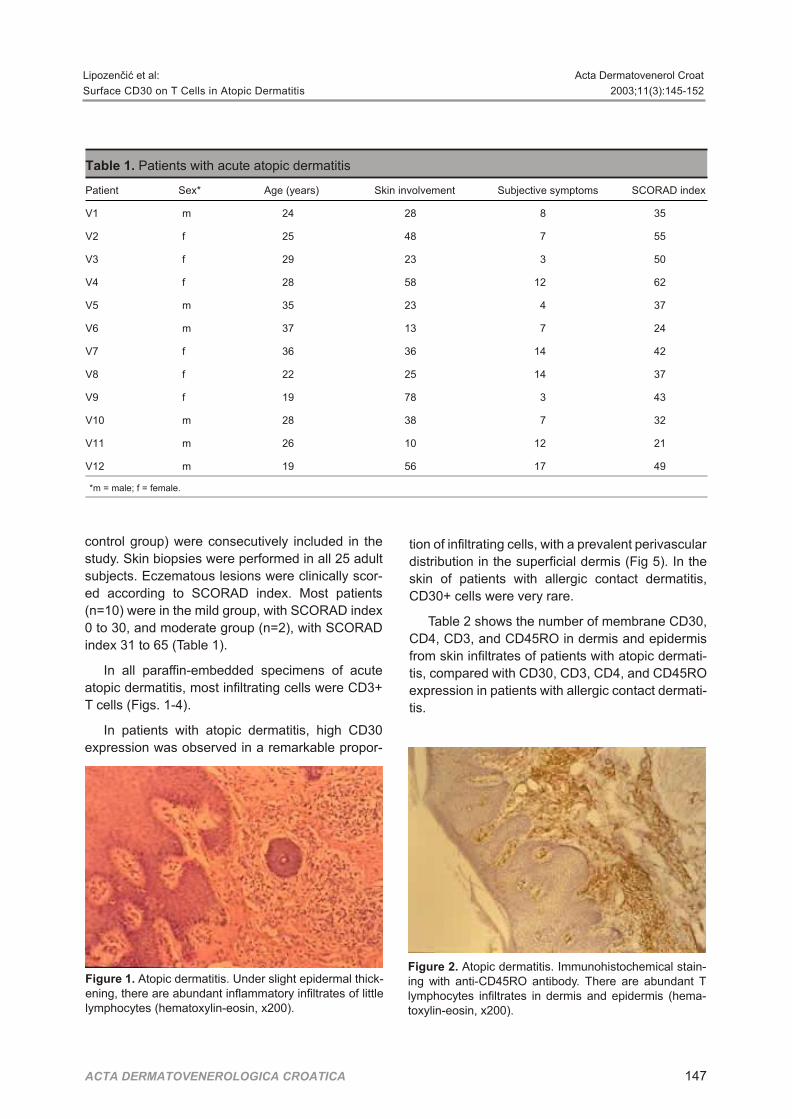
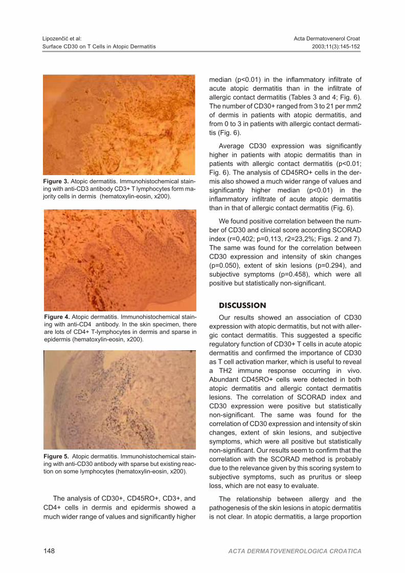
In all paraffin-embedded specimens of acute
atopic dermatitis, most infiltrating cells were CD3+
T cells (Figs. 1-4).
In patients with atopic dermatitis, high CD30
expression was observed in a remarkable pro por -
tion of infiltrating cells, with a prevalent perivascular
distribution in the superficial dermis (Fig 5). In the
skin of patients with allergic contact dermatitis,
CD30+ cells were very rare.
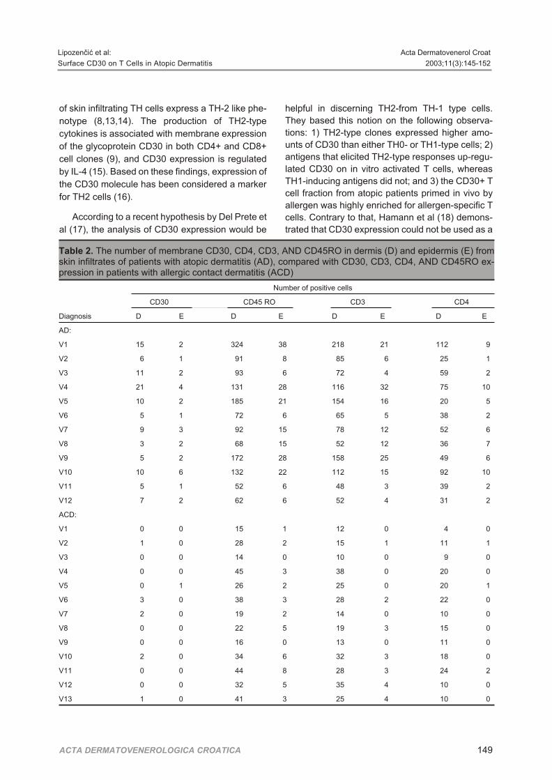
Table 2 shows the number of membrane CD30,
CD4, CD3, and CD45RO in dermis and epidermis
from skin infiltrates of patients with atopic der ma ti -
tis, compared with CD30, CD3, CD4, and CD45RO
expression in patients with allergic contact der ma ti -
tis.
ACTA DERMATOVENEROLOGICA CROATICA 147
Lipozenèiæ et al: Acta Dermatovenerol Croat
Surface CD30 on T Cells in Atopic Dermatitis 2003;11(3):145-152
Ta ble 1. Pa tients with acute atopic der ma ti tis
Patient Sex* Age (years) Skin involvement Subjective symptoms SCORAD index
V1 m 24 28 8 35
V2 f 25 48 7 55
V3 f 29 23 3 50
V4 f 28 58 12 62
V5 m 35 23 4 37
V6 m 37 13 7 24
V7 f 36 36 14 42
V8 f 22 25 14 37
V9 f 19 78 3 43
V10 m 28 38 7 32
V11 m 26 10 12 21
V12 m 19 56 17 49
*m = male; f = female.
Fig ure 1. Atopic der ma ti tis. Un der slight epi der mal thick -en ing, there are abun dant in flam ma tory in fil trates of lit tlelym pho cytes (hematoxylin-eosin, x200).
Fig ure 2. Atopic der ma ti tis. Immunohistochemical stain -ing with anti-CD45RO an ti body. There are abun dant Tlym pho cytes in fil trates in dermis and epi der mis (hema -toxylin-eosin, x200).
The analysis of CD30+, CD45RO+, CD3+, and
CD4+ cells in dermis and epidermis showed a
much wider range of values and significantly higher
median (p<0.01) in the inflammatory infiltrate of
acute atopic dermatitis than in the infiltrate of
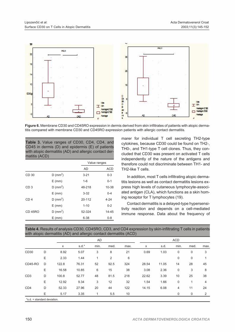
allergic contact dermatitis (Tables 3 and 4; Fig. 6).
The number of CD30+ ranged from 3 to 21 per mm2
of dermis in patients with atopic dermatitis, and
from 0 to 3 in patients with allergic contact der ma ti -
tis (Fig. 6).
Average CD30 expression was significantly
higher in patients with atopic dermatitis than in
patients with allergic contact dermatitis (p<0.01;
Fig. 6). The analysis of CD45RO+ cells in the der -
mis also showed a much wider range of values and
significantly higher median (p<0.01) in the
inflammatory infiltrate of acute atopic dermatitis
than in that of allergic contact dermatitis (Fig. 6).
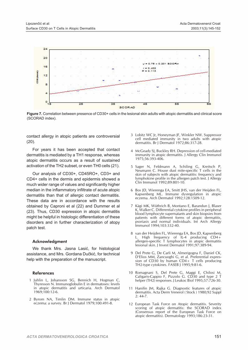
We found positive correlation between the num -
ber of CD30 and clinical score according SCORAD
index (r=0,402; p=0,113, r2=23,2%; Figs. 2 and 7).
The same was found for the correlation between
CD30 expression and intensity of skin changes
(p=0.050), extent of skin lesions (p=0.294), and
subjective symptoms (p=0.458), which were all
positive but statistically non-sig nif i cant.
DISCUSSION
Our results showed an association of CD30
expression with atopic dermatitis, but not with al ler -
gic contact dermatitis. This suggested a spe cific
regulatory function of CD30+ T cells in acute atopic
dermatitis and confirmed the im por tance of CD30
as T cell activation marker, which is useful to reveal
a TH2 immune response occurring in vivo.
Abundant CD45RO+ cells were detected in both
atopic dermatitis and allergic contact dermatitis
lesions. The correlation of SCORAD index and
CD30 expression were positive but statistically
non- sig nif i cant. The same was found for the
correlation of CD30 expression and intensity of skin
changes, extent of skin lesions, and subjective
symptoms, which were all positive but statistically
non-significant. Our results seem to confirm that the
correlation with the SCORAD method is probably
due to the relevance given by this scoring system to
subjective symptoms, such as pruritus or sleep
loss, which are not easy to evaluate.
The re la tion ship be tween al lergy and the
pathogenesis of the skin le sions in atopic der ma ti tis
is not clear. In atopic der ma ti tis, a large pro por tion
148 ACTA DERMATOVENEROLOGICA CROATICA
Lipozenèiæ et al: Acta Dermatovenerol Croat
Surface CD30 on T Cells in Atopic Dermatitis 2003;11(3):145-152
Fig ure 4. Atopic der ma ti tis. Immunohistochemical stain -ing with anti-CD4 an ti body. In the skin spec i men, thereare lots of CD4+ T-lym pho cytes in dermis and sparse inepi der mis (hematoxylin-eosin, x200).
Fig ure 3. Atopic der ma ti tis. Immunohistochemical stain -ing with anti-CD3 an ti body CD3+ T lym pho cytes form ma -jor ity cells in dermis (hematoxylin-eosin, x200).
Fig ure 5. Atopic der ma ti tis. Immunohistochemical stain -ing with anti-CD30 an ti body with sparse but ex ist ing re ac -tion on some lym pho cytes (hematoxylin-eosin, x200).
of skin in fil trat ing TH cells ex press a TH-2 like phe -
no type (8,13,14). The pro duc tion of TH2-type
cytokines is as so ci ated with mem brane ex pres sion
of the glycoprotein CD30 in both CD4+ and CD8+
cell clones (9), and CD30 ex pres sion is reg u lated
by IL-4 (15). Based on these find ings, ex pres sion of
the CD30 mol e cule has been con sid ered a marker
for TH2 cells (16).
Ac cord ing to a re cent hy poth e sis by Del Prete et
al (17), the anal y sis of CD30 ex pres sion would be
help ful in dis cern ing TH2-from TH-1 type cells.
They based this no tion on the fol low ing ob ser va -
tions: 1) TH2-type clones ex pressed higher amo -
unts of CD30 than ei ther TH0- or TH1-type cells; 2)
an ti gens that elic ited TH2-type re sponses up-reg u -
lated CD30 on in vi tro ac ti vated T cells, whereas
TH1-in duc ing an ti gens did not; and 3) the CD30+ T
cell frac tion from atopic pa tients primed in vivo by
al ler gen was highly en riched for al ler gen- spe cific T
cells. Con trary to that, Hamann et al (18) dem on s -
trated that CD30 ex pres sion could not be used as a
ACTA DERMATOVENEROLOGICA CROATICA 149
Lipozenèiæ et al: Acta Dermatovenerol Croat
Surface CD30 on T Cells in Atopic Dermatitis 2003;11(3):145-152
Ta ble 2. The num ber of mem brane CD30, CD4, CD3, AND CD45RO in dermis (D) and epi der mis (E) fromskin in fil trates of pa tients with atopic der ma ti tis (AD), com pared with CD30, CD3, CD4, AND CD45RO ex -pres sion in pa tients with al ler gic con tact der ma ti tis (ACD)
Number of positive cells
CD30 CD45 RO CD3 CD4
Diagnosis D E D E D E D E
AD:
V1 15 2 324 38 218 21 112 9
V2 6 1 91 8 85 6 25 1
V3 11 2 93 6 72 4 59 2
V4 21 4 131 28 116 32 75 10
V5 10 2 185 21 154 16 20 5
V6 5 1 72 6 65 5 38 2
V7 9 3 92 15 78 12 52 6
V8 3 2 68 15 52 12 36 7
V9 5 2 172 28 158 25 49 6
V10 10 6 132 22 112 15 92 10
V11 5 1 52 6 48 3 39 2
V12 7 2 62 6 52 4 31 2
ACD:
V1 0 0 15 1 12 0 4 0
V2 1 0 28 2 15 1 11 1
V3 0 0 14 0 10 0 9 0
V4 0 0 45 3 38 0 20 0
V5 0 1 26 2 25 0 20 1
V6 3 0 38 3 28 2 22 0
V7 2 0 19 2 14 0 10 0
V8 0 0 22 5 19 3 15 0
V9 0 0 16 0 13 0 11 0
V10 2 0 34 6 32 3 18 0
V11 0 0 44 8 28 3 24 2
V12 0 0 32 5 35 4 10 0
V13 1 0 41 3 25 4 10 0
marer for in di vid ual T cell se cret ing TH2-type
cytokines, be cause CD30 could be found on TH2-,
TH0-, and TH1-type T cell clones. Thus, they con -
cluded that CD30 was pres ent on ac ti vated T cells
in de pend ently of the na ture of the an ti gens and
there fore could not dis crim i nate be tween TH1- and
TH2-like T cells.
In ad di tion, most T cells in fil trat ing atopic der ma -
ti tis le sions as well as con tact der ma ti tis le sions ex -
press high lev els of cu ta ne ous lym pho cyte-as so ci -
ated an ti gen (CLA), which func tions as a skin hom -
ing re cep tor for T lymphocytes (19).
Contact dermatitis is a delayed-type hy per sen si -
tiv ity reaction and depends on a cell-mediated
immune response. Data about the frequency of
150 ACTA DERMATOVENEROLOGICA CROATICA
Lipozenèiæ et al: Acta Dermatovenerol Croat
Surface CD30 on T Cells in Atopic Dermatitis 2003;11(3):145-152
Ta ble 3. Value ranges of CD30, CD4, CD4, andCD45 in dermis (D) and epi der mis (E) of pa tientswith atopic der ma ti tis (AD) and al ler gic con tact der -ma ti tis (ACD)
Value ranges
AD ACD
CD 30 D (mm2) 3-21 0-3
E (mm) 1-6 0-1
CD 3 D (mm2) 48-218 10-38
E (mm) 3-32 0-4
CD 4 D (mm2) 20-112 4-24
E (mm) 1-10 0-2
CD 45RO D (mm2) 52-324 14-45
E (mm) 6-38 0-8
Ta ble 4. Re sults of anal y sis CD30, CD45RO, CD3, and CD4 ex pres sion by skin-in fil trat ing T cells in pa tients with atopic der ma ti tis (AD) and al ler gic con tact der ma ti tis (ACD)
AD ACD
x s.d.* min. med. max. x s.d. min. med. max.
CD30 D 8.92 5.07 3 8 21 0.69 1.03 0 0 3
E 2.33 1.44 1 2 6 0 0 1
CD45-RO D 122.8 76.31 52 92.5 324 28.54 11.05 14 28 45
E 16.58 10.85 6 15 38 3.08 2.36 0 3 8
CD3 D 100.8 52.77 48 81.5 218 22.62 3.39 10 25 38
E 12.92 9.34 3 12 32 1.54 1.66 0 1 4
CD4 D 52.33 27.96 20 44 122 14.15 6.08 4 11 24
E 5.17 3.35 1 5.5 10 0 0 2
*s.d. = standard deviation.
Fig ure 6. Mem brane CD30 and CD45RO ex pres sion in dermis de rived from skin in fil trates of pa tients with atopic der ma -ti tis com pared with mem brane CD30 and CD45RO ex pres sion pa tients with al ler gic con tact der ma ti tis.
contact allergy in atopic patients are controversial
(20).
For years it has been accepted that contact
dermatitis is mediated by a TH1 response, whereas
atopic dermatitis occurs as a result of sustained
activation of the TH2 subset, or even TH0 cells (21).
Our analysis of CD30+, CD45RO+, CD3+ and
CD4+ cells in the dermis and epidermis showed a
much wider range of values and significantly higher
median in the inflammatory infiltrate of acute atopic
dermatitis than that of allergic contact dermatitis.
These data are in accordance with the results
obtained by Caproni et al (22) and Dummer et al
(23). Thus, CD30 expression in atopic dermatitis
might be helpful in histologic differentiation of these
disorders and in further characterization of atopy
patch test.
Acknowledgment
We thank Mrs. Jasna Lasiæ, for histological
assistance, and Mrs. Gordana Duèkiæ, for technical
help with the preparation of the manuscript.
References
1 Juhlin L, Johansson SG, Bennich H, Hogman C,Thyresson N. Immunoglobulin E in dermatoses: levelsin atopic dermatitis and urticaria. Arch Dermatol1969;100:12-6.
2 Byrom NA, Timlin DM. Immune status in atopiceczema: a survey. Br J Dermatol 1979;100:491-8.
3 Lobitz WC Jr, Honeyman JF, Winkler NW. Suppressorcell mediated immunity in two adults with atopicdermatitis. Br J Dermatol 1972;86:317-28.
4 McGeady SJ, Buckley RH. Depression of cell-mediated immunity in atopic dermatitis. J Allergy Clin Immunol1975;56:393-406.
5 Sager N, Feldmann A, Schiling G, Kreitsch P,Neumann C. House dust mite-specific T cells in theskin of subjects with atopic dermatitis: frequency andlymphokine profile in the allergen patch test. J AllergyClin Immunol 1992;89:801-10.
6 Bos JD, Wierenga EA, Smitt JHS, van der Heijden FL,Kapsenberg ML. Immune dysregulation in atopiceczema. Arch Dermatol 1992;128:1509-12.
7 Kägi MK, Wüthrich B, Montano E, Barandun J, BlaserK, Walker C. Differential cytokine profiles in peripheral blood lymphocyte supernatants and skin biopsies frompatients with different forms of atopic dermatitis,psoriasis and normal individuals. Int Arch AllergyImmunol 1994;103:332-40.
8 van der Heijden FL, Wierenga EA, Bos JD, KapsenbergL. High frequency of IL-4 producing CD4+allergen-specific T lymphocytes in atopic dermatitislesional skin. J Invest Dermatol 1991;97:389-94.
9 Del Prete G, De Carli M, Almerigogna F, Daniel CK,D’Elios MM, Zancuoghi G, et al. Preferential ex pres -sion of CD30 by human CD4+ T cells producingTH2-type cytokines. FASEB J 1995;9:81-6.
10 Romagnani S, Del Prete G, Maggi E, Chilosi M,Caligaris-Cappio F, Pizzolo G. CD30 and type 2 Thelper (TH2) responses. J Leukoc Biol 1995;57:726-30.
11 Hanifin JM, Rajka G. Diagnostic features of atopicdermatitis. Acta Derm Venerol ( Stock ) 1980;92 Suppl2: 44-7.
12 European Task Force on Atopic dermatitis. Severityscoring of atopic dermatitis: the SCORAD index(Consensus report of the European Task Force onatopic dermatitis). Dermatology 1993;186:23-31.
ACTA DERMATOVENEROLOGICA CROATICA 151
Lipozenèiæ et al: Acta Dermatovenerol Croat
Surface CD30 on T Cells in Atopic Dermatitis 2003;11(3):145-152
Fig ure 7. Cor re la tion be tween pres ence of CD30+ cells in the lesional skin adults with atopic der ma ti tis and clin i cal score (SCORAD in dex).
13 Wierenga EA, Snoek M, Bos JD, Jansen HM,Kapsenberg ML. Comparison of diversity and functionof house dust mite-specific T lymphocyte clones fromatopic and non-atopic donors. Eur J Immunol 1991;201519-26.
14 Rheinold U, Kukel S, Goeden B, Neuman U, KreyselHW. Functional characterization of skin-infiltratinglymphocytes in atopic dermatitis. Clin Exp Immunol1991;86:444-8.
15 Nakamura T, Lee RK, Nam SY, Al-Ramady BK, KoniPA, Bottomly K, et al. Reciprocal regulation of CD30expression on CD4+ T cells by IL-4 and IFN-gamma. JImmunol 1997;158:2090-8.
16 Cavagni G, Caffarelli C, Facchetti F, Brugnoni D,Notaragelo LD, Tosoni C, et al. Cutaneous CD30+cells in children with atopic dermatitis. Int Arch Allergy Immunol 2000;121:224-8.
17 Del Prete G, De Carli M, D’Elios MM, Daniel KC,Almerigogna F, Alderson M, et al. CD30 dedicatedsignaling promotes the development of human Thelper type 2-like T cells. J Exp Med 1995;182:1655-61.
18 Hamann D, Hilkens CM, Grogan J, Lens SM,Kapsenberg ML, Yazdanbakhsh M, et al. CD30expression does not discriminate between human TH1and TH2-type T cells. J Immunol 1996;156:1387-91.
19 Gelb AB, Smoller BR, Wamke RA, Picker LJ.Lymphocytes infiltrating primary cutaneous neoplasms express selectively the cutaneous lym pho cyte- as so ci -ated antigen (CLA). Am J Pathol 1993;142:1556-64.
20 Szepietowski JC, Mckenze RC, Keohane SG, AldridgeRD, Hunter JA. Atopic and non-atopic individuals react to nickel challenge in a similar way. A study of thecytokine profile in nickel-induced contact dermatitis.Br J Dermatol 1997;137:195-200.
21 Romagnani S. Lymphokine production by human Tcells in disease states. Annu Rev Immunol 1994;12:227-57.
22 Caproni M, Bianchi B, D’Elios MM, De Carli M,Amedei A, Fabbri P. In vivo relevance of CD30 inatopic dermatitis. Allergy 1997;52:1063-70.
23 Dummer W, Rose C, Brocker EB. Expression of CD30on T helper cells in the inflammatory infiltrate of acuteatopic dermatitis but not of allergic contact dermatitis.Arch Dermatol Res 1998;290:598-602.
152 ACTA DERMATOVENEROLOGICA CROATICA
Lipozenèiæ et al: Acta Dermatovenerol Croat
Surface CD30 on T Cells in Atopic Dermatitis 2003;11(3):145-152

HEAD AND NECK SQUAMOUS CELL CARCINOMA SKIN
METASTASES BELOW OF THE DIAPHRAGM
Davorin Ðaniæ, Ana Ðaniæ1
Department of Otorhinolaryngology and Cervicofacial Surgery, Slavonski Brod GeneralHospital, Slavonski Brod, and Osijek University School of Medicine, Osijek;1Zagreb University School of Medicine, Zagreb, Croatia
Corresponding author:
Prof. Davorin Ðaniæ, M.D., Ph.D.
Department of Otorhinolaryngology and
Cervicofacial Surgery
Dr. Josip Benèeviæ General Hospital
35000 Slavonski Brod, Croatia
Received: 25. 02. 2003
Accepted: 18. 06. 2003.
INTRODUCTION
Metastasis is one of the most critical prognostic
factors of malignances arising in the head and neck
(1,2). In general, prognoses for patients with distant
metastases are poor, with many patients surviving
only a few months (1,3). Distant metastases from
squamous cell carcinoma of the mucosa of the
head and neck have a complex biologic nature. The
incidence of distant metastases of the head and
neck squamous cell carcinoma is relatively small in
comparison with other malignancies. In 1923, Crile
(4) was the first to report that the incidence of
metastases in patients with head and neck carci -
noma was approximately 1%. During the past
years, better local control of the malignant disease
have caused distant metastases to become a
common occurrence. Recent clinical data have
shown increased incidence of distant metastases,
up to 30% or even 50% at autopsy, most commonly
involving the lung, bone, and liver (5-7). The
incidence of distant metastases is influenced by the
location of the primary tumor, initial T and N stage of
the neoplasm, and presence or absence of regional
involvement above clavicula (8). Skin metastases
are a form of distant metastases. They are defined
as an isolated or multiple intradermal collection of
tumor cells remote from the primary tumor or
153
Acta Dermatovenerol Croat 2003;11(3):153-157 CLINICAL ARTICLE
SUMMARY Cutaneous metastases from carcinoma are relatively
uncommon in clinical practice. Metastasis to skin sites from squamous
cell carcinoma of the mucosa of the head and neck are also very rare.
However, skin metastases may be the first clinical evidence of the
malignant disease or its loco-regional recurrence. Early recognition of
skin metastasis can lead to an accurate and prompt diagnosis and timely
treatment. Patients with skin meta stases have very poor prognosis. We
report on four such patients, one of them with multiple skin metastases
from the squamous cell carcinoma in the cervical part of the esophagus
above and below the level of the diaphragm. In reviewing the literature,
only two cases of solitary skin metastases below the diaphragm from
laryngeal squamous cell carcinomas have been reported.
KEY WORDS carcinoma, squamous cell; neoplasm meta stasis; skin
loco-regional disease. Skin metastases in patients
with metastatic disease, including melanoma, from
internal malignant tumors are not uncommon; we
previously found that they accounted for <10% of all
distant metastatic lesions (9). In patients with
squamous cell carcinoma of the head and neck,
skin metastases are rare, with incidence ranging
from 0.76% to 2.4%. Most are sporadic cases, with
the exception of two studies reporting a significant
number of the cases (1,2). The site and the number
of metastases vary, with the neck and chest being
the most common sites. Yoskovich et al (2) reported
on watershed characteristics of metastases distri -
bu tion; however, they did not report any occurring
below the level of the diaphragm (2). The deve -
lopment of skin metastases may be a consequence
of aggressive nature of the tumor or may indicate
patient’s local resistance at typical distant metasta -
tic sites.
We present three cases of the skin metastases
of the squamous cell carcinoma from the head and
neck as well as a patient with multiple nodular skin
metastases above and below the level of the
diaphragm. To the best of our knowledge, the latter
is the first such case ever described.
SUBJECTS AND METHOD
There were 372 consecutive patients with
squamous cell carcinoma hospitalized at the
Department of Otorhinolaryngology and Cervicofa -
cial Surgery, Slavonski Brod General Hospital, in
the 1992-2002 period. Data collected from their
medical records for the needs of analysis were the
following: age, sex, tumor site and size, nodal
status and stage, histologic grading, time of
presentation of skin and distant metastases, and
localization of metastases (Table 1). Skin meta -
stases were differentiated clinically and histopatho -
logically from other forms of skin involvement.
Lesions of the epidermis, dermis, and immediate
subcutaneous tissue were morphologically and
histochemically compared with the primary squa -
mo us cell carcinoma.
RESULTS
Out of 372 patients with squamous cell car -
cinoma of the head and neck, four (0.93%, three
men and a woman) developed skin metastases. At
the time of presentation of the primary tumor, the
average age of the patients was 56 years. Two of
the patients who developed skin metastases had
poorly differentiated invasive squamous cell carci -
noma (G3) and the other two patients had mode -
rately differentiated carcinoma (G3) with respect to
their primary tumor. All of them had stage IV of the
disease. They presented with skin meta stases in
the period between 0 and 40 months. None of them
were alive a year after the development of skin
metastases. The sites of development of the skin
metastases included the neck, chest, face, back,
and arm, above the level of the diaphragm; and
abdomen, pelvis, and legs below the level of the
diaphragm. The patient with poorly differentiated
squamous cell carcinoma (G3) in the cervical part
of the esophagus developed multiple skin metasta -
ses above and below the diaphragm (Figs. 1 and 2).
In this patient, no other tumors but esophageal
carcinoma were found on additional clinical exa -
mination. He had no clinically evident local or regi -
onal metastasis, but developed distant metastases
in liver. His general condition was very poor
because he had very serious associated illnesses.
The patient did not receive any kind of therapy and
154 ACTA DERMATOVENEROLOGICA CROATICA
Ðaniæ and Ðaniæ: Acta Dermatovenerol Croat
Squamous Cell Carcinoma Subdiaphragmatic Metastases 2003;11(3):153-157
Table 1. Clinical features of the primary carcinoma and skin metastases*
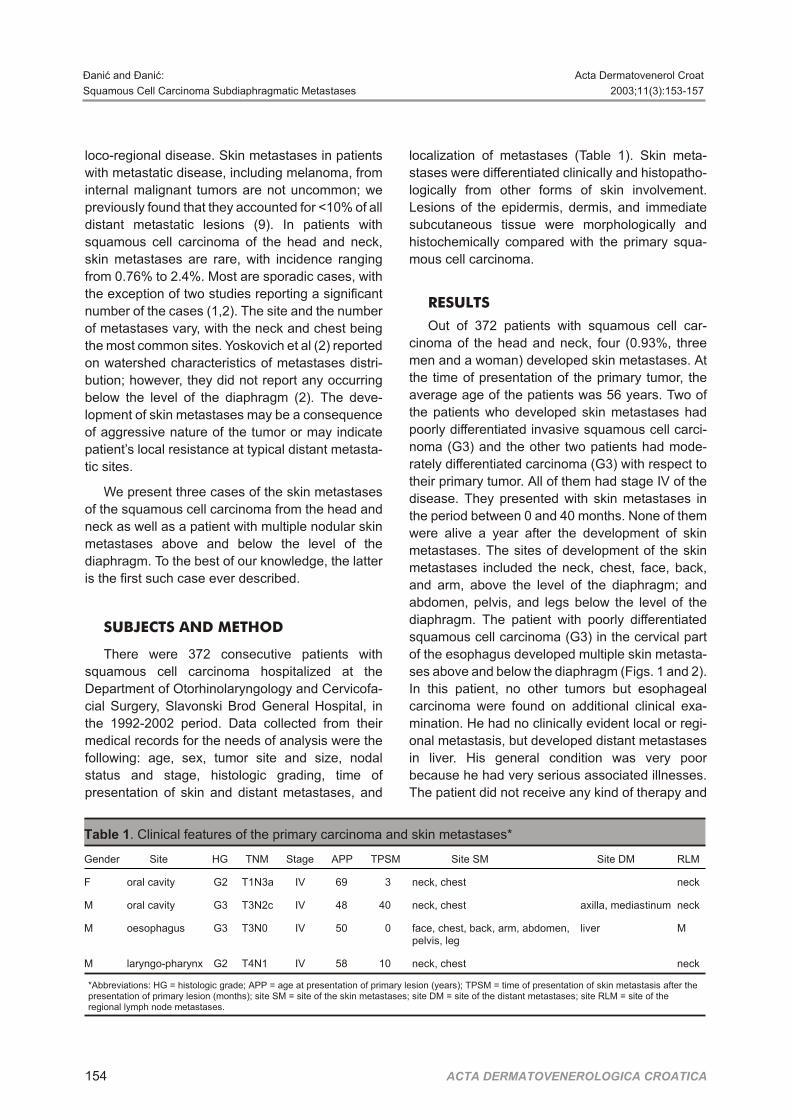
Gender Site HG TNM Stage APP TPSM Site SM Site DM RLM
F oral cavity G2 T1N3a IV 69 3 neck, chest neck
M oral cavity G3 T3N2c IV 48 40 neck, chest axilla, mediastinum neck
M oesophagus G3 T3N0 IV 50 0 face, chest, back, arm, abdomen,pelvis, leg
liver M
M laryngo-pharynx G2 T4N1 IV 58 10 neck, chest neck
*Abbreviations: HG = histologic grade; APP = age at presentation of primary lesion (years); TPSM = time of presentation of skin metastasis after thepresentation of primary lesion (months); site SM = site of the skin metastases; site DM = site of the distant metastases; site RLM = site of theregional lymph node metastases.

died 4 months later. Two patients with the primary
carcinoma of oral cavity had neck metastases in the
time of the presentation of primary tumor. They
were first treated with radiotherapy and chemo -
therapy. One of them died after 6 months due to the
progression of the metastatic disease, and the
other developed metastasis in the neck and distant
metastases in the axilla 23 months later. He
underwent radical dissection of the neck and lym -
ph adenectomy of the axilla and received another
round of radiotherapy with chemotherapy. After 17
months, the patient developed distant meta sta ses
in the mediastinum and skin metasta ses on the
neck and chest, and died 3 months later. Patient
with laryngeal carcinoma died 10 months after
laryngectomy and radiotherapy because of local
tumor progression and regional metastases in the
neck and distant in the chest.
DISCUSSION
The mechanism of the development and appe -
arance of skin metastases is not completely under -
stood. Prior retrospective clinical study suggested
that skin metastases might evolve by different
mechanism. When metastases arise in the head
and neck region, the spread of tumor cells can
occur through local dermal lymphatic network.
Distant metastases are thought to spread by blood.
Perineurial spread can also be a possible route for
metastases to the skin (2,10). «Metastasis from
metastasis» presumes aggressive nature of tumor
and high frequency of associated distant metastatic
lesions (11). New and highly sensitive immuno -
histo chemical methods, molecular analyses, and
techniques of cell culture may improve the detec -
tion of distant micrometastases in head and neck
cancer (12,13).
Skin metastases may occur in local, regional,
and distant sites, but most frequently they occur in
the vicinity of the primary tumor and some tumors
metastasize with predilection to specific areas (14).
ACTA DERMATOVENEROLOGICA CROATICA 155
Ðaniæ and Ðaniæ: Acta Dermatovenerol Croat
Squamous Cell Carcinoma Subdiaphragmatic Metastases 2003;11(3):153-157
Figure 1. Tumor (skin metastasis) on the apex of the nose (a) is the first presentation of the esophageal carcinoma andskin metastases above (b) and below (c) the level of the diaphragm.
a b c
Figure 2. X-ray finding of primary squamous cell carci -noma of the esophagus.
Local skin metastases may develop in scars at a
surgical incision site (9). Skin metastases from
squamous cell carcinoma of the head and neck
appear to have watershed characteristics; extreme -
ly rare cases have been reported with skin meta -
stases below the level of the diaphragm. Reviewing
the literature, we found two such cases (9,15). In
both reports, primary tumors were squamous cell
carcinoma of the larynx and skin metastases
occurred in the anterior wall of the abdomen like
solitary subcutaneous nodule. Our patient with the
carcinoma of the cervical part of the esophagus
developed multiple skin metastases that presented
as subcutaneous nodules above the level of the
diaphragm (on the face, chest, and arms) and
below the level of the diaphragm (on the abdomen,
pelvis, and leg). No other tumors, except for the
carcinoma of the cervical esophagus, were found in
this case. There was no evidence of loco-regional
spread of tumor, but the patient developed distant
metastases in liver. Skin metastases were the first
clinical indicator of disease in apparently disease-
free patient, leading to the earlier discovery of the
local malignant disease. Multiple skin metastases
above and below the level of the diaphragm may
develop due to aggressive nature of the tumor or
indicate patient’s local resistance at typical distant
metastatic sites, such as the lung and liver. In a very
small percentage of cases, metastases may be
discovered at the some time or prior to the dia -
gnosis of a primary tumor. Lookingbill et al (9) found
that 0.6% patients developed skin metastases as
the first sign of internal carcinoma. The develop -
ment and appearance of skin metastases is similar
in severity to the development of other distant
metastases in other cases. The histology of the
metastases is similar to the primary tumor, although
metastases may be more anaplastic and less
differentiated.
The true skin metastases from squamous cell
carcinoma from the head and neck must be
differentiated from other skin malignancies or
malignancies associated with or affecting the skin,
because they have different prognosis. These
malignancies included tumors with direct spread in
skin or metastasis from cutaneous squamous cell
carcinoma. Recurrence in previously operated
neck portends a poor prognosis (15). Inadequate
clearance of lymph-bearing tissue from dissected
levels in the neck may also account for tumor
recurrence. One possible complication of the
aspiration biopsy of malignant tumor is dissemi -
nation of tumor cells along the needle track or tumor
implantation into the incision site at the time of
surgery. As the use of endoscopic surgical tec -
hnique for the management of malignances has
increased over the last years, metastases develop -
ing at the troacar insertion site became an
emerging problem (16,17).
In conclusion, skin metastases from squamous
cell carcinoma of the head and neck are uncommon
and in some cases, the first sign of the cancer
disease. They can occur above and below the level
of the diaphragm and early recognition can lead to
timely treatment. The treatment is palliative, it must
be individualized, and prognosis for patients with
skin metastases is poor. A better understanding of
the biology of tumor invasion and metastasis
occurrence may lead to the development of new,
more effective strategies in prevention of secondary
tumors.
Acknowledgments
We thank Prof. Ana Marušiæ M.D. Ph.D. and
Prof. Matko Marušiæ M.D. Ph.D. for their as sis tance
with the prep a ra tion of this manu script.
References
1 Pitman KT, Johnson JT. Skin matastases from head andneck squamous cell carcinoma: incidence and impact.Head Neck 1999;21:560-5.
2 Yoskovitch A, Hier MP, Okrainec A, Black MJ. Skinemetastases in squamous cel carcinoma of the head andneck. Otolaryngol Head Neck Surg 20001;124:248-52.
3 Kowalski LP, Medina JE. Nodal metastases: predictivefactors: Otolaryngol Clin North Am 1998;31:621-37.
4 Crile GW. Carcinoma of the jaws, tongue, cheek andlips. Surg Gynecol Obstet 1923;36:159-84.
5 Calhoun KH, Fulmer P, Weiss R, Hokanson JA. Distantmetastases from head and neck squamous cellcarcinomas. Laryngoscope 1994;104:1199-205.
6 Papac RJ. Distant metastases from head and neckcancer. Cancer 1984;53:342-5.
7 Zbaren P, Lehmann W. Frequency and sites of distantmetastses in head and neck squamous cell carcinoma.Arch Otolaryngol Head Neck Surg 1987;113:762-4.
156 ACTA DERMATOVENEROLOGICA CROATICA
Ðaniæ and Ðaniæ: Acta Dermatovenerol Croat
Squamous Cell Carcinoma Subdiaphragmatic Metastases 2003;11(3):153-157
8 Ferlito A, Shaha AR, Silver CE, Rinaldo A, Mondin V.Incidence and sites of distant metastases from head and neck cancer. ORL J Otorhinolaryngol Relat Spec2001;63:202-7.
9 Lookingbill DP, Spangel N, Helm KF. Cutaneousmetastases in patients with metastatic carcinoma: aretrospective study of 4020 patients. J Am AcadDermatol 1993;29:228-36.
10 Kmucha ST, Troxel J. Dermal metastases in epidermoid carcinoma of the head and neck. Arch OtolaryngolHead Neck Surg 1993;119:326-30.
11 Scwartz RA. Cutaneous metastatic disease. J Am AcadDermatol 1995;33:161-82.
12 Ferlito A, Partridge M, Brennan Hamakawa H. Lymphnode micrometastases in head and neck cancer: areviwe. Acta Otolaryngol 2001;121:660-5.
13 Wirtschafter A, Benninger MS, Moss TJ, Umiel T,Blazoff K, Worsham MJ. Micrometastatic tumor
detection in patients with head and neck cancer. ArchOtolaryngol Head Neck Surg 2002;128:40-3.
14 Lee TC, Schwartz R, Wells MJ, Heymann WR, Quirk C, Elston DM. Metastatic carcinoma of the skin.eMedicine J 2001;2(7).
15 Horiuchi N, Tagami H. Skin metastasis in laryngealcarcinoma. Clin Exper Dermatol 1992;17:282-3.
16 Myers EN, Fagan JJ. Treatment of the N+ neck insquamous cell carcinoma of the upper aerodigestivetract. Otolaryngol Clin North Am 1998;31:671-86.
17 Shinohara S, Yamamoto S, Maetani T, Kim T.Implantation metastasis of the head and neck cancerafter fine needle aspiration biopsy. Auris Nasus Larynx2001;28:377-80.
18 Hertel H, Fleck M, Kuhne-Heid R, Schneider A.Troacar-site metastasis in not always due tolaparoscopy. Surg Endosc 2001;15:896.
ACTA DERMATOVENEROLOGICA CROATICA 157
Ðaniæ and Ðaniæ: Acta Dermatovenerol Croat
Squamous Cell Carcinoma Subdiaphragmatic Metastases 2003;11(3):153-157
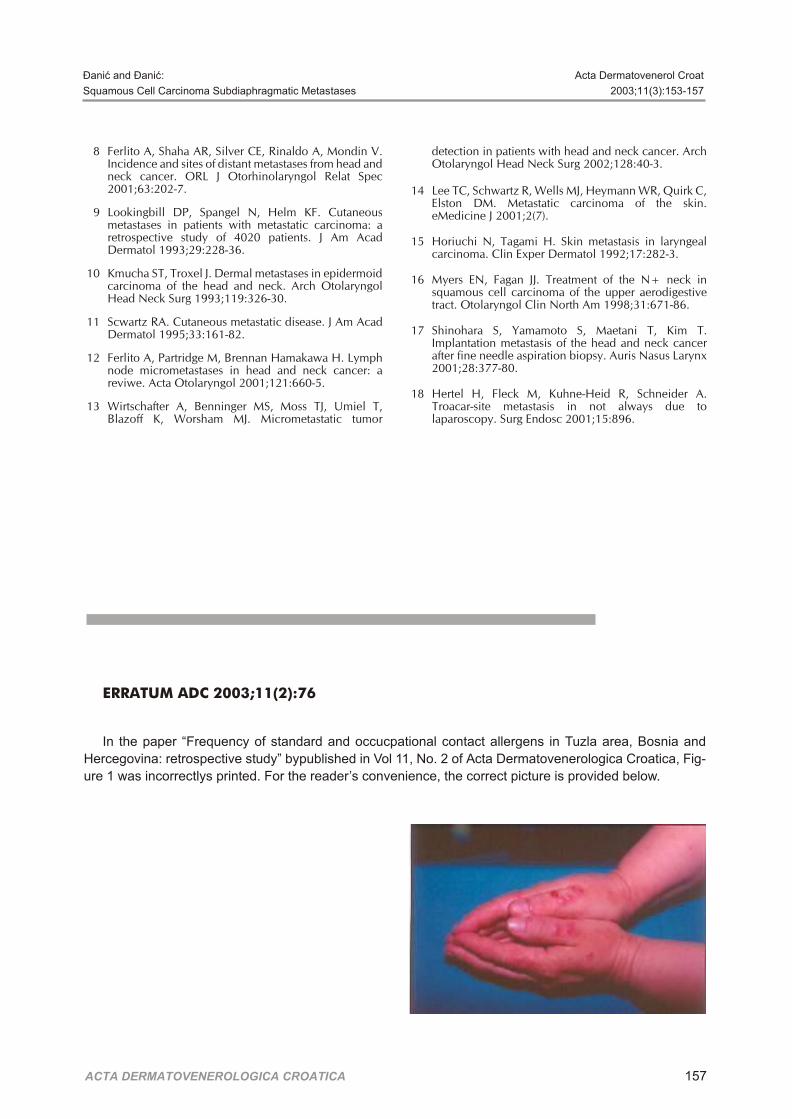
ER RA TUM ADC 2003;11(2):76
In the pa per “Fre quency of stan dard and occucpational con tact al ler gens in Tuzla area, Bosnia and
Hercegovina: ret ro spec tive study” bypublished in Vol 11, No. 2 of Acta Dermatovenerologica Croatica, Fig -
ure 1 was incorrectlys printed. For the reader’s con ve nience, the cor rect pic ture is pro vided below.
SUNSCREENS – THE ULTIMATE COSMETIC
Ronni Wolf1, Hagit Matz1,2, Edith Orion1, Jasna Lipozenèiæ3
1Dermatology Unit, Kaplan Medical Center, Rechovot, Israel;2Department of Dermatology, Tel-Aviv Sourasky Medical Center and the Sackler Faculty ofMedicine, Tel-Aviv University, Tel-Aviv, Israel;3Department of Dermatology and Venerology, Zagreb University Hospital Center, Zagreb,Croatia
Corresponding author:
Prof. Ronni Wolf, M.D., Ph.D.
Head of Dermatology Unit
Kaplan Medical Center
76100 Rechovot, Israel
Re ceived: 20. 06. 2003.
Ac cepted: 15. 07. 2003.
158
Acta Dermatovenerol Croat 2003;11(3):158-162 RE VIEW
SUMMARY One decade ago, a sun protection factor (SPF) of 15 was considered a
complete blocker of ultraviolet radiation (UV). The logic behind that cutoff point was
that sunscreens with this SPF number would always prevent erythema and that
preventing erythema would prevent all the ill effects of UV exposure. Today, we know
that both of these assumptions were wrong and we tend to recommend higher SPF.
Consumers apply only about one-quarter to one-half thickness of the layer of
sunscreen material used to measure the SPF in the laboratory. That means that less
than 50% of the SPF number claimed on the label is spread on the consumer’s skin,
meaning that a sunscreen with an SPF 30 will give the real protection of an SPF of
15. Therefore, recommend 60 when you want a real protection of 30! Significant
injury, DNA damage, mutations, and carcinogenesis can and do occur also with
cumulative suberythemal UV exposure. Thus, erythema induction, a criterion that
defines SPF, is not a good indicator of UV damage. We also need higher SPF values
to prevent the damage caused by suberythemal doses of UV. The value of the SPF
claimed on the label is diminished by environmental factors that are not taken into
account during SPF measurements in the laboratory, such as sweating, water
immersion, rubbing off, and photodegradation. There are some misunderstandings
and confusion about the mode of action of physical sunscreens. It was originally
considered that, in contrast to organic sunscreens, the inorganic metal oxides (zinc
oxide and titanium dioxide) acted as scatterers or reflectors of UV light, as a mirror.
This is not the case with modern micronized forms of metal oxides. It has been
shown that both zinc oxide and titanium dioxide mobilize electrons within their atomic
structure while absorbing UV radiation. Thus, although metallic oxides are not inert
per se, in their coated form they are stable, non-toxic, and safe and they act as highly
efficient UV attenuators. Therefore, we recommend our patients to use this type of
sunscreens. We should exert all our influence upon our patients not to expose
themselves to excessive sunlight, to routinely use generous layers of sunscreen
agents, and to wear protective clothing. To wait for the dust to settle around the issue
of the effectiveness of sunscreens in preventing melanoma, while the ideal
sunscreens - topical, systemic, whatever - are at our disposal, is a luxury we cannot
afford.
KEY WORDS sunlight; sunscreening agents; skin; ultraviolet rays

INTRODUCTION
Our paper will be brief, but it will touch upon one
of the hottest subjects in the universe, the sun, and
how to shield the protective covering and most
extensive organ of the human body, the skin,
against the damaging effects of sunlight.
For hundreds of years, it was social snobbery –
not sunscreens – that protected the rich from
having their skin damaged by sun. A bronzed face
and hands were the telltale signs of poor, of those
who labored beneath the sun. Then one day in the
1920s, the doyen of fashion, Coco Chanel, returned
from a Palm Beach vacation with a suntan, and the
bronzed look suddenly ceased to be lower class
stigma and became upper class chic. It was another
French Revolution!
The parasols were put away and the beaches
and poolsides packed with sun worshippers. The
bathing suits became skimpier and skimpier, and
the bikini, let alone the topless, exposed more and
more flesh to be baked and fried under the sun. As
economic conditions improved, even “ordinary”
people had more leisure time, took longer holidays,
and became involved in outdoor activities. Such
newly defined signs of success, appeal, and beauty
turned out to be a lethal combination too often when
it came to health.
Then, around two decades ago, the men in the
laboratories who were apparently insensitive to
social pressures had the temerity to cast shadows
on “healthy” sunshine and even went so far as to
emphasize its damaging, harmful, and carcinoge -
nic effects. The golden tan not only paled, but
became an enemy. The data were so irrefutable
that the public had no other choice but either to give
up their long-cherished solar rituals and protect
themselves or risk major health problems. The
overwhelming majority finally became convinced
that there was no “healthy” tan, and no “healthy
sunshine”, and accepted the verdict of medical
research that it was a matter of “fry now and pay
later”.
For most of us who made outdoor activities an
integral part of a normal, healthy lifestyle, the
change has been easier said than done. Count
Dracula had to go to great lengths to manage his
schedule and escape the sun, but few of us
ordinary mortals could, or would even want to, do
anything similar. This created an urgent need for
effective photoprotective measures. To our good
luck, the promise of enormous profits from photo -
protective agents galvanized the sunscreen manu -
facturers. The discovery of the solutions, however,
is no less a real testimony to the use of chemistry for
improving the quality of life of millions of people all
over the world.
Although sunscreen formulation and production
are guided by well-based and proven scientific and
theoretical considerations for the sake of the users
and their well-being, the process is also vigorously
motivated by economic, commercial, and business
interests that form the backbone of a worldwide,
multibillion-dollar industry. The shelves offer such a
wide range of choices that recommending a sun -
screen is like finding one’s way alone through a
minefield in no-man’s land. We will try to help you to
navigate through this minefield and we will highlight
the essential points of the rational use of sunscre -
ens.
HOW HIGH SPF SHOULD WE
RECOMMEND TO OUR PATIENTS?
One decade ago, an SPF of 15 was considered
a complete UV blocker. The logic behind that cutoff
point was that these sunscreens would always
prevent erythema and that preventing erythema
would prevent all the ill effects of UV exposure (1).
Today we know that both of these assumptions
were wrong and we tend to recommend higher
factors. Most currently popular sunscreens have an
SPF between 15 to 35, and it is not uncommon to
find products claiming to have a factor of 50 or 70,
or even higher.
The Food and Drug Administration has recently
set a limit of 30 as the optimally recommended SPF
value and the upper limit for approved SPF labeling.
Products with the SPF above 30 will be labeled as
having SPF “30 plus” (2), because the additional
benefit derived from using sunscreen formulations
with an SPF above 30 is outweighed by the po -
tential risks of exposing the skin to a higher concen -
tration of sunscreen ingredients as well as the
increase in the costs of the products. The mathe -
matics here are actually very deceptive: increasing
the SPF of a product over 15 theoretically adds very
little to its blocking capacity! For example, in crea -
ACTA DERMATOVENEROLOGICA CROATICA 159
Wolf et al: Acta Dermatovenerol Croat
Sunscreens – The Ultimate Cosmetic 2003;11(3):158-162

sing the SPF from 30 to 40 will increase its capacity
to block UV by only 1%, i.e., from 96.7% to 97.5%.
But that difference in 10 points and increased
benefit of 1% means far more chemicals on the skin
and much higher prices.
These numbers are theoretical extrapolations
and it would be careless not to defend the belief that
sunscreens with the SPF 15 are optimal blockers
and the ones we should recommend. This
reasoning is based on the following considerations:
1. A number of studies have shown that con -
sumers apply only about one-quarter to one- half
thickness of the layer of sunscreen material used to
measure the SPF in the laboratory (3). This means
that the actual SPF spread on the con sumer’s skin
is less than 50% of the SPF number appearing on
the label, meaning that a sunscreen with an SPF 30
will give the real protection of an SPF 15. So,
recommend 30 when you want a real protection of
15!
2. We now know that significant injury, DNA
damage, mutations, and carcinogenesis can and
do occur with cumulative suberythemal UV expo -
sure. Thus, erythema induction, a criterion that de -
fines the SPF, is not a good indicator of UV damage.
We need higher SPF values to prevent the damage
caused also by suberythemal doses of UV (1).
3. The value of the SPF number stated on the
label is diminished by environmental factors not
taken into consideration during SPF measurements
in the laboratory, such as sweating, water
immersion, rubbing off, and photodegradation of
the active ingredients that is not related to their
fading from the skin (4,5).
CHEMICAL VS. PHYSICAL
INGREDIENTS – WHICH TO
ADVOCATE?
Sunscreen chemicals contain conjugate double
bonds. This configuration permits electron delocali -
zation to occur throughout the molecule. This
process absorbs light at comparatively low
energies in the UV wavelength range. At this point,
the electromagnetic energy is converted into
chemical energy, which is stored in the molecule.
The molecule is now in an excited and chemically
unstable state and might react with other ingredi -
ents of the sunscreen or with organic tissue
compounds, and cause damage. As the molecule in
the excited state returns to the ground state, energy
is emitted in lower magnitude, for example, in the
infrared region.
There are some misunderstandings and confu -
sion about the mode of action of physical sun -
screens. It was originally considered that, in
contrast to organic sunscreens, the inorganic metal
oxides (zinc oxide and titanium dioxide) acted as
mere scatterers or reflectors of UV light, as a mirror.
This is not the case with modern micronized forms
of metal oxides. Another and more far-reaching
misconception is that they are inert materials that
do not undergo any chemical change while attenu -
ating UV light. Things go so far that some of these
products bear the claim of being “chemical-free”,
which is absurd.
Both zinc oxide and titanium dioxide mobilize
electrons within their atomic structure while absorb -
ing UV radiation. The absorbed energy results in
mobilization and transition of electrons from one
part of the molecule (creating a “hole”) to the other
(creating excited electrons), both of which are
chemically unstable and active. When the electron
returns to its lower energy band, it emits energy in a
lower frequency than the excitation energy, and this
is exactly what happens with organic sunscreens.
Although more than 90% of these electrons return
back to their original band within nanoseconds,
some do not, and these might react with water and
organic compounds, forming free radicals. Again, it
is similar to what happens with organic sunscreens
(6-8).
So if the physical or particulate sunscreens ab -
sorb light by the same mechanism as the chemical
ones and are not inert materials that just reflect and
scatter light, what is the difference between the two
and do the physical sunscreens have any ad -
vantage? The answer is – there is a difference in
favor of the physical sunscreens!
The reason is that the metallic oxides in sun -
screens are always coated with silicone or other
materials. In this form, their photoreactivity and their
ability to react with living tissues is nearly non -
existent. Furthermore, it has been shown that
metal lic oxides do not penetrate the stratum cor -
neum and thus do not cause any harm to living cells
160 ACTA DERMATOVENEROLOGICA CROATICA
Wolf et al: Acta Dermatovenerol Croat
Sunscreens – The Ultimate Cosmetic 2003;11(3):158-162
of the epidermis. So, although metallic oxides are
not inert per se, they are stable, non-toxic, and safe
in their coated form as well as they are highly
efficient UV attenuators. Although they are
“newcomers” to the market, they have already
become considerably popular and will most
probably continue to enjoy the fine reputation they
deserve in future.
THE QUESTION OF REAPPLICATION
Sunscreen performance is often tested in air-
con di tioned laboratories under artificial light. It is
inevitable, then, that the most frequently used
models have only limited ability to incorporate
behavioral and environmental variables. Therefore,
they have limited reliability and predictive value.
There are few studies on the efficacy of sunscreen
reapplication, and even fewer involved in compar -
ing the effect of various regimens and their
reapplication (9).
While it is quite clear that one cannot gain extra
or additional SPF values from reapplication of a
sunscreen, the reapplication might well assure the
presence of a given SPF on the skin for a longer
period of time, since the SPF of sunscreens decre -
ases due to environmental factors and photodegra -
dation (4,10).
The use of sunscreens, however, carries risks
that could be explained by the theory on human
behavior and risk management, called “risk com -
pen sation” or “risk homeostasis”(11). This means
that the use of seat belts would be compensated by
riskier driving; the use of condoms would increase
the number of different sexual partners; the im -
prove ment in the treatment of human immunode -
ficiency virus (HIV) infection would lead to the
higher exposure to unsafe sex (11); and the
morning after pill would allow carelessness the
night before. Several convincing studies have
shown that the use of sunscreens was associated
with an increase in the duration of recreational sun
exposure (12). It was suggested that sunscreen
use might even encourage prolonged sun exposure
because it delays sunburn. This tendency might
also explain why there are a so many epidemio -
logical studies showing that the use of sunscreens
did not reduce the incidence of melanoma, but even
increased it.
There is a clear and urgent need for a wise and
appropriate education policy to avoid the scenario
in which behavioral adaptation may decrease the
influence and benefits of sunscreen-promotion
policy. Also, the authorities should prohibit the ap -
pea rance of gorgeous suntanned models in sun -
screen advertisements and replace them with
gorgeous pale models...
CONCLUSION
Sunscreens are and will remain the ultimate
cosmetic and we should not reverse a decade-long
policy or stop recommending sunscreens on the
basis of some epidemiological studies whose
results indicated that sunscreen use may not
protect against melanoma, or may even increase its
risk. We should instruct our patients not to expose
themselves to excessive sunlight, to routinely use
generous layers of sunscreen agents, and to wear
protective clothing.
References
1 Naylor M, Kevin C. The case of sunscreens. A review of their use in preventing actinic damage and neoplasia.Arch Dermatol 1997;133:1146-54.
2 U.S. Food and Drug Administration. Sunscreen drugproducts for over-the-counter human use: Finalmonograph. In: Anonymous. Federal Register.Washington (DC): Department of Health, Educationand Welfare, Food and Drug administration; 1999. p.27666-93.
3 Bech-Thomsen N, Wulf H. Sunbathers’ application ofsunscreen is probably inadequate to obtain the sunprotection factor assigned to the preparation.Photodermatol Photoimmunol Photomed 1993;9:242-4.
4 Flindt-Hansen H, Nielsen C, Thune P. Measurementsof photodegradation of PABA and some PABAderivatives. Photodermatol 1988;5:257-60.
5 Tarras-Wahlberg N, Stenhagen G, Larko O, Rosen A,Wennberg A, Wennerstrom O. Changes in ultravioletabsorption of sunscreens after ultraviolet irradiation. JInvest Dermatol 1999;113:547-53.
6 Serpone N, Lawless D, Khairutdinov R. Subnano -second relaxation dynamics in TiO2 colloidal sols(particle sizes Rp = 1.0-13.4 nm). Relevance toheterogenous photocatalysis. J Phys Chem 1995;99:16655-61.
7 Lawless D, Serpone N, Meisel D. Role of OH radicalsand trapped holes in photocatalysis. A pulse radiolysisstudy. J Phys Chem 1991;95:5166-70.
ACTA DERMATOVENEROLOGICA CROATICA 161
Wolf et al: Acta Dermatovenerol Croat
Sunscreens – The Ultimate Cosmetic 2003;11(3):158-162
8 Jaeger C, Bard A. Spin trapping and electron spinresonance detection of radical intermediates inphotodecomposition of water at Tio2 particulatesystems. J Phys Chem 1979;83:3146-52.
9 Diffeey B. When should sunscreen be reapplied? J AmAcad Dermatol 2001;45:882-5.
10 Pruim B, Green A. Photobiological aspects of sun -screen re-application. Australas J Dermatol 1999;40:14-8.
11 Richens J, Imrie J, Copas A. Condoms and seat belts:the parallels and the lessons. Lancet 2000;355:400-3.
12 Autier P, Dore J, Negrier S, Lienard D, Panizzon R,Lejeune FJ, et al. Sunscreen use and duration of sunexposure: a double-blind, randomized trial. J NatlCancer Inst. 1999;91:1304-1309.
162 ACTA DERMATOVENEROLOGICA CROATICA
Wolf et al: Acta Dermatovenerol Croat
Sunscreens – The Ultimate Cosmetic 2003;11(3):158-162
Ad ver tise ment for KRAMER ther mom e ters, from 1937.From the col lec tion of Stella Ferenèiæ-Fatoviæ, M.D., Ph.D.
TREATMENT OF VITILIGO: CURRENT METHODS AND NEW
APPROACHES
Krešimir Kostoviæ, Ivana Nola, Željana Buèan, Mirna Šitum
Department of Dermatology and Venerology, Sisters of Mercy University Hospital, Zagreb,Croatia
Corresponding author:
Krešimir Kostoviæ, M.D.
Department of Dermatology and Venerology
Sisters of Mercy University Hospital
Vinogradska cesta 29
HR-10000 Zagreb, Croatia
Received: 03. 03. 2003.
Ac cepted: 18. 05. 2003.
INTRODUCTION
Vitiligo is an acquired idiopathic hypomelanotic
disorder characterized by circumscribed depig -
ment ed macules. Histologically, involved skin
shows a loss of functional melanocytes and mela -
nin within the epidermis (1). Vitiligo affects people of
all races, with the incidence of 1-2% without sexual
predilection (2).
There are several hypotheses on the pathoge -
nesis of the disease, but not a single one is fully
explanatory. The autoimmune hypothesis stems
from the association of vitiligo with autoimmune
disorders and the finding of antimelanocyte auto -
antibodies in some individuals (3-5). The neuronal
theory suggests that a neurochemical mediator is
responsible for destroying the melanocytes (6,7). In
the self-destruction theory, it is proposed that me -
lanocytes destroy themselves due to a defect in the
natural protective mechanism that removes toxic
melanin precursors (8,9).
The disease is categorized according to the
extent of involvement and the distribution of de -
pigmentation. Generalized vitiligo is the most
common presentation, with bilateral, symmetric
depigmentation of the face (especially periorificial
163
Acta Dermatovenerol Croat 2003;11(3):163-170 RE VIEW
SUMMARY Vitiligo is an acquired idiopathic hypomelanotic disorder
characterized by circumscribed depigmented maculae. It can be
treated in many ways. The choice of therapy is individually adjusted
depending on various factors, such as the patient age, type and stage
of disease, and affected body site. Current treatment modalities include
psoralen with exposure to ultraviolet A (PUVA) radiation therapy,
narrow-band UVB therapy, topical corticosteroids, depigmentation
therapy with monobenzylether of hydroquinone, and surgical treat -
ments (minigrafting, thin split-thickness grafting, suction blister
grafting, micropigmentation). There are also some new treatment
modalities, such as 308-nm excimer laser, vitamin D analogues, tacro -
limus, depigmentation with Q-switched ruby laser, and grafting of
cultured melanocytes.
KEY WORDS immunosuppressive agents; lasers; photo chemothera -
py; phototherapy; vitiligo
areas); neck; torso; extensor surfaces or bony
prominences of the hands, wrists, and legs; axillae;
and orifices or mucosal surfaces. Acrofacial vitiligo
encompasses depigmentation of the distal fingers
and facial orifices. Focal vitiligo describes depig -
mented maculae in a localized, non-dermatomal
distribution. Segmental vitiligo occurs in a derma -
tomal, asymmetric distribution. Because of its
earlier onset, recalcitrant course, and decreased
association with autoimmune disease, segmental
vitiligo is considered a special type of the disease.
Universal vitiligo implies loss of pigment over the
entire body surface area (10).
PHOTOTHERAPY
PUVA
Photochemotherapy is a therapeutic method
that uses psoralen and exposure to ultraviolet (UV)
A radiation (PUVA). Psoralens can be applied either
topically (“topical PUVA”) or orally (“oral PUVA”),
followed by exposure to artificial UVA radiation. The
most widely used photosensitizers for oral PUVA
therapy are 8-methoxypsoralen (8-MOP) and 5-me -
tho xypsoralen (5-MOP). 8-MOP (0.6 mg/kg body -
weight) should be given two hours and 5-MOP (1.2
mg/kg bodyweight) one hour prior to UVA radiation
exposure. 5-MOP is a less phototoxic agent. This
reduced phototoxicity is important when treating a
disease like vitiligo. The incidence and severity of
adverse events, such as nausea, vomiting, pruritus,
and erythema, is 2 to 11 times more frequent with
8-MOP than with 5-MOP (11). Both treatments are
administered two to three times per week. An initial
dose of UVA radiation is approximately 0.5 J/cm2
(2). Alternatively, the initial dose can also be based
on minimal phototoxic dose (MPD) and it is about
75% of the MPD. PUVA is the most useful for
extensive vitiligo, and the areas that respond most
favorably are the face and trunk (10,12).
Although 70-80% of patients will experience the
induction of pigment with oral psoralen treatments,
less than 20% of patients have total repigmentation,
and 30-40% of patients can expect to have only a
partial treatment response (13-16).
Topical PUVA can be used in patients with less
than 20% total body surface area depigmentation
and has to be done very carefully because of photo -
toxicity (17). The preparation, which is usually in a
form of solution or cream, is applied directly to the
lesions typically 20 minutes before exposure to
UVA. Initial UVA doses are about 0.25 J/cm2, with
the same increments until mild erythema of the
lesions is achieved (18). The advantages of topical
PUVA are lack of gastrointestinal (nausea, vomi -
ting) and hepatic (increased liver transaminases)
side effects and no need for post-treatment eye
photoprotection because there is no systemic
photosensitization.
PUVASOL, which is commonly used in countries
with abundance of sunlight and lack of the facilities
for artificial sources of light, works on the same
principle except that natural sunlight is used instead
of UVA. The same types of oral and topical pre -
parations are used. PUVASOL should be pre -
scribed only to very conscientious patients who
comply with dermatologist’s instructions (2).
For all types of PUVA the recommended upper
limit for the total number of treatments is 100-150,
with a cumulative dose of 1,000-1,500 J/cm2 for
white-skinned individuals (1).
Stimulation of melanocytes in the outer root
sheath of the hair follicle in areas affected by vitiligo
is the mechanism by which PUVA acts in repigmen -
ting the white areas (19,20). It has been determined
that this repigmentation occurs through the action
of immune cytokines and inflammatory mediators
released in the skin by keratinocytes in particular,
as a consequence of PUVA therapy (20-25). These
cytokines and mediators act as signals for
melanocyte migration from the hair follicles.
Narrow-Band UVB
Narrow-band UVB is a more recent form of
phototherapy. Narrow-band UVB lamp delivers
almost exclusively 311-nm radiation. Treatments
are given two to three times every week. The start
dose is 0.10-0.25 J/cm2, with increments of 20% in
each subsequent exposure until satisfactory ery -
thema in the lesions is achieved (2, 26).
The advantages of narrow-band UVB over oral
PUVA therapy include the following: shorter trea -
tment time; no systemic effects since oral drugs are
not required; less burning incidents; less contrast
formation between depigmented and normal pig -
mented skin; no need for post-treatment eye photo -
164 ACTA DERMATOVENEROLOGICA CROATICA
Kostoviæ et al: Acta Dermatovenerol Croat
New Treatment Approaches to Vitiligo 2003;11(3):163-170
protection; and allowed use in children and preg -
nant and lactating women (27).
Westerhof and Nieuweboer-Krobotova (28)
com pared narrow-band UVB with topical PUVA.
Repigmentation showed after 4 months in 46% of
their patients in the group treated with topical PUVA
and 67% of their patients in the 311-nm UVB
treatment group. Two recent studies also showed
favourable results with narrow-band UVB as a
monotherapy (29,30). Scherschun et al (29) treated
7 patients with narrow-band UVB three times per
week. Five of the seven patients achieved more
than 75% repigmentation after a mean of 19 treat -
ments. The remaining two patients had 50% and
40% repigmentation after 46 and 48 treatments,
respectively. Njoo et al (30) treated 51 children with
generalized vitiligo twice a week with narrow-band
UVB for the maximum period of one year. The
treatment resulted in more than 75% overall
repigmentation in 53% of their patients.
Nowadays, narrow-band is considered as a first
choice therapy for adults and children with genera -
lized vitiligo (26).
308-nm Excimer Laser
The most recent form of phototherapy is 308-nm
xenon chloride excimer laser, which emits cohe -
rent, monochromatic UV-B light in short pulses and
delivers high doses of light to localized area (31). It
allows treatment of hard-to-reach lesions, and
unne ces sary exposure of the surrounding, unin -
volved, normal skin can be avoided. Baltas et al
(31) treated four patients twice a week with excimer
laser during six months, achieving 50-95% re -
pigmentation in all four patients. In the most recent
study, 18 patients with vitiligo were treated with
308-nm excimer laser three times per week for a
maximum of 12 treatments (32). Twenty-three
vitiligo patches from 12 patients received at least 6
treatments, which resulted in some degree of re -
pigmentation in 57% of the treated patches. Eleven
vitiligo patches from 6 patients received all 12 trea -
tments and resulted in some degree of repigmen -
tation in 82% of the treated patches. The degree of
repigmentation in the period of two (57%) to four
(82%) weeks was much higher than that achieved
with any other present vitiligo therapy (32).
PAUVA
Phenylalanine, which is not phototoxic, has
been tried for the treatment of vitiligo along with
UVA (PAUVA). Camacho and Mazuecos (33) re -
ported in their retrospective study on 193 patients
treated with a combination of topical phenylalanine
gel and oral phenylalanine an overall improvement
rate of 83.1% with the maximum response on the
face. Other reports reported lower success rates
ranging from 14-44% (34,35).
IMMUNOMODULATING AGENTS
Corticosteroids
Topical corticosteroids are useful for the treat -
ment of small, localized vitiliginous patches of
recent onset. The best results are achieved on face
and neck (26,36). High-potency steroids are used
for the treatment of localized, depigmented areas
for the period of 1 to 2 months, during which the
therapy is slowly tapered to a lower-strength
preparation (37). Several investigators reported
that 32-58% of patients experienced full repigmen -
tation of the vitiliginous areas and that an additional
16-40% of patients experienced partial repigmen -
tation (38- 41).
Systemic corticosteroids can be helpful in
arresting rapidly spreading disease, but they are
less effective in inducing repigmentation (42,43).
They are used in high-dose pulsed therapy. The
role of systemic corticosteroids in the treatment of
vitiligo remains controversial given the potential of
causing serious adverse effects (2).
The mechanism of action of corticosteroids in
vitiligo is unclear. It is often assumed that cortico -
steroids suppress inflammatory processes freq -
uent ly observed in active progressive lesions (44).
The use of oral corticosteroids was associated
with decreased serum levels of antimelanocyte
antibodies among patients with active vitiligo
(45,46).
Levamisole
Levamisole is an immunomodulating agent
used for controlling slowly progressive disease.
The mechanism of its action remains unknown, but
it seems to be a safe mode of treatment. It is ad -
ministered at doses of 150 mg on two consecutive
days per week during 2-4 months. In an un con -
ACTA DERMATOVENEROLOGICA CROATICA 165
Kostoviæ et al: Acta Dermatovenerol Croat
New Treatment Approaches to Vitiligo 2003;11(3):163-170

trolled study on Indian patients (who overall res -
pond better than lighter skinned patients to vitiligo
remedies), the disease progression was arrested in
34 of 36 (94%) patients with active disease. A
variable degree of repigmentation was seen in 64%
of patients treated with levamisole (47).
Tacrolimus
Tacrolimus (FK 506) is the first topical
immunomodulator to be approved for the therapy of
atopic dermatitis. In a recent uncontrolled study, 6
patients with generalized vitiligo were treated with
tacrolimus ointment. Excellent repigmentation was
achieved only in a single patient, whereas
moderate repigmentation of 50-75% was observed
in other four patients after a treatment period of 1-5
months (48). In their case report, Smith et al (49)
described a man with atopic dermatitis and vitiligo
treated with tacrolimus ointment. Over 90%
repigmentation of face and scalp was noted after 18
month of therapy, although the truncal areas
showed no discernible improvement.
This treatment requires further confirmation,
and control studies are needed to determine the
efficacy and safety of topical tacrolimus in the
treatment of vitiligo.
VITAMIN D ANALOUGES
Two recent studies have shown that topical
calcipotriol potentiates efficacy of PUVA in the
treatment of vitiligo (50,51). In another study, 17 our
of 22 patients treated twice daily with calcipotriol as
monotherapy showed 30-100% improvement after
3-9 months of therapy (52). However, Chiaverini et
al (53) concluded on the basis of their results that
topical calcipotriol in monotherapy was not an
effective treatment of vitiligo.
It should be emphasized that most studies
performed so far were small and poorly designed.
Thus, the use of vitamin D analogues in vitiligo
treatment remains a highly controversial issue.
COMBINATION TREATMENTS
Topical Corticosteroids With UVA
A combination therapy using a potent cortico -
steroid (fluticasone propionate) applied once daily
and UVA irradiation performed twice weekly is an
effective and well-tolerated method for repigmenta -
tion of localized vitiligo lesions (54). The combina -
tion therapy can lead to a higher percentage of
repigmentation than either fluticaso ne propionate
or UVA alone.
Pseudocatalase With Narrow-BandUVB
It was found that the concentration of catalase in
the epidermis of patients with vitiligo is decreased
(55,56). This finding led to the use of an interesting
molecule, pseudocatalase, in vitiligo therapy, as
pseudocatalase removes hydrogen peroxide from
the depigmented epidermis.
Topical pseudocatalase, used in combination
with UVB and a topical calcium preparation, has
been reported in an open study to lead to complete
repigmentation in 90% of treated patients (57).
However, these results have not been confirmed in
controlled studies. In an open single-center trial,
Patel et al (58) found no clear evidence of the
efficacy of the topical pseudocatalase (applied
twice daily) with narrow-band UVB (twice weekly),
but a slight tendency of worsening of vitiligo in
almost all patients (58).
DEPIGMENTATION THERAPY
For patients with extensive areas of depigmen -
ta tion (more than 80%) and/or disfiguring lesions on
the face, who do not respond to repigmentation
therapies, depigmentation of the residual melanin
should be considered (26).
Monobenzylether of Hydroquinone
Monobenzylether of hydroquinone (MBEH) is
the most commonly used agent to remove residual
melanin in patients with vitiligo universalis. MBEH is
a potent melanocytotoxic agent (59-62). It is used at
20% concentration and applied twice daily. De -
pending on the percentage of the residual pigmen -
ta tion, 6 months to 2 years may be required to
complete the therapy (26). There is a risk of
depigmentation at distance sites and 15% of pa -
tients develop contact dermatitis (1,2,63); ochro no -
sis is a rare complication (64).
Q-Switched Ruby Laser
Another form of depigmentation therapy for
vitiligo uses a Q-switched ruby (QSR) laser appara -
tus. The QSR laser is capable of selectively dest -
166 ACTA DERMATOVENEROLOGICA CROATICA
Kostoviæ et al: Acta Dermatovenerol Croat
New Treatment Approaches to Vitiligo 2003;11(3):163-170
roying melanin and melanin-containing structures
in the skin (26). Depigmentation by laser therapy is
reported to achieve faster depigmentation than
depigmentation with a bleaching agent (65,66).
SURGICAL TREATMENTS
Surgical treatments are undertaken only for
small, localized, recalcitrant lesions in vitiligo that
bas been stable for many months. All procedures
can be performed under local anesthesia. The
general selection criteria for surgical treatments are
non-responsiveness to medical therapy; stable
vitiligo; absence of the Koebner phenomenon;
positive minigrafting test; no tendency for scar or
keloid formation; and age over 12 years (67,68).
Minigrafting
Multiple, small-punch biopsy specimens (1-2
mm) are obtained from a pigmented donor site
(buttocks or hip) close together to minimize scarring
and at the recipient treatment site, where the biopsy
specimens are separated by 4 to 5 mm of
vitiliginous skin (69-71). Erbium YAG laser can be
used to prepare the recipient site (72). Sub seq -
uently, grafted areas are irradiated with PUVA to
promote the outgrowth of pigment cells from the
minigrafts (2,73). Complications at donor site may
include light scarring, postinflammatory hyper- or
hypopigmentation, and infection. At the recipient
site, cobblestone effect (wrinkles in graft) and in -
fection have been observed as adverse effects
(71).
Thin Split-Thickness Grafting
This technique allows the grafting of large areas
in a relatively short time, with a minimum scarring in
treated areas. It involves obtaining superficial
normally pigmented skin by using a dermatome
(74,75). The tissue obtained is then placed on vitil -
iginous patches, which have been prepared in a
similar way or by dermabrasion. The success rate
of this technique is >80% (2).
Suction Blister Grafting
In this technique, a blister is made on normally
pigmented donor skin by the application of negative
pressure. To produce a negative pressure of 200
mm for 2-3 h, special apparatus is required (76).
The depigmented areas are denuded with liquid
nitrogen or dermabrasion (12,77). Erbium YAG
laser or CO2 laser can be used to prepare the
recipient site (72-79). The roof of the blister, which
contains the melanocytes, is snipped off with fine
scissors and the harvested skin is simply placed on
the recipient site and covered with non-adherent
dressings. This technique is non-scarring since
only the epidermis is used, whereas the dermis is
left untouched (2,26). The mean success rate of
this procedure is 87% (80).
Grafting of Cultured Melanocytes
During this procedure, autologous melanocytes
are expanded by in vitro culturing techniques and
transplanted into a previously denuded achromic
skin area. It may represent an adequate method to
repigment larger vitiliginous skin areas in the future.
The major disadvantage of melanocyte transplanta -
tion lies in the complexity and cost of the culture
systems and equipment needed to obtain the
culture (12,26). Also, there is a concern about the
possible effects the chemicals (tetradecanoyl phor -
bol acetate, or TPA) used in culturing the cells might
have on a post-transplantation transformation of
normal melanocytes into malignant ones (81).
Therefore, culturing of melanocytes in physiologic
reagents is highly recommended, although it is very
expensive (82,83).
Micropigmentation
This technique is an adaptation of the technique
of permanent tattooing and may be helpful for very
stable, recalcitrant, small lesions, especially on the
lips, areolae, and hands (84). Pigments based on
iron oxides are mixed to obtain a match as close as
possible with the normal skin color (2,12).
CONCLUSION
Vitiligo can be treated in many ways. Un -
fortunately, not all patients respond to current
treatment methods. The choice of therapy depends
of individual characteristics of the patient: age, type
and stage of disease, and affected body site.
According to a recent meta-analysis, the guidelines
for vitiligo should include topical corticosteroids for
localized disease (excimer laser is a potential new
therapy); narrow-band UVB (alternatively PUVA)
for generalized vitiligo; surgical interventions for
segmental, stable, and lip-tip vitiligo (around the
ACTA DERMATOVENEROLOGICA CROATICA 167
Kostoviæ et al: Acta Dermatovenerol Croat
New Treatment Approaches to Vitiligo 2003;11(3):163-170
lips); and depigmentation with monobenzylether of
hydroquinone or Q-switched ruby laser for residual
pigmented maculae in vitiligo universalis (2,85).
References
1 Shaffrali F, Gawkrodger DJ. Management in vitiligo.Clin Exp Dermatol 2000;25:575-9.
2 Taneja A. Treatment of vitiligo. J Dermatolog Treat2002;13:19-25.
3 Naughton GK, Eisinger M, Bystryn JC. Antibodies tonormal melanocytes in vitiligo. J Exp Med 1983;158:246-51.
4 Kemp EH, Waterman EA, Gawkrodger DJ, Watson PF,Weetman AP. Identificaton of epitopes on tyrosinasewhich are recognised by autoantibodies from patientswith vitiligo. J Invest Dermatol 1999;113:267-71.
5 Bystryn JC. Immune mechanisms in vitiligo. ClinDermatol 1997;15:853-61.
6 Al’ Abadie MS, Senior HJ, Bleehen SS, Gawkrodger DJ. Neuropeptide and neuronal marker studies in vitiligo.Br J Dermatol 1994;131:160-5.
7 Lerner AB. Vitiligo. J In vest Dermatol 1959;32:285-310.
8 Lerner AB. On the aetiology of vitiligo and gray hair.Am J Med 1971;51:141-7.
9 Schallreuter KU, Wood JM, Ziegler I, Lemke KR,Pittelkow MR, Lindsey NJ, et al. Defective tetrahy -drobiopterin and catecholamine biosynthesis in thedepigmentation disorder vitiligo. Biochim BiophysActa. 1994;1226:181-92.
10 Kovacs SO. Vitiligo. J Am Acad Dermatol 1998;38:647-66.
11 McNeely W, Goa KL. 5-methoxypsoralen. A review ofits effects in psoriasis and vitiligo. Drugs 1998;56:667-90.
12 Halder RM, Young CM. New and emerging therapiesfor vitiligo. Dermatol Clin 2000;18:79-89.
13 Wildfang IL, Jacobsen FK, Thestrup-Pedersen K. PUVAtreatment of vitiligo: a retrospective study of 59patients. Acta Derm Venereol 1992;72:305-6.
14 Grimes PE. Psoralen photochemotherapy for vitiligo.Clin Dermatol 1997;15:921-6.
15 Kwok YK, Anstey AV, Hawk JL. Psoralen photo -chemotherapy (PUVA) is only moderately effective inwidespread vitiligo: a 10-year retrospective study. ClinExp Dermatol 2002;27:104-10.
16 Ortonne J. Psoralen therapy in vitiligo. Clin Dermatol1989;7:120-35.
17 Parrish J, Fitzpatrick T, Shea C, Pathak MA. Photo -chemotherapy of vitiligo. Use of orally administeredpsoralens and a high-intensity long-wave ultravioletlight system. Arch Dermatol 1976;112:1531-4.
18 British Photodermatology Group. British photo -dermatology group guidelines for PUVA. Br J Dermatol 1994;130:246-55.
19 Arrunategui A, Arroyo C, Garcia L, Covelli C, EscobarC, Carrascal E, et al. Melanocyte reservoir in vitiligo. Int J Dermatol 1994;33:484-7.
20 Norris DA, Horikawa T, Morelli JG. Melanocytedestruction and repopulation in vitiligo. Pigment CellRes 1994;7:193-203.
21 Imokawa G, Yada Y, Miyagishi M. Endothelinssecreted from human keratinocytes are intrinsicmitogens for human melanocytes. J Biol Chem1992;267:24675-80.
22 Yohn J, Morelli J, Walchak S, Rundell KB, Norris DA,Zamora MR, et al. Cultured human keratinocytessynthesize and secrete endothelin-1. J Invest Dermatol1993;100:23-6.
23 Halaban R, Langdon R, Birchall N, Cuono C, Baird A,Scott G, et al. Basic fibroblast growth factor fromhuman keratinocytes is a natural mitogen formelanocytes. J Cell Biol 1988;107:1611-9.
24 Gordon P, Mansur C, Gilchrest B. Regulation of human melanocyte growth, dendricity, and melanization bykeratinocyte derived factors. J Invest Dermatol1989;92:565-72.
25 Morelli JG, Yohn JJ, Lyons MB, Murphy RC, Norris DA.Leukotrienes C4 and D4 as potent mitogens forcultured human neonatal melanocytes. J InvestDermatol 1989;93:719-22.
26 Njoo MD, Westerhof W. Vitiligo. Pathogenesis andtreatment. Am J Clin Dermatol 2001;2:167-81.
27 British Photodermatology Group. An appraisal ofnarrow-band (TL-01) UV-B phototherapy. BritishPhotodermatology Group Workshop Report (April1996). Br J Dermatol 1997;137:327-30.
28 Westerhof W, Nieuweboer-Krobotova L. Treatment ofvitiligo with UV-B radiation vs topical psoralen plusUV-A. Arch Dermatol 1997;133:1525-8.
29 Scherschun L, Kim JJ, Lim HW. Narrow-bandultraviolet B is a useful and well-tolerated treatment forvitiligo. J Am Acad Dermatol 2001;44:999-1003.
30 Njoo MD, Bos JD, Westerhof W. Treatment ofgeneralized vitiligo in children with narrow-band(TL-01) UVB radiation therapy. J Am Acad Dermatol2000;42:245-53.
31 Baltas E, Csoma Z, Ignacz F, Dobozy A, Kemeny L.Treatment of vitiligo with the 308-nm xenon chlorideexcimer laser. Arch Dermatol 2002;138:1619-20.
32 Spencer JM, Nossa R, Ajmeri J. Treatment of vitiligowith the 308-nm excimer laser: a pilot study. J Am Acad Dermatol 2002;46:727-31.
33 Camacho F, Mazuecos J. Treatment of vitiligo with oraland topical phenylalanine: 6 years of experience. ArchDermatol 1999;135:216-7.
168 ACTA DERMATOVENEROLOGICA CROATICA
Kostoviæ et al: Acta Dermatovenerol Croat
New Treatment Approaches to Vitiligo 2003;11(3):163-170
34 Greiner D, Ochsendorf FR, Milbradt R. Vitiligo therapy with phenylalanine/ UV-A. Catamnestic studies afterfive years. Hautarzt 1994;45:460-3.
35 Rosenbach T, Wellenreuther U, Nurnberger F,Czarnetzki BM. Treatment of vitiligo with phenylala -nine and UV-A. Hautarzt 1993;44:208-9.
36 Koga M. Vitiligo: a new classification and therapy. Br JDermatol 1977;97:255-61.
37 Grimes P. Vitiligo. An overview of therapeuticapproaches. Dermatol Clin 1993;11:325-38.
38 Kandil E. Vitiligo: response to 0.2% betamethasone17-valerate in flexible collodion. Dermatologica 1970;141:277-81.
39 Koopmans-van Dorp B, Goedhart-van Dijjk B, Neering H, van Dijk E. Treatment of vitiligo by local application of betamethsone 17-valerate in a dimethyl sulfoxidecream base. Dermatologica 1973;146:310-4.
40 Kandil E. Treatment of vitiligo with 0.1% beta -methasone 17-valerate in isopropyl alcohol: a dou ble- blind trial. Br J Dermatol 1974;91:457-60.
41 Kumari J. Vitiligo treated with topical clobetasolpropionate. Arch Dermatol 1984;120:631-5.
42 Radakovic-Fijan S, Fürnsinn-Friedl AM, HönigsmannH, Tanew A. Oral dexamethasone pulse treatment forvitiligo. J Am Acad Dermatol 2001;44:814-17.
43 Seiter S, Ugurel S, Tilgen W, Reingold U. Use ofhigh-dose methylprednisolone pulse therapy inpatients with progressive and stable vitiligo. Int JDermatol 2000;39:624-7.
44 Liu XQ, Shao CG, Jin PY, Wang HQ, Ye GY, YawalkarS. Treatment of localized vitiligo with Ulobetasolcream. Int J Dermatol 1990;29:295-7.
45 Hann SK, Kim HI, Im S, Park YK, Cui J, Bystryn JC. Thechange of melanocyte cytotoxicity after systemicsteroid treatment in vitiligo patients. J Dermatol Sci1993;6:201-5.
46 Hann SK, Chen D, Bystryn JC. Systemic steroidssuppress antimelanocyte antibodies in vitiligo. J CutMed Surg 1997;14:193-5.
47 Pasricha JS, Khera V. Effect of prolonged treatment with levamisole on vitiligo with limited and slow-spreadingdisease. Int J Dermatol 1994;33:584-7.
48 Grimes PE, Soriano T, Dytoc MT. Topical tacrolimusfor repigmentation of vitiligo. J Am Acad Dermatol2002;47:789-91.
49 Smith DA, Tofte SJ, Hanifin JM. Repigmentation ofvitiligo with topical tacrolimus. Dermatology 2002;205:301-3.
50 Yalcin B, Sahin S, Bukulmez G, Karaduman A, AtakanN, Akan T, et al. Experience with calcipotriol asadjunctive treatment for vitiligo in patients who do notrespond to PUVA alone: a preliminary study. J AmAcad Dermatol 2001;44:634-7.
51 Ermis O, Alpsoy E, Cetin L, Yilmaz E. Is the efficacy ofpsoralen plus ultraviolet A therapy for vitiligoenhanced by concurrent topical calcipotriol? Aplacebo-controlled double-blind study. Br J Dermatol2001;145:472-5.
52 Ameen M, Exarchou V, Chu AC. Topical calcipotriol as monotherapy and in combination with psoralen plusultraviolet A in the treatment of vitiligo. Br J Dermatol2001;145:476-9.
53 Chiaverini C, Passeron T, Ortonne JP. Treatment ofvitiligo by topical calcipotriol. J Eur Acad DermatolVenereol 2002;16:137-8.
54 Westerhof W, Nieuweboer-Krobotova L, Mulder PG,Glazenburg EJ. Left-right comparison study of thecombination fluticasone propionate and UV-A vseither fluticasone propionate or UV-A alone for thelong-term treatment of vitiligo. Arch Dermatol 1999;135:1061-6.
55 Falabella R. What’s new in the treatment of vitiligo? JEur Acad Dermatol Venereol 2001;15:287-9.
56 Schallreuter KU, Moore J, Wood JM, Beazley WD,Peters EM, Marles LK, et al. Epidermal H2O2accumulation alters tetrahydrobiopterin (6BH4)recycling in vitiligo: Identification of a generalmechanism in regulation of all 6BH4-dependentprocesses? J Invest Dermatol 2001;116:167-74.
57 Schallreuter KU, Wood JM, Lemke KR, Levenig C.Treatment of vitiligo with a topical application ofpseudocatalase and calcium in combination withshorter UVB exposure: a case study on 33 patients.Dermatology 1995;190:223-9.
58 Patel DC, Evans AV, Hawk JL. Topical pseudocatalasemousse and narrowband UVB phototherapy is noteffective for vitiligo: an open, single-centre study. ClinExp Dermatol 2002;27:614-4.
59 Morrone A, Picardo M, de Luca C, Terminali O, Passi S, Ippolito F. Catecholamines and vitiligo. Pigment CellRes 1992;5:65-9.
60 Denton D, Lerner A, Fitzpatrick T. Inhibition ofmelanin formation by chemical agents. J InvestDermatol 1951;18:119-35.
61 Passi S, Nazzaro-Porro M. Molecular basis of substrateand inhibitory specificity of tyrosinase: phenoliccompounds. Br J Dermatol 1981;104:659-65.
62 Jimbow K, Obata H, Pathak MA, Fitzpatrick TB.Mechanism of depigmentation by hydroquinone. JInvest Dermatol 1974;62:436-49.
63 Nordlund JJ, Forget B, Kirkwood J, Lerner AB.Dermatitis produced by applications of monobenzonein patients with active vitiligo. Arch Dermatol1985;121:1141-4.
64 Snider RI, Thiers BH. Exogenous ochronosis. J AmAcad Dermatol 1993;28:662-4.
65 Njoo MD, Vodegel RM, Westerhof W. Depigmen -tation therapy in vitiligo universalis with 4-methoxy -phenol and Q-switched ruby laser. J Am AcadDermatol 2000;42:760-9.
ACTA DERMATOVENEROLOGICA CROATICA 169
Kostoviæ et al: Acta Dermatovenerol Croat
New Treatment Approaches to Vitiligo 2003;11(3):163-170
66 Thissen M, Westerhof W. Laser treatment for furtherdepigmentation in vitiligo. Int J Dermatol 1997;36:386-8.
67 Falabella R, Arrunategui A, Barona MI, Alzate A. Theminigrafting test for vitiligo: detection of stable lesionsfor melanocyte transplantation. J Am Acad Dermatol1995;32(2 Pt 1):228-32.
68 Westerhof W, Boersma B. The minigrafting test forvitiligo: detection of stable lesions for melanocytetransplantation. J Am Acad Dermatol 1995;33:1061-2.
69 Falabella R. Treatment of localized vitiligo byautologous minigrafting. Arch Dermatol 1988;124:1649-55.
70 Falabella R. Repigmentation of segmental vitiligo byautologous minigrafting. J Am Acad Dermatol1983;9:514-21.
71 Boersma B, Westerhof W, Boj J. Repigmentation invitiligo vulgaris by autologous minigrafting: Results innineteen patients. J Am Acad Dermatol 1995;33:990-5.
72 Pai GS, Vinod V, Joshi A. Efficacy of erbium YAGlaser-assisted autologous epidermal grafting in vitiligo.J Eur Acad Dermatol Venereol 2002;16:604-6.
73 Skouge J, Morison W, Diwan R, Rotter S. Autograftingand PUVA. A combination therapy for vitiligo. JDermatol Surg Oncol 1992;18:357-60.
74 Agrawal K, Agrawal A. Vitiligo: repigmentation withdermabrasion and thin split-thickness skin graft.Dermatol Surg 1995;21:295-300.
75 Kahn A, Cohen M. Vitiligo: treatment by dermabrasionand epithelial sheet grafting. J Am Acad Dermatol1995;33:646-8.
76 Koga M. Epidermal grafting using the tops of suctionblisters in the treatment of vitiligo. Arch Dermatol1988;124:1656-8.
77 Falabella R. Surgical therapies for vitiligo. ClinDermatol 1997;15:927-39.
78 Oh CK, Cha JH, Lim JY, Jo JH, Kim SJ, Jang HS, et al.Treatment of vitiligo with suction epidermal grafting by the use of an ultrapulse CO2 laser with a computerized pattern generator. Dermatol Surg 2001;27:565-8.
79 Kim HU, Yun SK. Suction device for epidermal graftingin vitiligo: employing a syringe and a manometer toprovide an adequate negative pressure. Dermatol Surg2000;26:702-4.
80 Njoo MD, Westerhof W, Bos JD, Bossuyrt PM. Asystematic review of autologous transplantationmethods in vitiligo. Arch Dermatol 1998;134:1543-9.
81 Brysk M, Newton R, Rajaraman S, Plott T, Barlow E,Bell T, et al. Repigmentation of vitiliginous skin bycultured cells. Pigment Cell Res 1989;2:202-7.
82 Lerner AB, Halaban R, Klaus SN, Moellmann GE.Transplantation of human melanocytes. J InvestDermatol 1987;89:219-24.
83 Olsson MJ, Juhlin L. Transplantation of melanocytes invitiligo. Br J Dermatol 1995;132:587-91.
84 Halder RM, Pham HN, Breadon JY, Johnson BA.Micropigmentation for the treatment of vitiligo. JDermatol Surg Oncol 1989;15:1092-8.
85 Njoo MD, Westerhof W, Bos JD, Bossuyrt PM. Thedevelopment of guidelines for the treatment of vitiligo.Clinical Epidemiology Unit of the Istituto Dermopaticodell’Immacolata-Istituto di Recovero e Cura a Carattere Scientifico (IDI-IRCCS) and the Archives of Der ma tol -ogy. Arch Dermatol 1999;135:1514-21.
170 ACTA DERMATOVENEROLOGICA CROATICA
Kostoviæ et al: Acta Dermatovenerol Croat
New Treatment Approaches to Vitiligo 2003;11(3):163-170
CANDIDA INFECTIONS TODAY – HOW BIG IS THE
PROBLEM?
Ivana Nola, Lenka Oremoviæ, Krešimir Kostoviæ, Antica Soldo-Beliæ, Liborija Lugoviæ
De part ment of Dermatology and Venerology, Sis ters of Mercy Uni ver sity Hos pi tal, Zagreb,Croatia
Cor re spond ing au thor:
Ivana Nola, M.D., M.S.
De part ment of Der ma tol ogy and Venerology
Sis ters of Mercy Uni ver sity Hos pi tal
Vinogradska cesta 29
10000 Zagreb, Croatia
Re ceived: 02. 05. 2003.
Ac cepted: 15. 06. 2003.
INTRODUCTION
Eighty per cent of nor mal in di vid u als har bor
yeasts as com men sals in the oropharynx, gas tro in -
tes ti nal tract, or va gina. Yeasts are rarely found on
the nor mal skin, ex cept in oc cluded intertriginous
ar eas (subaxillary, groins, submammary, and inter -
gluteal re gion) (1-5).
About thirty years ago, sys temic fun gal in fec -
tions were ex tremely rare. Now a days, the in ci -
dence of lo cal and sys temic fun gal in fec tions in -
creases and the rea sons for this are many (6). Due
to ad vances in med i cine hu man life span has ex -
tended. Many in di vid u als suf fer from immunode -
ficiencies. Ad min is tra tion of chemotherapeutical
drugs, trans plan ta tion of or gans, parenteral feed -
ing, and parenteral drugs ad min is tra tion, in fec tions
with hu man im mu no de fi ciency vi rus (HIV), and
wide ad min is tra tion of an ti bi ot ics are the main rea -
sons of im mu no log i cal dis or ders and sys temic fun -
gal in fec tions to day. The most com mon pre dis pos -
ing fac tors for de vel op ing fun gal in fec tions are skin
and mucosal dam ages, dys func tion of T-cells and
171
Acta Dermatovenerol Croat 2003;11(3):171-177 RE VIEW
SUM MARY Thirty years ago, su per fi cial fun gal in fec tions were com -mon, but sys temic fun gal in fec tions were not as fre quent as to day.Since that time in ci dence in both su per fi cial and sys temic fun gal in fec -tion has been in creas ing. The rea sons are many. Due to ad vances inmed i cine, hu man life span is ex tended and many peo ple suf fer fromvar i ous immunodeficiencies. Trans plan ta tion of or gans and tis sues,wide ap pli ca tion of parenteral feed ing and parenteral ad min is tra tion ofdrugs, in fec tion with hu man im mu no de fi ciency vi rus (HIV), and long- term peroral ad min is tra tion of an ti bi ot ics are the main rea sons for ap -pear ance of many im mu no logic dysfunctions and thereby sys temic fun -gal in fec tions. The most usual pre dis pos ing fac tors for sys temic fun galin fec tion are skin and mucosal dam age, hypofunction of T-cell-me di -ated im mu nity, de creased func tion of neutrophiles, long-term ad min is -tra tion of corticosteroids, as well as dys func tion of mi cro bial flora. Sys -temic fun gal in fec tions are a great prob lem, be cause they are very dif fi -cult to prove and to treat. This is why pre ven tion of sys temic in fec tions is ex tremely im por tant to day, in clud ing the re moval of pre dis pos ing fac -tors as well as ra tio nal drug ad min is tra tion.
KEY WORDS antifungal agents; Candida; Candida albicans; candidia -sis; cau sal ity
neutrophiles, long-term ad min is tra tion of cortico -
ste ro ids, and dis tur bance of mi cro bial flora. Sys -
temic fun gal in fec tions rep re sent a great prob lem in
the clin i cal prac tice as they are very severe
diseases, sometimes very hard to prove, and often
difficult to treat (3,5).
DEF I NI TION OF CANDIDOSIS
Candidosis (or candidiasis) is an in fec tion, with
pro tean clin i cal man i fes ta tions, caused by the yeast
Candida (C.) albicans or oc ca sion ally by other spe -
cies of the ge nus Candida. The in fec tions are usu -
ally con fined to the skin, nails, mu cous mem branes,
and gas tro in tes ti nal tract but can also be sys temic
and in volve mul ti ple in ter nal or gans (1-6).
ETI OL OGY OF CANDIDOSIS
Among var i ous spe cies of the ge nus Candida,
C. albicans is the most com mon cause of su per fi cial
and sys temic candidiasis. C. albicans and C.
tropicalis to gether make about 80% of the spe cies
iso lated from med i cal spec i mens (7,8). The spe cies
of Candida have been graded by de scend ing de -
gree of patho ge nic ity as fol lows: C. albicans, C.
stellatoidea, C. tropicalis, C. parapsilosis, C. pseu -
do tro picalis, C. glabrata, C. dubliniensis, C. guillier -
mondii, and C. krusei (1,9).
C. albicans lives in the hu man di ges tive sys tem.
Candida is kept at low lev els by “friendly” bac te ria
that feed on Candida, thus a bal ance in the body is
main tained. Once the “friendly” bac te ria have been
de stroyed, the yeast be gins to over grow and takes
over the di ges tive sys tem. It may break through the
in tes ti nal walls and spread to other ar eas of the
body, such as si nuses, throat, re pro duc tive or gans,
lungs, and skin. Candida also be comes a patho gen
when, de pend ing on its vir u lence and the state of
the host’s im mune sys tem, it mul ti plies greatly and
con verts from the saprophytic form to the par a sitic
“my ce lium phase”. Then it causes dis eases of the
skin, mu cous mem branes, and in ter nal or gans
(1,8).
PATHOGENESIS OF CANDIDOSIS
The de vel op ment of the dis eases due to
Candida spe cies de pends on the com plex in ter ac -
tion be tween the in nate patho ge nic ity of the yeast
and the de fense mech a nisms of the host. Other im -
por tant fac tors in the ini ti a tion of in fec tion with
Candida in clude ad her ence of the yeast to ep i the lial
cells and sub se quent in va sion (10). Nor mal in tact
skin with its con stant slough ing and re gen er a tion
pro vides an ef fec tive bar rier against Candida. Skin
sur face lipids are also partly in hib i tory. The dam age
of this bar rier by me chan i cal means or oc clu sion fa -
cil i tates in fec tion. The mech a nism of in va sion is un -
clear but may in volve elab o ra tion of keratinolytic
en zymes – phospholipases, or strain-spe cific pro -
teo lytic en zymes (11,12). Ultrastructurally, pseudo -
hyphae can be seen pen e trat ing intracellularly into
corneocytes in clin i cal le sions con sis tent with
candidiasis. Around the or gan isms a prom i nent
clear space can be seen, sug gest ing an on go ing
pro cess of ep i the lial tis sue lysis (13,14). It seems
that mycelial growth pre dom i nates in in va sive dis -
ease states, while the blastospore growth phase
pre dom i nates in saprophytic states. Highly an ti -
genic or toxic prod ucts of Candida are able to in -
duce vig or ous host re sponse mech a nisms that limit
in fec tion and pro duce the typ i cal cu ta ne ous man i -
fes ta tions of the dis ease at the same time. Such a
mech a nism may ex plain cer tain find ings in sys -
temic candidiasis (1-4).
The main part of host de fense is pre ven tion of
ad her ence. Fur ther more, in healthy peo ple neutro -
philes, eozinophiles, and monocytes that phago -
cytose and kill hyphe also have an im por tant role in
de fense. There fore, in host de fense against Can -
dida in fec tion, cel lu lar im mu nity is con sid ered more
im por tant than humoral im mu nity (15,16). Re cently,
it has been found that neutrophiles ex crete candi -
dacide fac tors, e.g. myeloperoxidase. In the field of
the humoral im mu nity great in ter est is ded i cated to
spe cific se cre tory im mu no glob u lin-A (anti- can dida
s-IgA) in saliva and serum (16).
Prog ress in med i cine has also brought about im -
mu no log i cal and sys temic dis or ders re lated to very
early or very ad vanced age. Cer tain sys temic dis or -
ders (di a be tes mellitus, Cush ing’s dis ease, ure -
mia), che mo ther apy of ma lig nant dis eases, or gan
trans plan ta tion, wide ap pli ca tion of parenteral feed -
ing and parenteral ther apy, HIV in fec tions, and ad -
min is tra tion of an ti bi ot ics are the main rea sons of
in creased incidence of systemic fungal infections
(3,4).
172 ACTA DERMATOVENEROLOGICA CROATICA
Nola et al: Acta Dermatovenerol Croat
Candida In fec tions To day 2003;11(3):171-177
Main Pathogenetic Fac tors ofCandidosis
Some peo ple de velop candidosis due to a com -
bi na tion of causes. Fac tors that fa vor candidosis
are nu mer ous. The most com mon are skin and
mucosal dam ages, immunosuppression, neutrope -
nia, endocrinopathies, ma lig nant dis eases, ad -
vanced age, preg nancy, and iat ro genic fac tors (Ta -
ble 1.)(1-6,17).
Skin dam ages and lo cally re duced mi cro bial re -
sis tance of the skin and mu cous mem branes due to
mac er a tion and ero sive intertrigo, nap kin rash,
dam age to the acid man tle of the skin (par tic u larly in
obese peo ple), and me chan i cal ir ri ta tion of the mu -
cous mem branes by, for ex am ple, ill-fit ting den tures
fa cil i tate Candida in fec tion (1-4).
Any con di tion that weak ens im mune sys tem can
fa cil i tate the on set of candidosis (e.g., in trin sic im -
mu no de fi ciency states, such as DiGeorge syn -
drome, myeloperoxidase de fi ciency, Chadiak-
Higashi syn drome, hyperimmunoglobulinemia E
syn drome, chronic granulomatous dis eases, AIDS)
(18). Im mu no de fi ciency, humoral and es pe cially
cell-me di ated (which is the ma jor fault in the rare
dis ease of chronic mucocutaneous candidosis), is a
fre quent pathogenetic cause (4).
Al though Candida is known to in duce Th-1
clones that sup press IgE syn the sis (19), se rum
con cen tra tion of IgE an ti bod ies against Candida is
of ten in creased in atopic pa tients (20). IgE syn the -
sis may be pre cip i tated not only by en hanc ing
interleukin 4 pro duc tion, but also by re duc ing in ter -
feron gamma se cre tion (20). Thus, at ten tion has re -
cently been fo cused on fungi as ag gra vat ing fac tors
in atopic der ma ti tis due to the fre quent de tec tion of
IgE an ti bod ies to fungi in these pa tients. In pa tients
with atopic der ma ti tis, the rate of pos i tive skin prick
tests for Candida is high, like in other fun gal in fec -
tions, e.g., in Malassezia in fec tion anti-Malassezia
IgE an ti bod ies are also high (21,22). Antifungal
drugs im proved the skin man i fes ta tions in pa tients
with atopic der ma ti tis who had IgE an ti bod ies
against Candida; the se rum IgE lev els against
Candida in these pa tients also de creased (23,24).
Neutropenic pa tients are par tic u larly vul ner a ble
be cause phagocytosis of the yeast de pends on
func tion ing of polymorphonuclear leu ko cytes and
macrophages (4).
Endocrinological fac tors can be pre dis pos ing
fac tors for Candida in fec tions, which oc cur more of -
ten in pa tients with di a be tes mellitus and Cush ing’s
dis ease (1-4). The mech a nism by which di a be tes
mellitus is be lieved to in crease in fec tion rates is
through in creased tis sue glu cose, al tered yeast ad -
he sion, and de creased phagocytosis (25).
Lack of proper di ges tive se cre tions can cause
re duc tion in “friendly” bac te ria. Con sti pa tion is also
com mon in hypothyroidism. A drop in body tem per -
a ture stops many dif fer ent chem i cal re ac tions,
some of which pre vent Candida over growth (1-4).
Ma lig nant dis eases (lym phoma, leu ke mia, and
ma lig nant tu mors) pre dis pose pa tients to Candida
in fec tions.
In old age, in fancy or dur ing preg nancy candi -
dosis of ten in di cates a di min ished state of de fense
of the host (1-3).
The cop per intrauterine de vice (IUD) is an other
pos si ble yeast pro moter. Cop per IUD us ers of ten
de velop ex ces sive lev els of cop per in their tis sues.
Ex cess cop per can de press the ad re nal, thy roid,
and im mune sys tems of the body, mak ing it more
dif fi cult for the body to re sist yeast (8).
Now a days iat ro genic fac tors are com mon
causes of candidosis, such as long-term ad min is -
tra tion of an ti bi ot ics, oral birth con trol pills, cytosta -
tics, and glucocorticoids (1-3).
An ti bi ot ics are a com mon cause of Candida in -
fec tions, be cause they de stroy “friendly” as well as
harm ful bac te ria. When an ti bi ot ics de stroy “friendly”
bac te ria, Candida has a chance to be gin to mul ti ply.
Any one who has been treated with an ti bi ot ics for
ACTA DERMATOVENEROLOGICA CROATICA 173
Nola et al: Acta Dermatovenerol Croat
Candida In fec tions To day 2003;11(3):171-177
Ta ble 1. Most com mon fac tors that fa vor candidosis
Oc clu sion
Dam age to the stra tum corneum
Immunosuppression
Neutropenia
Endocrinopathies (di a be tes mellitus and Cush ing’s dis ease)
Ure mia and ma lig nant dis eases
Ex tremes of age, men stru a tion, and preg nancy
Iat ro genic fac tors (an ti bi ot ics, oral con tra cep tives, ste roids, X – ir ra di a tion, in dwell ing cath e ters, and i.v. drug abuse) (4).

acne, ma jor den tal work, or any con di tion where an -
ti bi otic use has been fre quent, i.e., for more than
7-10 days, is a can di date for Candida in fec tion
(1-3).
Cytostatics and glucocorticoids are also a com -
mon cause of candidosis to day due to their im -
muno suppressive ef fect (2).
Oral birth con trol pills mostly con tain the hor -
mone es tro gen. Sup ple men tal es tro gen in the syn -
thetic form has been found to pro mote the growth of
yeast (8).
Es tro gen also plays role in women’s sus cep ti bil -
ity to Candida in fec tions. Candida col o ni za tion of
vag i nal mu cosa is es tro gen-de pend ent and de -
creases sharply af ter meno pause. In con trast, the
like li hood of col o ni za tion in creases dur ing preg -
nancy by 25-33% (25). The wide spread use of hor -
mone re place ment for re duc tion of os teo po ro sis
and heart dis ease may cause an in creas ing trend in
vulvovaginitis caused by Candida among older
women (25).
Other causes of Candida in fec tions are com mon
tap wa ter con sump tion, par a sites and in tes tine
worms, con sti pa tion, al co hol and ex ces sive bowel
cleans ing as well as gen er al ized mal nu tri tion (Ta ble
2.)(1-6,8).
Com mon tap wa ter is rich in chlo rine, which has
been found to de stroy “friendly” in tes ti nal bac te ria,
leav ing space for Candida to grow (8).
Par a sites and in tes ti nal worms de stroy “friendly”
bac te ria in the in tes tines, mak ing yeast over growth
pos si ble (8).
Con sti pa tion can lead to Candida in fec tion, be -
cause a slow mov ing di ges tive tract be comes very
al ka line and an al ka line en vi ron ment is per fect for
Candida over growth (8).
Ex ces sive use of alcohol can di rectly de stroy
“friendly” bac te ria and al low yeast to grow. Beer, for
ex am ple, can be a par tic u lar prob lem not be cause
of it de struc tive ef fect on the in tes ti nal bac te ria, but
pri mar ily be cause of its malt ose con tent. Yeast cells
feed on malt ose ex tremely well and grow very
quickly (1-3).
CLIN I CAL MAN I FES TA TIONS OF
CANDIDA IN FEC TIONS
Candida in fec tions may be su per fi cial but also
may in volve in ter nal or gans or cause candidemia
(1-3).
Su per fi cial Candidosis
Su per fi cial candidosis is re stricted to skin and
mu cosa. Symp toms are mul ti far i ous, but in most
cases char ac ter is tic (2).
Cu ta ne ous candidiasis oc curs on moist cu ta ne -
ous sites as lo cal ized in fec tion (intertrigo, folliculitis,
di a per der ma ti tis, pustular folliculitis, or paronychia)
(1-3).
Mucosal candidiasis oc curs in the oral cav ity
and/or oropharynx as acute pseudomembranous
candidiasis or thrush, acute atro phic candidiasis,
chronic atro phic candidiasis, cheilosis, chronic
hyper plastic candidiasis, black hairy tongue, or me -
dian rhom boid glossitis; or in gen i tal re gion as
vulvovaginitis or balanitis (1-6,26-28). Thrush may
pres ent early in life, as Candida, pre sum ably orig i -
nat ing from the ma ter nal birth ca nal, col o nizes the
mouth of the new born. Thrush is the most com mon
form of oral candidiasis and may af fect up to 5% of
new borns and 10% of hos pi tal ized el derly pa tients,
as well as pa tients with di a be tes mellitus, ma lig nan -
174 ACTA DERMATOVENEROLOGICA CROATICA
Nola et al: Acta Dermatovenerol Croat
Candida In fec tions To day 2003;11(3):171-177
Ta ble 2. Other fac tors that fa vor candidosis
Cop per intrauterine de vice (IUD)
Tap wa ter con sump tion
Par a sites and in tes ti nal worms
Con sti pa tion
Drugs and al co hol
Ex ces sive bowel cleans ing
Oc cu pa tion
Hyperhidrosis
Mac er a tion
Heat
Iron de fi ciency
Gen er al ized mal nu tri tion
Acrodermatitis enteropathica
In trin sic im mu no de fi ciency states (DiGeorge syn drome,myeloperoxidase de fi ciency, Chëdiak-Higashi syn drome,hyperimmunoglobulinemia E syn drome, chronicgranulomatous dis ease, and ac quired im mu no de fi ciencysyn drome) (1-6,8).
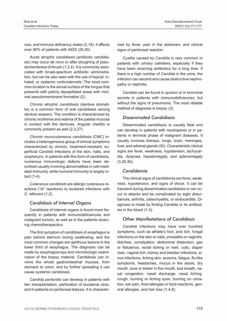
cies, and im mune de fi ciency states (2,18). It af fects
over 90% of pa tients with AIDS (29,30).
Acute atro phic candidiasis (an ti bi otic candidia -
sis) may oc cur de novo or af ter slough ing of pseu -
do membrane of thrush (1,2,4). It is com monly as so -
ci ated with broad-spec trum an ti bi otic ad min is tra -
tion, but can be also seen with the use of top i cal, in -
haled, or sys temic corticosteroids. The most com -
mon lo ca tion is the dor sal sur face of the tongue that
pres ents with patchy depapillated ar eas with min i -
mal pseudomembrane for ma tion (2).
Chronic atro phic candidiasis (den ture stomati -
tis) is a com mon form of oral candidiasis among
den ture wear ers. The con di tion is char ac ter ized by
chronic er y thema and edema of the pal a tal mu cosa
in con tact with the den tures. An gu lar cheilitis is
com monly pres ent as well (2,3,27).
Chronic mucocutaneous candidiasis (CMC) in -
cludes a het er o ge neous group of clin i cal symp toms
char ac ter ized by chronic, treat ment-re sis tant, su -
per fi cial Candida in fec tions of the skin, nails, and
oropharynx. In pa tients with this form of candidiasis,
nu mer ous im mu no logic de fects have been de -
scribed usu ally in volv ing ab nor mal i ties in cell-me di -
ated im mu nity, while humoral im mu nity is largely in -
tact (1-4).
Cu ta ne ous candidods are al ler gic cu ta ne ous re -
ac tions (“id” re ac tions) to lo cal ized in fec tions with
C. albicans (1,2).
Candidosis of In ter nal Or gans
Candidosis of in ter nal or gans is found more fre -
quently in pa tients with immunodeficiencies and
ma lig nant tu mors, as well as in the pa tients re ceiv -
ing chemotherapeutics.
The first symp tom of candidosis of esoph a gus is
pain be hind ster num dur ing swal low ing, and the
most com mon changes are aphthous le sions in the
lower third of esoph a gus. The di ag no sis can be
made by esophagoscopy and microbiologic ex am i -
na tion of the bi opsy ma te rial. Candidosis can in -
volve the whole gas tro in tes ti nal mu cosa, from
stom ach to co lon, and by fur ther spread ing it can
cause sys temic candidosis.
Candida peri to ni tis can de velop in pa tients with
lien trans plan ta tion, per fo ra tion of du o de nal ul cer,
and in pa tients on peritoneal di al y sis. It is char ac ter -
ized by fe ver, pain in the ab do men, and clin i cal
signs of peritoneal re ac tion.
Cys ti tis caused by Candida is very com mon in
pa tients with uri nary cath e ters, es pe cially if they
have been re ceiv ing an ti bi ot ics for a long time. If
there is a high num ber of Candida in the urine, the
in fec tion can as cend and cause ob struc tive nephro -
pathy or ne phri tis.
Candida can be found in spu tum or in bron chial
se crets in pa tients with immunodeficiencies, but
with out the signs of pneu mo nia. The most re li able
method of di ag no sis is bi opsy (3).
Dis sem i nated Candidiasis
Dis sem i nated candidiasis is usu ally fa tal and
can de velop in pa tients with neutropenia or in pa -
tients in ter mi nal phase of ma lig nant dis eases. It
usu ally in volves kid neys, lungs, brain, meninges,
liver, and ad re nal glands (30). Char ac ter is tic clin i cal
signs are fe ver, weak ness, hypotension, tachy car -
dia, dyspnea, hepatomegaly, and splenomegaly
(3,29,30).
Candidemia
The clin i cal signs of candidemia are fe ver, weak -
ness, hypotension, and signs of shock. It can be
tran sient dur ing dis sem i nated candidosis or can oc -
cur in at tacks and be com pli cated by sight dis tur -
bances, ar thri tis, osteomyelitis, or endocarditis. Di -
ag no sis is made by find ing Candida or its an ti bod -
ies in the blood (1-3).
Other Man i fes ta tions of Candidosis
Candida in fec tions may have over hun dred
symp toms, such as ath lete’s foot, jock itch, fun gal
in fec tions on the skin or nails, pros ta ti tis or vag i ni tis,
di ar rhea, con sti pa tion, ab dom i nal dis ten sion, gas
or flat u lence, rec tal itch ing or rash, colic, di a per
rash, vag i nal itch, kid ney and blad der in fec tions, si -
nus in fec tions, itch ing skin, ec zema, fa tigue, flu-like
symp toms, head aches, mu cus in the stools, dry
mouth, sore or blis ter in the mouth, bad breath, na -
sal con ges tion, na sal dis charge, na sal itch ing,
cough, burn ing or itch ing eyes, burn ing on uri na -
tion, ear pain, food al ler gies or food re ac tions, gen -
eral al ler gies, and hair loss (1-4,8).
ACTA DERMATOVENEROLOGICA CROATICA 175
Nola et al: Acta Dermatovenerol Croat
Candida In fec tions To day 2003;11(3):171-177
HISTOPATHOLOGY OF CANDIDOSIS
Com mon su per fi cial candidiasis of the skin has
as pri mary le sion a subcorneal pus tule re sem bling
that of im pe tigo. The fun gal or gan isms are pres ent
in small amounts in the stra tum corneum. They con -
sist of mycelia and ovoid spores, some in a bud ding
stage.
Candida granuloma is char ac ter ized by pro -
nounced papillomatosis and hyperkeratosis, and a
dense in fil trate of lym pho cytes, plasma cells, neu tro -
philes, and for eign-body gi ant cells in the dermis
(1-3).
DI AG NO SIS OF CANDIDOSIS
Di ag no sis of su per fi cial cu ta ne ous Candida in -
fec tion can be made on the ba sis of typ i cal ap pear -
ance of the clin i cal le sions and the pres ence of sat -
el lite vesicopustules, and con firmed by KOH ex am i -
na tion and cul ture of skin scrap ings (1,2).
Di ag no sis of dis sem i nated in fec tions is not easy
and can be made only in 10-40% of the pa tients. The
iden ti fi ca tion of the patho gen by hemoculture and/or
tis sue bi opsy is the most rel e vant method. Other op -
tion is by serological meth ods, by which an ti bod ies,
an ti gens, or met a bolic prod ucts of fungi are traced in
blood. Serological meth ods in clude in di rect immuno -
fluorescent method (IgG and IgM) of de ter mi na tion
of Candida an ti gens (an ti gen of cell mem brane,
cytoplasmatic an ti gen), de ter mi na tion of me tab o lites
of fun gus (arabinitol), and de ter mi na tion of mi to -
chon drial and chro mo somal DNA of fungi (3).
TREAT MENT OF CANDIDOSIS
Drugs used in man age ment of cu ta ne ous
candidiasis in clude polyene an ti bi ot ics (nystatin
cream, so lu tion, or pow der) and imidazoles (mico -
na zole, clotrimazole or econazole, ketoconazole,
itraconazole, and fluconazole). For lo cal treat ment,
very con ve nient is 0.1% gen tian vi o let, es pe cially
for mucosal in fec tion in chil dren and adults
(25,34-36). Gen tian vi o let is not used for new borns
be cause it may ir ri tate the infant’s mouth (25).
Sys temic Candida in fec tions re quire parenteral
ad min is tra tion of amphotericin, ad min is tra tion of
ketoconazole per os or fluconazole per os or in tra -
ve nously (3).
Amphotericin (fungizone) is a polyene an ti bi otic
prep a ra tion de rived from Streptomyces nodosus
(1-3). It is in sig nif i cantly resorbed from the gut and
there fore ad min is tered orally only in the treat ment
of fun gal dis eases of the gut. Whether ad min is tered
in tra ve nously or by in fu sion, amphotericin can
reach the nec es sary con cen tra tion and spread in all
tis sues. It passes blood-brain bar rier poorly, ex cept
in case of men in gi tis. Amphotericin is ex creted very
slowly, mainly by the kid neys. It is ad min is tered in -
tra ve nously in in fu sion form and its ad min is tra tion is
in di cated in all forms of deep mycosis. In the treat -
ment of mycotic men in gi tis it should be ad min is -
tered intrathecally. Treat ment usu ally takes sev eral
weeks (3).
PRE VEN TION OF CANDIDOSIS
Pre ven tion of candidosis con sists of the re moval
of all pre dis pos ing fac tors. In su per fi cial candidosis
it con sists of wear ing suit able clothes, main tain ing
intertriginous re gions dry, clean ing mouth and den -
tures prop erly, chang ing di a pers fre quently, and so
on (1-3).
In pre ven tion of sys temic Candida in fec tion, it is
im por tant to ra tio nal ize an ti bi otic ad min is tra tion,
de crease ap pli ca tion of corticosteroids (as much as
pos si ble), con trol di a be tes mellitus, and re duce the
use of in tra ve nous and uri nary cath e ters (3).
Ref er ences
1 Braun-Falco O, Plewig G, Wolff HH, Winkelmann RK.Dermatomycoses. In: Braun-Falco O, Plewig G, WolffHH, Winkelmann, ed i tors. Der ma tol ogy. Berlin:Springer Verlag; 1991. p. 219-47.
2 Fitzpatrick TB, Eisen AZ, Wolff K, Freedberg JM,Frak-Austen K. Der ma tol ogy in gen eral Med i cine. 4thed. New York: McGraw-Hill; 1993.
3 Soldo I. Sistemne gljiviène infekcije. In: Vrhovac B, ed i -tor. Interna medicina. Zagreb: Naprijed; 1991. p.1565-7.
4 Du Vivier A, McKee PH. So lar dam age and skin can -cer. In: An thony du Vivier. At las of clin i cal der ma tol -ogy. 2nd ed. Lon don: Der ma tol ogy Intrenational Ltd;1998. p. 9.1-9.37.
5 Hay RJ, Moore M. My col ogy. In: Rook A, WilkinsonDS, Ebling FJ, Cham pion RH, Bur ton JL, BreathnackSM, ed i tors. Text book of der ma tol ogy. 6th ed.Blackwell Sci en tific Pub li ca tions; 1998. p. 1335-50.
6 Garber G. An over view of fun gal in fec tions. Drugs2001;61 Suppl:11-2.
176 ACTA DERMATOVENEROLOGICA CROATICA
Nola et al: Acta Dermatovenerol Croat
Candida In fec tions To day 2003;11(3):171-177
7 Rippon JW. Candidiasis and the patho genic yeast. In:Med i cal my col ogy: the patho genic and the patho genicactinomycetes. Phil a del phia: WB Saunders; 1982. p.484.
8 Hopfer Rl. My col ogy of Candida in fec tions. In: BodeyGP, Fainstein V, ed i tors. Candidiasis. New York: Ra ven Press; 1985. p. 1.
9 De Bernardis F, Mondello F, San Millan R, Ponton J,Cassone A. Biotyping and vir u lence prop er ties of skiniso lates of Candida parapsilosis. J Clin l Microbiol1999;37:3481-6.
10 Cot ter G, Kavanagh K. Ad her ence mech a nisms ofCandida albicans. Br J Biomed Sci 2000;57:241-9.
11 Okeke CN, Tsuboi R, Kawai M, Yamazaki M,Reangchainam S, Ogawa H. Re verse tran scrip tion -3’rapid am pli fi ca tion of cDNA ends-nested PCR of ACT1and SAP2 mRNA as a means of de tect ing vi a bleCandida albicans in an in vi tro cu ta ne ous candidiasismodel. J In vest Dermatol 2000;114:95-100.
12 Kvaal C, Lachke SA, Srikantha T, Daniels K, Mc Coy J,Soll DR. Misexpression of the opaque-phase-spe cificgene PEP1 (SAP1) in the white phase of Candidaalbicans con fers in creased vir u lence in a mouse modelof cu ta ne ous in fec tion. In fect Immun 1999;67:6652-62.
13 Scherwitz C. Ultrastructure of hu man cu ta ne ouscandidosis. J In vest Dermatol 2000;198:278.
14 Mon tes LF, Wilborn WH. Fun gus-host re la tion ship incandidiasis: a brief re view. Arch Dermatol 1985;121:119.
15 Simark-Mattsson C, Jontell M, Bergenholtz G,Dahlgren UI. Re duced in vivo cell-me di ated im munere sponses to mumps, tu ber cu lin, and strep to ki -nase/streptodornase but not to Candida albicans in oral li chen planus. J Dent Res 1999;78:1704-10.
16 Savolainen J, Kosonen J, Lintu P, Viander M, Pene J,Kalimo K, et al. Candida albicans mannan- and pro -tein-in duced humoral, cel lu lar and cytokine re sponsesin atopic der ma ti tis pa tients. Clin Exp Al lergy 1999;29:824-31.
17 Abu-Elteen KH. In ci dence and dis tri bu tion of Candidaspe cies iso lated from hu man skin in Jor dan. Mycoses1999;42:311-7.
18 Velegraki A, Nicolatou O, Theodoridou M, Mostrou G, Legakis NJ. Pae di at ric AIDS-re lated lin ear gingival er y -thema: a form of erythematous candidiasis? J OralPathol Med 1999;28:178-82.
19 Kimura M, Tsuruta S, Yoshida T. Mea sure ment ofCandida-spe cific lym pho cyte pro lif er a tion by flowcytometry in chil dren with atopic der ma ti tis. Arerugi1998;47:449-56.
20 Kimura M, Tsuruta S, Yoshida T. IFN-gamma plays adom i nant role in upregulation of Candida-spe cific IgEsyn the sis in pa tients with atopic der ma ti tis. Int Arch Al -lergy Immunol 2000;122:195-9.
21 Kimura M, Maeng DJ, Kobayashi M, Suto H, Ogawa H.Fungi and atopic der ma ti tis. Nip pon Ishinkin GakkaiZasshi 1999;40:79-83.
22 Arzumanyan VG, Magarshak OO, Semenov B. Yeastfungi in pa tients with al ler gic dis eases: spe cies va ri etyand sen si tiv ity to antifungal drugs. Bull Exp Biol Med2000;19:601-4.
23 Morita E, Hide M, Yoneya Y, Kannbe M, Tanaka A,Yamamoto S. An as sess ment of the role of Candidaalbicans an ti gen in atopic der ma ti tis. J Dermatol1999;26:282-7.
24 Adachi A, Horikawa T, Ichihashi M, Takashima T,Komura A. Role of Candida al ler gen in atopic der ma ti -tis and ef fi cacy of oral ther apy with var i ous antifungalagents. Arerugi 1999;48:719-25.
25 Lambiase MC. Candidiasis, cu ta ne ous. Avail able at:http://www.emedicine.com/derm/topic67.htm. Ac ces -sed: July 3, 2003.
26 Dreizen S. Oral candidiasis. Am J Med 1984;77:28.
27 Nyquist G. A study of den ture sore mouth: an in ves ti ga -tion of trau matic, al ler gic and toxic le sions of the oralmu cosa aris ing from the use of full denotures. ActaOdontal Scand 1952;10(Suppl 9):11.
28 Nyiresy P. Chronic vulvovaginal candidiasis. Am FamPhy si cian 2001;63:697-701.
29 Vazquez JA. Ther a peu tic op tions for the man age mentof oropharyngeal and esoph a geal candidiasis inHIV/AIDS pa tients. HIV Clin Tri als 2000;1:47-59.
30 Vazquez JA. Op tions for menagement of mucosalcandidosis in pa tients with AIDS and HIV in fec tions.Pharmacotherapy. 1999;19:76-87.
31 Grouhi M, Dalal I, Nisbet-Brown E, Roifman CM. Ce re -bral vasculitis as so ci ated with chronic mucocutaneouscandidiasis. J Pediatr 1998;133:571-4.
32 Del Pozo MM, Bermejo F, Molina JA, de la Fuente EC,Mar ti nez-Mar tin P, Benito-Leon J. Chronic neutrophilic men in gi tis caused by Candida albicans. Neurologia1998;13:362-6.
33 De Andrade MF, Nishinari K, Puech-Leao P. Intertrigoin pa tients with lower limb lymphedema: clin i cal andlab o ra tory cor re la tion. Revista do Hos pi tal dasClinicas; Faculdade de Medicina Da Universidade deSao Paulo 1998;53:3-5.
34 Schaller M, Preidel H, Januschke E, Korting HC. Lightand elec tron mi cro scopic find ings in a model of hu man cu ta ne ous candidosis based on re con structed hu manepi der mis fol low ing the top i cal ap pli ca tion of dif fer enteconazole for mu la tions. J Drug Tar get 1999;6:361-72.
35 Hazen KC. Fun gi cidal ver sus fungistatic ac tiv ity ofterbinafine and itraconazole: an in vi tro com par i son. JAm Acad Dermatol 1998;38:S37-41.
36 Bryan MG. Candidiasis, chronic mucocutaneous.E-med i cine. Avail able at: http//www.emedicine.com/derm/topic569.htm. Ac cessed: July 3, 2003.
ACTA DERMATOVENEROLOGICA CROATICA 177
Nola et al: Acta Dermatovenerol Croat
Candida In fec tions To day 2003;11(3):171-177

NAPHTHALAN – A NAT U RAL ME DIC I NAL PROD UCT
Pero Vržogiæ, Želimir Ostrogoviæ, Anða Alajbeg
Naftalan Spe cial Hos pi tal for Med i cal Re ha bil i ta tion, Ivaniæ Grad, Croatia
Cor re spond ing au thor:
Pero Vržogiæ, M.D.
Naftalan Spe cial Hos pi tal for
Med i cal Re ha bil i ta tion
Omladinska 23
10310 Ivaniæ Grad, Croatia
Re ceived: 30. 06. 2003.
Ac cepted: 20. 07. 2003.
IN TRO DUC TION
Ev ery thing spring ing from the earth is termed
wa ter or oil. Ac cord ing to this clas si fi ca tion, naph -
tha lan be longs to oils, or more pre cisely to naph -
thene-based oils. To day, the term “oil” re minds us of
a power source, a fuel in our car tanks, and seems
to be less con ceiv able as a me dic i nal agent. There -
fore, it should be em pha sized that oil was first rec -
og nized as a me dic i nal prod uct, and only much
later as a power source.
As early as the 1930’s, ac a de mi cian T. G.
Pašaev tried to iso late naphthalan from in dus trial
par af fin and naph thene oils and pro posed the term
”naphthalan oil” for the prod uct (1). Ac cord ing to
cur rent con cepts, how ever, the term “earth min eral
oil” seems most ap pro pri ate (1,2).
Naphthalan – An cient Cure
The use of oil as a me dic i nal agent dates back to
an cient Egyp tians, Assyrians, and Bab y lo nians (2).
There are his tor i cal re cords on a num ber of oils that
were used as me dic i nal agents (2). For ex am ple, on
the Apsheron Pen in sula, wan der ing sur geons used
the “white or red Surahan oil”; the oil ob tained from
the Tagern Lake Bogem monks named “St.
Quirinus oil”; North Amer i can In di ans also used oil
as a me dic i nal agent in wound heal ing and man -
age ment of skin dis eases (2). Pliny, a Ro man nat u -
ral ist and his to rian, pro vided ini tial in for ma tion on
the me dic i nal prop er ties of oil in his book Naturalis
historia, writ ing about the min eral oil used by the cit -
i zens of Agrigento (Sic ily) in the man age ment of
178
Acta Dermatovenerol Croat 2003;11(3):178-184 RE VIEW
SUM MARY Naphthalan has long been known for its me dic i nal prop er ties and ben e fi cial ef fect in in flam ma tory dis eases such as pso ri a sis, atopic der ma ti -tis, and psoriatic der ma ti tis. Physicochemical anal y ses of the naphthalanfound at the Križ oil field near Ivaniæ Grad in Croatia have shown it to be iden -ti cal to the naphthalan from Baku, Azerbaijan, which has been used in thetreat ment of vul gar pso ri a sis since the be gin ning of the 20th cen tury. AtNaftalan Spe cial Hos pi tal for Med i cal Re ha bil i ta tion, naphthalan ther apy hasbeen used for 14 years. Ex pe ri ence ac quired to date in the use of naphthalanin the man age ment of squamous dermatoses and atopic der ma ti tis as well as in flam ma tory rheu matic dis eases has shown fa vor able re sults that have been sci en tif i cally and pro fes sion ally ver i fied. Naphthalan is def i nitely a po tent nat -u ral me dic i nal agent, which should be fur ther in ves ti gated to con firm its us -age in the treat ment of these dis eases as fully jus ti fied.
KEY WORDS naphthalanotherapy; baths; dermatologic agent; pso ri a sis; tars
skin le sions (1); the heavy Penn syl va nian oil,
known as “Sen eca oil”, was suc cess fully used in the
treat ment of skin dis eases not so long ago; and the
“light Par me san oil”, oleum pe tro leum Italicum, from
Amian near Parma was used in the Mid dle Ages not
only in It aly but through out Eu rope (2,3).
A num ber of writ ten doc u ments dat ing from the
Mid dle Ages deal with the re sults, in di ca tions, and
con tra in di ca tions of oil treat ment, e.g., “Tafta” and
“Mehzan – Uol Aravie” (1-3). Naphthalan wells
were found be tween the Large and Small Cau ca -
sus Moun tains, along the Kuru River in Azerbaijan.
Naphthalan was also men tioned in a 700 years old
doc u ment de scrib ing the great Hin dus’ rit ual of of -
fer ing sac ri fice “for cure from awful diseases” (3).
There are many leg ends on the me dic i nal prop -
er ties of naphthalan, the ru mor on its cu ra tive ef -
fects hav ing spread far and wide. Sick peo ple fre -
quented naphthalan wells to take naphthalan baths
or sim ply ap ply it all over the body. Camel car a vans
pass ing naphthalan wells on their Silk Road, filled
the goat skins with naphthalan and car ried it on their
way back to the Far East to sell it at fairs. That is
why naphthalan first ap peared as a me dic i nal prod -
uct in the Ori en tal med i cine. The term “naphthalan”
has been pos tu lated to de rive from ‘naft o lan’,
which in the Ori ent means ‘there where oil is” (2,3).
In the 13th cen tury, the world trav eler Marco
Polo ran across naphthalan wells on his way to
China and de scribed them in his travel-book Mil lion,
or Li bre des merveilles du monde, as large wells
filled with oil that is not ed ible but is use ful in the
treat ment of skin and joint dis eases (4).
Be gin nings of Naphthalan Prod ucts
En gi neer Jeger, who stayed in Baku dur ing the
oil boom in 1895, learned about the me dic i nal prop -
er ties of naphthalan by word of mouth, and went to
see the wells (2). He de cided to take them on lease
and soon or ga nized a tub treat ment, also ar rang ing
for ini tial drills to pro vide ad e quate sup ply with
naphthalan. In this way he en sured higher naphtha -
lan sup plies than those pro vided at the nat u ral wells
and soon started trans port ing it to Dresden, where
he built a small plant for naphthalan pro cess ing and
man u fac ture of naphthalan oint ments and other
prod ucts. At the same time, Pro fes sor List from
Magdeburg em barked upon similar activities (1-8).
Thus, there were two naphthalan com pa nies in
Ger many at the be gin ning of the 20th cen tury: Na -
ph thalan in Dresden and Naphthalan in Magde -
burg, both man u fac tur ing var i ous stan dard ized
naphthalan-based prod ucts, such as well known
Dresden oint ment “skinlan”, cos metic creams, oint -
ments for the treat ment of burns, sun tan oils, tooth -
paste, and so on. These prod ucts soon be came
very pop u lar all over Eu rope, America, and Japan.
The Naphthalan Com pany from Dresden con -
trib uted greatly to wide knowl edge about naphtha -
lan by is su ing a hand book on the use of naphthalan.
Be side Jeger’s com mer cial pa pers, this book
brought more than 600 brief re ports by the re -
nowned phy si cians of the time on their ex pe ri ence
with naphthalan. Pro fes sor Unna, a fa mous Ger -
man der ma tol o gist, said: “The one who has na -
phtha lan has ev ery thing” (2).
At that time, naphthalan was in tro duced in the
phar ma co poeia of many coun tries un der the terms
Naphthalanum, Naphthalanum liquidum, Vas e line
oduste saponifice, and oth ers (2). It was al most reg -
u larly avail able in phar ma cies, mostly for mu lated
as oint ment, oil, or some other prep a ra tion. In the
anthological phar ma col ogy books by Martindale
and Haagar, it is found un der the terms “na -
phthalan” and “naphalan”, re fer ring to the same
earth oil from the same well, only one of them was a
prod uct from Dresden, and the other from Magde -
burg. Kottmaier’s Hand book of Prac ti cal Med i cine,
pub lished in 1967, rec om mends a magistral prod -
uct con tain ing 20% of naphthalan (9).
Af ter the Oc to ber Rev o lu tion in 1917, the bor -
ders be tween the USSR and the rest of the world
were closed, and naphthalan fell into obliv ion. How -
ever, it should be noted that der ma tol o gists from the
Zagreb Uni ver sity De part ment of Der ma tol ogy
(Kogoj, Èajkovac, and oth ers) con tin ued pre scrib -
ing magistral naphthalan-con tain ing prod ucts that
some phar ma cies still used to prepare (10,12).
And what hap pened to naphthalan in Azerbaijan
af ter the Oc to ber Rev o lu tion?
Sci en tific elab o ra tion of the use of naphthalan oil
was launched as early as 1920, a health re sort with
33 beds was opened in 1933, and In sti tute for the
Study of Naphthalan Ac tion and Uti li za tion was
founded at the De part ment of Balneology and
Phys i cal Med i cine, School of Med i cine in Baku in
ACTA DERMATOVENEROLOGICA CROATICA 179
Vr´ogiæ: Acta Dermatovenerol Croat
Naphthalan – a Nat u ral Me dic i nal Prod uct 2003;11(3):178-184
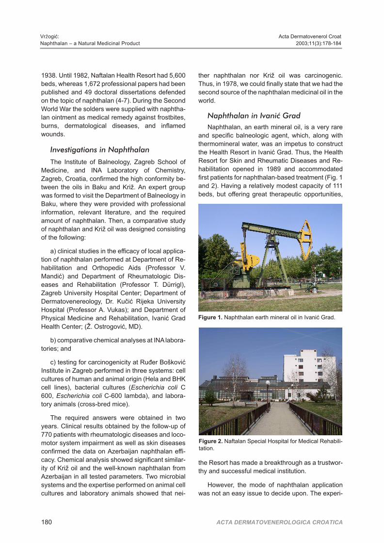
1938. Un til 1982, Naftalan Health Re sort had 5,600
beds, whereas 1,672 pro fes sional pa pers had been
pub lished and 49 doc toral dis ser ta tions de fended
on the topic of naphthalan (4-7). Dur ing the Sec ond
World War the sol ders were sup plied with naphtha -
lan oint ment as med i cal rem edy against frost bites,
burns, dermatological diseases, and inflamed
wounds.
In ves ti ga tions in Naphthalan
The In sti tute of Balneology, Zagreb School of
Med i cine, and INA Lab o ra tory of Chem is try,
Zagreb, Croatia, con firmed the high con for mity be -
tween the oils in Baku and Križ. An ex pert group
was formed to visit the De part ment of Balneology in
Baku, where they were pro vided with pro fes sional
in for ma tion, rel e vant lit er a ture, and the re quired
amount of naphthalan. Then, a com par a tive study
of naphthalan and Križ oil was de signed consisting
of the following:
a) clin i cal stud ies in the ef fi cacy of lo cal ap pli ca -
tion of naphthalan per formed at De part ment of Re -
ha bil i ta tion and Or tho pe dic Aids (Pro fes sor V.
Mandiæ) and De part ment of Rheumatologic Dis -
eases and Re ha bil i ta tion (Pro fes sor T. Dürrigl),
Zagreb Uni ver sity Hos pi tal Cen ter; De part ment of
Dermatovenereology, Dr. Kuèiæ Rijeka Uni ver sity
Hos pi tal (Pro fes sor A. Vukas); and De part ment of
Phys i cal Med i cine and Re ha bil i ta tion, Ivaniæ Grad
Health Center; (Ž. Ostrogoviæ, MD).
b) com par a tive chem i cal anal y ses at INA lab o ra -
to ries; and
c) test ing for car ci no ge nic ity at Ruðer Boškoviæ
In sti tute in Zagreb per formed in three sys tems: cell
cul tures of hu man and an i mal or i gin (Hela and BHK
cell lines), bac te rial cul tures (Esch e richia coli C
600, Esch e richia coli C-600 lambda), and lab o ra -
tory an i mals (cross-bred mice).
The re quired an swers were ob tained in two
years. Clin i cal re sults ob tained by the fol low-up of
770 pa tients with rheumatologic dis eases and lo co -
mo tor sys tem im pair ment as well as skin dis eases
con firmed the data on Azerbaijan naphthalan ef fi -
cacy. Chem i cal anal y sis showed sig nif i cant sim i lar -
ity of Križ oil and the well-known naphthalan from
Azerbaijan in all tested pa ram e ters. Two mi cro bial
sys tems and the ex per tise per formed on an i mal cell
cul tures and lab o ra tory an i mals showed that nei -
ther naphthalan nor Križ oil was car ci no genic.
Thus, in 1978, we could fi nally state that we had the
sec ond source of the naphthalan medicinal oil in the
world.
Naphthalan in Ivaniæ Grad
Naphthalan, an earth min eral oil, is a very rare
and spe cific balneologic agent, which, along with
thermomineral wa ter, was an im pe tus to con struct
the Health Re sort in Ivaniæ Grad. Thus, the Health
Re sort for Skin and Rheu matic Dis eases and Re -
ha bil i ta tion opened in 1989 and ac com mo dated
first pa tients for naphthalan-based treat ment (Fig. 1
and 2). Hav ing a rel a tively mod est ca pac ity of 111
beds, but of fer ing great ther a peu tic op por tu ni ties,
the Re sort has made a break through as a trust wor -
thy and successful medical institution.
How ever, the mode of naphthalan ap pli ca tion
was not an easy is sue to de cide upon. The ex pe ri -
180 ACTA DERMATOVENEROLOGICA CROATICA
Vr´ogiæ: Acta Dermatovenerol Croat
Naphthalan – a Nat u ral Me dic i nal Prod uct 2003;11(3):178-184
Fig ure 1. Naphthalan earth min eral oil in Ivaniæ Grad.
Fig ure 2. Naftalan Spe cial Hos pi tal for Med i cal Re ha bil i -ta tion.

ence from the Naphthalan Health Re sort in
Azerbaijan could not be used be cause, ac cord ing
to Eu ro pean cri te ria, ap pli ca tion of na tive oil was
not ac cept able. There fore, we have adopted Pro -
fes sor Pašaev’s in struc tions from the lit er a ture.
Pašaev as cribed naphthalan bioactive com po nent
to polycyclic naph thene car bo hy drates and sug -
gested re moval of tar and ar o matic con tent. Na -
phthalan ther apy prac ticed at the Naphthalan Re -
sort in Ivaniæ Grad is based on this prin ci ple. The re -
fined naphthalan oil dis til late of 283-432 °C is free
from tar and other un de sired sub stances, while the
con cen tra tion of me dic i nal com po nents is in -
creased, be cause 4 li ters of na tive naphthalan yield
1 li ter of the pu ri fied agent. The use of this me dic i nal
prod uct is as so ci ated with a min i mal rate of con tra -
in di ca tions and pos si ble side ef fects, whereas the
ap pli ca tion meets the re quired cos metic and hy -
gienic con di tions. We use naphthalan tubs in tended
for sit ting and ly ing pos ture (Fig. 3), where
naphthalan is be ing re gen er ated, rinsed, and ther -
mally “ster il ized” dur ing the ses sion; there fore, the
process of naphthalan application is automated
(10,12,15-17).
PHYSICOCHEMICAL PROP ER TIES OF
NAPHTHALAN
Pe tro leum (oil) and its de riv a tives are rather
com plex mix tures of or ganic com pounds, pre dom i -
nantly com posed of car bon and hy dro gen, and
some heteroatoms (mostly ni tro gen, sul fur, and ox -
y gen) (18,19). Oil con stit u ents be long to dif fer ent
com pound classes: sat u rates (com posed of acy clic
and cy clic naph thene moi eties), aromatics (com -
pounds con tain ing at least one ar o matic ring), and
polars (con tain ing at least one heteroatom be sides
car bon and hy dro gen). Oil com po nents with high
mo lec u lar mass, which pre cip i tate in n-alkanes, are
classified as asphaltenes.
To day, there are plenty of pub lished data on the
com po si tion of oil (18-20). As an a lyt i cal tools be -
come ever more so phis ti cated re gard ing sep a ra -
tion and sen si tiv ity, the num ber of com po nents
thought to com pose pe tro leum in creases. Some
classes of oligocyclicity, which be long to the
so-called bi o log i cal mark ers, are ex ten sively in ves -
ti gated (21-24). These are com plex or ganic com -
pounds, which, dur ing the pro cess of pe tro leum
gen er a tion in na ture, have re tained the chem i cal
skel e ton char ac ter is tics of the par ent or ganic mol e -
cules (of bi o logic or i gin). They of ten oc cur among
the va ri ety of stereoisomers, which sensitively
indicate the different stresses they had suffered
(24).
For cen tu ries pe tro leum has been con sid ered to
have cu ra tive prop er ties, to ac cel er ate skin and mu -
cous mem brane cell re gen er a tion. Baths and prep -
a ra tions as oint ments, pastes, lin i ments, and
creams have been ap plied. Azerbaijani pe tro leum
called “naphthalan” was used as antimicrobial and
heal ing prep a ra tion for burns and in ju ries, for the
treat ment of ec zema, ar thri tis, neurodermitis, skin
fungi, and par a sites (25). Ther a peu tic ef fects of
naphthalan oil and its prep a ra tions have been in -
ten sively stud ied (26-30). Naphthalan baths are
used for rheu ma tism and gout treat ment and have
also been re ported ef fec tive in the treat ment of
some gynecologic dis eases. Sat u rated pe tro leum
oligocycles (naph thenes) were con sid ered to be the
main active ingredients in disease treatment.
Some en cour ag ing re sults with prep a ra tions de -
rived from the Cro atian naph tha lene oil have been
re ported in the treat ment of pso ri a sis vulgaris and
atopic ec zema (24). The ne ces sity of pre cau tion in
health ser vice makes in ten sive re search oblig a tory
in or der to en joy the nat u ral ad van tages and to min i -
mize pre dic tive haz ards. To eval u ate the Cro atian
naph tha lene oil, a re al is ti cally based in ter dis ci plin -
ACTA DERMATOVENEROLOGICA CROATICA 181
Vr´ogiæ: Acta Dermatovenerol Croat
Naphthalan – a Nat u ral Me dic i nal Prod uct 2003;11(3):178-184
Fig ure 3. Naphthalan tub ap pli ca tion in psoriatic pa tients.
ary study has been launched to bring, hope fully,
some elucidation to the subject complexity.
Naphthalan oil be longs to “youn ger” oils than the
in dus trial oil; it is of plant or i gin and is found in su -
per fi cial lay ers of the earth (100-700 m depth). Na -
tive naphthalan springs as a thick, dark-brown liq -
uid of a spe cific ar o matic odor. It is clas si fied among
heavy oils for its high spe cific weight (0.93-0.98 at
15 °C). Naphthalan con tains neg li gi ble con cen tra -
tions of light ben zine, ligroine and ker o sine frac -
tions, whereas con densed par af fins are only found
in traces or not at all. It is char ac ter ized by high vis -
cos ity, 6.45 °E at 50 °C ac cord ing to Engler, and
con tains 0.46% of sul fur and 0.11% of ni tro gen, ac -
cord ing to Kjeldal. Na tive oil con tains 28% of tars,
with con den sa tion point at 21 °C and in flam ma ble
point at 110 °C. Naphthalan con tains a rel a tively
high con cen tra tion of naph thene ac ids (3%),
among them a biostimulant cyc lo pen tane acid and
1.32% of or ganic acids, which is why it is
characterized by acidic reaction.
Naphthalan also con tains a high per cent age of
naph thene car bo hy drates (55%), 15% of ar o matic
car bo hy drates, and no meth ane car bo hy drates.
Naph thene oil is char ac ter ized by high polycyclicity
of naph thene car bo hy drates, with a small num ber
of car bon at oms in lat eral chains con sti tut ing three-,
tetra-, and pentacyclic com pounds of the cyclopen -
ta no perhydrophenanthrene struc ture, the most im -
por tant be ing isoprenanes, steranes, and triter -
panes. By their chem i cal struc ture, they closely re -
sem ble hor mones, vi ta mins, and bioactive sub -
stances. The me dic i nal prop er ties have just been
as cribed to these polycyclic naph thene car bo hy -
drates. Fi nally, naphthalan is neither skin irritant nor
carcinogenic.
THER A PEU TIC EF FECTS OF
NAPHTHALAN
In ad di tion to me chan i cal and ther mal ef fects,
naphthalan has a very pro nounced bioactive ef fect
on the hu man body. The main bioactive com po -
nents of naphthalan are polycyclic naph thene car -
bo hy drates, con sist ing of 4, 5, or more rings of a
char ac ter is tic cyclopentanoperhydrophenanthrene
nu cleus. This struc ture is very sim i lar to some en -
do crine com pounds, e.g., vi ta mins, hor mones, and
bioactive sub stances. Thus, it is pos tu lated that
naphthalan en ters the body by the prin ci ple of sub -
sti tu tion only to be in volved in var i ous phys i o logic
and patho logic pro cesses, i.e. in the humoral-hor -
mone chains of the pathogenesis of various dis e -
ases.
Based on the 10-year clin i cal fol low-up and ob -
ser va tion of 10,000 pa tients, lit er a ture data, and
lab o ra tory and ex per i men tal stud ies, a num ber of
naphthalan ef fects have been iden ti fied and
described.
Anti-in flam ma tory Ef fect
The ac tive sub stances from naphthalan pen e -
trate through the skin to form com pounds sim i lar to
the suprarenal gland me di a tors, which stim u late the
func tion of the glands and act on corticosteroid syn -
the sis. Lab o ra tory find ings con firm this ac tiv ity,
point to nor mal iza tion of the in flam ma tory ac tiv ity
pa ram e ters, and sug gest an ef fect on im mune func -
tion. Clin i cally, sup pres sion of in flam ma tory ac tiv ity,
re duc tion of edema and rubor, pain al le vi a tion, gen -
eral state im prove ment, and re duced se ver ity and
du ra tion of “morn ing stiff ness” are re corded in pa -
tients with in flam ma tory rheu ma tism who were
treated with naphthalan. In skin diseases, reduction
of infiltration and erythema is observed (29).
Antiproliferative Ef fect
Naphthalan ex erts antiproliferative ac tiv ity and
de creases the immunocompetent cell counts in
psoriatic le sions (30).
Stim u la tion of Ter mi nal KeratinocyteDif fer en ti a tion
Stud ies of the ef fects of naphthalan on in vi tro
pro lif er a tion and dif fer en ti a tion of keratinocytes
were car ried out at De part ment of Der ma tol ogy and
Venereology, J. W. Goe the Uni ver sity Hos pi tal in
Frank furt, Ger many. The re sults pointed to the in hi -
bi tion of keratinocyte pro lif er a tion, with a ten dency
to nor mal iza tion in the psoriatic skin. These two
facts sug gest the naphthalan ther apy to be a prom -
is ing con tri bu tion in the man age ment of psoriasis
and atopic dermatitis (31).
No Toxic Ef fects
The ex pe ri ence from sev eral-year fol low-up of
pa tients un der go ing naphthalan ther apy showed no
182 ACTA DERMATOVENEROLOGICA CROATICA
Vr´ogiæ: Acta Dermatovenerol Croat
Naphthalan – a Nat u ral Me dic i nal Prod uct 2003;11(3):178-184

ther apy-re lated im pair ment of he ma to log i cal and
bio chem i cal parameters (32).
METH ODS OF NAPHTHALAN
UTI LI ZA TION
Naphthalan Bath
The pa tient is im mersed up to the shoul ders in
the oil me dium, at the tem per a ture of 34-38 °C, for
8-14 min utes. Also, only parts of the body can be
im mersed in the bath or, in stead of bath, na -
phthalan can be ap plied all over body surface.
Oc clu sive Naphthalan Dress ing
This method is usu ally ap plied over night on the
psoriatic body ar eas with se vere in flam ma tion, in fil -
tra tion, and thick scal ing patches.
Iontophoresis with Naphthalan
The me dic i nal agent is in tro duced from the cath -
ode into the af fected area by gal vanic cur rent.
Sonophoresis
Naphthalan is ap plied as a con tact me dium be -
tween the ul tra sonic head and tar get body area.
Sonophoresis for small joints of the hand and foot is
per formed in naphthalan bath into which the ul tra -
sound head is im mersed.
Mas tic Ther apy
Mas tic is a solid prep a ra tion of 30% naphthalan,
which is ap plied in the form of com press or soak ing
in dis solved me dium, as in the ap pli ca tion of par af -
fin. This thermotherapeutic pro ce dure pro duces hy -
per emia, con sid er ably pro longed ther mal ef fect,
and pro nounced con sen sual re ac tion. The mea -
sure ments of skin tem per a ture on the palm and dor -
sum of both hands be fore, dur ing, and 20 and 60
min utes af ter ther apy re vealed that the tem per a ture
in creased dur ing ther apy by 5.5 °C, ex ceed ing ini -
tial tem per a ture by 4.3 °C im me di ately upon ther -
apy, by 2.5 °C at 20 min utes, and by 1.5 °C at 60
min utes. Upon the ap pli ca tion of mas tic, pa tients
feel pleas ant warmth, less warm than par af fin of the
same tem per a ture, which allows for the use of
higher temperatures for therapeutic purpose.
Spe cific ef fects in re ha bil i ta tion in in flam ma tory
rheu ma tism should also be em pha sized, as this
ther apy re sults in pain al le vi a tion, edema re duc tion,
re lax ation of mus cu la ture ten sion, and in crease in
pre vi ously re stricted move ments. The rate of so lid i -
fi ca tion, i.e. lower or higher pro gres sive re sis tance
dur ing re ha bil i ta tion treat ment, is ad justed by mod i -
fy ing the volume ratio of the mastic product con stit -
u ents.
IN DI CA TIONS FOR THE USE OF
NAPHTHALAN THER APY
Naphthalan ther apy can be used in the treat -
ment of var i ous dis eases af fect ing dif fer ent or gans
and or gan sys tems. Skin dis eases that re spond
well to the naphthalan ther apy are atopic der ma ti tis,
pso ri a sis, prurigo, ich thy o sis, and scleroderma.
Among musculoskeletal dis eases, there are some
in flam ma tory rheu matic dis eases (rheu ma toid ar -
thri tis, psoriatic ar thri tis, and Bechterew’s dis ease),
de gen er a tive dis eases of joints and spine (arthrosis
and vertebrogenous syn dromes), and ex tra-ar tic u -
lar rheu ma tism that can be treated with naphthalan.
Naphthalan can also be ap plied in the treat ment of
pe riph eral ner vous sys tem dis eases, such as neu ri -
tis, radiculitis, neuropathies, and pa re sis, and in the
posttraumatic and post op er a tive re ha bil i ta tion of
the lo co mo tor sys tem (contractures, post op er a tive
in fil trates, circulatory disturbances, Sudeck syn -
drome, burns, and frostbites).
CON CLU SION
Naphthalan has long been known for its me dic i -
nal prop er ties. Physicochemical anal y ses of the
naphthalan found at the Križ oil field near Ivaniæ
Grad have shown it to be iden ti cal to the naphthalan
from Baku, Azerbaijan, which has been used in the
treat ment of vul gar pso ri a sis since the be gin ning of
the 20th century.
At Naftalan Spe cial Hos pi tal for Med i cal Re ha -
bil i ta tion, naphthalan ther apy has been used for 14
years, dur ing which pe riod nu mer ous pro fes sional
and sci en tific stud ies were per formed. The ex pe ri -
ences ac quired to date in the use of naphthalan in
the man age ment of squamous dermatoses and
atopic der ma ti tis as well as of in flam ma tory rheu -
matic dis eases have shown fa vor able re sults that
have been sci en tif i cally and professionally verified.
Naphthalan is def i nitely a po tent nat u ral me dic i -
nal agent call ing for ad di tional stud ies to con firm its
ACTA DERMATOVENEROLOGICA CROATICA 183
Vr´ogiæ: Acta Dermatovenerol Croat
Naphthalan – a Nat u ral Me dic i nal Prod uct 2003;11(3):178-184
fur ther us age in the treat ment of these dis eases as
fully justified.
Ref er ences
1 Pašaev TG. Naftalanskaja neft i ee leèebnoe dejstvie.Izdateljstvo: Medgiz, Moskva 1959; 3-14
2 Karaev AI, Aliev RK, Babaev AZ. Naftalanskaja neft.Moskva: Izdatel’stvo Akademii nauk SSSR; 1959; 5-11
3 Gulieva SA. Unikalnaja leèebnaja naftalanovaja neft.Baku; 1981.
4 Krust TA. Von Venedig nach China. Darmstadt:Wissenschaftliche Buch Gesellschaft; 1996.
5 Gulieva SA. Naftalanoleèenie. Baku; 1979.
6 Sultanov AA. Revmatoidnj artrit v Azerbejdžane.Baku-Maarif, Maarif ed.; 1971;44-52.
7 Kuliev AH. Naftalan i metodika ego leèebnogoprimenenia. Baku; 1973.
8 Jeger EI. Naftalan, Sbornik sobranija otzivov i dokladov vraèej o leèenii i dejsvii naftalana, prevod s nemec -kogo. Tbilisi 1904:1-8.
9 Kottmaier J. Priruènik za praktiènu medicinu. Beograd,Zagreb: Medicinska knjiga; 1967.
10 Ostrogoviæ Ž, Perin B. Primjena naftalanske nafte –naftalana u lijeèenju psorijaze i drugih kožnih bolesti.Pso ri a sis 1985;17:14-9.
11 Kogoj F. Pomasti. In: Kogoj F, ed i tor. Dermato venero -loška propedeutika. Zagreb: Nakladni zavod Hrvatske;1947: 400-40.
12 Èajkovac V. Otvoreno ljeèilište “Naftalan” za kožne ireumatske bolesti. Acta Dermatol Iug 1989;3:157.
13 Ostrogoviæ Ž, Perin B. Primjena naftalanske nafte umedicini. Reumatizam 1984;1-2:17-22.
14 Èajkovac V, Skrbin-Smeh A. Lijeèenje psorijazenaftalanom – dosadašnja iskustva. Pso ri a sis 1990;29:13-4.
15 Ostrogoviæ Ž. Naftalanoterapija psorijaze i psorijatiè -nog artritisa. MEJAD6 1990;20(Suppl):68-71.
16 Ostrogoviæ Ž. Naftalan – zemno mineralno ulje kaobalneološki agens. Medix 1999;25:83-6.
17 Ostrogoviæ Ž, Èaèiæ K, Skrbin-Smeh A. Naše iskustvo ulijeèenju psorijaze i psorijatiènog artritisa naftalano -terapijom. MEJAD6 1990;20(Suppl):132-8.
18 Li MW, Lin RZ, Liao YS, Snowdon LR, Wang PR, Li YI.Or ganic geo chem is try of oils and con den sates in theKekeya Field, South west De pres sion of the Tarim Ba sin (China). Org Geochem 1999;30:15-37.
19 Hunt JM. In: Gilluly J, ed i tor. Pe tro leum in the res er -voir.. Pe tro leum, geo chem is try and ge ol ogy. 2nd ed.San Fran cisco (CA): WH Free man; 1996. p. 617.
20 Alajbeg I, Dinter G, Alajbeg A, Telen S, Prostenik M.Study of Cro atian non-ar o matic naphthalane con stit u -ents with skel e tons anal o gous to bioactive com -pounds. J Chromatogr A 2001;918:127-34.
21 Sum mons RE, Ca pon RJ. Iden ti fi ca tion and sig nif i cance of 3-ethyl steranes in sed i ments and pe tro leum. Geo -chim Cosmochim Acta 1991;55:2391-5.
22 Stoilov I, Smith SL, Watt DS, Carlson RM, Fago FJ,Moldowan JM. Syn the sis of bi o log i cal mark ers in fos silfu els .8. H-1 and C-13 NMR anal y sis of C-23 and C-24diastereomers of 5-al pha-dinosterane. Magn ResonChem 1994;32:101-6.
23 Volkman JK, Barett SM, Blackburn SI, Mansour MP,Sikes EL, Gelin F. Microalgal biomarkers: A re view ofre cent re search de vel op ments. Org Geochem 1998;298:1163-79.
24 Lapidus J, ed i tor. The biomarket guide, in ter pret ingmo lec u lar fos sils in pe tro leum and an cient sed i ments.Englewood Cliffs (NJ): Prentice Hall; 1993;363
25 Huseinov DY, Rustamov AI. Naphthalan – the oil thatheals. Azerbaijan In ter na tional 1995;3,4:1-6.
26 Musaev AV, Guseinova SG. The naphthalan ther apy ofpa tients with gun shot trauma to the pe riph eral ner voussys tem [in Rus sian]. Vopr Kurortol Fizioter Lech FizKult 1998;5:23-7.
27 Musaev AV, Nasrullaeva SN, Namazov DZ. Aclinico-thermographic as sess ment of the ef fi cacy ofnaphthalan ther apy in pa tients with di a betic micro -angio pathies of the lower ex trem i ties [in Rus sian]. Vopr Kurortol Fizioter Lech Fiz Kult 1998;4:26-9.
28 Insanov AB, Musaev AV, Gazvinova EA. The ef fect ofthe ultraphonophoesis of deresinated naphthalan in pa -tients with a his tory of exudative pleu risy [in Rus sian].Vopr Kurortol Fizioter Lech Fiz Kult 1995;6:18-20.
29 Grigor’eva VD, Badalov NG. Deresinated naphthalanin the treat ment of psoriatic ar thri tis [in Rus sian]. VoprKurortol Fizioter Lech Fiz Kult 1994;4:13-5.
30 Vržogiæ P, Jakiæ-Razumoviæ J, Pašiæ A. Ef fect ofnaphthalan on epi der mal pro lif er a tion ac tiv ity andCD3, CD4 and CD8 lym pho cyte count. Acta Dermato -venerol Croat 2003;11:65-9.
31 Thaci D, Schindewolf M, Smeh-Skrbin A,Krnjeviæ-Peziæ G, Vržogiæ P, Dobriæ I, et al. Heavynaphthen oil ex hib its antipsoriatic ef fi cacy in vivo andantiproliferative as well as differentation-in clud ing ef -fects on keratinocytes in vi tro. Arch Dermatol 2000;136:678-9.
32 Krnjeviæ-Peziæ G, Vržogiæ P, Ostrogoviæ Ž,Smeh-Skrbin A, Dobriæ I. Some he ma to log i cal and bio -chem i cal pa ram e ters in psoriatic pa tients treated withnaphthalan. Acta Dermatovenereol Croat 1997;5:49-53.
184 ACTA DERMATOVENEROLOGICA CROATICA
Vr´ogiæ: Acta Dermatovenerol Croat
Naphthalan – a Nat u ral Me dic i nal Prod uct 2003;11(3):178-184

In Memoriam
THOMAS BERNHARD FITZPATRICK
(1919-2003)
E-mails from the ADA, sadly but nec es sar ily,may be har bin gers of bad news. When I opened mymail box in the wee hours last night, I learned aboutthe de mise of Pro fes sor Thomas B. Fitzpatrick lastSat ur day. I was aware of Tom’s suf fer ing but the im -mu ta ble fi nal ity of death com mands awe. This iswhen his tory places a full stop in its nar ra tive. TBF,as he was fre quently re ferred to, is no more.
I knew Tom for 35 years, al most to the day, and it is just over thirty years that I stayed with the Fitzeswhile en route from Buf falo back to Vi enna, about topay my first visit to Har vard. My fam ily and I couldex pe ri ence the fine hos pi tal ity and charm of theFitzes’ won der ful old home and the warmth Bea ra -di ates at all times. And not much later we had theFitzes in our home out side Vi enna, with FranzGreiter, when the women went shop ping for somevery Cen tral-East ern Eu ro pean item, namely, aplumeau. The de cades passed, the cen tury ended,and ever so many per sonal en coun ters had fol -lowed.
Tom was a fru gal per son al ity, very cu ri ous andquick-witted. In con ver sa tion he did not weigh ev ery sen tence, nay, he weighed ev ery sin gle word andput in vis i ble ques tion marks be hind, be fore hewould be lieve it. Not too long ago, I heard Wal terShel ley say that, when he was a res i dent, therewere but four MD-PhD’s in US der ma tol ogy, TomFitzpatrick, Aaron Lerner, Al bert Kligman, and Shel -ley him self.
One of the Four Grand Old Men Has Gone.
I do not feel en ti tled to write a full obit u ary, oth ers and wor thier col leagues will do that. But let me elab -o rate for the read er ship of the Acta Dermatovenero -logica Croatica what are some of the strong pil larsto up hold his mem ory in the field. Tom was four de -cades with Har vard, twenty-seven years of whichas chair man and Wigglesworth Pro fes sor of der ma -tol ogy. He was a ded i cated teacher, an al most om -ni scient der ma tol o gist, a fine in ves ti ga tor and men -tor to many. In tan dem with Aaron Lerner, in neigh -
bor ing Yale, he serendipitously formed sort of a tan -dem in pig ment cell re search. He was also ac tive inan other such trio, with Klaus Wolff and JohnParrish, in the in tro duc tion of PUVA ther apy withsys tem i cally ad min is tered psoralens. I well re mem -ber the Acad emy Meet ing in De cem ber 1974 whenthe news about this reg i men was bro ken on thefront page of the New York Times. In this verymonth, TBF cel e brated his 54th, and Klaus Wolff his 39th birth day. Lastly, the edi tion of Der ma tol ogy inGen eral Med i cine, the DIGM as it is called col lo qui -ally, is now in its 6th edi tion. This en cy clo pe dic vol -ume, two in fact per edi tion by now, has be come abi ble for in ves ti ga tors in, be low, and be yond theskin. These achieve ments guar an tee that his leg -acy will last many de cades to come. Too bad thatthe late flower of the nine ties, Fitzpatrick´s Jour nalof Clin i cal Der ma tol ogy, with ered away af ter a shortpe riod, be ing so much a mir ror of Tom se nior and ju -nior.
In nu mer a ble hon ors have been be stowed uponTom, which will not be de tailed here. The Fitzpatrick Festschrift in the JID (Sup ple ment 1983), is a goodplace to look up and most cer tainly now, upon hisde mise, other such texts will fol low.
Tom’s ap proach to prob lems, as much as hisface and the sound of his voice, will re main be foremy mind as long as I can think. And so will be theim age of his per son al ity, the cab o chon ruby peek -ing from the end of his watch chain run ning throughthe but ton holes of his gilet.
To Bea and the Ju nior Fitzes all our love andcon do lences go at this mo ment. May G-d grantthem so lace in these bit ter days of mourn ing.
R.I.P.
Au gust 19, 2003.
Karl Holubar, MD FRCP GSE
In sti tute for the His tory of Med i cine
Uni ver sity of Vi enna
Vi enna, Aus tria
185
Acta Dermatovenerol Croat 2003;11(3):185-188 NEWS AND COMMENTS
An nounce ment by the Ed i tor
Just out: The Sixth Edi tion of Fitzpatrick’s Der ma tol ogy in Gen eral Med i cine (DIGM)
Ev ery dis ci pline has its heroes and its bi bles. In
der ma tol ogy, for most of us Thomas B. Fitzpatrick
at Har vard was more than that. Es tab lished as one
of the lead ing au thor i ties in mod ern in ves ti ga tive
der ma tol ogy, he be came a sym bol and the role
model. Un for tu nately, no body is im mor tal. Heroes
die, too. We learned about his death on Au gust 16,
2003. The light of his work, how ever, re mains a so -
lace, and thus, the new est, sixth edi tion of DIGM in
two vol umes is the best way to keep his mem ory
and ideas alive. The ed i tors of this edi tion are some
of the most prom i nent der ma tol o gists (Irwin M.
Freedberg, Ar thur Z. Eisen, Klaus Wolff, K. Frank
Austen, Lowell A. Gold smith, and Ste phen I. Katz),
who were partly his close col lab o ra tors. For many
of the con tri bu tors, it was and is both a con fir ma tion
of good rep u ta tion and an act of honor to write a
chapter in this encyclopedia.
The sixth edi tion of DIGM cov ers ad vances in
clin i cal der ma tol ogy as much as in ba sic sci ences
re lated to skin, and is also in tended for phy si cians
of other spe cial ties for whom changes of the skin
are im por tant. The DIGM is truly en cy clo pe dic in its
width and depth, of which half a cen tury ago no body
could even dream. It in cludes a new sec tion on ev i -
dence-based der ma tol ogy, deal ing with dermatolo -
gic changes through the stages of life. No ta bly so,
il lus tra tions are in color. The ther a peu tic sec tions
have all been up dated. Dis or ders thought to be only
of his toric im por tance, such as antrax and variola,
are dis cussed, as well as dermatologic dis eases as
po ten tial agents of mass de struc tion. Thus, the
sixth edi tion of DIGM re flects the com plex i ties of
our age. There are 280 chap ters, grouped into 37
sec tions cov er ing all as pects of the science and
care of the skin in health and disease.
Many of the chap ters are com pletely new, and
many re writ ten. Part one con sists of two sec tions –
Gen eral Con sid er ations and In tro duc tion; part two
– Bi ol ogy and De vel op ment of Skin, of four sec -
tions; part three is en ti tled Dis or ders Pre sent ing in
the Skin and Mu cous Mem branes; part four – Der -
ma tol ogy and Med i cine; and part five – Dis eases
Due to Mi cro bial Agents, In fes ta tions, Bites and
Stings, where Sex u ally trans mit ted dis eases are in -
cluded. Part six – Ther a peu tics, is pre sented in four
sec tions di vided in ev i dence-based der ma tol ogy,
top i cal therapy, systemic therapy, and surgery in
dermatology.
No ta bly, this is the sec ond out of six edi tions of
DIGM that car ries a chap ter on the his tory of der ma -
tol ogy, which is in cluded at the be gin ning of vol ume
I. It should be con sid ered a mat ter of small pride to
the Cro atian med i cal com mu nity that this chap ter is
co-authored by Stella Fatoviæ-Ferenèiæ, as an other
Cro atian au thor, our col league Mihael Skerlev, was
a co-au thor of the chap ters on vi ral dis eases in the
Fourth and Fifth edi tion of this great textbook.
Any way, Fitzpartick’s sixth edi tion, the DIGM, re -
mains a tes ta ment to his vi sion of der ma tol ogy and
a leg acy of his ac tiv i ties.
A gen tle reader and con nois seur of der ma tol ogy
may won der if the DIGM is to be pre ferred to the
Brit ish en cy clo pe dia, the Rook-Wilkinson. There is
only one an swer to that ques tion: both should be on
the shelves of any ma jor dermatologic library.
Prof. Jasna Lipozenèiæ, MD., PhD.
186 ACTA DERMATOVENEROLOGICA CROATICA
Acta Dermatovenerol Croat
News and Comments 2003;11(3):185-188

ANNUAL MEETING OF THE CROATIAN DERMATOVENEROLOGICAL SOCIETY
The An nual Meet ing of the Cro atian Dermato -
vene ro logical So ci ety (CDS) was headed by its
Pres i dent Prof. Jasna Lipozenèiæ on May 31, 2003,
at Plitvice Lakes. High lights in cluded the ret ro spec -
tive itin er ary of con gresses and sym po siums or ga -
nized by the Ex ec u tive Board of CDS since 1999
and the de ci sion to or ga nize three Meet ings in
2004. At the meet ing on No vem ber 21, 2003, the
same CDS Board will be cho sen for the next four
years.
The dis cus sions were pos i tive, such as those
led by Prof. Mirna Šitum M.D., Ph.D. sec re tary and
vice pres i dent, and Branka Marinoviæ, M.D., Ph.D.
vice pres i dent, and the fu ture suc cess of CSD was
shared.
The meet ings to be held in 2004 were pro posed,
as fol lows: Up date of Atopic Der ma ti tis, April
2004, pres i dent Prof. Jasna Lipozenèiæ; Psycho -
derma tology in Sep tem ber 2004, pres i dent Prof.
Šitum; and Immunosuppressants in Der ma tol -
ogy in De cem ber, 2004, pres i dent Branka
Marinoviæ M.D, Ph.D., and Prof. Franjo Gruber
M.D., Ph.D.
In ter na tional Con gress on “Top i cal Pro ce dures,
In no va tions, and Mis treat ments” held in Plitvice,
May 29-31, 2003, was a big suc cess, with over 260
in at ten dants from all over the world (17 countries).
Prof. Jasna Lipozenèiæ, M.D., Ph.D.
Pres i dent of the CDS
ACTA DERMATOVENEROLOGICA CROATICA 187
Acta Dermatovenerol Croat
News and Comments 2003;11(3):185-188
RAVE com pany: Very gen tle Lily soap rec om mended for sen si tive skin of la dies and chil dren.From the col lec tion of Stella Ferenèiæ-Fatoviæ, M.D., Ph.D.

188 ACTA DERMATOVENEROLOGICA CROATICA
Acta Dermatovenerol Croat
News and Comments 2003;11(3):185-188
Un der the aus pices of the Cro atian Acad emy of Med i cal Sci ences
In ter na tional Symposium
UP DATE ON ATOPIC EC ZEMA/DER MA TI TIS SYN DROME
or ga nized by
Sec tion Dermatology of the Eu ro pean Acad emy of Al lergy and Clin i cal Im mu nol ogy
in co op er a tion with
Cro atian Dermatovenerological So ci ety of the Cro atian Med i cal As so ci a tion
Ho tel Croatia, Cavtat/Dubrovnik, Croatia
www.cybermed.hr/4dermkh
April 25-28, 2004
Con tact:
Jasna Lipozenèiæ (Zagreb)
De part ment of Der ma tol ogy and Venerology, Zagreb Uni ver sity Hos pi tal Cen ter,
Šalata 4, 10000 Zagreb, Croatia
Phone/Fax: + 385-1-4920-014; e-mail: [email protected]
International Symposium on
PSYCHODERMATOLOGY
Cavtat, Croatia
September 22-26, 2004
is organized by
Croatian Dermatovenerological Society of the Croatian Medical Association
Contact:
Mirna Šitum
Department of Dermatology and Venerology
Sisters of Mercy University Hospital
Vinogradska 29, 10000 Zagreb, Croatia
Tel. +385-1-3787-422; Fax. +385-1-3769-067
e-mail: [email protected]; [email protected]
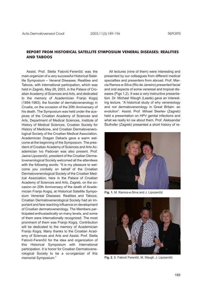
RE PORT FROM HIS TOR I CAL SAT EL LITE SYM PO SIUM VENERAL DIS EASES: RE AL I TIES
AND TABOOS
As sist. Prof. Stella Fatoviæ-Ferenèiæ was the
main or ga nizer of a very suc cess ful His tor i cal Sat el -
lite Sym po sium – Veneral Dis eases: Re al i ties and
Ta boos, with in ter na tional par tic i pa tion, which was
held in Zagreb, May 28, 2003, in the Pal ace of Cro -
atian Acad emy of Sci ences and Arts, and ded i cated
to the mem ory of Ac a de mi cian Franjo Kogoj
(1894-1983), the founder of dermatovenerology in
Croatia, on the oc ca sion of the 20th An ni ver sary of
his death. The Sym po sium was held un der the aus -
pices of the Cro atian Acad emy of Sci ences and
Arts, De part ment of Med i cal Sci ences, In sti tute of
His tory of Med i cal Sci ences, Cro atian So ci ety for
His tory of Med i cine, and Cro atian Dermatovenero -
logical So ci ety of the Cro atian Med i cal As so ci a tion.
Ac a de mi cian Dragan Dekaris gave a warm wel -
come at the be gin ning of the Sym po sium. The pres -
i dent of Cro atian Acad emy of Sci ences and Arts Ac -
a de mi cian Ivo Padovan was also pres ent. Prof.
Jasna Lipozenèiæ, pres i dent of the Cro atian Derma -
to venerological So ci ety wel comed all the at ten dees
with the fol low ing words: “It is my plea sure to wel -
come you cor dially on be half of the Cro atian
Dermatovenerological So ci ety of the Cro atian Med -
i cal As so ci a tion, here in the Pal ace of Cro atian
Acad emy of Sci ences and Arts, Zagreb, on the oc -
ca sion on 20th An ni ver sary of the death of Ac a de -
mi cian Franjo Kogoj, at His tor i cal Sat el lite Sym po -
sium Ve ne real Dis eases: Re al i ties and Ta boos.
Cro atian Dermatovenerological So ci ety had an im -
por tant and fare reach ing in flu ence on de vel op ment
of Cro atian dermatovenerology. The Mem bers par -
tic i pated en thu si as ti cally on many lev els, and some
of them were in ter na tion ally rec og nized. The most
prom i nent of them was Franjo Kogoj. Con tri bu tion
will be ded i cated to the mem ory of Ac a de mi cian
Franjo Kogoj. Many thanks to the Cro atian Acad -
emy of Sci ences and Arts and As sist. Prof. Stella
Fatoviæ-Ferenèiæ for the idea and organization of
this Historical Symposium with International
participation. It is honor for Croatian Dermatove ne -
ro logical Society to be a co-organizer of this
memorial Symposium.”
All lec tures (nine of them) were in ter est ing and
pre sented by our col leagues from dif fer ent med i cal
spe cial ties and pre sent ers from abroad. Prof. Mar -
cia Ramos-e-Silva (Rio de Ja neiro) pre sented fa cial
and oral as pects of some ve ne real and trop i cal dis -
eases (Figs 1,2). It was a very in struc tive pre sen ta -
tion. Dr. Mi chael Waugh (Leeds) gave an in ter est -
ing lec ture, “A his tor i cal study of why venereology
and not dermatovenerology in Great Brit ain: an
evo lu tion”. As sist. Prof. Mihael Skerlev (Zagreb)
held a pre sen ta tion on HPV gen i tal in fec tions and
what we re ally kn ow about them. Prof. Aleksandar
Štulhofer (Zagreb) pre sented a short his tory of re -
189
Acta Dermatovenerol Croat 2003;11(3):189-196 REPORTS
Fig. 1. M. Ramos-e-Silva and J. Lipozenèiæ
Fig. 2. S. Fatoviæ Ferenèiæ, M. Waugh, J. Lipozenèiæ

search in sex ual risk-tak ing in Croatia and so cial
im pact in terms of hu man im mu no de fi ciency vi rus
(HIV) and ac quired im mu no de fi ciency syn drome
(AIDS) pre ven tion cam paigns and sex ed u ca tion
pro grams. Ana Marija Dürrigl and Stella Fatoviæ-
Ferenèiæ (Zagreb) shed light on some as pects of
sexuall-re lated top ics based on ev i dence from Cro -
atian Glagolitic writ ing. Prof. Josip Begovac
(Zagreb) with his pre sen ta tion, “The suc cess and
fail ures in com bat ing the HIV/AIDS ep i demic”, in -
formed us on new ad vances in ba sic and clin i cal re -
search in HIV in fec tion. “The Skerlievo disease:
between myth and reality” was presented by A.
Muzur and A. Škrabonja (Rijeka).
Very im pres sive were lec tures of two young stu -
dents of his tory of med i cine – Agata Makoviæ
(Zagreb) – “Aids in Pub lic Jour nals; and M. Mišir
(Osijek) – “AIDS in Biomedicine Jour nals in
Croatia”. Most Cro atian med i cal jour nals were a
good source of qual ity in for ma tion about AIDS in
our coun try long be fore first pa tients ap peared in
our hospitals.
In spite of multidisciplinary par tic i pa tion, we be -
lieve that this His tor i cal Sym po sium proved his full
suc cess. Con grat u la tions to As sist. Prof. Stella
Fatoviæ-Ferenèiæ for or ga ni za tion of the Sym po sium.
Prof. Jasna Lipozenèiæ, M.D., Ph.D.
190 ACTA DERMATOVENEROLOGICA CROATICA
Acta Dermatovenerol Croat
Reports 2003;11(3):189-196
RAVE com pany: cream against com mon cold (left) and pas tilles for dis in fec tion of the mouth (right).An ad ver tise ment from 1937.
From the col lec tion of Stella Ferenèiæ-Fatoviæ, M.D., Ph.D.

ACTA DERMATOVENEROLOGICA CROATICA 191
Acta Dermatovenerol Croat
Reports 2003;11(3):189-196
WEL COME OF PRES I DENT OF THE CRO ATIAN DERMATOVENEROLOGICAL SO CI ETY
OF THE CRO ATIAN MED I CAL AS SO CI A TION AND CON GRESS PRES I DENT, TOP I CAL
PRO CE DURES, IN NO VA TIONS, AND MIS TREAT MENTS IN DERMATOVENEROLOGY,
Plitvice, May 29, 2003
On be half of the Or ga niz ing and Sci en tific Com -
mit tees of the In ter na tional Con gress “Top i cal Pro -
ce dures, In no va tions, and Mis treat ments in Derma -
tovenerology”, pres i dent cor dially wel comed ev ery -
one to the Plitvice Lake Na tional Park, in the heart
of Croatia. At the be gin ning of the open ing cer e -
mony, Prof. Jasna Lipozenèiæ read the fol low ing ex -
cuses: “Be cause of un cer tain ties, I would be un -
able to at tend the Con gress. I’m very sorry that I am
un able to do this in 2003. Per haps I can try to at tend
it next year”, Larry C. Millikan. “I deeply re gret that,
due to an un for tu nate cri sis which has arisen, I
have to can cel my trip to Plitvice. I hope for your
kind un der stand ing in this very se ri ous sit u a tion. I
sin cerely wish you all the very best for a suc cess ful
con gress and once again apol o gize for this sud den
change of events. I hope for an other op por tu nity in
the fu ture and re main yours, with warm est re -
gards”, Andreas Katsambas. “I am so sorry that un -
for tu nately I can’t come to Plitvice be cause of se -
ries of ad min is tra tive re quire ments that have made
me can cel my par tic i pa tion. I do wish you a suc -
cess ful meet ing and I hope to en joy in par tic i pat ing
at the Con gress next year, wish warm re gards”,
Prof. At tila Horvath.
“I hope you will en joy 16 beau ti ful blue-green
lakes, linked by a se ries wa ter falls and cas cades
form ing a chain through a wooden val ley. In 1979,
UNESCO Of fice in cluded the Plitvice Lake in the list
of World Cul tural and Nat u ral In her i tance. Held on
the pre mises of the Ho tel Jezero in this lovely am bi -
ent, the In ter na tional Con gress ”Top i cal Pro ce -
dures, In no va tions, and Mis treat ments in Dermato -
venerology" is a sci en tific and travel ex pe ri ence to
be trea sured for ever,” said Prof. Lipozenèiæ.
Like a year ago at the Con gress in Opatija, we
were proud to have again at this meet ing our good
old friends, Prof. Ring (Mu nich), Prof. Nakayama
(To kyo), Prof. Kapp (Hannover), Prof. Heghyi
(Brno), Prof. Arslanagiæ (Sarajevo), Prof. Ramos-
e- Silva (Rio de Ja neiro), Dr. Celebi (Tur key), Prof.
Stary (Vi enna), Prof. Trevisan (Trieste), Dr. Kokelj
(Trieste), and Head doc tor Potoènik (Ljubljana). We
also wel comed new friends of our So ci ety and
coun try, Prof. Gollnick (Magdeburg), Pres i dent of
UEMS, Prof. Wolf (Tel Aviv) from Kaplan Med i cal
Cen ter, a very im por tant pub lisher in der ma tol ogy
world wide, and Dr. Law rence, Fujisawa se nior in -
ves ti ga tor from the USA. We wished them a very
en joy able stay that will make them to come to
Croatia again in May 2004 on In ter na tional Sym po -
sium on Atopic der ma ti tis.
(Figures 1-6)
Prof. Jasna Lipozenèiæ, M.D., Ph.D.

192 ACTA DERMATOVENEROLOGICA CROATICA
Acta Dermatovenerol Croat
Reports 2003;11(3):189-196
Fig. 2. Pro fes sors at the Wel come din ner from left handside: H. Behrendt-Ring, J. Ring, Mr. and Mrs. Stary, H.Gollnick and A. Kapp.
Fig. 3. Wel come din ner from right hand side: Prof. Heghy, his sun and wife, Pro fes sors F. Gruber, M. Skerlev and M. Ramos e Silva
Fig. 4. Wel come din ner from left hand side: Dr. C. Celebi, Prof. R. Wolf with his wife, Doc tors M. Potoènik and M.Sirotkoviæ-Skerlev
Fig. 5. Wel come din ner, from left hand side: Prof. J.
Lipozenèiæ, G. Trevisan, Dr. F. Kokelj, Prof. M. Šitum, Dr.
A. Pašiæ, As sist. Prof. V. Barišiæ-Druško and Prof. M.
Waugh
Fig. 6. Welcomce din ner, from left side hand: As sist. Prof. V. Milavec-Puretiæ, Prof. J. Lipozenèiæ, Mrs. and Dr. I.Law rence, Prof. A. Basta-Juzbašiæ
Fig. 1. Prof. Lipozenèiæ, Con gress pres i dent at the Open -
ing cer e mony

FI NAL CON CLU SION
of the In ter na tional Con gress “Top i cal Pro ce dures, In no va tions, and
Mis treat ments” Plitvice, May 31, 2003
It’s a plea sure to close such a suc cess ful meet -
ing as it was In ter na tional Con gress «Top i cal Pro -
ce dures, In no va tions, and Mis treat ments», said
Prof. Jasna Lipozenèiæ.
Prom i nent lec tur ers came from 17 coun tries. We
have suc cess fully re solved 41 oral pre sen ta tions, 6
Sat el lite Sym po siums with 12 oral pre sen ta tions,
and we have awarded the best three post ers pre -
sented at the Con gress. Der ma tol ogy is play ing an
in creas ingly vi tal role in pa tient care. Through out
this Con gress, we have con cen trated on the fol low -
ing ini tia tives: ed u cat ing and in creas ing the avail -
abil ity of pri mary skin care, pro vid ing pa tients with
high est qual ity of care through di rect ac cess to
dermatologic lo cal ther apy, and in creas ing sup port
to ba sic and ap plied re search with phar ma ceu ti cal
in dus try. The Cro atian Dermatovenerological So ci -
ety (CDS) was very ac tive in the last 5 years (16
meet ings). The CDS is pur su ing a full agenda in the
com ing year. Prog ress we have made thus far in
each area in the field of der ma tol ogy and vene -
reology was dis cussed, along with anticipation of
what the future holds in dermatologic health care.
I be lieve the Con gress pro gram was in ter est ing
and in for ma tive, and proved to be suc cess ful and
use ful for all of us. (Figs 1-3)
In the six main top ics of the Con gress, we heard
the fol low ing:
1) That the lo cal treat ment is very im por tant in
der ma tol ogy as well as in ap pro pri ate in di ca tions;
2) That op ti mi za tion of top i cal ther apy is
needed;
3) That cos metic prod ucts, sunscreens, and
many new top i cal prod ucts are on the mar ket and
must be used ap pro pri ately;
4) An up date on ex tem po ra ne ous pre scrip tions
in der ma tol ogy with qual ity man age ment, pre -
sented by Prof. Gloor in Spirig Sat el lite Sym po sium;
ACTA DERMATOVENEROLOGICA CROATICA 193
Acta Dermatovenerol Croat
Reports 2003;11(3):189-196
Fig. 1. Pro fes sors from left hand side: M. Skerlev, V. Heghy,J. Lipozenèiæ, M. Ramos-e-Silva, J. Ring, H. Gollnick
Fig. 2. Prof. M. Gloor in Spring Sattelite Symposium
Fig. 3. As sist. Prof. I. Bartenjev in Vichy – Sat el lite Sym po -
sium

5) New ap proaches in the treat ment of atopic
der ma ti tis;
6) About new pso ri a sis ther apy, photo therapy
and photochemotherapy with own ex pe ri ences
from dif fer ent coun tries;
7) Re sults of Sun Pre ven tion cam paign in
Slovenia and Croatia, with photoprotection em pha -
sized in Vichy Sat el lite Sym po sium;
8) Treat ment of vi ral, bac te rial, and par a sitic dis -
eases and sex u ally trans mit ted dis eases (STDs) as
a part of our spe cial iza tion – venereology was im -
por tant part on this congress;
9) The Con gress was or ga nized be cause of mis -
treat ments in dermatologic cos me tol ogy, and there
were many pre sen ta tions and dis cus sions about
that;
10) Top i cal corticosteroids (old and new) were
pre sented and guide lines for all dermatovenero -
logists were very in struc tive;
11) La ser in der ma tol ogy was pres ent and the
fu ture of dermatotherapy not only as in no va tion in
Croatia. The ex pe ri ence from Slovenia and Croatia
was very im pres sive;
12) Dermatosurgery is im por tant part of lo cal
dermatotherapeutic meth ods and ex cel lently per -
formed in Sis ters of Mercy Uni ver sity Hos pi tal,
Zagreb;
13) Ap pro pri ate treat ments and mis treat ments
in dermatomycology are very of ten seen in daily
praxis and of ten un rec og nized;
14) It is nec es sary to ed u cate not only gen eral
prac ti tio ners, but also dermatovenerologists;
15) Itraconazole (ITRAC3) is a novel treat ment
for onychomycosis and tinea capitis and was suc -
cess fully pre sented at Belupo Sat el lite Sym po sium
and ex hi bi tion;
16) Atopic der ma ti tis treat ment and skin care
was pro vided by Uriage-Formasana, with great pal -
ette of prod ucts in skincare of dry and atopic skin;
17) The Skin Scare Re ducer ther apy – Hansa -
plast was pre sented by Beiersdorf as the only prod -
uct in Croatia for the treat ment of new and old scars
and kel oids;
18) Hair and scalp prob lems and im por tance of
cos metic der ma tol ogy was pre sented in Sat el lite
Sym po sium by Oktal-Pharma.
The Con gress at tended 260 par tic i pants from
17 dif fer ent coun tries all over the world.
Prof. Jasna Lipozenèiæ, M.D., Ph.D.
Pres i dent of the CDS
194 ACTA DERMATOVENEROLOGICA CROATICA
Acta Dermatovenerol Croat
Reports 2003;11(3):189-196
Fare well Re marks
In ter na tional Con gress of Cro atian So ci ety for Der ma tol ogy and Venerology,
Plitvice, May 07-14, 2003
Dear Pro fes sor Lipozenèiæ, La dies and Gen tle -
men,
At the end of this won der ful con gress I would like
to speak on be half of the for eign guests, who came
from 17 coun tries to the 16 lakes and 100 wa ter falls
of beau ti ful Plitvice. We want to thank the or ga niz -
ers for this ex cel lent con gress and their won der ful
hos pi tal ity.
We have heard a lot of ex cel lent lec tures, seen
in ter est ing post ers, and got new and ex cit ing in for -
ma tion for our pa tients. I want to con grat u late the
Cro atian Dermatological So ci ety on many en thu si -
as tic young der ma tol o gists in this coun try!
At this event, I also would like to say one word on
be half of the Eu ro pean Acad emy of Dermato- Vene -
rology (EADV). Yes ter day, we had some per sonal
dis cus sions and I learned that many peo ple in East -
ern Eu ro pean coun tries have the feel ing that EADV
is re stricted to the po lit i cal body of the Eu ro pean
Un ion. This is not true: the EADV is not a po lit i cal
body and com prises the whole Eu rope with all its
rich ness and di ver sity in cul ture and his tory. So the
EADV is also your acad emy and I would like to ask
you to join it. For res i dents there is the pos si bil ity to
be come a ju nior mem ber with re duced fees.
Again, let me thank Pro fes sor Jasna Lipozenèiæ
and her splen did team for this out stand ing meet ing!
Prof. Johannes Ring, M.D., Ph.D.
SEC TION DER MA TOL OGY OF THE EU RO PEAN ACAD EMY OF ALLERGOLOGY AND
CLIN I CAL IM MU NOL OGY
Sec tion Der ma tol ogy of the Eu ro pean Acad emy
of Allergology and Clin i cal Im mu nol ogy (EAACI)
busi ness meet ing in Paris was held on Sat ur day,
June 7, 2003, at 5.00 p.m. A new Board of Der ma -
tol ogy Sec tion for the 2003-2005 pe riod was con sti -
tuted. The pres i dent cho sen was Prof. Bindslev-
Jensen Carsten (Den mark), and sec re tary Prof.
Thomas Bieber (Ger many). New mem bers of the
Sec tion are Girolomoni Gianpiero (It aly), Hauser
Conny (Swit zer land), Lipozenèiæ Jasna (Croatia),
Kristine Turjanmaa (Fin land), and Törsten
Zuberbier (Ger many).
The time ta ble was as fol lows: 1) Re port of ac tiv -
ity last year (past pres i dent Prof. A. Kapp and sec -
re tary Prof. T. Reunala); 2) New board sug ges tion
and vot ing – Prof. A. Kapp; 3) Fu ture ac tiv i ties –
new pres i dent Prof. Bindslev-Jensen, and 4) other.
The next EAACI Meet ings will be held in Am ster -
dam, 2004; in Mu nich, 2005; in Mos cow, 2007, and
in Gothenburg, 2008.
It was sug gested that Post grad u ate course in
Pathophysiology, Di ag no sis, and Ther apy of Con -
tact Al ler gic Der ma ti tis will be held by Eu ro pean So -
ci ety of Con tact Der ma ti tis (Diepgen). Task Forces:
Dermatologic Sec tion plus Pe di at ric Sec tion: Atopy
Patch Test ing and Der ma tol ogy Sec tion: Ur ti caria.
It was pro posed that In ter na tional Sym po sium
about Atopic Der ma ti tis un der spon sor ship of
EAACI Der ma tol ogy Sec tion will be or ga nized in
Croatia (by Prof. Lipozenèiæ) in 2004.
Prof. Jasna Lipozenèiæ, M.D., Ph.D.
ACTA DERMATOVENEROLOGICA CROATICA 195
Acta Dermatovenerol Croat
Reports 2003;11(3):189-196
RE PORT FROM XXII CON GRESS OF THE EU RO PEAN ACAD EMY OF ALLERGOLOGY
AND CLIN I CAL IM MU NOL OGY,
PARIS, JUNE 7-11, 2003
This was the fifth time that Eu ro pean Acad emy
of Allergology and Clin i cal Im mu nol ogy (EAACI)
was held in France (1950 Paris, 1971 Mar seille,
1981 Clermont Ferrand and again in Paris, 1992) in
one of the most at trac tive cit ies in the world. The
venue of the Con gress was Palaise des Congres,
one of the larg est Con gress cen ters in Eu rope.
On June 7, 2003 Prof. Lipozenèiæ was ac tive in
Der ma tol ogy Sec tion Meet ing at 5.00 p.m.
At Sunday June 8, 2003, a Com pany spon sored
Sym po sium Aventis, An ti his ta mines Eval u a tion or
Rev o lu tion? took place. There were lec tures pre -
sent ing fexofenadin as a better med i ca tion than
loratadine, which has same ef fect as levocetirizin.
In seasonal and pe ren nial allergic rhi ni tis and
chronic id io pathic ur ti caria, levocetizin 5 mg
(Xyzal®) is ef fec tive. Montelukast (Singulair, tbl.) ef -
fec tively treats the night time im pact of sea sonal al -
ler gic rhi ni tis.
Very good state ment was in Sym po sium Drug
Al lergy; Food Al lergy; Pe di at ric Al ler gies; New Lan -
guage and Medias Al lergy and Asthma and the pa -
tients. On Mon day June 9, 2003, there were many
pre sen ta tions on asthma, whereas al lergy prob -
lems in der ma tol ogy were given less at ten tion at the
EAACI, ex cept for ur ti caria, angioedema, and
atopic der ma ti tis. Very successfull pre sen ta tions
were the fol low ing: in Mains Sym po sium 1 – Al lergy
to Cos me tics and Per fumes: Al ler genic in gre di ents
in cos me tics and per fumes; New clin i cal as pects
and al lergy test ing and How sen si tiz ing po ten tial is
eval u ated. In Main Sym po sium 5, the most suc -
cess ful pre sen ta tion was on drug allergies by H. F.
Merk (Ger many), en ti tled Drug me tab o lism in the
skin, and Di ag no sis of drug-in duced skin re ac tions
by Annick Burband.
Rhi ni tis and asthma in chil dren and adults, Com -
pany spon sored sym po sia and Main Sym po sia,
were maybe given too much space for the pro file of
the au di ence at tend ing the Con gress.
Al lergy Gene to Dis ease in Ple nary Ses sion 3
in cluded the fol low ing in ter est ing ses sions: “Genes
if tol er ance” , “How do genes af fect the re sponse do
drugs?”, and “Gene ther apy is here to day and is
here to stay”.
Main Sym po sia 25, New Trends in Atopic Der -
ma ti tis was prom is ing, with lec tures ti tled Ep i de mi -
ol ogy and Ge net ics (Zuberbier); Are there new
mech a nisms? (Bieber), Is IgE re ally im por tant in
atopic der ma ti tis? (Werfel); New treat ments
(Reitamo); Hot top ics in in sect al lergy from nat u ral
his tory of in sect al lergy in chil dren; In ter face be -
tween in sect al lergy and mastocytosis; Im mu no log -
i cal mech a nisms of venom immunotherapy, and
Recent development in venom immunotherapy.
There were 1,405 reg is tered pos ters and about
5,000 par tic i pants, 100 ses sions (4 ple nary ses -
sions, 28 main sym po sia, 4 mini sym po sia, 14 oral
ab stract ses sions, 12 poster dis cus sion ses sions, 8
com pany spon sored sym po sia and a se ries of sat -
el lite sym po sia) cov er ing all as pects of al lergy, from
ba sic sci ence to di ag no sis, treat ment, and socio-
econ omy.
We en joyed in Paris dur ing the 22nd EAACI Con -
gress.
Prof. Jasna Lipozenèiæ, M.D., Ph.D.
196 ACTA DERMATOVENEROLOGICA CROATICA
Acta Dermatovenerol Croat
Reports 2003;11(3):189-196
Marko Polo´s Di ary
Stella Fatoviæ-Ferenèiæ, ESHDV rep re sen ta tive of Croatia
On the Iraq War, the Mu nich Clinic and a His -
tor i cal sat el lite sym po sium in Zagreb
It has been more then 700 years since Marko
Polo passed the land between the Tigris and
Euphrates Rivers on his way to China. It is hard to
believe, looking at the Tigris and Euphrates today,
the area where Western civilisation was rooted, that
once it was the site of the biblical Garden of Eden.
Instead, we witness war in the name of liberty and
democracy. Tons of bombs are being dropped.
Mu nich April 10th: Hos pi tal of Lud wig-
Maximilian–Uni ver sity
Mu nich was my des ti na tion in April. I had an ap -
point ment with Pro fes sors Plewig and Holubar and
Blackwell pub lish ers at the Hos pi tal of the Lud -
wig-Maximilian–Uni ver sity. We con tin ued with pre -
p a ra tions of our book Skin in Wa ter Col ors, which
will hope fully be pre sented at the Bar ce lona Con -
gress. Af ter a work fruit ful, our host took us late in
the af ter noon to the Asamkirche (of fi cially known as
St-Johann-Nepomuk), one of the most en chant ing
ex am ples of the Ba var ian Roccoco style. Af ter a
pleas ant though rainy walk around Sendlinger-Tor-
Platz, we fin ished our day at an en joy able Ba var ian
res tau rant. I left Ger many by train. An early spring
land scape with a crisp or der of the fields framed my
win dow sight. The solid moun tains ris ing steeply
away from the train tracks, hid ing for ests and cab -
ins, as well as se cretes and sto ries, which I was
familiar with from Grimm’s fairy tales.
Zagreb May 28th – Ve ne real dis eases: Re al i -
ties & Ta boos
This his tor i cal sat el lite sym po sium within a
dermatological con gress was held at the Con fe r -
ence Hall in the Pal ace of the Cro atian Acad emy of
Sci ences and Arts. The or ga niz ers, the Cro atian
Acad emy of Sci ences and Arts (the De part ment for
Med i cal Sci ences as well as the De part ment for the
His tory of Med i cal Sci ences) and two so ci et ies of
the Cro atian Med i cal As so ci a tion (the Dermato -
vene rolo gical and the Med ico-shis tor i cal) ded i -
cated the event to the mem ory of Franjo Kogoj, on
oc ca sion of the 20th an ni ver sary of his death. By to -
day, in ves ti ga tions of ve ne real dis eases have
clearly sur passed the lim its of med i cine and re -
vealed the com plex ity of life and sick ness, in bi ol -
ogy as much as in so ci ol ogy and cul ture. Dur ing the
sym po sium, we learned that, re gard ing ve ne real
dis eases, the Brit ish Isles did not fol low the con ti -
nen tal tra di tions. Knowl edge about syph i lis came to
Eng land only in 1686, and the first who wrote on
syph i lis was Dan iel Turner in 1717. From an other
cor ner of the world, Brazil, we heard about fa cial
and oral as pects of ve ne real and trop i cal dis eases,
whereas data on HPV gen i tal in fec tions were pre -
sented from the De part ment of Der ma tol ogy and
Venereology at Zagreb Uni ver sity School of Med i -
cine. The sym po sium of fered a multidis ciplinary ap -
proach aim ing to clar ify var i ous as pects of ve ne real
dis eases in dif fer ent times. For that rea son, not only
dermatovenerologists, but also spe cial ists of in fec -
tious dis eases, med i cal his to r i ans phi lol o gists, and
197
Acta Dermatovenerol Croat 2003;11(3):197-198 MARKO POLO‘S DIARY
so ci ol o gists were in vited to speak. Suc cesses and
fail ures in com bat ing the HIV/AIDS ep i demic were
out lined, and risks posed by STD re search and its
so cial im pact in Croatia were ad dressed. Causes
and con se quences of Skerlievo dis ease two hun -
dred years ago were de scribed by med i cal his to ri -
ans from Rijeka, and the at ti tudes vis-á-vis sex u al -
ity and so cial stigma in the me di eval pe riod were
pin pointed. The lec tur ers Mi chael Waugh, Mar cia
Ramos-e-Silva, Mihael Skerlev, Josip Begovac,
Aleksandar Štulhofer, Marija Ana Dürrigl, Amir
Muzur, and Ante Škrobonja com pe tently and clearly
out lined their top ics. Agata Makoviæ and Mihael
Mišir, two young phy si cians, par tic i pated in such a
sym po sium for the first time. Their re search con -
cerned the ap pear ance of AIDS as a topic in pub lic
and bio med i cal jour nals. As their men tor, I was
proud of their work. In the pro cess of glob al iza tion
to which we are so strongly, and some times un will -
ingly, ex posed, knowl edge should be come a dom i -
nant power. Young peo ple must there fore be stim u -
lated more and con sid ered the most prom is ing and
important force in a knowl edge- based society in the
making. The following day, the Congress of der ma -
tol o gists started in Plitvice. I stayed in Zagreb,
making preparations for my next des ti na tion – Lon -
don.
*********************************
The world is wait ing, sretan vam put !
*********************************
198 ACTA DERMATOVENEROLOGICA CROATICA
Acta Dermatovenerol Croat
Marko Polo‘s Diary 2003;11(3):197-198

Dear Ed i tor-in-Chief,
I would like to of fer my com ment on the ar ti cle pub lished in your Jour nal (Ljubojeviæ S. Se vere acute re -
spi ra tory syn drome, Acta Dermatovenerologica Croatia 2003;11:129-30). As jour nals have also in for ma tive
and ed u ca tive pur pose, I may say that Acta Dermatovenerologica Croatica undoubtfully rep re sents such a
strat egy in the best pos si ble way by pub lish ing the in for ma tion of the high est pri or ity. I would like to con grat u -
late you as the Ed i tor-in-Chief of Acta Dermatovenerologica Croatica.
Cor dially,
Mihael Mišir, MD
Acta Dermatovenerologica Croatica: Pri or ity in Re port ing on
New Ep i demic
To the ed i tor. It is quite a while that I have been
in volved with an in ves ti ga tion on the ear li est news
and in for ma tion as so ci ated with ep i dem ics dealt
with in bio med i cal jour nals in Croatia. Al though I
was mainly deal ing with ac quired im mu no de fi -
ciency syn drome (AIDS), the new chal lenge, Se -
vere Acute Re spi ra tory Syn drome (SARS), that we
be came aware of in early 2003, prompted me to fol -
low also what kind of in for ma tion was pre sented on
that topic. I checked the same se lec tion of jour nals,
which I used in the AIDS in ves ti ga tion. I was dis ap -
pointed to learn that Cro atian med i cal jour nals
come to li brar ies with in cred i ble de lay. This is op po -
site to the pol icy jour nals should fol low, i.e. reach ing
a high im pact fac tor, and pre vents prompt in for ma -
tion to the read ers. The ed i to rial staff of each jour nal
should mind that apart from internet pos si bil i ties
and the pri or ity list of jour nal’s sub scrib ers, there
are many researchers, who just need a variety of
periodicals at hand.
Since the time of the ear li est jour nals on our ter -
ri tory pro fes sional in for ma tion in med i cine was
avail able. The first most fas ci nat ing news, such as
that on X-rays in the ear li est is sues of Lijeènièki
vjesnik; out breaks of cer tain dis eases, new di ag -
nos tic tools and ther a pies, were thus trans planted
from other jour nals into ours (1,2). Con se quently,
knowl edge, ther a peu tic reg i mens and health pol icy
was in flu enced fa vor ably. Acta Dermatovenerolo -
gica Iugoslavica, for ex am ple, whose ed i to rial
board in cluded nu mer ous prom i nent dermatovene -
rologists of Croatia, brought one of the first re ports,
e.g., on a new en tity al ready in 1982 – re port ing on
sar coma Kaposi in young ho mo sex u als in New
York. Acta Dermatovenerologica Iugoslavica pub -
lished it be fore Epidemiološki vjesnik or any similar
journal of infectious diseases (3).
New dis eases seem to be re ported speed ily in
Cro atian pub lic rather than in bio med i cal jour nals.
Ac cord ing to an other line of in ves ti ga tion, car ried
out by Makoviæ (4), it is ev i dent that pub lic jour nals
were as prompt bring ing the news on AIDS in
Croatia as else where in the world. Re gard ing SARS
the re sults are quite sim i lar. Be sides, the jour nal of
the Cro atian Med i cal Cham ber, Lijeènièke novine,
which some how bridges the in for ma tion gap be -
tween pub lic and bio med i cal jour nals, ap peared to
be quite in for ma tive in this as pect. Re gard ing
199
Acta Dermatovenerol Croat 2003;11(3):199-200 LET TER TO THE ED I TOR
SARS, it ad e quately ful filled its task bring ing suf fi -
cient in for ma tion on new out break. Three ar ti cles
were thus pub lished in its pages in April and May
2003 (5-7). The first text con tained a short his tory of
the dis ease, num ber of af fected peo ple and coun -
tries in volved, and fur ther, ep i de mi ol ogy, di ag nos tic
and eti o log i cal data as well as pro tec tive mea sures.
The sec ond text re ported that there are no SARS
cases in our coun try but in case of appearance, no
provisions for their isolation are available as well
(6).
My in ves ti ga tion showed that there is no in for -
ma tion on SARS within Cro atian bio med i cal jour -
nals, un til June 2003 when two ar ti cles ap peared.
The first one was pub lished in Farmaceutski
glasnik, trans fer ring the in for ma tion from Lijeènièke
Novine, men tioned above (8). The sec ond was
Ljubojeviæ’s com ment on SARS pub lished in Acta
Dermatovenerologica Croatica aim ing to in form the
read er ship on the new and puz zling out break and
of fer ing a list of use ful web sites for those who need
to know more and pre fer vir tual re al ity (9). The only
de tail which I would add to sup ple ment Ljubojeviæ’s
data is that ac cord ing to The Lan cet, SARS oc -
curred in Guandong Prov ince, in China al ready in
No vem ber, 2001 which moves back the be gin ning
of the out break one year and three months before
February 1, 2003 (10).
In con clu sion I may say that a dermatological
jour nal was again the first among the Cro atian bio -
med i cal jour nals to shed light on a new dis ease
even though one would ex pect to find such an in for -
ma tion in the pages of some gen eral med i cal jour -
nal, or more likely, in an ep i de mi o log i cal jour nal.
The fact that the SARS is sue, al though not strictly
be long ing to sphere of der ma tol ogy, ap peared in
Acta Dermatovenerologica Croatica dem on strates
the sen si tiv ity and open ness of its ed i to rial staff. It
also leaves us hope for the fu ture bio med i cal jour -
nal ism not frag mented and not just tar get ing ev i -
dence based medicine but committed to new
dangers and risks.
Mihael Mišir, M.D.
Ref er ences
1 Fatoviæ-Ferenèiæ S. The old est Cro atian Med i cal Jour -nal. Croat Med J 2002;43:355-8.
2 Fatoviæ-Ferenèiæ S. Lijeènièki vjesnik in the set ting ofroent gen ol ogy be gin nings in Croatia. Lijeè Vjesn1996;118:84-5.
3 Mišir M. AIDS in Cro atian med i cal jour nals. Book ofAb stracts, Acta Dermatovenerol Croat 2003;11:42.
4 Makoviæ A. AIDS in Cro atian pub lic jour nals. Book ofAb stracts, Acta Dermatovenerol Croat 2003;11:41-2.
5 Marton I. SARS – se vere acute re spi ra tory syn drome.Lijeènièke novine 2003;18:26-7.
6 Cafuk B. SARS na èetiri kontinenta. Lijeènièke novine2003;18:27.
7 Cafuk B. SARS jaše dalje Lijeènièke novine 2003;19:18-21.
8 Marton I. SARS – se vere acute re spi ra tory syn drome.Farm Glas 2003;59:165-7.
9 Ljubojeviæ S. Se vere acute re spi ra tory syn drome. ActaDermatovenerol Croat 2003; 11:129-30.
10 Chotani RA, LaPorte RE, Linkov F, Dodani S, Danyal A, Khan MI. Just-in-time lec tures: SARS. Lan cet 2003;361: 1531.
200 ACTA DERMATOVENEROLOGICA CROATICA
Acta Dermatovenerol Croat
Letter to the Editor 2003;11(3):199-200

ANNOUNCEMENTS
XVIII World Al lergy Or ga ni za tion Con gress ICACI, Vancuver, Can ada, Sep tem ber 7-12, 2003. Con tact: Sally Kolf,
611 East Wells Street, Mil wau kee, WI 53202, USA; e-mail: con [email protected]; www.worldallergy.org
Cur rent State on Pso ri a sis and Naphtalanotherapy, Sym po sium of Cro atian Dermatovenerological So ci ety of theCro atian Med i cal As so ci a tion, Ivaniæ Grad, Croatia, Sep tem ber 19, 2003. Con tact: Prof. Jasna Lipozenèiæ, Šalata 4,10000 Zagreb, Croatia. Tel./Fax: +385-1-4920-014; e-mail: jasna.lipozenèiæ@zg.tel.hr
9th Con gress of the Eu ro pean Con fed er a tion of Med i cal My col ogy and 7th Trends in In va sive Fun gal In fec -tions, Joint Meet ing, Am ster dam, The Neth er lands, Sep tem ber 28-Oc to ber 1, 2003. Con tact: Con gress Care,Muntelbolwerk 1, P.O. Box 440, 5201 AK’s-Hertogenbosch, The Neth er lands; e-mail: [email protected] ,www.congresscare.com
12th Con gress of Eu ro pean Acad emy of Der ma tol ogy and Venerology, Bar ce lona, Spain, Oc to ber 15-18, 2003.Con tact: Unicongress, Calvet, 55, Baixos, 08021 Bar ce lona, Spain; e-mail: [email protected];www.eadv.org
6th Tergestinum Sym po sium on Pso ri a sis and 2nd Alpe Adria Meet ing on Pso ri a sis, Bibione, It aly, No vem ber7-8, 2003. Con tact: Or gan is ing Sec re tar iat, Via San Nicolo 14, 34121 Trieste, It aly. Tel +39 40 368343, Fax: +39 40368808
Sex u ally Trans mit ted Dis eases, Con tin u ing Med i cal Ed u ca tion Course or ga nized by the Uni ver sity School of Med i -cine Zagreb and Cro atian Dermatovenerological So ci ety of the Cro atian Med i cal As so ci a tion, Šubiæeva 9, 10000Zagreb, Croatia, No vem ber 21-22, 2003. Con tact: Prof. Jasna Lipozenèiæ, Šalata 4, 10000 Zagreb, Croatia. Tel./Fax:+385-1-4920-014; e-mail: jasna.lipozenèiæ@zg.tel.hr
9th Alple-Adria-Dan ube Con gress of Sex u ally Trans mit ted Dis eases and In fec tions of the Skin, Prague, No -vem ber 27-30, 2003. Con tact: [email protected]
8th World STI/AIDS Con gress, Punta del Este, Uru guay, De cem ber 2-5, 2003
3rd World Con gress of the In ter na tional Acad emy of Cos metic Der ma tol ogy, Beijing, China, De cem ber 7-10,2003. Con tact: IACD2003 Sec re tar iat, Chi nese Med i cal Meet ings In ter na tional, 42 Dongsi Xidajie, Beijing 100710,China; e-mail: [email protected]; www.chinamed.com.cn/IACD
6th In ter na tional Con gress of the Eu ro pean So ci ety for La ser Aes thetic Sur gery (ESLAS), What La ser: whenand why, Rome, It aly, De cem ber 12-14, 2003. Con tact: [email protected]
In ter na tional Mas ter Course of Age ing Skin, Paris, France, Jan u ary 8-10, 2004. Con tact www.web-imcas.com
“Al lergy and Ec zema”, Mi lan, It aly, Jan u ary 22-24, 2004. Con tact: e-mail: [email protected],www.mcaevents.org
62nd An nual meet ing of Acad emy of Der ma tol ogy, Wash ing ton DC, USA, Feb ru ary 6-11, 2004.
201
Acta Dermatovenerol Croat 2003;11(3):201-202 ANNOUNCEMENTS
4th World Con gress of IACD, Cairo, Egypt, April 12-18, 2004.
Up date on Atopic Ec zema/Dermatits Syn drome, Cavtat, Croatia, April 25-28, 2004. Con tact: Prof. JasnaLipozenèiæ, De part ment of Der ma tol ogy and Venerology, Zagreb Uni ver sity Hos pi tal Cen ter, Šalata 4, 10000 Zagreb;e-mail: [email protected]
Sec ond EADV In ter na tional Spring Sym po sium, Bu da pest, Hun gary, April 29-May 1, 2004. Con tact:www.eadvbudapest2004.com; e-mail: [email protected]
9th In ter na tional Con gress of Der ma tol ogy, Beijing, China, May 2004. Con tact: ICD2004 Sec re tar iat, Dept. of For -eign Re la tions, Chi nese Med i cal As so ci a tion, 42 Dongsi Xidajie, Beijing 100710, China. e-mail:[email protected]; www.chinamed.com.cn/der ma tol ogy
15th Ljudevit Jurak In ter na tional Sym po sium on Com par a tive Pa thol ogy; Main Topic: Head & Neck Pa thol ogy,Zagreb, Croatia, June 4-5, 2003. Con tact: www.kbsm.hr/jurak/sym po sium.htm
23rd Con gress of the Eu ro pean Acad emy of Allergology and Clin i cal Im mu nol ogy, Am ster dam, Neth er lands,June 12-16, 2004. Con tact: Dept. Allergology, Uni ver sity Hos pi tal Rot ter dam, dr. Molewaterplein 40, NL-3015 GD Rot ter -dam, The Neth er lands; e-mail: [email protected]; www.congrex.com/eaaci2004
10th Con gress of the Eu ro pean Con fed er a tion of Med i cal My col ogy, June 17-20, 2004, Wroclaw, Po land. Con -tact: Con gress Care, Muntelbolwerk 1, P.O. Box 440, 5201 AK’s-Hertogenbosch, The Neth er lands; e-mail:[email protected] , www.congresscare.com
X World Con gress of Pe di at ric Der ma tol ogy, Rome, It aly, July 7-10, 2004. Con tact: Tri umph Congressi, ViaLucilio, 60, 00136 Rome, It aly; e-mail: der [email protected]; www.gruppotriumph.it
Amer i can Acad emy of Der ma tol ogy, Acad emy ’04, New York, USA, July 28-Au gust 1, 2004. Con tact: Amer i canAcad emy of Der ma tol ogy, De part ment of Meet ings & Con ven tions, 930 E Wood field Road, Schaumburg, IL 60173; fax:847 330 1090
Deut sche Gesellschaft fur Allerologogie und Klinische Immunologie e.V. Tagung, Aachen, Ger many, Sep tem -ber 15-19, 2004. Con tact: [email protected]
In ter na tional Sym po sium on Psychodermatology, Cavtat, Croatia, Sep tem ber 22-26, 2004. Con tact: Prof. MirnaŠitum, De part ment of Der ma tol ogy and Venerology, Clin i cal Hos pi tal “Sestre milosrdnice”, Vinogradska 29, 10000Zagreb, Croatia; e-mail [email protected], [email protected]
4th International Con gress on Autoimmunity, Bu da pest, Hun gary, No vem ber 3-7, 2004. Con tact: fax:0041 22 7322850; phone 0041 22 908 0488
13th Con gress of the Eu ro pean Acad emy of Der ma tol ogy and Venerology, Flor ence, It aly, No vem ber 17-21,2004. Con tact: e-mail: pres i [email protected]; [email protected]
10th World Con gress on Can cers of the Skin, Vi enna, Aus tria, March 19-23, 2005. Con tact: Elfriede Pomp, De -part ment of Der ma tol ogy, Uni ver sity of Vi enna, Vi enna Gen eral Hos pi tal, Waehringer Guertel 18-20, A-1090 Vi enna,e-mail: [email protected]; www.wccs.at
World Al lergy Con gress – 19th In ter na tional con gress of Allergology and Clin i cal Im mu nol ogy and 24th Con -gress of the Eu ro pean Acad emy of Allergology and Clin i cal Im mu nol ogy, Mu nich, Ger many, June 26-June 1, 2005. Con tact: e-mail: [email protected] www.congrex.com/wac2005
6th World Con gress on Mel a noma, Van cou ver, B.C., Can ada, Sep tem ber 2-9, 2005. Con tact: Venue West Con fer -ence Ser vices Ltd., Van cou ver, B.C., Can ada; e-mail: con [email protected]
202 ACTA DERMATOVENEROLOGICA CROATICA
Acta Dermatovenerol Croat
Announcements 2003;11(3):201-202

IN STRUC TIONS TO AU THORS
ACTA DERMATOVENEROLOGICA CROATICA (ADC)is a quar terly peer-re viewed jour nal, in dexed in In dex Medi -cus/MEDLINE and Excerpta Medica/EMBASE. It pub -lishes orig i nal sci en tific ar ti cles, short sci en tific com mu ni -ca tions, clin i cal ar ti cles, case re ports, re views, re ports,news and comments, and an nounce ments in the fields ofder ma tol ogy and venerology.
Gen eral Guide lines
Type the com plete manu script dou ble-spaced, on one side of A4 bond pa per, with a left side mar gin of at least 4cm.
The manu scripts should not ex ceed 12-15 typedpages in case of orig i nal sci en tific pa pers, and 6-8 pagesin case of short com mu ni ca tions, clin i cal ar ti cles, case re -ports, and re views.
The manu scripts should be writ ten in Eng lish. The au -thors are re spon si ble for en sur ing that the Eng lish used is suit able for pub li ca tion. All ma te rial is as sumed to be sub -mit ted ex clu sively to this jour nal.
All manu scripts are sub ject to peer re view.
Prep a ra tion of Manu scripts for Sub mis sion
Ti tle Page
The ti tle page should carry (a) the ti tle of the pa per,which should be con cise but in for ma tive; (b) full name ofeach au thor, with in sti tu tional af fil i a tion; (c) name(s) of de -part ment(s) and in sti tu tion(s) to which the work should beat trib uted; (d) name and ad dress (with tele phone and faxnum bers as well as the e-mail adress) of the au thor towhom re quests for re prints should be ad dressed; (f)source(s) of sup port in the form of grants, equip ment,drugs, or all of these; and (g) a short run ning head of notmore than 40 char ac ters (count let ters and spaces) at thefoot of the ti tle page.
Sec ond Page
The sec ond page should carry a sum mary of not more than 250 words, fol lowed by three to six key words fromthe Med i cal Sub ject Head ings (MeSH) list of In dexMedicus.
Manu script
The text of ob ser va tional and ex per i men tal is usu ally,but not nec es sar ily, di vided into sec tions with the head -ings In tro duc tion, Ma te rial (Pa tients) and Methods, Re -sults, and Dis cus sion. Long ar ti cles may need sub head -ings within some sec tions to clar ify their con tents, es pe -
cially Re sults and Dis cus sion sec tions. Other types of ar -ti cles, such as case re ports, re views, and ed i to ri als, arelikely to need other for mat.
Ab bre vi ated terms should be writ ten in full the firsttime they are used in the text, with ab bre vi a tion in pa ren -the ses.
Un der line the words that must be printed in italic.
Ref er ences should be iden ti fied in the text by arabicnu mer als in pa ren the ses, and be num bered and listedcon sec u tively at the end of the manu script in the or der inwhich they are first cited in the text.
In di cate in the text where the il lus tra tions (fig ures andta bles) should be in serted.
Ta bles and fig ures should be pro vided each on a sep -a rate sheet of pa per af ter the ref er ences. De scrip tive leg -ends to fig ures should be typed dou ble-spaced on a sep -a rate sheet of pa per, whereas fig ures should be sub mit -ted in an en ve lope, with the num ber, the name of the (first) au thor, and ti tle of the manu script on the back: each ta bleshould be typed on a sep a rate sheet of pa per, num beredin the or der in which they are first cited in the text, with a ti -tle and de scrip tive leg end. Terms used in ta bles shouldnot be ab bre vi ated.
Eth ics
When re port ing ex per i ments on hu man sub jects, in di -cate whether the pro ce dures were in ac cor dance with theeth i cal stan dards of the re spon si ble com mit tee on hu man ex per i men ta tion (in sti tu tional or re gional) or with the Hel -sinki Dec la ra tion from 1975 as re vised in 1983. Do notuse pa tients, names, ini tials or hos pi tal num bers, es pe -cially any il lus tra tive ma te rial.
Sta tis tics
De scribe sta tis ti cal meth ods and pro vide enough data to en able a knowl edge able reader to as sess the re portedre sults him or her self. Please state the sta tis ti cal pack age (ver sion, man u fac turer) used for sta tis ti cal anal y sis.
Ac knowl edge ments
Please spec ify: (a) con tri bu tions that need ac knowl -edg ing but do not jus tify au thor ship, such as gen eral sup -port by a de part men tal chair man; (b) ac knowl edge mentsof tech ni cal help; (c) ac knowl edge ments of fi nan cial andma te rial sup port, spec i fy ing the na ture of sup port; (d) fi -nan cial re la tion ship that may be a source of con flict of in -
203
Acta Dermatovenerol Croat 2003;11(3):203-204 INSTRUCTIONS TO AUTHORS
204 ACTA DERMATOVENEROLOGICA CROATICA
Instructions Acta Dermatovenerol Croat
to Authors 2003;11(3):203-204
ter est. Tech ni cal help should be ac knowl edged in a sep a -rate para graph as well as other con tri bu tions.
Ref er ences
Ref er ences should be typed dou ble-spaced on a sep -a rate sheet of pa per. The Van cou ver style, pro posed bythe In ter na tional Com mit tee of Med i cal Jour nal Ed i tors, isused (Engl J Med 1991,324:421-8, BMJ 1991,302:338-41, or www.icmje.org) . Ex am ples of cor rect forms of ref er -ences are given be low:
Jour nal ar ti cle
Stan dard jour nal ar ti cle (list all au thors, but if theirnum ber ex ceeds six, give six fol lowed by et al.)
You CH, Lee KY, Chey RY, Menguy R. Electrogastro -graphic study of pa tients with un ex plained nau sea, bloat -ing and vom it ing. Gastroenterology 1989;79:311-4.
Chap ter in a book
Weinstein L, Swartz MN. Patho logic prop er ties of in -vad ing mi cro or gan isms. In: Sodeman WA Jr, SodemanWA, ed i tors. Patho logic phys i ol ogy: mech a nisms of dis -ease. Phil a del phia: Saunders; 1974. p. 457-72.
Ar ti cle not in Eng lish
Massone L, Borghi S, Pestarino A, Piccini R, GambiniC. Lo cal isa tions palmaires purpuriques de la dermatiteherpetiforme. Ann Dermatol Venerol 1987;114:1545-7.
Con fer ence pa per
Harley NH. Com paring ra don daugh ter dosimetricand risk mod els. In: Gammage RB, Kaye SV, ed i tors. In -door air and hu man health. Pro ceed ings of the Sev enthLife Sci ences Sym po sium; 1984 Oct 29-31; Knox ville(TN). Chelsea (MI):Lewis, 1985:69-78.
Disertation
Youssef NM. School ad just ment of chil dren with con -gen i tal heart dis eases (dis ser ta tion). Pitts burgh (PA):Uni ver sity of Pitts burgh; 1988.
Sub mis sion of Manu scripts
Manu scripts should be printed on pa per and sub mit -ted in trip li cate, with one copy on a floppy disk, and sentto:
Ed i to rial Of fice
De part ment of Der ma tol ogy and Venerology
Zagreb Uni ver sity Hos pi tal Cen ter
Šalata 4, 10000 Zagreb, Croatia
e-mail: [email protected]
Manu scripts on Disks
Floppy disks should be 3.5-inch (1.44 MB) IBM for -mat ted and la beled with the name of the au thor.
The file, the word-pro cess ing pro gram, and ver sionused should be in di cated by pen. MS-Word for Win dowsis pre ferred, al though manu scripts pre pared us ing anyother IBM-com pat i ble word-pro ces sor are ac cept able.
Avoid com plex for mat ting; the text will be styled ac -cord ing to the ADC de sign spec i fi ca tions. Do not usebold, cap i tal ized text, or a run ning head. Do not use foot -notes or endnotes. Sub mit the text, ta bles, and il lus tra -tions as sep a rate files. For ta bles, al ways use ta ble ed i torfunc tion; en sure that each data point is con tained within aunique cell, i.e. do not use car riage re turns within cells.For il lus tra tions, the pre ferred for mats are TIFF of 300 dpi res o lu tion, al though any for mat in gen eral use is ac cept -able pro vided it is not ap pli ca tion-spe cific. If MS Excell isused for charts make sure to en close orig i nal Excell file.
ACTA DERMATOVENEROLOGICA CROATICA 205
Air Fresh ener: 4711 Eau du Co logne, a world fa mous blue-golden trade mark.
Wel come es pe cially in win ter time, when it’s too cold to air the room.
Your skin de serves only the best – se ries “4711” Eau du Co logne cos me tics and per fumes.
From the col lec tion of Stella Ferenèiæ-Fatoviæ, M.D., Ph.D.
206 ACTA DERMATOVENEROLOGICA CROATICA
Mar riage Al low ance and Sex u ally Trans mit ted Dis eases
When it co mes to syph i lis, it is dif fi cult to is sue a mar riage al low ance on the ba sis of neg a tive
seroreaction, if it is not known from the med i cal his tory of the per son how the treat ment and the dis ease
it self have passed off. Seroreaction may be come neg a tive af ter a sin gle treat ment, which does not
mean that the pa tient is cured. This is why the au thor Schmidt-La Baume thinks that the mar riage al -
low ance can not be granted be fore 1.5–2 years elapse from the in fec tion, if the ther apy has started in
the seronegative stage of the pri mary in fect and the pa tient has en dured two in ten sive com bined treat -
ments. Dur ing that pe riod of time seroreaction should not be come pos i tive, and nei ther should the
signs of the sec ond ary syph i lis ap pear. It is more dif fi cult to is sue the al low ance if seroreaction is pos i -
tive and signs of the sec ond ary or ter tiary syph i lis are pres ent. In such cases, the mar riage al low ance is
is sued only af ter sev eral com bined treat ments, with li quor and se rum re main ing seronegative for a
1.5–2 years af ter the last treat ment. It usu ally takes 4-5 years be fore the al low ance can be granted. Dif -
fi cul ties in mak ing the de ci sion to is sue the mar riage al low ance are en coun tered when the re ac tion
does not be come neg a tive even after several treatments as well as in cases of congenital syphilis.
(Med. Welt. 1936, 763.)
(Cro atian trans la tion of the text was is sued in the jour nal Medicinsko farmaceutska pošta, 1937.)
From the col lec tion of Stella Ferenèiæ-Fatoviæ, M.D., Ph.D.