adaptation to cellular stress: role in severity and ... · adaptation to cellular stress: role in...
TRANSCRIPT
Neil Kaplowitz, MD University of Southern California, Research Center for Liver Diseases, Keck School of Medicine of USC Los Angeles, CA
Adaptation to Cellular Stress: Role in severity and
susceptibility to DILI?
Conflict of Interest
I have no conflicts with regard to this presentation.
Consulting Agreements: GSK, Merck, Roche, Takeda, Pfizer, Acorda
Collaborations: Ionis, Genentech
SAB: Hepregen, Dili sym
Current understanding of IDILI
There are known knowns
There are known unknowns
There are unknown unknowns
D. Rumsfeld

WHAT IS HEPATOCELLULAR STRESS?
Liver disease promoting triggers (drugs, viruses, alcohol,
fatty acids), often mediated by organelle and oxidative
stress, activate signal transduction pathways and
transcription factors which promote gene expression
programs or post-translation modifications which
mitigate or promote injury, the final outcome being cell
survival, death, or an inflammatory response.

Susceptibility to occurrence and/or severity of idiosyncratic DILI (IDILI)?
IDILI mainly mediated by adaptive immunity.
Most patients with susceptible HLA polymorphisms do not
develop IDILI or develop only mild injury. ? Immune Tolerance ? Hepatocellular adaptation to stress
Most drugs associated with IDILI cause
organelle/biochemical stress in model systems (cell culture,
isolated organelles).
Predictive in vitro toxicology vs. clinical experience??
Why?

Significance of Hazards in Immune DILI
DRUG
? Mechanistically Integral to Development of Immune DILI
HAZARDS In Model Systems
? Surrogate for Immunogenic Parent Drug or Reactive Metabolites
Upstream Danger Downstream Sensitization to Immune Killing

Hepatocellular Stress and IDILI: Conceptual Framework
Drugs (reactive metabolites) or Parent drug
Covalent binding (ER, etc) Sequestration (mitochondria, lysosomes) Transporter inhibition – bile acid retention ROS
Organelle stress Mitochondria ER
Adaptive Responses
Lethal or
Nonlethal
Innate + Adaptive Immunity
DAMPS

Adaptation to Hepatocellular Stress
Stress
NRF2 transcription of antioxidant genes UPRER chaperones and ↓client proteins UPRMT chaperones, import machinery Autophagy/mitophagy remove damaged organelles Mitochondrial biogenesis Mitochondrial fission/fusion
Post-translational Modifications Redox: disulfides, sulfenic acid, gluthionylation, nitrosylation, methylation, acetylation phosphorylation, ubiquitinylation (and reversal)
MicroRNAs - unexplored

Transcription Responses to Bile Acid retention (stress)
Bile Acid Adaptive responses BSEP (canalicular export) NTCP ( uptake) MRP 3/4 (sinusoidal export)
CYP7A1 ( BA synthesis)
FXR
PXR NRF2 NF-kB
others
Bile acid detoxification (sulfation, hydroxylation) Indirect – survival and antioxid response

covalent binding, misfolded protein ROS, bile acids
ER Stress
Unfolded Protein Response
(Grp78 displaced) Activation of ER memb sensors
P-JNK
Mitoch ROS
Inflammation NF-kB
Bcl-2
CHOP
IRE1α
ATF6
PERK
sXBP1
Degrade mRNAs (secretory proteins)
nucleus chaperones
sATF6 nucleus ERAD proteins
ATF4
P-eIF2α
nucleus chaperones + Nrf2
translational inhibition
Apoptosis Inflammation Adaptive

Triggers of Mitochondrial Stress
Inhibition of mitoch DNA synthesis (e.g. nucleosides)
Drug (cationic) accumulation (e.g. amiodarone)
Reactive metabolites (covalent binding) (e.g. valproic acid, APAP)
Signal transduction (e.g. APAP)
e.g. P-JNK ROS (mitoch)
Others: bile acids, ER stress

Adaptation to Mitochondrial Stress UPRMT
ATFS1 transcription factor: taken up and degraded by mitochondria mitoch depolarization diverts ATFS1 to nucleus mitochondrial chaperones + protein import machinery
Mitophagy (removal of ROS producing mitochondria) stress/damage stabilization of PINK1 which then binds + activates Parkin mitophagy Mitochondrial biogenesis retrograde signaling mitochondrial stress CRTC3 CREB PGC1α nuclear genes
Fission (needed for mitophagy) Mitoch stress stabilizes MFF target of DRP1 in outer membrane
destabilizes OPA1 (fusion factor) in inner membrane Fusion mediated by outer membrane MFN 1/2 and inner membrane OPA-1 enhances mitochondrial function
Ubiq. p62
transcription factor
co-activator co-activator (master regulator)

Complex I + III
O2
H2O2
H2O
“Canary in the mine” (stress warning and maintenance
of defense)
Signaling Activate kinases (ERK) Antioxidant Defense KEAP, Nrf2, ARE Hypoxia response HIF-1α
PHYSIOLOGICAL (ADAPTIVE)
PATHOLOGICAL
MAPK (JNK) Inflammation Cell death
Intensity and Duration of ROS
Determine Outcome
GSH
GP
GSH
GSSG
GR
O2
Mitochondrial ROS in Health and Disease

P-JNK2 + ATPP-JNK1 + ATP
JNK1 + ATPJNK2 + ATP
ADP Oligo CCCP AA
Effect of JNK1 versus JNK2 on isolated mitochondria
Hepatology in press 2016
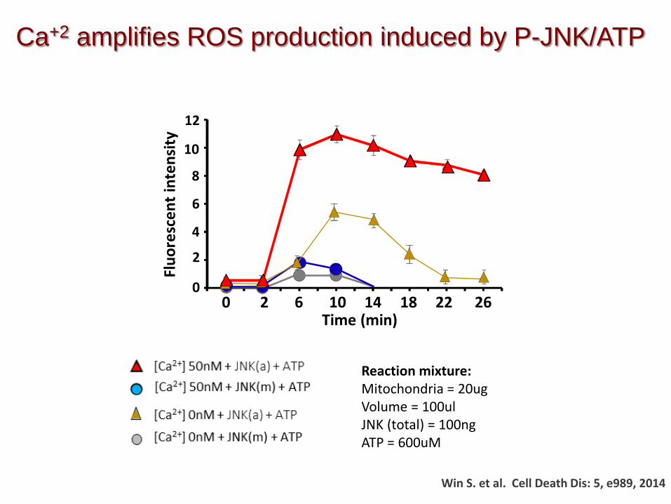
[Ca2+] 50nM + P-JNK + ATP [Ca2+] 0nM + P-JNK + ATP
[Ca2+] 50nM + JNK + ATP [Ca2+] 0nM + JNK + ATP
8Fl
uore
scen
t int
ensi
ty 12
10
6
4
2
0
Time (min)0 2 6 10 14 18 22 26
Reaction mixture: Mitochondria = 20ug Volume = 100ul JNK (total) = 100ng ATP = 600uM
Ca+2 amplifies ROS production induced by P-JNK/ATP
Win S. et al. Cell Death Dis: 5, e989, 2014

Model of JNK-Sab Mediated Mitochondrial Impairment and Cell Death
APAP TNF/galactosamine
Tunicamycin Palmitic Acid
Sab P-JNK OCR ?
Mito ROS Necrosis MPT
Bcl2 family mediated MOMP
Apoptosis
ROS
Win S. et al. JBC, 286: 35071, 2011 Win S. et al. Cell Death Dis, 5:, e989, 2014 Win S. et al. J Hepatol, 62: 1367,2015 Win S. et al. Hepatology in press 2016
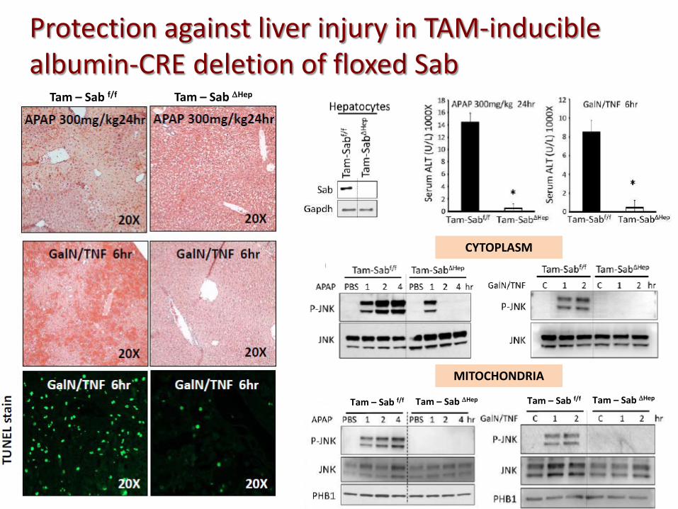
Protection against liver injury in TAM-inducible albumin-CRE deletion of floxed Sab
Tam – Sab f/f Tam – Sab ∆Hep
MITOCHONDRIA
CYTOPLASM
Tam – Sab f/f Tam – Sab ∆Hep Tam – Sab f/f Tam – Sab ∆Hep
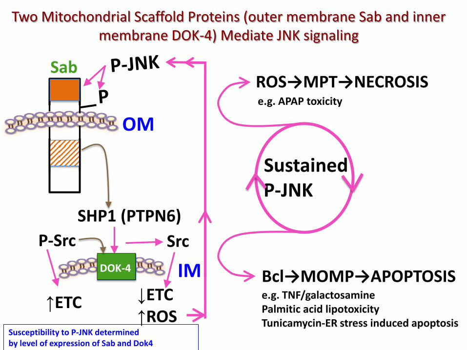
Two Mitochondrial Scaffold Proteins (outer membrane Sab and inner membrane DOK-4) Mediate JNK signaling
SHP1 (PTPN6) P-Src
OM
Src
IM ↓ETC ↑ROS
↑ETC
DOK-4
Sab
Sustained P-JNK
ROS→MPT→NECROSIS e.g. APAP toxicity
Bcl→MOMP→APOPTOSIS e.g. TNF/galactosamine Palmitic acid lipotoxicity Tunicamycin-ER stress induced apoptosis Susceptibility to P-JNK determined
by level of expression of Sab and Dok4
Intrinsic Stress Mitoch
ER DNA
(ROS, palmitic acid, bile acids)
Survival responses Caspase inhibitors
cFLIP XIAP
Anti-apoptosis Bcl members Bcl-XL Mcl1
Anti-oxidant response NF-kB NRF2
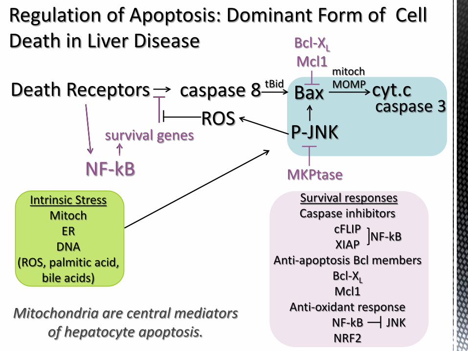
Regulation of Apoptosis: Dominant Form of Cell Death in Liver Disease
Death Receptors caspase 8 Bax cyt.c
NF-kB
survival genes ROS
P-JNK caspase 3
MKPtase
mitoch tBid
Bcl-XL Mcl1
JNK Mitochondria are central mediators
of hepatocyte apoptosis.
MOMP
NF-kB
NF-kB
survival genes

1. Chemicals stress hepatocytes in a variety of ways.
2. Many intricate adaptive responses dampen the adverse effects of stress and protect hepatocytes.
3. Stress can affect the “FITNESS” of hepatocytes leading to increased susceptibility to the lethal consequences of IMMUNE ATTACK (or generate sublethal DANGER SIGNALS).
4. The balance of injurious versus adaptive responses to drug-induced stress may be modulated by genetic and environmental factors.
5. Analogous to the yin-yang nature of immunity, the injurious stress versus adaptive responses in hepatocytes may be an important contributor to the occurrence of IDILI, even if immune mediated.
Conclusions