acute renal failure - usmf · acute renal failure •rapid decline in the gfr over days to weeks....
TRANSCRIPT
Acute Renal Failure
S. Popa,Conferentiar universitar
Disciplina Reumatologie & Nefrologie

•AKF – Acute Kidney Failure•CKD – Chronic Kidney Disease•ISN - International Society of Nephrology•K/DOQI – Kidney Disease Outcomes Quality Initiative
•KDIGO - Kidney Disease: Improving Global Outcomes
•MDRD - Modification of Diet in Renal Disease Study
•NKF – National Kidney Foundation

Renal Physiology Review
The Kidneys:
a. Control the fluid/electrolyte balance for the body
b. Remove metabolic wastes from the blood & excrete them to the outside
c. Regulate red-blood cell production
d. Regulate blood-pressure
e. Important in calcium ion absorption
f. Control volume, composition and pH of the blood

Renal Hormone Regulation
Synthesis and activation of hormones by the kidney include:
• Active form of Vitamin D
• Erythropoietin
Renal blood flow regulated by:
Renin-angiotensin aldosterone system (RAAS)

Fluid and Electrolyte Control Mechanisms
•RAAS – Renin-Angiotensin Aldosterone System
•Aldosterone
•ADH – Anti-Diuretic Hormone


Aldosterone
• Increases rate of sodium ion absorption
•Chloride moves along with sodium because of + charge of sodium
• Increases rate of potassium & hydrogen ion secretion
Result:
Fluid and sodium retention increases blood-pressure

Vasopressin (Antidiuretic Hormone, ADH)
• Vasopressin or antidiuretic hormone (ADH) is a nonapeptide that is synthesized in the hypothalamus.
• It has long been known to play important roles in the control of the body’s osmotic balance, blood pressure regulation, and proper kidney function.
• It is released from the posterior pituitary in response to hypertonicity and causes the kidneys to reabsorb solute-free water and return it to the circulation from the tubules of the nephron, thus returning the tonicity of the body fluids toward normal. An incidental consequence of this renal reabsorption of water is concentrated urine and reduced urine volume.
• Several disease states arise when the body loses control of ADH secretion or responds to its presence.


Epidemiology
•≈ 5-10% in hospitalized pts
•≈ 70% in critically ill pts
•5-6% ICU pts require RRT
•Once AKI occurred, the treatment is supportive, at an annual cost $10 billion in the US.

Acute Renal Failure•Rapid decline in the GFR over days to weeks. This
can occur in the setting of previously normal kidney function or as an acute-on-chronic phenomenon. Expressed clinically as the retention of nitrogenous waste products in the blood
• Acute kidney injury is defined when one of the following criteria is met:
*Serum creatinine rises by ≥ 26µmol/L within 48 hours or*Serum creatinine rises ≥ 1.5 fold from the reference value, which
is known or presumed to have occurred within one week or urine
output is < 0.5ml/kg/hr for >6 consecutive hours
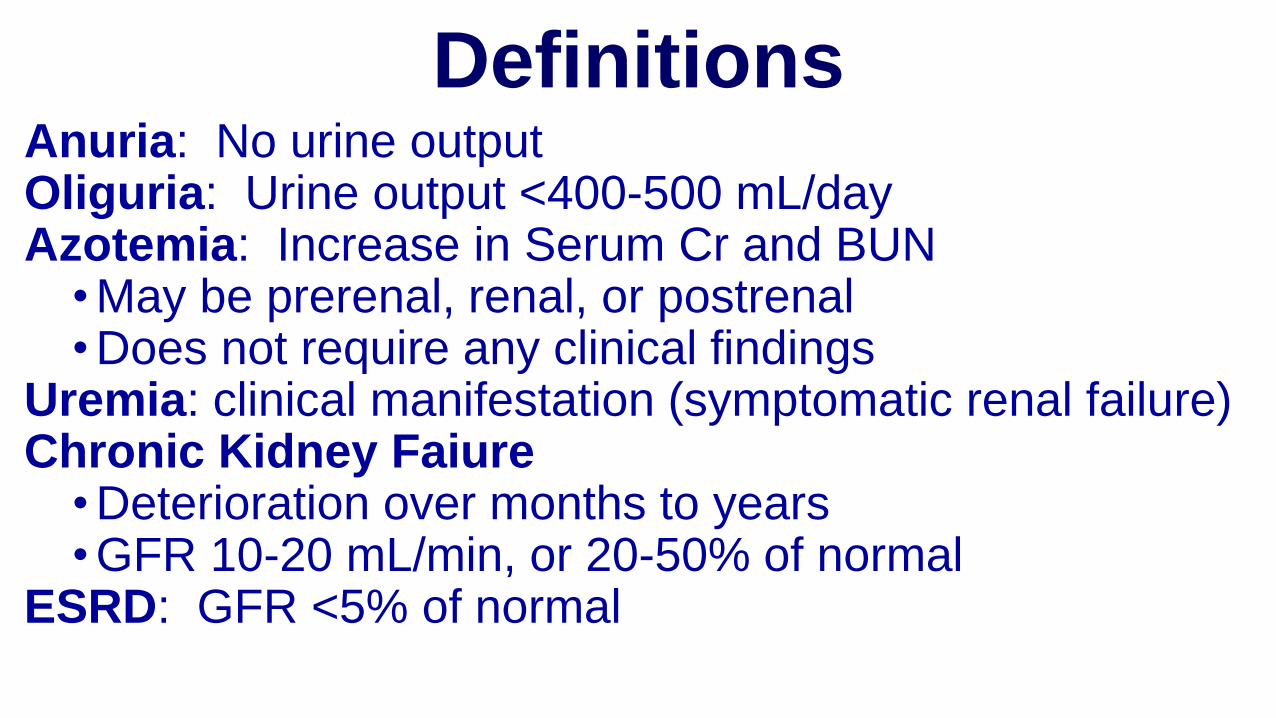
DefinitionsAnuria: No urine outputOliguria: Urine output <400-500 mL/dayAzotemia: Increase in Serum Cr and BUN
• May be prerenal, renal, or postrenal• Does not require any clinical findings
Uremia: clinical manifestation (symptomatic renal failure)Chronic Kidney Faiure
• Deterioration over months to years• GFR 10-20 mL/min, or 20-50% of normal
ESRD: GFR <5% of normal

Acute Renal Failure.Causes
A. Pre-renal = 55%
B. Post-renal = 5-15%
C. Renal parenchymal
(intrinsic)= 40%

Hilton, R. BMJ 2006;333:786-790
Causes of acute renal failure

Classification of the Etiologies of Acute Renal Failure

Volume depletionAcute urinary obstructionUse of diuretics, ACE or ARB Use of NSAIDs, iodinated contrast agents, or other nephrotoxic agentsHeart failureAcute glomerulonephritis or acute tubulo-interstitial nephritis Liver failureMalignancy (e.g., myeloma)
Common Causes of Acute or Acute on
Chronic Kidney Injury

Hypovolemia Peripheral vasodilation
Decreased cardiac output
Decrease in BP
Decrease of effective circulating volume
Compensations mechanisms:RAASsympathetic NS, NAArg-Vasopresine (ADH)
Decrease of tissue perfusion
Increase of renal reabsorption of water and sodium, thirst
vasoconstriction in some organs (skin, muscles, viscera - inclusive kidney)

Decrease of effective circulating volume
Oliguria,
Functional ARF
•Myogenic vasodilation
(“self-regulation”)
•Prostaglandins PGE2
•Vasodilatation effect of angiotensin on afferent arterioles
Compensations mechanisms:RAASsympathetic NS, NAArg-Vasopresine (ADH)

Oliguria,
Functional ARF
Decrease of effective circulating volume
Increase of renal reabsorption of water and sodium
Dense, hyperosmolar urine, with low sodium concentration
Decrease of glomerular and tubular perfusion
Tubular lesions (“organic ARF”)
Isostnenuric, isoosmolar urine, with high sodium concentration

Pathogenesis of Prerenal Azotemia
Renal
Vasoconstriction
Decreased
GFR
Angiotensin II
Adrenergic nerves
Vasopressin
+
+
+
Nitric oxide
Prostaglandins
-
-
Volume
Depletion
Congestive
Heart Failure Liver
Failure
Sepsis

Prerenal Acute Renal Failure
• Volume Depletion
• Decreased effective blood volume• congestive heart failure
• cirrhosis
• nephrotic syndrome
• sepsis
• Renal vasoconstriction• Hepato-renal syndrome
• Hypercalcemia
• Nonsteroidal anti-inflammatory drugs

Classification of acute kidney injury / acute renal failure

Relationship Between GFR and Serum Creatinine in ARF
120
40
80
0
GFR
(mL/min)
0 7 14 21 28
4
Days
2
0
6
Serum
Creatinine
(mg/dL)

Symptoms of ARF
• Decrease urine output (70%)
• Edema, esp. lower extremity
• Mental changes
• Heart failure
• Nausea, vomiting
• Pruritus
• Anemia
• Tachypenic
• Cool, pale, moist skin

ARF: Signs and Symptoms
•Hyperkalemia•Nausea/Vomiting•Arterial Hypertension
•Pulmonary edema•Ascites•Asterixis•Encephalopathy
• Decrease urine output (70%)
• Edema, esp. lower extremity
• Mental changes• Heart failure• Pruritus• Anemia• Tachypneaa• Cool, pale, moist skin

Hyperkalemia Symptoms
•Weakness
•Lethargy
•Muscle cramps
•Paresthesias
•Dysrhythmias

Hyperkalemia & EKG
•K > 5.5 -6
•Tall, peaked T’s
•Wide QRS
•Prolong PR
•Diminished P
•Prolonged QT
•QRS-T merge – sine wave


Clinical picture
•ARF clinically evolves into four phases:1. Pre-anuric phase2. Anuric phase3. Polyuric phase4. Recovery of renal function phase.
•Pre-anuric phase lasts 24-36 hours and is characterized by clinical signs of disease which generated ARF.
•Slight increase of BUN (50-80mg/dl) and creatinine (1,2-1,4mg/dl) •Proteinuria is discrete.

Anuric phase•It lasts 9-17 days to wide ranges from 36 hours to 40 days. It begins with the installation of oligo-anuria. Diuresis in patients with preserved GFR is reduced.•Symptoms:•fever at onset, then the temperature is normal;•respiratory symptoms: acidotic shortness of breath (Kussmaul), ammonia halitosis, lungs uremic syndrome (interstitial edema with progression to alveolar radiological nodular micro- and macro- opacities, imprecisely delineated, bilateral, responding favorably to ultrafiltration)•Cardiovascular manifestations: tachycardia, arterial hypotension, hypertension usually due to hyperhydratation, arrhythmia secondary to electrolyte disbalance, pericarditis, pulmonary edema•Mucocutaneous manifestations: skin pallor, jaundice, bleeding diathesis (mucocutaneous ecchymosis, bruising, skin necrosis), pruritic rash

Anuric phase
•Digestive symptoms: nausea, vomiting, hiccups, diarrhea, diffuse abdominal pain, upper gastrointestinal bleeding, acute pancreatitis (may be cause and effect ARF);•Neurological symptoms: restlessness, drowsiness, seizures, coma;•Renal manifestations: oligo-anuria is the cardinal sign. Diuresis is less than 400ml/day. Anuria suggests either an obstruction cause or a vascular occlusion. There are types of ARF with normal urine output characteristic for acute toxic tubulointerstitial nephritis (non-oliguric).

Anuric phaseUrinalysis:
urine may be cloudy, hematuric or purulent; urinary osmolarity is low;
urinary sediment is different depending on the cause ARF:
•Pre-renal ARF – non-active urinary sediment
•Post-renal ARF - the sediment is reach in Erythrocytes and Leukocytes,
•In nephrotoxic and ischemic ARF – epithelial and granulous casts, usually
with proteinuria <1g/day and micro-hematuria.
Biochemistry:
urea increases rapidly at the initial phase (50-100mg/dl/day), then slowly
(20mg/dl/day), creatinine increases by 0.5 mg/day, uric acid increases up to 12
mg/dl
Drops in Na - 130 mEq/L, increase in K to 7 mEq/L, bicarbonates decreased to
20 mEq/L

Anuric phase
•Ultrasound reveals normal or slightly enlarged size of kidneys and can diagnose obstructive uropathy;
• Intravenous urography should be avoided in these patients due to overlapping another nefrotoxic - contrast nephropathy substances;
•MRI is extremely useful in detecting renal artery stenosis, his role was expanded in evaluation of acute renovascular crisis.
•Renal biopsy is indicated for emergency needs in case of suspicion of rapidly progressive glomerulonephritis and vasculitis

Polyuric and functional recovery phase
•Polyuric phase starts with resumption of diuresis and ends with normalization of kidney function.
•polyuria can be installed quickly (diuresis 3000-4000 ml/day) or slowly (diuresis increases progressively 300-400 ml/day).
•clinical status of patients improves,
•paraclinical nitrogen retention products have a paradoxical increase in the first 5 days of polyuric phase, followed by a plateau for five days, and finally decreased rapidly.
•Functional recovery phase.
•polyuria gradually diminishes until the installation of normal diuresis –glomerular filtration rises progressively. The phase lasts about a year.

Physical Exam
•Volume Status •Mucus membranes, orthostatic hypotension,
•Cardiovascular•murmurs, pericardial rub, hypertension
•Pulmonary•Decreased breath sounds•Rales
•Rash (Allergic interstitial nephritis)•Large prostate •Extremities (Skin turgor, Edema)

Prerenal Acute Renal Failure:Clinical Presentation
•History•volume loss (e.g., diarrhea, acute blood loss)
•heart disease
• liver disease
•evidence of infection
•diuretic use
• thirst
•orthostatic symptoms

Prerenal Acute Renal Failure:Clinical Presentation
•Physical Examination•Blood pressure and pulse
•Orthostatic changes in blood pressure
•Skin turgor
•Dryness of mucous membranes and axillae
•Neck veins
•Cardiopulmonary exam
•Peripheral edema

Prerenal Acute Renal Failure:Clinical Presentation
• BUN : Creatinine ratio• > 20:1
• Urine indices• Oliguria
• usually < 500 mL/24 hours; but may be non-oliguric
• Elevated urine concentration• UOsm > 700 mmol/L• specific gravity > 1.020
• Evidence of high renal sodium avidity• UNa < 20 mmol/L• FENa < 0.01
• Inactive urine sediment

Fractional Excretion of Sodium
FENa = Filtered Sodium
Excreted Sodium

Fractional Excretion of Sodium
•Etiologies of a fractional excretion of sodium <0.01•normal renal function
•prerenal azotemia
•Hepato-renal syndrome
•early obstructive uropathy
•contrast nephropathy
• rhabdomyolysis
•acute glomerulonephritis

Treatment of Prerenal Acute Renal Failure
•Correction of volume deficits
•Discontinuation of antagonizing medications•NSAIDs/COX-2 inhibitors
•Diuretics
•Optimization of cardiac function

Postrenal Acute Renal Failure
•Urinary tract obstruction•level of obstruction
•upper tract (ureters)
•lower tract (bladder outlet or urethra)
•degree of obstruction•partial
•complete

Pathophysiology of Renal Failure in Obstructive Uropathy
•Early•Increased intratubular pressure
•Initial increase followed by decrease in renal plasma flow
•Late•Normal intratubular pressure
•Marked decrease in renal plasma flow
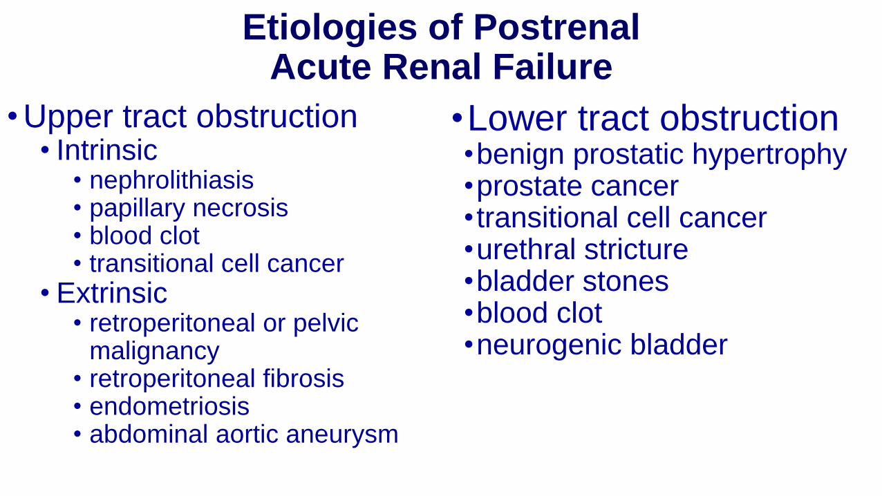
Etiologies of PostrenalAcute Renal Failure
• Upper tract obstruction• Intrinsic
• nephrolithiasis• papillary necrosis• blood clot• transitional cell cancer
• Extrinsic• retroperitoneal or pelvic
malignancy• retroperitoneal fibrosis• endometriosis• abdominal aortic aneurysm
•Lower tract obstruction•benign prostatic hypertrophy•prostate cancer• transitional cell cancer•urethral stricture•bladder stones•blood clot•neurogenic bladder

Postrenal Acute Renal Failure:Clinical Presentation
•History•Symptoms of bladder outlet obstruction
•urinary frequency
•urgency
• intermittency
•hesitancy
•nocturia
• incomplete voiding
Postrenal Acute Renal Failure:Clinical Presentation
•History•Changes in urine volume
•anuria•polyuria• fluctuating urine volume
•Flank pain•Hematuria•History of pelvic malignancy

Postrenal Acute Renal Failure:Clinical Presentation
•Physical Examination•Suprapubic mass•Prostatic enlargement•Pelvic masses•Adenopathy

Postrenal Acute Renal Failure:Clinical Evaluation
•Diagnostic studies•BUN: Creatinine ratio > 20:1•Unremarkable urine sediment•Variable urine chemistries

Postrenal Acute Renal Failure:Clinical Evaluation
•Diagnostic studies• Post-void residual bladder volume
• > 100 mL consistent with voiding dysfunction
• Radiologic studies• Ultrasound
• CT scan
• Nuclear medicine
• Retrograde pyelography
• Antegrade nephrograms

Intrinsic Acute Renal Failure
•Acute tubular necrosis (ATN)
•Acute interstitial nephritis (AIN)
•Acute glomerulonephritis (AGN)
•Acute vascular syndromes
•Intratubular obstruction
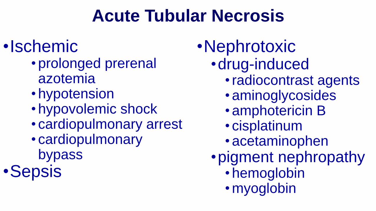
Acute Tubular Necrosis
•Ischemic• prolonged prerenal azotemia
• hypotension• hypovolemic shock• cardiopulmonary arrest• cardiopulmonary bypass
•Sepsis
•Nephrotoxic•drug-induced
• radiocontrast agents • aminoglycosides• amphotericin B• cisplatinum• acetaminophen
•pigment nephropathy• hemoglobin• myoglobin

Pathophysiology of ATN:Tubular Epithelial Cell Injury and Repair
Loss of polarityNormal Epithelium
Migration, Dedifferentiation of
Viable Cells
Differentiation &
Reestablishment of
polarity
Sloughing of viable and dead cells with
luminal obstruction
Ischemia/ Reperfusion ApoptosisNecrosis
Cell death
Adhesion molecules
Na+/K+-ATPase
Proliferation

Pathophysiology of Acute Tubular Necrosis
•Mechanisms of decreased renal function•Vasoconstriction
•Tubular obstruction by sloughed debris
•Back leak of glomerular filtrate across denuded tubular basement membrane

Phases of Ischemic ATNG
FR
Time

Pathophysiology of ATN
Ischemia
Endothelial Injury
Capillary Obstruction
&
Continued Ischemia
Inflammation
Tubular Injury
Disruption of Cytoskeleton
Loss of Cell Polarity
Desquamation of Cells
Tubular Obstruction &
Backleak
Apoptosis &
Necrosis
Activation of Vasoconstrictors
Impaired Vasodilation
Increased Leukocyte Adhesion

Acute Tubular Necrosis:Clinical Presentation
• History• Acute illness• Exposure to nephrotoxins• Episodes of hypotension
• Physical examination• Hemodynamic status• Volume status• Features of associated illness
• Laboratory data• BUN : Creatinine ratio < 10:1• Evidence of toxin exposure

Acute Tubular Necrosis:Clinical Presentation
• Urine indices• Urine volume
• may be oliguric or non-oliguric
• Isosthenuric urine concentration• UOsm 300 mmol/L• specific gravity 1.010
• Evidence of renal sodium wasting• UNa > 40 mmol/L• FENa > 0.02
• Urine sediment• tubular epithelial cells• granular casts

Acute Interstitial Nephritis
• Drug-induced• penicillins
• cephalosporins
• sulfonamides
• rifampin
• phenytoin
• furosemide
• NSAIDs
• Malignancy
• Idiopathic
• Infection-related• bacterial
• viral
• rickettsial
• tuberculosis
• Systemic diseases• SLE
• sarcoidosis
• Sjögren’s syndrome
• tubulointerstitial nephritis and uveitis

Acute Interstitial Nephritis:Clinical Presentation
•History• preceding illness or drug exposure
•Physical examination• fever
• rash
•Laboratory Findings• eosinophilia

Acute Interstitial Nephritis:Clinical Presentation
•Urine findings•non-nephrotic protinuria
•hematuria
•pyuria
•WBC casts
•eosinophiluria

Acute Glomerulonephritis
•Nephritic presentation•proteinuria
•may be in nephrotic range (> 3 g/day)
•hematuria
•RBC casts
•Diagnosis usually requires renal biopsy

Acute Glomerulonephritis
•Etiologies• poststreptococcal glomerulonephritis
• postinfectious glomerulonephritis
• endocarditis-associated glomerulonephritis
• systemic vasculitis
• thrombotic microangiopathy • hemolytic-uremic syndrome
• thrombotic thrombocytopenic purpura
• rapidly progressive glomerulonephritis

Acute Vascular Syndromes
•Renal artery thromboembolism
•Renal artery dissection
•Renal vein thrombosis
•Atheroembolic disease
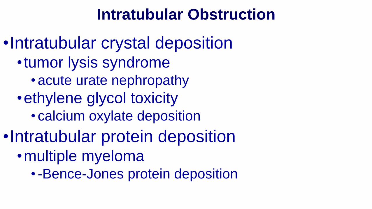
Intratubular Obstruction
•Intratubular crystal deposition• tumor lysis syndrome
• acute urate nephropathy
•ethylene glycol toxicity • calcium oxylate deposition
•Intratubular protein deposition•multiple myeloma
• -Bence-Jones protein deposition

Diagnostics
•Diagnosis is based on:•History (existence of clinical suspicion for an acute condition which involves decrease of GFR - sepsis, arterial hypotension, nephrotoxic, etc.),•Clinical features•Paraclinical signs – azotemia is increasing progressively day by day.

Differential Diagnosis of ARF
•Prerenal ARF•Postrenal ARF•Intrinsic ARF
•acute tubular necrosis•acute interstitial nephritis•acute glomerulonephritis•acute vascular syndromes•intratubular obstruction

Hilton, R. BMJ 2006;333:786-790
Differential diagnosis of acute renal failure

Sonographic appearance of the renal parenchyma. (A) Normal right kidney (longitudinal view). (B) Normal right kidney (transverse view). (C) Echogenic right kidney with prominent medullary pyramids (arrows). (D) Atrophic right kidney (longitudinal view) with thin parenchyma and containing mostly sinus fat. L, liver.

Sonographic appearance of the upper and lower urinary tract. (A) Hydronephrosis of a left kidney (longitudinal
view) with dilated renal pelvis and major and minor calyces. (B) Another longitudinal view of the same kidney showing
a dilated ureter (arrows) tracking underneath the lower pole. (C) Transverse view of the urinary bladder (B) showing
dilated distal ureters (arrows). (D) Transverse view of the urinary bladder with an enlarged prostate gland (P).

Simple renal cyst with posterior enhancement in an adult kidney.
Advanced polycystic kidney disease with multiple cysts

End-stage hydronephrosis with cortical thinning
Staghorn calculi filling the entire
collecting system and creating pronounced shadowing

Acute vs Chronic Renal Failure
History•Known Chronic
•Recent Toxic Exposure
•Recent Hypoxic Insult
•Recent Trauma
•Known Diseases Associated with ARF
•Prev. Abnormal Lab Results Suggesting Chronic

Acute vs Chronic Renal Failure
•Rapidly Rising Creatinine = Acute•Kidney Size
•Small = Chronic•Renal Ultrasound
•Increased Echogenicity = Chronic•Urine Flow Rate
•Oliguric or Anuric usually = Acute

History & physical exam in ARF

Differential diagnosis•To differentiate between diverse forms of Intrinsic ARF the urinalysis is needed:
•in glomerulonephritis, vasculitis: hematuria, erythrocytes casts, proteinuria •in ATN: epithelial casts, epithelial cells, minimal proteinuria•in interstitial nephritis: leukocyturia, eosinophiluria •proteinuria below 1g/day – in ischemic and nephrotoxic ARF, •proteinuria >1g/day - in glomerular disease, multiple myeloma, allergic interstitial nephritis caused by NSAIDs•hemoglobinuria, myoglobinuria: hemolysis, rhabdomyolysis.

Differentiation of pre-renal ARF from intrinsic ARF
• Pre-renal ARF: - Fractional Excretion of Sodium (FeNa+) <1%•Concentration of urinary sodium (mEq/L)<10•Urinary density – 1.020•Urinary osmolality (mOsm/kg H
2O) >500
•urinary creatinine/plasmatic creatinine ratio >4•Urinary sediment – hyalinic casts
• Intrinsic ARF: - FeNa+ (%) >1•Concentration of urinary sodium(mEq/L) >20•Urinary density <1.010•Urinary osmolality (mOsm/kg H
2<250
•urinary creatinine/plasmatic creatinine ratio <20•Urinary sediment – granular casts

Differentiation with exacerbation of chronic kidney disease and ESRD
•ARF is characterized by missing of:•Suggestive symptoms of chronic renal disease (asthenia, loss of appetite, polyuria, nocturia)•HTN,•History of nitrogen retention (increased urea & creatinine)•Skin symptoms (pallor, scratching lesions)•Features of uremic sensitive-motoric polyneuropathy•X-ray symptoms of renal bone disease,
•Normal dimensions of kidneys by ultrasound.

Diagnostic Evaluation of ARF
Form of ARF BUN:CrUNa
(mEq/L)FENa Urine Sediment
Prerenal >20:1 <20 <1% Normal
Postrenal >20:1 >20 variable Normal or RBC’s
Intrinsic
ATN <10:1 >40 >2% Muddy brown casts; tubular epithelial cells
AIN <20:1 >20 >1% WBC’s, WBC casts, RBC’s, eosinophils
AGN variable <40 <1% RBC’s, RBC casts
Vascular variable >20 variable Normal or RBC’s

Management of ARF
Management depends on the etiology and severity of the ARF.• A. “Pre-renal” ARF: Improve renal blood flow by replacing lost
volume and/or optimizing perfusion. Be very careful about using potassium in this setting!
• B. Post-renal ARF: Relieve the identified obstruction. This may require placement of a Foley catheter or a percutaneous nephrostomy, removal of a stone, etc. Following relief of obstruction, there may be a very brisk post-obstructive diuresis with very high urine output and electrolyte loss, putting your patient at risk for pre-renal ARF.
• C. Renal causes of ARF: Specific therapy depends on the diagnosis.

Treatment of Prerenal Acute Renal Failure
•Correction of volume deficits
•Discontinuation of antagonizing medications•NSAIDs/COX-2 inhibitors
•Diuretics
•Optimization of cardiac function

Treatment of pre-renal ARF
•Pre-renal ARF is reversible after resumption of renal perfusion, with normalization of BUN and Creatinine within 24-48 h.•Patients with hypovolemic shock should be monitored by measuring of CVP:
•CVP < 2 cm H2O, hypovolemia are corrected by vascular repletion with isotonic fluids (sodium chlorate 0,9%), high isotonic fluids (blood, plasma, albumin).•Fluids are administered until BP is normalized and urinary flux is 30 ml/h, avoiding overload and acute pulmonary edema.•If CVP >8 cm H
2O the administration of fluids is discontinued and
•the cardiac failure should be corrected with cardiac tonics, diuretics, vasodilators to reduce cardiac afterload.

Treatment of intrinsic ARF
Preventive Treatment •Rapid restoration of intravascular volume reduces the incidence of ATN after major surgical interventions, severe trauma, burns. •The importance to maintain euvolemia, is better demonstrated in contrast induced nephropathy: - prophylactic use of saline solutions , pre- and post- procedure, is more efficient in prevention of ARF in comparison with Manitol & Furosemide
•N-acetilcisteine is useful in prevention of contrast induced nephropathy in doses of 600 mg t.d., pre- and post- procedure, associated with hydration.•use of iso-osmolare contrasts or less nephrotoxic contrasts (gadolinium), reduces the incidence of contrast nephropathy.
NSAIDs, diuretics and ACEI should be administered with caution in patients with chronic kidney disease and hypovolemia, because they could convert a pre-renal ARF into ischemic acute tubular necrosis.

Treatment of intrinsic ARF
Specific treatment•Dopamine, in renal dosis,1-3 mg/kg/min, in prospective, clinical trials did not demonstrate the influence of ARF course•Fenodolpam, a selective dopamine agonist which determines better renal vasodilation and o natriuresiscompared to dopamine•Loop diuretics are frequent administration in oliguric ARF in high doses to convert oliguric ARF into non-oliguric. Furosemide and Thorasemide rise diuresis, and reduce the need for dialysis. Thus, their use is recommended only in ARF associated with hypervolemia.

Treatment of intrinsic ARF
Specific Treatment•Manitol – osmotic diuretic which reduces tissue edema and causes intrarenal vasodilatation via stimulating prostaglandin production. •Atrial natriuretic peptide (ANP) - polypeptide synthesized in atrial muscle which increases ultrafiltration rate via decreasing vascular resistance in afferent arterioles. It is efficient in prevention of ischemic and nephrotoxic ARF.•Insulin-like grow factor - efficient in prevention of ATN, not in installed ARF. It stimulates production of NO, protein synthesis, decrease of cell apoptosis.

Acute Tubular Necrosis:Treatment
•Supportive therapy•No specific pharmacologic treatments•Acute dialysis for:
• volume overload • metabolic acidosis • hyperkalemia• uremic syndrome
• pericarditis• encephalopathy
• azotemia

Acute Interstitial Nephritis:Treatment
•Discontinue offending drug
•Treat underlying infection
•Treat systemic illness
•Glucocorticoid therapy may be used in patients who fail to respond to more conservative therapy

Treatment of Postrenal Acute Renal Failure
•Relief of obstruction• Lower tract obstruction
• bladder catheter
• Upper tract obstruction• ureteral stents• percutaneous nephrostomies
•Recovery of renal function dependent upon duration of obstruction
•Risk of post-obstructive diuresis

Hyperkalemia Treatment
•Calcium gluconate (carbonate)
•Sodium Bicarbonate
•Insulin/glucose
•Diuretics (Furosemide)
•Albuterol
•Hemodialysis

Renal Replacement Therapy for ARF
Consider dialysis or hemofiltration if:•Uremic symptoms (encephalopathy, pericarditis)
•Oligoanuria >48 hours•Hyperkalemia >6.5mEq/l•Severe acidosis•Volume overload unresponsive to diuretic (pulmonary edema)

Renal Replacement Therapy
•Intermittent hemodialysis•Hemofiltration and continued hemodiafiltration•Peritoneal dialysis

Treatment of complications
•Acute pulmonary edema is secondary to hyperhydratation in oligo-anuria.• This is a major clinical emergency:
•Oxigenotherapy with high debit•Furosemide in high doses: 240-500 mg i.v. within - 30-60 min (or Thorasemide) •Nitroglycerine i.v.•Hypotensive: sodium nitropruside.
•ACE inhibitors should be avoided…

Treatment of hyperkalemia:If K+= 6-6.5 mEq/l - stop medicines potent to rise potasemia(Spironolactone, ACE inhibitors, sartans) and give ion-changing resinsand Furosemide (Thorasemide).•If K+> 6.5 mEq/l due to a risk of cardiac arrest, urgently:
•Furosemide:120-240 mg i.v;•Sodium Bicarbonate 8,4‰, 40-100ml i.v. within 30-60 min, •ß-mimetics: Isoprenaline i.v. if complete AV block or Albuterol spray•Hypertonic glucose 50%, 50 ml + Insulin 10 UI i.v.•Calcium gluconate, 20-60 ml i.v. prevents cardiac arrhythmia;•Ion-changing resins: Kayexalat, binds the intestinal K+,15-30 g 4/day po, or via intestinal tube;•In oliguria with K>7 mEq/l hemodialysis is most efficient treatment.

Nutrition:•Caloric intake - 30-50 cal/kg/day, accordingly to grade of hypercatabolism.•Protein intake - 0,3-0,5 g/kg/day in non-dialyzed patients and 1-1,3 g/kg/day in dialyzed patients . •Liquids intake: diuresis + 500ml (lost from perspiration) + extra-renal lost (diarrhea, vomiting) + 50 ml for each Celsius degree above normal.•Salt intake - 1g/day in non-dialyzed patients and 2-3 g/day in dialyzed patients .