acute liver failure · acute liver failure a. w. holt department of critical care medicine,...
TRANSCRIPT

Acute Liver Failure A. W. HOLT Department of Critical Care Medicine, Flinders Medical Centre, Adelaide, SOUTH AUSTRALIA
ABSTRACT Objective: To consider the classification and to present an approach to the diagnosis and management of complications associated with acute liver failure. Data sources: A review of studies reported from 1966 to 1998 and identified through a MEDLINE search on treatment of acute liver failure. Summary of review: Acute liver failure can be subdivided into hyperacute, (encephalopathy within 7 days of onset of jaundice) acute (8-28 days from jaundice to encephalopathy) and subacute (29 to 72 days from jaundice to encephalopathy) forms. Management of all forms involves largely supportive care until hepatocyte regeneration and recovery occurs (predominantly in the hyperacute group), or bridging supportive therapy until orthotopic liver transplantation can be performed. New therapies such as bioartificial liver support devices and ex-vivo liver perfusion offer exciting possibilities for this bridging therapy. While orthotopic liver transplantation remains the definitive treatment for many patients with acute liver failure, N- acetyl-cystine (150 mg/kg over 15 minutes followed by 12.5 mg/kg/hour) and PGE1 (10 – 40 µg/hour) are reported to have an additional role and are being used increasingly in the management of all forms of acute liver failure. Conclusions: Acute liver failure is the end stage of many acute viral and drug induced hepatic diseases. Management is largely supportive until hepatic repair or transplantation can be performed. Recently, additional hepatic protective, regenerative and supportive therapies have been successfully used. (Critical Care and Resuscitation 1999; 1: 25-38)
Key words: Acute liver failure, hepato-renal syndrome, hepatic encephalopathy, cerebral oedema, liver assist device, orthotropic liver transplantation
Classification Two different classifications of acute liver failure (ALF) have been proposed by two prominent groups who work in this field1,2 (Table 1). Both seek to describe the clinical spectrum of ALF within subgroups to highlight the association of aetiology and prognostic features, to facilitate the delineation of management (particularly with respect to the need for and timing of orthotopic liver transplant). The French (Hopital Beaujon classification) advocate that ALF occurs at the pre-encephalopathy stage and that the most sensitive marker is synthetic function.2 This approach means that ALF is not a clinical diagnosis, and as most patients with ALF have factor V levels well below 50% by the time encephalopathy ensues, this results in more patients with acute liver injury being diagnosed with ALF. Unlike the
King’s group, the French do not recognise the ‘hyperacute’group. The reason for this is probably due to their casemix, with ALF due to paracetamol poisoning being much less common in France. Using the King’s classification,1 the difference between the forms of ALF is reflected in the aetiology, incidence of life-threatening cerebral oedema and prognosis without orthotopic liver transplant (Table 2). Given the substantial contribution to the literature of ALF made by the King’s unit, and the lack of need from a critical care point of view to include less severe forms of ALF, I would advocate the use of the King’s definition of ALF. The clinical syndrome of hyperacute liver failure reflects largely the features of paracetamol injury with a significant proportion surviving without orthotopic liver transplant (this is likely to be greater than the 36%
Correspondence to: Dr. A. W. Holt, Department of Critical Care Medicine, Flinders Medical Centre, Bedford Park, South Australia 5042
25

A. W. HOLT Critical Care and Resuscitation 1999; 1: 25-38
quoted from the King’s unit, with recent advances in critical care). Table 1. Classifications of Acute liver failure Classification Features King’s a) Hyperacute liver failure encephalopathy within 7 days of onset of jaundice. b) Acute liver failure 8-28 days from jaundice to encephalopathy c) Subacute liver failure 29-72 days from jaundice to encephalopathy Hopital Beaujon Acute liver failure prothrombin or factor V below 50% of normal a) Fulminant early encephalopathy (<28days from onset of jaundice) b) Subfulminant late encephalopathy (>28 days from onset of jaundice) Recent data from a large cohort of ALF patients drawn from 12 transplant centres, 295 patients over 2 years (1994-96) demonstrated a spontaneous survival rate in paracetamol induced-ALF of 57%.3 However, cerebral oedema with life-threatening intra-cranial hypertension is a major determinant of poor prognosis. Acute and subacute liver failure syndromes define patients in whom orthotopic liver transplant (OLT), if possible, is the treatment of choice due to their poor prognosis. With patients who have a clear subacute syndrome, less emphasis is required to detect and treat cerebral oedema due to a much lower incidence and severity of the disorder. Table 2. Acute liver failure data from King’s unit Classification Hyperacute Acute Subacute Aetiology Paracetamol Hepatitis C Hepatitis C Hepatitis B NANBNC* NANBNC Hepatitis B Cerebral Common Common Infrequent oedema (69%) (59%) (14%) Survival without 36% 7% 7% orthotopic liver transplant * NANBNC = presumed viral non A non B non C Aetiology The spectrum of causes of ALF varies between countries and centres, although direct comparisons are
made difficult by differing definitions. In Australia the best data available are from patients who have received an hepatic transplant. Cryptogenic/non-ABC group is by far the most common cause of ALF in Australia, whereas drug-induced ALF (particularly paracetamol-induced) appears to be less common in Australia4 compared with United States of America3 (Table 3). Table 3. Causes of acute liver failure and incidence in Australia and USA Australia USA Drug-Induced (Total) 21% 32% Paracetamol 20% Idiosyncratic 12% Cryptogenic/NonABC 43% 15% Hepatitis B 16% 10% Hepatitis A 0% 7% Wilson’s Disease 6% 12% Treatment Treatment of patients with ALF generally involves supportive care until hepatocyte regeneration and recovery occurs (predominantly in the hyperacute group) or bridging therapy until OLT can be performed (Table 4). The major reasons why patients fail to survive to OLT (or are precluded from OLT) are brain injury from intra-cranial hypertension, acute lung injury, pulmonary oedema and overwhelming sepsis. Table 4. Treatment of Acute Liver Failure System Support CNS - Hepatic encephalopathy - Cerebral oedema Respiratory Circulatory Renal Haematological Gastrointestinal Metabolic Prevention of Sepsis Hepatic Rescue Hepatic Support Liver Transplant Orthotopic Auxiliary A. SYSTEM SUPPORT 1. Central nervous system The main issues in treatment are:
26

Critical Care and Resuscitation 1999; 1: 25-38 A. W. HOLT
a) Protection of airway The rapid progression to high-grade encephalopathy with airway obstruction, hypoventilation and aspiration is a common preventable sequence leading to death or unsuitability for OLT. b) Treatment of hepatic encephalopathy Naso-gastric antibiotics. Gentamicin (80mg via a nasogastric tube 8-hourly) has replaced neomycin as the aminoglycoside of choice for hepatic encephalopathy as it is cheaper and plasma levels are measurable compared with neomycin. Lactulose. While lactulose is the mainstay of therapy for chronic hepatic encephlopathy, gastrointestinal stasis associated with ventilation and sedation results in a high risk of gaseous abdominal distension due to fermentation, particularly with aggressive oral regimes (the addition of cisapride in this regard may be helpful in emptying the bowel). Lactulose enemas (100 ml in 100 ml of water) appear to be at least as effective as oral lactulose and circumvents the problem of gastric stasis. The appropriateness of combining gastrointestinal antibiotics and lactulose is argued. The problem of rapidly gaining control with lactulose makes the addition of nasogastric gentamicin initially advisable.5 While plasma ammonia levels do not correlate well with the severity of encephalopathy (or indeed with severity of ALF) within the individual patient, the trend is a reasonable guide to the adequacy of intestinal decontamination and worsening or resolution of the ALF. Flumazenil. Flumazenil has been used in ALF, based on the hypothesis that excessive gamma-aminobutyric acid (GABA) receptor stimulation by endogenous agonists is a major cause of hepatic encephalopathy. In patients dying from paracetamol induced ALF with grade IV encephalopathy, substances which inhibit the binding of labeled flumazenil are found in cerebral tissue, which are absent in controls.6 In chronic liver disease with portal systemic encephalopathy, the doses of flumazenil quoted vary from 0.3-5 mg.7 However, there appears to be a decreased response in ALF compared with chronic hepatic encephalopathy, although the non-responders in high grade encephalopathy are likely to have raised intra-cranial pressure (ICP). Nevertheless, in patients with ALF any resolution of encephalopathy with flumazenil is rarely of great benefit in reducing the need for airway protection. c) Detection and treatment of cerebral oedema The prevention of neurological injury and death due to intra-cranial hypertension with subsequent cerebral ischaemia and transtentorial herniation, is crucial to recovery from severe ALF or to a successful bridge to
OLT. The pathophysiology of cerebral oedema of ALF is being increasingly defined using animal models. Both cytotoxic and vasogenic mechanisms have been described8 and include the following, 1) inhibition of Na-K ATPase with the resulting accumulation of intracellular sodium and consequent cerebral swelling, 2) toxic disturbance of the blood-brain barrier, and 3) increase in osmotically active molecules, particularly glutamine and other amino acids. Glutamine accumulation (which is the result of ammonia detoxification in astrocytes) has been emphasized.9 The accumulation of this amino acid correlates with the increase in brain water in animal models. However, a major shortcoming of the glutamine hypothesis is the lack of brain oedema in patients with chronic liver disease who have similar elevations of brain glutamine levels. It is possible that rapidity of onset and elevation of organic osmolar particles, particularly glutamine and myo-inositol, overwhelm the compensatory mechanisms that regulate cerebral cell volume. CEREBRAL HAEMODYNAMIC AND METABOLIC PROFILE IN ALF Despite this understanding of the mechanisms of cerebral oedema in ALF, considerable uncertainty surrounds much of the clinical data dealing with cerebral haemodynamics, and the implications for treatment of raised ICP resulting from this cerebral oedema. The pressure dependency and loss of autoregulation of cerebral blood flow (CBF) in severe ALF has been well demonstrated, and has been found in all patients studied with grade IV encephalopathy.10 The CBF autoregulation is restored rapidly (usually within 24 hours) following OLT. In ALF patients who have recovered without OLT, CBF autoregulation is usually restored prior to complete recovery of the encephalopathy. Clinical data from the King’s group appears to imply that inadequate CBF is the most common finding in high grade encephalopathy whereas the Pittsburgh group appear to regularly find normal or high CBF, and that low CBF is a pre-terminal event.11 The most recent data from the King’s group shows that 73% of patients with grade IV encephalopathy have a low CBF and that all patients who were studied had a low cerebral metabolic rate for oxygen.12 This reduced oxygen consumption was associated with evidence of anaerobic metabolism with increased cerebral lactate production. In these patients hyperventilation resulted in further fall in CBF and oxygen consumption, although lactate production was not increased. Prostaglandin I2 did not alter CBF but increased oxygen consumption. Mannitol and NAC significantly increased CBF and oxygen consumption.
27

A. W. HOLT Critical Care and Resuscitation 1999; 1: 25-38
In contrast, the Pittsburgh group found that only 52% of patients studied had reduced CBF.11 Importantly, 24% had an elevated CBF and these patients were more likely to show cerebral oedema on CT scan, and have a worse prognosis. In patients with an ICP greater than 24 mmHg, all were found to have high CBF. The CBF was also significantly higher in patients in grade IV encephalopathy. This work strongly suggests that cerebral swelling, intra-cranial hypertension and poor prognosis is associated with high CBF or cerebral hyperaemia. The obvious difference between these findings and the data from the King’s unit is difficult to reconcile, although it must be noted that only 21% of the Pittsburgh’s patients were in grade IV encephalopathy compared to all of the patients studied by the King’s unit. The Pittsburgh group has characterised five cerebral haemodynamic phases (Table 5) by measuring epidural ICP, CBF (Xe133 technique) and arterio-jugular venous oxygen content difference (AVDO2). The mean CBF in patients with ALF was lower (28.7ml/min/100g) than healthy controls (53ml/min/100g).13 However, from the Pittsburgh data the reduced flow was initially associated with reduced cerebral metabolic rate as the O2 extraction was normal. With increasing severity, CBF initially increased, although not to ‘healthy’ levels, and the reduced cerebral O2 consumption produced a low AVDO2 and effective luxury flow or hyperaemia. While the Pittsburgh group conclude from their data that patients with ALF progress through the five phases, it is perhaps more correct to conclude:
1. Patients who survive with medical therapy were never observed to be beyond phase 2 2. Patients observed beyond phase 2 and survived underwent OLT 3. Patients who died without OLT, all achieved phase 4 or 5.
The data in patients who died without OLT did not show a sequential deterioration through each phase. However these measurements were taken intermittently and it is reasonable to infer a progression
through each phase at variable rates, with the aim of therapy of course to prevent this progression. The increase in CBF with the progression from phase 1-3 was associated with a rise in ICP and it is tempting to conclude that the hyperaemia contributes to the cerebral oedema. The possibility that hyperaemia contributes to cerebral oedema has a number of important implications for treatment. For example,
1. As cerebral autoregulation is lost in ALF, unnecessary augmentation of mean arterial pressure (MAP) may be detrimental as it may exacerbate the hyperaemia.
2. Therapies that are associated with cerebral vasoconstriction may be of benefit in the early phases in the setting of increased ICP and luxury perfusion or hyperaemia.
3. Monitoring of cerebral blood flow (using Xe133 washout or transcranial doppler) or jugular bulb venous oxygen saturation (as an index of adequacy of global oxygenation), becomes important along side MAP and ICP. Measuring actual flow is largely confined to research protocols, whereas the insertion of a jugular bulb catheter is simple and safe even in coagulopathic patients.
Diagnosis of cerebral oedema Patients with hyperacute and acute ALF can be assumed to have cerebral oedema as it can occur in up to 80% of ALF patients with grade IV encephalopathy. Clinical signs of cerebral oedema (e.g. pupillary changes and motor posturing) are usually late features, and as most patients when they reach grade III encephalopathy are being sedated (to facilitate intubation and ventilation for airway protection), clinical assessment of neurological status is usually unhelpful. Cerebral CT scanning is also of little help as it has a low sensitivity in the detection of elevated ICP (although it will exclude intracranial haemorrhage). In a small series, 15 ALF patients in either stage III
Table 5. Five cerebral haemodynamic and metabolic phases of ALF Phase 1 Phase 2 Phase 3 Phase 4 Phase 5 ICP <25 <25 >25 >25 >25 mmHg CBF <29 >29 >29 10-29 <10 /min/100g AVDO2 high narrow narrow high high- normal eventually low, with no oxygen extraction Cerebral Normal Hyperaemic Hyperaemic Impending Ischaemia perfusion perfusion low compliance ischaemia herniation
28
Critical Care and Resuscitation 1999; 1: 25-38 A. W. HOLT
or IV encephalopathy, 80% had an elevated ICP of 32 + 22 mmHg (mean + SD) and range of 18 - 100 mmHg.14 Four patients had CT scan evidence of an elevated ICP (these patients had pressures of 100, 40, 27, and 23 mmHg). The 8 patients who had normal CT scans had ICP’s of 24 + 4 mmHg (mean + SD), with a range of 18 - 29 mmHg. For this reason many centres routinely measure ICP (at extradural, subarachnoid or intracerebral sites) in patients with ALF with high-grade encephalopathy who require mechanical ventilation. Benefits of an ICP monitor. The advantages of an ICP monitor include,
1. measurement and maintenance of cerebral perfusion pressure,
2. an ability to target treatment for cerebral oedema, to reduce ICP and prevent transtentorial herniation,
3. selection of salvageable patients for OLT (in one series all patients with sustained cerebral perfusion pressure of < 40 mmHg over 2 hours died. Even those that went on to OLT died post-operative from brain injury),
4. management of peri-operative surges in ICP associated with OLT (e.g. reperfusion of graft can be associated with brain threatening surges in ICP).15
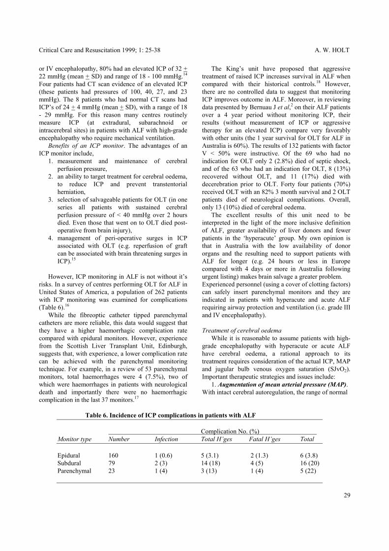
However, ICP monitoring in ALF is not without it’s risks. In a survey of centres performing OLT for ALF in United States of America, a population of 262 patients with ICP monitoring was examined for complications (Table 6).16 While the fibreoptic catheter tipped parenchymal catheters are more reliable, this data would suggest that they have a higher haemorrhagic complication rate compared with epidural monitors. However, experience from the Scottish Liver Transplant Unit, Edinburgh, suggests that, with experience, a lower complication rate can be achieved with the parenchymal monitoring technique. For example, in a review of 53 parenchymal monitors, total haemorrhages were 4 (7.5%), two of which were haemorrhages in patients with neurological death and importantly there were no haemorrhagic complication in the last 37 monitors.17
The King’s unit have proposed that aggressive treatment of raised ICP increases survival in ALF when compared with their historical controls.18 However, there are no controlled data to suggest that monitoring ICP improves outcome in ALF. Moreover, in reviewing data presented by Bernuau J et al,2 on their ALF patients over a 4 year period without monitoring ICP, their results (without measurement of ICP or aggressive therapy for an elevated ICP) compare very favorably with other units (the 1 year survival for OLT for ALF in Australia is 60%). The results of 132 patients with factor V < 50% were instructive. Of the 69 who had no indication for OLT only 2 (2.8%) died of septic shock, and of the 63 who had an indication for OLT, 8 (13%) recovered without OLT, and 11 (17%) died with decerebration prior to OLT. Forty four patients (70%) received OLT with an 82% 3 month survival and 2 OLT patients died of neurological complications. Overall, only 13 (10%) died of cerebral oedema. The excellent results of this unit need to be interpreted in the light of the more inclusive definition of ALF, greater availability of liver donors and fewer patients in the ‘hyperacute’ group. My own opinion is that in Australia with the low availability of donor organs and the resulting need to support patients with ALF for longer (e.g. 24 hours or less in Europe compared with 4 days or more in Australia following urgent listing) makes brain salvage a greater problem. Experienced personnel (using a cover of clotting factors) can safely insert parenchymal monitors and they are indicated in patients with hyperacute and acute ALF requiring airway protection and ventilation (i.e. grade III and IV encephalopathy). Treatment of cerebral oedema While it is reasonable to assume patients with high-grade encephalopathy with hyperacute or acute ALF have cerebral oedema, a rational approach to its treatment requires consideration of the actual ICP, MAP and jugular bulb venous oxygen saturation (SJvO2). Important therapeutic strategies and issues include: 1. Augmentation of mean arterial pressure (MAP). With intact cerebral autoregulation, the range of normal
Table 6. Incidence of ICP complications in patients with ALF Complication No. (%) Monitor type Number Infection Total H’ges Fatal H’ges Total Epidural 160 1 (0.6) 5 (3.1) 2 (1.3) 6 (3.8) Subdural 79 2 (3) 14 (18) 4 (5) 16 (20) Parenchymal 23 1 (4) 3 (13) 1 (4) 5 (22)
29

A. W. HOLT Critical Care and Resuscitation 1999; 1: 25-38
autoregulatory threshold is higher than commonly appreciated (i.e. 75 - 85 mmHg). MAP in patients with ALF being ventilated will often be significantly lower, and cerebral perfusion pressure (CPP) may be borderline or inadequate even without significant elevation of the ICP. Furthermore, as cerebral autoregulation is lost with high grade encephalopathy, unnecessary empirical augmentation of MAP may well exacerbate the hyperaemia (and perhaps ICP) if present in the early stages. Therefore the appropriate MAP will not only be dependent on the ICP (i.e. CPP) but also on the SJvO2. In ALF, data relating to adequacy of CPP is lacking. In the setting of neurotrauma a CPP of 70 mmHg has been found to be an important goal of therapy. In ALF maintenance of CPP above 50 mmHg was associated with full neurological recovery in all patients.19 Extrapolating from these data, patients with ALF, by stage III and IV encephalopathy have intracranial hypertension (mean ICP 32 mmHg), and therefore augmentation of MAP to levels of 80 - 85 mmHg may be required. The vasodilatation associated with ALF together with avoidance of excessive volume loading due to propensity to pulmonary oedema results in the near universal requirement of a catecholamine infusion to augment MAP. However to augment MAP in the setting of SJvO2 > 70% (indicating hyperaemia), is not only unnecessary but potentially deleterious. 2. Hyperventilation. Reducing ICP with the cerebral vasoconstriction produced by hyperventilation is a time-honored strategy. However, the potential to cause ischaemia in the setting of neurotrauma has seen it fall from favour.20 It may well be safe to hyperventilate patients in association with hyperaemia (high SJvO2), however as with neurotrauma, the data from King’s unit12 argues that hyperventilation should be avoided and normocapnia targeted. 3. Mannitol. Mannitol has been shown to be of consistent benefit in lowering ICP without eroding CPP. It’s use appeared to increase survival in ALF with increased ICP (using historical controls).21 The benefit in lowering ICP in this study was reduced in the setting of acute renal failure (although these data are recorded before continuous renal replacement techniques were used). 4. Thiopentone. In one study, thiopentone was of additional benefit in the setting of raised ICP refractory to mannitol. Once again survival in this group was greater than expected on historical grounds.18 5. N-Acetylcysteine (NAC). N-Acetylcysteine is associated with a reduction in the incidence of cerebral oedema in ALF due to paracetamol.22 NAC was also shown to increase CBF (effect on MAP was not mentioned) and cerebral metabolic rate (i.e. O2 and
glucose uptake) was increased. These changes were interpreted as resolving a covert O2 debit.12 6. Cerebral vasoconstrictors. The association between cerebral hyperaemia, intracranial hypertension and poor prognosis suggests the possible benefit of vasocontrictors. Indomethacin has been shown to be effective at normalizing severe intracranial hypertension in a single case report.23 7. Hepatic Support. Bridging to liver regeneration or OLT with some method of hepatic support will clearly be the way of the future. This is particularly true for Australia where waiting times for donor organs is variable and can be many days. While the aim of these therapies is to replace the entire liver function, pre-vention of cerebral oedema and brain injury will be crucial. Early clinical data using high volume plasmapheresis and bioartificial livers suggests that they are effective in reducing ICP. Neurological Prognosis Up to 10% of patients undergoing OLT for ALF become brain dead during or immediately after transplantation. To select which patients are not neurologically salvageable and avoid proceeding to OLT is important. The target CPP of 50 mmHg arose from the fact that any patient who had a CPP < 40 mmHg for 2 hours died a neurological death, with or without OLT.19 However, in a report of 4 cases of ALF due to paracetamol poisoning with an ICP > 35 mmHg for 24 hours or longer (with the peak ICP ranging between 44 - 101 mmHg) and CPP < 50 mmHg for over 2 hours (with a minimum CPP ranging between 15 - 39 mmHg), all survived neurologically intact without OLT.24 There are also cases described where pupillary response to light has been lost with subsequent full recovery. The only brainstem reflex, which has been shown to 100% specific for predicting neurological death with or without OLT, is the oculovestibular reflex.25 2. Respiratory Patients with ALF readily develop pulmonary oedema and the combination of high oxygen requirement and the circulatory consequences of ventilating stiff lungs often precludes OLT. The aetiology of the pulmonary oedema is frequently multifactorial (Table 7). While acute lung injury often occurs in ALF, pulmonary oedema in these patients is often very responsive to redressing fluid balance, suggesting a significant hydrostatic component. Even though ALF is considered a cause of acute lung injury, significant ARDS is usually a result of aspiration or sepsis.
30

Critical Care and Resuscitation 1999; 1: 25-38 A. W. HOLT
Table 7. Causes of pulmonary oedema in ALF Hydrostatic Myocardial depression Fluid overload due to, treatment of hypotension renal failure blood product requirement Acute lung injury/ARDS Aspiration Sepsis Acute liver failure Important issues in the management of these patients are; Early airway protection Judicious volume loading. Hypotension is often poorly responsive to volume loading and early catecholamine support is required. Early haemofiltration. Early haemofiltration is important not only for metabolic control but the emphasis on its early introduction highlights the importance of fluid balance control from a lung point of view. 3. Circulation The haemodynamic disturbance associated with ALF is characterised by: 1. hypotension, 2. high cardiac output-low systemic vascular resistance, 3. impaired oxygen extraction, and 4. depressed myocardial contractility The pattern of disturbance is similar to that of the systemic inflammatory response syndrome (SIRS), although increased pulmonary vascular resistance is usually not a feature. The pathogenesis is also likely to be multifactorial with pathological induction of inducible nitric oxide synthetase causing vasodilatation by increasing guanylyl cyclase activation,26 and impairment of mitochondrial energy generating enzyme systems playing a central role. Resuscitation, following judicious volume loading, will include a catecholamine infusion. The issues relevant to the choice of catecholamines are: Adrenaline: Increases splanchnic blood flow in healthy volunteers and in animal models of SIRS. However, the utility of adrenaline is reduced by the propensity of β2 stimulation to produce type B lactic acidosis. Noradrenaline: Reduces splanchnic blood flow in healthy volunteers. The data in animal models is
variable with most demonstrating no deleterious effects on splanchnic blood flow.27,28 However, others have failed to show noradrenaline restoring splanchnic flow despite restoration of blood pressure.29 In ALF, both adrenaline and noradrenaline restore MAP while maintaining cardiac index by increasing systemic vascular resistance. Adrenaline results in a significant rise in heart rate unlike noradrenaline. Both agents cause a fall in O2 consumption despite maintaining global O2 delivery.30 Dopamine: Dopamine has no redeeming features as an inotropic or vasopressor agent in ALF. In particular, dopamine’s indirect activity reduces its efficacy over time, and the demonstrated elevation in pulmonary capillary wedge pressure is clearly undesirable in this setting.31 Low dose dopamine does not confer any renal protective effects32 and the hastening in onset of bowel ischaemia in an animal model of haemorrhagic shock raises the issue of detrimental gastrointestinal effects.33 Adjunctive Agents
PGI2/PGE1: results in an increased delivery and consumption of O2 in patients with ALF.34 There is also reversal of the reduction in O2 consumption and increased extraction ratio associated with the use of adrenaline and noradrenaline to increase the MAP. In one study this was associated with an increase in cardiac index and O2 delivery without significant erosion of MAP.30 PGI2 at 5µg/kg/minute without a catecholamine infusion, achieves the same improvement in O2 flux at the expense of a significant rise in heart rate and fall in MAP.35 N-Acetylcysteine (NAC): N-acetylcysteine has had an interesting and evolving role in the management of acute liver disease. Following it’s role in preventing hepatic necrosis due to paracetamol poisoning, by replenishing depleted stores of glutathione, NAC has been shown to improve survival in patients with established liver injury.36 The mechanism of this beneficial effect is not well delineated. However observational studies have demonstrated circulatory benefits. NAC results in a significant elevation of plasma levels of cyclic 3’,5’-guanosine monophosphate (cGMP) in acute ALF. Levels of cGMP already elevated in ALF, increase by a further 200% after a NAC infusion. Interestingly, NAC has no significant effect on levels of cGMP in patients recovered from ALF.36 It is likely that NAC improves nutrient flow by microcirculatory vasodilation as a consequence of enhanced activity of the nitric oxide-soluble guanylate cyclase enzyme system. NAC at a dose of 150mg/kg over 15 minutes followed by 12.5 mg/kg/hour, results in an even greater improvement in O2 consumption and similar
31

A. W. HOLT Critical Care and Resuscitation 1999; 1: 25-38
Hepatorenal Syndrome increase in O2 delivery compared with PGE1, but without the rise in heart rate or decrease in MAP.35 More recent work in patients in circulatory failure, post OLT (due to poor graft function or sepsis), sepsis, and decompensation of chronic liver disease, confirms the beneficial circulatory effects of NAC above those of vasodilatory prostaglandins. In these patients PGI2 (5ηg/kg/minute) increased O2 delivery at the expense of MAP but this time without increase in O2 consumption. In contrast, NAC maintained MAP while increasing both O2 delivery and oxygen consumption. Importantly, only NAC increased liver blood flow as measured by indocyanine green clearance.37 While these benefits appear to be consistently demonstrated by the King’s group, not all units have been able to reproduce this benefit using the same dose.38
Pathogenesis
1. Vasoconstriction of interlobular and arcuate arteries and afferent arterioles.
2. Reduced glomerular capillary ultrafiltration coefficient.
While it is likely that the cause of the hepatorenal syndrome is multifactorial, circulating levels of endothelin correlate with the development and severity of hepatorenal syndrome.39 The hepatorenal syndrome can be considered conceptually as pre-renal renal failure in the setting of severe liver disease. Treatment, particularly with respect to avoidance of dialysis is a more common problem in decompensated cirrhosis rather than ALF. The kidney in hepatorenal syndrome is primed for ischaemic injury. The progression to renal injury and acute tubular necrosis requiring dialysis is associated with any additional ischaemic injury. With paracetamol induced ALF a significant component may be direct paracetamol nephrotoxicity.
In summary, NAC and (less reliably) PGE1/PGI2 appear to resolve the covert O2 debit frequently found in ALF (although the clinical significance of this is unclear). However, unlike PGE1/PGI2, NAC maintains the MAP and increases liver blood flow. From a total patient care consideration the haemodynamic therapeutic goals are, catecholamines and adjunctive agents titrated to optimise oxygen delivery and consumption, although adequate organ perfusion pressure is crucial and often hardest to achieve. For example, to improve or maintain specific organ function from a cerebral, cardiac and renal point of view, maintenance or augmentation of MAP is the major circulatory goal. From a splanchnic point of view, vasopressors require caution because of the absence of a reliable and practical measure of adequacy of splanchnic blood flow. Gastric tonometry has not been embraced in ALF patients.
Treatment The treatment of the hepatorenal syndrome includes:
1) Augmentation of the MAP 2) Reduction in the intra-abdominal pressure From first principles, reducing intra-abdominal
pressure by draining a tense ascites must improve not only renal but also hepatic blood flow. In animal models of elevated intra-abdominal pressure, the reduction of liver blood flow is of a greater magnitude than renal blood flow. If intra-abdominal pressure is greater than 30 cmH2O then the ascites should be drained. The results from studies are variable and drainage of ascites has been a common precipitating event for the hepatorenal syndrome. Obviously hypovolaemia needs to avoided, although in one study a large volume paracentesis was associated with a fall in creatinine clearance even though cardiac index, MAP, central venous pressure and pulmonary capillary wedge pressure were apparently maintained.40 In a more recent study, Luca et al found that ascitic drainage was associated with a significant improvement in liver blood flow and renal function.41
In conclusion, noradrenaline plus NAC are the agents of choice in ALF, although in the setting of ALF due to viral hepatitis noradrenaline plus PGE1 may be better. 4. Renal Renal impairment is almost universal in ALF and renal failure requiring dialysis is very common. The issues of metabolic and uraemia control often have a lower priority than fluid balance control. In severe liver disease, the kidney is ‘prostaglandin driven’ or very dependent on intrarenal vasodilatory reserve, particularly with respect to medullary blood flow. This is due to relative hypotension, hypovolaemia and in the case of chronic liver disease, ascitic fluid induced increase in intra-abdominal pressure. The kidney in this setting has no reserve for any minor insult and if it occurs, progression of liver disease and development of hepatorenal syndrome ensues.
3) Renal vasodilators Dopamine is often considered a renal vasodilator.
However, in clinical practice where medullary vasodilator reserve is already exhausted, dopamine has no specific renal vasodilator properties and is no better at increasing renal
32

Critical Care and Resuscitation 1999; 1: 25-38 A. W. HOLT
blood flow than any other inotrope or vasopressor.
PGE1 seems an ideal agent if the MAP is not reduced. In one study, intra-arterial PGE1 over 60 minutes was of no benefit (although MAP was not controlled).42 In contrast, misoprostal (i.e. an oral analogue of PGE1) was of benefit.43
Endothelin antagonists: are likely to be of benefit and studies are awaited.
4) Dialysis Continuous renal replacement techniques not only have the benefit of haemodynamic stability in the setting of ALF but also have been shown not to exacerbate intra-cranial hypertension compared with intermittent dialysis.44
5. Haematology There is much debate about the appropriateness of correction of any coagulopathy with fresh frozen plasma (FFP). Many prominent workers in the field strenuously advocate the avoidance of FFP, so that coagulation parameters can be used as a sensitive marker of hepatic recovery. It is my belief that procedures such as insertion of intra-cranial pressure monitors require aggressive coagulopathy correction, thereafter the INR should be maintained below 2.5. 6. Gastrointestinal Prevention of upper gastrointestinal bleeding due to stress ulceration and portal gastropathy, in the presence of coagulopathy, is important. While portal hypertension is normally considered a feature of chronic liver disease, significantly elevated portal pressures are often present in ALF. The portal hypertension correlates with a degree of hepatic architecture collapse and systemic vasodilation. In one study, the mean hepatic venous pressure gradients were significantly elevated in patients with ascites (15 mmHg) and renal failure (14 mmHg), above a mean hepatic venous pressure of 13 mmHg.45 While ALF patients will not have the mucosal and vessel changes of chronic liver disease, the potentiation of the incidence and severity of bleeding due to elevated portal pressures requires aggressive prophylaxis. Intravenous omeprazole (40 mg twice daily appears to be necessary for rapid acid suppression) and sucralfate is indicated for the prevention of erosive gastro-oesophagitis. Nasogastric tranexamic acid should be added to the regime if there is evidence of bleeding. Acute pancreatitis is an important complication of ALF. While the association between acute pancreatitis and acutely decompensating chronic liver disease is better appreciated, a recent study found evidence of pancreatitis in one third of patients with ALF.46 The presence of pancreatitis is associated with higher
mortality and may complicate or preclude progression to OLT. 7. Metabolic Fastidious monitoring of blood glucose is required and intravenous dextrose infusions should be provided. B. PREVENTION OF SEPSIS Many patients with ALF present with, or develop severe circulatory failure and exclusion of supervening sepsis is impossible. Furthermore, animal models readily demonstrate intestinal bacterial translocation in acute liver injuries.47 Following a thorough culture screen, empiric broad spectrum antibiotics are indicated until culture results are known. The King’s unit has advocated the combination of ceftazidine and flucloxacillin,48 although this regimen seems to lack adequate anaerobic cover. In these patients there is little room for error and as they are often hospitalised with intravenous catheters prior to the critical care episode, I would advocate the combination of vancomycin (monitoring blood levels in the presence of renal failure) and meropenem (500 mg i.v. 8-hourly) until cultures are known. Continued broad cover in a prophylactic sense is indicated until recovery of liver function or transplantation. During this phase, prevention of and frequent surveillance for nosocomial infection is crucial. Selective decontamination of the digestive tract appears to offer no benefit to prophylactic parenteral antibiotics.48 In patients receiving broad spectrum antibiotics, fungal prophylaxis is important. Regimes proposed vary from fluconazole, to treatment with liposomal amphotericin B. Given that many of these patients will proceed to OLT and subsequent immunosuppression, the risk of selecting non-candida fungi with fluconazole is a concern. I would advocate a low intravenous daily dose (e.g. 10mg) or alternate daily (e.g. 20mg) amphotericin B as the strategy of choice. Significant colonisation demands the full dose of amphotericin B treatment. Nebulised amphotericin (30 mg in 5 ml of 0.9% saline) should be added for fungal colonisation of the respiratory tract. C. HEPATIC RESCUE The administration of both PGE1 and NAC has been shown to improve liver function and survival in some cases of ALF. PGE1: Prostaglandin E1 has been shown to have an hepatic cytoprotective effect.49 In animal models of hepatic ischaemic injury,50 and viral injury,51 PGE1 has been shown to reduce the hepatic injury. In patients with acute or subacute ALF due to viral hepatitis A, B or C;
33

A. W. HOLT Critical Care and Resuscitation 1999; 1: 25-38
High volume plasmapheresis PGE1 was associated with improved liver function and an improved survival rate. This study was a small and uncontrolled study (e.g. 17 patients) where 71% survived without OLT. In the group with grade IV encephalopathy, 45% survived without OLT (normally the survival in this group in this setting is less than 10%). The results suggested it is important to start PGE1 early as the responders had lower grade encephalopathy and the non-responders had grade IV encephalopathy. There was also a convincing temporal relationship between improved liver function with commencement, relapse with withdrawal and improvement again with recommencement of PGE1.52
Preliminary data from Clemmesen et al, suggests that a 10 - 16 liter plasmapheresis with FFP may be associated with improved indices of cerebral oxygenation.56 However, this effect may be just a function of increased CPP (which increase from 62 to 92 mmHg). In this study, the ICP fell from 19 to 11 mmHg in 8 patients (although this did not reach statistical significance). The benefits appear to be sustained for about 12 hours. Until other liver assist devices are widely available, short term gains may be of benefit in critical cases in the lead up to an OLT. Bioartificial liver support devices. In a subsequent small randomised, double-blind
placebo-controlled trial, PGE1 (10 – 40 µg/hour) was not associated with clinical or biochemical improvement. However there were only 18 patients included, all excluded from OLT for various reasons and the overall survival was surprisingly high at 55%. Duration of ALF was the major predictor of survival, and PGE1 appeared to be ineffective if started more than 10 days after the onset of ALF.53
There are currently two systems that have been introduced into clinical trials. 1. Bioartificial liver (Demetrious, LA, USA).57 This system involves a plasma filter which separates the cellular and plasma components (to remove biocompatability problems with platelets and white cells). The plasma is then passed over a charcoal column, followed by a column containing suspended pig hepatocytes. Thereafter, the plasma is remixed with the cellular component and returned to the patient. Using this technique for 6 hours per day in 7 patients with ALF, the ICP normalised and ammonia fell in all patients and they all survived to OLT. Further results from phase 1 clinical trials have yielded encouraging results and a prospective randomised controlled trial has been initiated.58
NAC: The King’s group have shown that in paracetamol induced ALF, continuation of NAC beyond 20 hours until there is recovery (or OLT), is associated with improved survival compared with historical controls,54 this has been confirmed subsequently in a randomised controlled study.22 Whether NAC mediates it’s beneficial effect via improved hepatic function or reduced end organ dysfunction, is not clear. 2. Extracorporeal liver assist device (Sussman,
Houston, USA). This device utilises a column of suspended cultured human hepatoblastoma cells which are unique in retaining full synthetic function. This system functions similarly to a continuous renal replacement circuit. However, the mass of hepatocytes per cartridge in this system is significantly less than the bioartificial liver, and continuous application is crucial. Successful bridging to recovery in an animal model of paracetamol induced ALF has been reported.59 In a human pilot-controlled trial the biocompatability of the device was satisfactory and no adverse effects on the platelet count or haemodynamic stability was detected.60
D. HEPATIC SUPPORT The use of an extracorporeal system for blood purification is not a new idea and charcoal haemoperfusion has a long history in the treatment of ALF. While this technique has not been shown to be of any benefit, it highlights the problem of the adverse effects of maintaining these circuits over a prolonged period of time, which readily erodes any benefit of purification. Recently, a new system which separates blood cellular components from a sorbent suspension using a membrane dialyser system was developed to overcome the adverse effects of charcoal haemoperfusion. However, early data from an evaluation of this BioLogic-DT sorbent-suspension dialyser was disappointing with no significant effect on ammonia levels and adverse effects on the coagulation system.55
Ex-vivo liver perfusion The use of animal livers for ex-vivo perfusion is not a new idea and has been performed. The utility of this procedure is poor due to the rapid destruction of the liver and adverse systemic effects of the hyperacute rejection. The development of the ‘transgenic’ pig which has hidden the immunogenic galactose haptens of the endothelial adhesion molecules, thereby minimising hyperacute rejection, offers hope for a successful ex-
The development of a successful hepatic support strategy will offer a tremendous potential to bridge patients in ALF to hepatic recovery or OLT.
34

Critical Care and Resuscitation 1999; 1: 25-38 A. W. HOLT
vivo pig liver perfusion over a more effective period. In a report of outcome of 548 patients with paracetamol induced liver injury referred to the King’s unit over a 6-year period, the utility of using these criteria for progression to OLT was evaluated.62 The OLT criteria were fulfilled by 124 patients. Of these, 56 patients were not actually listed and received medical treatment because of rapid progression to multiple organ dysfunction precluding OLT (37 patients), advanced age and comorbidities (4 patients) and psychiatric contraindications (15 patients). Only 5 (9%) of these patients survived. In the 68 patients who were listed for OLT, 24 patients did not receive a transplant due to their deteriorating condition (24 patients), 4 of these patients survived. The total number of patients who met the criteria, but who received medical treatment only were 80. Only 9 (11%) of these patients survived.
E. LIVER TRANSPLANT Orthotopic liver transplantation This remains the treatment of choice for many patients with ALF. The decision to proceed to OLT is made on the basis of likely aetiology, clinical presentation and progression of biochemical markers of hepatic injury, synthetic function and degree of end organ dysfunction. For this reason it is important that patients with ALF be transferred to a transplant centre as soon as possible to enable early assessment of this progression. Survival in Australia following OLT for ALF is 60 - 70% at 1 year, with very little ‘drop-off’ after the early post-operative period, and remaining above 60% at 5 years.4 Therefore, the challenge is to be able to pick those patients with ALF who have a survival of less than 60% with supportive care only.
There are other proposed prognostic indicators for patients with ALF.63 Factor V levels, long advocated by Bernuau et al, has not been found to be as useful as the King’s criteria for paracetamol induced ALF.64 In patients presenting with acute and subacute
hepatic failure syndromes, once progression of the injury is established, and in cases of presumed viral aetiology following a trial of PGE1, the decision to proceed to OLT is often straight forward given the poor prognosis in these patients with medical management. In patients presenting with hyperacute hepatic failure syndromes, the prognosis with OLT needs to be balanced against the possibility of survival without OLT, and free of a lifetime of immunosuppression. The King’s unit has published criteria for predicting death (< 20% survival) and need for OLT (Table 8).61
However these are guidelines only and need to be taken in context with other features. For example, clinical information (ICP, circulatory support) and perceived course of disease at the time a donor liver becomes available. Auxillary liver grafts The implantation of an auxiliary allograft liver in patients in whom the recovery of their native liver is expected, provides time for the recovery to occur. The technique currently used involves a left hepatectomy and implantation of a cut down liver graft. Currently, this orthotopic positioning is preferred despite the morbidity of partial hepatectomy, because the venous outflow obstruction and the need for prosthetic abdominal wall closures of the heterotopic technique precludes its use. When the native liver recovery occurs, immunosuppression is tapered and the graft atrophies.65 In a report of 30 cases of auxiliary liver transplant from 12 European centers, where the aetiology of ALF was hepatitis A, hepatitis B, paracetamol, autoimmune hepatitis, preeclampsia, ecstasy and other hepatoxic drugs,66 19 (63%) patients survived and 13 (43%) had resumed normal native liver function. Patients who had complete hepatic regeneration had mainly paracetamol induced hepatic injury or were affected by hepatitis A or B.
Table 8. Predictors of mortality in hyperacute liver failure Paracetamol pH < 7.3 or prothrombin time > 100sec, creatinine > 300 µmol/l and grade III or IV encephalopathy
All other causes prothrombin time >100seconds or any 3 of the following:
1. age < 10 or > 40 2. Hepatitis C, NANBNC,
halothane or drug reaction 3. duration of jaundice > 7 days Received: 8 January 1999 before encephalopathy Accepted: 22 January 1999 (i.e. acute or subacute syndromes)
4. prothrombin time > 50 seconds REFERENCES 5. bilirubin >300 µmol/l 1. O’Grady JG, Schalm SW,. Williams R. Acute liver
failure: redefining the syndromes. Lancet 1993;342:273-
35

A. W. HOLT Critical Care and Resuscitation 1999; 1: 25-38
275. 2. Bernuau J, Benhamou JP. Classifying acute liver failure.
Lancet 1993;342:252-253. 3. Schiodt FV, Atillasoy E, Shakil AO, et al. Etiology and
outcome for 295 patients with acute liver failure in the United States.Liver Transpl Surg 1999;5:29-34.
4. Strong R. Australian Liver Transplant Registry. Queensland Liver Transplant Service, Princess Alexandra Hospital, Woolloongabba, Queensland 1996.
5. Capocaccia L, Riggio O. Nonabsorbable disaccharides plus neomycin in hepatic encephalopathy: do they enhance each other? Hepatology 1990;12:368-370.
6. Basile AS, Hughes RD, Harrison PM, et al. Elevated brain concentrations of 1,4-benzodiazepines in fulminant hepatic failure. N Engl J Med 1991;325:473-478.
7. Gyr K, Meier R. Flumazenil in the treatment of portal systemic encephalopathy - an overview. Intens Care Med 1991;17:S39-S42.
8. Blei AT. Cerebral edema and intracranial hypertension in acute liver failure: distinct aspects of the same problem. Hepatology 1991;13:376-379.
9. Cordoba J. Glutamine, myo-inositol, and brain edema in acute liver failure. Hepatology 1996;23:1291-1292.
10. Strauss G, Adel Hansen B, Kirkegaard P, Rasmussen A, Hjortrup A, Larsen FS. Liver function, cerebral blood flow autoregulation, and hepatic encephalopathy in fulminant hepatic failure. Hepatology 1997;25:837-839.
11. Aggarwal S, Kramer D, Yonas H, et al. Cerebral hemodynamic and metabolic changes in fulminant hepatic failure: a retrospective study. Hepatology 1994;19:80-87.
12. Wendon JA, Harrison PM, Keays R, Williams R. Cerebral blood flow and metabolism in fulminant liver failure. Hepatology 1994;19:1407-1413.
13. Almdal T, Schroeder T, Ranek L. Cerebral blood flow and liver function in patients with encephalopathy due to acute and chronic liver diseases. Scand J Gastroenterol 1989;24:299-303.
14. Munoz SJ, Robinson M, Northrup B, et al. Elevated intracranial pressure and computed tomography of the brain in fulminant hepatic failure. Hepatology 1991;13:209-212.
15. Lidofsky SD, Bass NM, Prager MC, et al. Intracranial pressure monitoring and liver transplantation for fulminant hepatic failure.Hepatology 1992;16:1-7.
16. Blei AT, Olafsson S, Webster S, Levy R. Complications of intracranial pressure monitoring in fulminant hepatic failure. Lancet 1993;341:157-158.
17. Philips B, Hopton P, Armstrong I, Pollok A, Lee A. Fiberoptic intracranial pressure monitoring in fulminant hepatic failure. Proceedings of the Joint Congress on Liver Transplantation, September 1995, p3.
18. Forbes A, Alexander GJM, O’Grady JG, et al. Thiopental infusion in the treatment of intracranial hypertension complicating fulminant hepatic failure. Hepatology 1989;10:306-310.
19. Inagaki M, Shaw B, Schafer D, et al. Advantages of intracranial pressure monitoring in patients with
fulminant liver failure. Gastroenterology 1992;102: A826.
20. Minzelaar JP, Marmarou A, Ward J, et al. Adverse effects of prolonged hyperventilation in patients with severe head injury: a randomized clinical trial. J Neurosurg 1991;75:731-739.
21. Canales J, Gimson AES, Davis C, Mellon PJ, Davis M, Williams R. Controlled trial of dexamethasone and mannitol for the cerebral oedema of fulminant hepatic failure Gut 1982;23:625-629.
22. Keays R, Harrison PM, Wendon JA, et al. Intravenous acetylcysteine in paracetamol induced fulminant hepatic failure: a prospective controlled trial. BMJ 1991;303:1026-1029.
23. Clemmesen JO, Hansen BA, Larsen FS. Indomethacin normalizes intracranial pressure in acute liver failure: a twenty-three-year-old woman treated with indomethacin. Hepatology 1997;26:1423-1425.
24. Davies MH, Mutimer D, Lowes J, Elias E, Neuberger J. Recovery despite impaired cerebral perfusion in fulminant hepatic failure. Lancet 1994;343:1329-30.
25. Hanid MA, Silk DBA, Williams R. Prognostic value of the oculovestibular reflex in fulminant hepatic failure. BMJ 1978;1:1029.
26. Schneider F, Lutun P, Boudjema K, Wolf P, Tempe JD. In vivo evidence of enhanced guanylyl cyclase activation during the hyperdynamic circulation of acute liver failure. Hepatology 1994;19:38-44.
27. Bersten AD, Hersch M, Cheung H, Rutledge FS, Sibbald WJ. The effect of various sympathomimetics on the regional circulations in hyperdynamic sepsis Surgery 1992;112:549-561.
28. Breslow MJ, Miller CF, Parker SD, Walman AT, Traystman RJ. Effect of vasopressors on organ blood flow during endotoxin shock in pigs. Am J Physiol 1987;252:H291-H300.
29. Hussain SNA, Rutledge F, Roussos C, Magder S. Effects of norepinephrine and fluid administration on the selective blood flow distribution in endotoxic shock. J Crit Care 1988;3:32-42.
30. Wendon JA, Harrison PM, Keays R, Gimson AE, Alexander GJ, Williams R. Effects of vasopressor agents and epoprostenol on systemic hemodynamics and oxygen transport in fulminant hepatic failure. Hepatology 1992;15:1067-1071.
31. Leier CV, Heban PT, Huss P, Bush CA, Lewis RP. Comparative systemic and regional hemodynamic effects of dopamine and dobutamine in patients with cardiomyopathic heart failure. Circulation 1978;58:466-475.
32. Duke GJ, Bersten AD. Dopamine and renal salvage in the critically ill patient. Anaesth Intens Care 1992;20:277-302.
33. Segal JM, Phang PT, Walley KR. Low-dose dopamine hastens onset of gut ischemia in a porcine model of haemorrhagic shock. J Appl Physiol 1992;73:1159-1164.
34. Wendon JA, Harrison PM, Keays R, Gimson AE, Alexander G, Williams R. Arterio-venous pH
36

Critical Care and Resuscitation 1999; 1: 25-38 A. W. HOLT
differences and tissue hypoxia in patients with fulminant hepatic failure. Crit Care Med 1991;19:1362-1364.
35. Harrison PM, Wendon JA, Gimson AES, Alexander GJM, Williams R. Improvement by acetylcysteine of hemodynamics and oxygen transport in fulminant hepatic failure. N Engl J Med 1991;324:1852-1857.
36. Harrison P, Wendon J, Williams R. Evidence of increased guanylate cyclase activation by acetylcysteine in fulminant hepatic failure. Hepatology 1996;23:1067-1072.
37. Devlin J, Ellis AE, McPeake J, Heaton N, Wendon JA, Williams R. N-acetylcysteine improves indocyanine green extraction and oxygen transport during hepatic dysfunction. Crit Care Med 1997;25:236-242.
38. Walsh TS, Hopton P, Philips BJ, Mackenzie SJ, Lee A. The effect of N-acetylcysteine on oxygen transport and uptake in patients with fulminant hepatic failure. Hepatology 1998;27:1332-1340.
39. Moore K, Wendon J, Frazer M, Karani J, Williams R, Badr K. Plasma endothelin immunoreactivity in liver disease and the hepatorenal syndrome. N Engl J Med 1992;327:1774-1778.
40. Simon DM, McCain JR, Bonkovsky HL, Wells JO, Hartle DK, Galambos JT. Hepatology 1987;7:423-429.
41. Luca A, Feu F, Garcia-Pagan JC, et al. Favourable effects of total paracentesis on splanchnic hemodynamics in cirrhotic patients with tense ascites. Hepatology 1994;20:30-33.
42. Zusman RM, Axelrod L, Tolkoff-Rubin N. The treatment of the hepatorenal syndrome with intra-renal administration of prostaglandin E1. Prostaglandins 1977; 13:819.
43. Fevery J, Van-Cutsem E, Nevens F, Von-Steenbergen W, Verberckmoes R, De-Groote J. Reversal of hepatorenal syndrome in four patients by peroral misoprostil (prostacyclin E1 analogue) and albumin administration. Hepatology 1990;11:153-158.
44. Davenport A, Will EJ, Davison AM, et al. Changes in intracranial pressure during haemofiltration in oliguric patients with grade IV hepatic encephalopathy. Nephron 1989;53:142-146.
45. Navasa M, Garcia-Pagan JC, Bosch J, et al. Portal hypertension in acute liver failure. Gut 1992;33:965-968.
46. Kuo PC, Plotkin JS, Johnson LB. Acute pancreatitis and fulminant hepatic failure. J Am Coll Surg 1998;187:522-528.
47. Kasravi FB, Wang L, Wang XD, Molin G, Bengmark S, Jeppsson B. Bacterial translocation in acute liver injury induced by D-galactosamine. Hepatology 1996;23:97-103.
48. Rolando N, Wade JJ, Stangou A, et al. Prospective study comparing the efficacy of prophylactic parenteral antimicrobials, with or without enteral decontamination, in patients with acute liver failure. Liver Transpl Surg 1996;2:8-13.
49. Peltekian KM, Makowka L, Williams R, Blendis LM, Levy GA. Prostaglandins in liver failure and transplantation: regeneration, immunomodulation, and
cytoprotection. Prostaglandins in Liver Transplantation Research Group. Liver Transpl Surg 1996;2:171-184.
50. Araki H, Lefer AM. Cytoprotective actions of prostacyclin during hypoxia in the isolated perfused cat liver. Am J Physiol 1980; 238:H176-H181.
51. Abecassis M, Falk JA, Makowka L, Dindzans VJ, Falk RE, Levy GA. 16,16 dimethyl prostaglandin E2 prevents the development of fulminant hepatitis and blocks the induction of monocyte/macrophage procoagulant activity after murine hepatitis virus strain 3 infection. J Clin Ivest 1987; 80:881-886.
52. Sinclair SB, Levy GA. Treatment of fulminant hepatic failure with prostaglandin E1. A preliminary report. Dig Dis Sc 1991; 36:791-800.
53. Sterling RK, Luketic VA, Sanyal AJ, Shiffman ML. Treatment of fulminant hepatic failure with intravenous prostaglandin E1. Liver Transpl Surg 1998;4:424-431.
54. Harrison PM, Keays R, Bray GP, Alexander GJ, Williams R. Improved outcome of paracetamol-induced fulminant hepatic failure by late administration of acetylcysteine. Lancet 1990; 335:1572-1573.
55. Hughes RD, Pucknell A, Routley D, Langley PG, Wendon JA, Williams R Evaluation of the BioLogic-DT sorbent-suspension dialyser in patients with fulminant hepatic failure. Int J Artif Organs 1994;17:657-662.
56. Clemmesen JO, Gerbes AL, Glberg V, et al. Hepatic blood flow and splanchnic oxygen consumption in patients with liver failure. Effect Of high-volume plasmapheresis. Hepatology 1999;29:347-355.
57. Rozga J, Williams F, Ro MS, et al. Development of a bioartificial liver: properties and function of a hollow-fiber module inoculated with liver cells. Hepatology 1993;17:258-265.
58. Watanabe FD, Mullon CJ, Hewitt WR, et al. Clinical experience with a bioartificial liver in the treatment of severe liver failure. A phase I clinical trial. Ann Surg 1997;225:484-489.
59. Sussman NL, Chong MG, Koussayer T, et al. Reversal of fulminant hepatic failure using an extracorporeal liver assist device. Hepatology 1992;16:60-65.
60. Ellis AJ, Hughes RD, Wendon JA, et al. Pilot-controlled trial of the extracorporeal liver assist device in acute liver failure. Hepatology 1996;24:1446-1451.
61. O’Grady JG, Alexander GJ, Hayllar KM, Williams R. Early indicators of prognosis in fulminant hepatic failure. Gastroenterology 1989; 97:439-445.
62. Bernal W, Wendon J, Rela M, Heaton N, Williams R. Use and outcome of liver transplantation in acetaminophen-induced acute liver failure. Hepatology 1998;27:1050-1055.
63. Lake JR, Sussman NL. Determining prognosis in patients with fulminant hepatic failure: when you absolutely, positively have to know the answer. Hepatology 1995;21:879-882.
64. Izumi S, Langley PG, Wendon J, et al. Coagulation factor V levels as a prognostic indicator in fulminant hepatic failure. Hepatology 1996;23:1507-1511.
65. Shaw BW Jr. Auxiliary liver transplantation for acute liver failure. Liver Transpl Surg 1995;1:194-200.
37

A. W. HOLT Critical Care and Resuscitation 1999; 1: 25-38
66. Chenard-Neu MP, Boudjema K, Bernuau J, et al. Auxiliary liver transplantation: regeneration of the native liver and outcome in 30 patients with fulminant
hepatic failure--a multicenter European study. Hepatology 1996;23:1119-1127.
38