acute hepatitis including acute liver failure · acute presentation of autoimmune hepatitis -...
TRANSCRIPT

Acute Hepatitis
Including Acute Liver Failure
Ahmed ElsharkawyLiver Unit, Queen Elizabeth Hospital, Birmingham
Stefan HübscherInstitute of Immunology & Immunotherapy, University of Birmingham
Department of Cellular Pathology, Queen Elizabeth Hospital, Birmingham

Case 1

38 year old lady
• Had been unwell with gastroenteritis 2 months prior to admission after holiday in Ireland
• 1 month after that started on fluoxetine
• 2 days later noticed that she was jaundiced
• Was admitted at that stage and then discharged
• Readmitted with worsening jaundice, peripheral oedema and ascites
• PMH of asthma and fibroids
• Alcohol – half a bottle of wine five days a week for last 2 years

Admission Bloods and Liver Screen
• Hb 13.8, WCC 7.6, MCV 91.4, plts 195
• INR 1.8
• ALT 585, Bili 261, ALP 279, GGT 273, Alb 34, Na 135, K 4.1, Ur 2.7, Creat 56
• Hepatitis A, B, C and E negative
• Autoimmune profile and immunoglobulins negative
• Other liver screen all negative
• Leptospirosis negative

CT Performed at Day 1
• Patchy enhancement in the liver but no focal lesions
• Portal vein and hepatic vein patent
• Normal CBD
• Rest of organs normal
• No ascites

Progress Days 1 - 8
• Liaised daily with QE
• Started on prednisolone on day 1
• No significant change in LFTs
• Prednisolone stopped day 8
• Transferred to QE in view of on-going coagulopathy and jaundice
• No suggestion of hepatic encephalopathy

Day 9 Admission to QE
• Bilirubin 236
• ALT 225
• Albumin 25
• INR 2.6
• USS – fatty infiltration of the liver
• Small amount of ascites seen
• No liver flap

Days 10 - 14
• In status quo
• No sign of hepatic encephalopathy – number connection tests all less than 30 seconds
• History reviewed fully on multi-consultant ward round
• Some concerns about alcohol intake although no suggestion of dependency clinically
• Also AST (250) higher than ALT (150 at this stage)
• Caeruloplasmin low – ? Wilson’s
• Transjugular liver biopsy arranged

Biopsy Planned
• Questions –
1. Is there evidence of acute alcoholic hepatitis?
2. Is there any suggestion of Wilson disease?
3. Could this be DILI or seronegative hepatitis?
4. How severe is the amount of necrosis in the liver?
5. Is there any suggestion of spontaneous recovery?

Case 1
Liver Biopsy

Liver Biopsy in Acute Hepatitis
Histological Approach
1. Is this acute or chronic damage?
2. How severe is the damage?
3. What is the cause?


HVG
Panacinar Necrosis (Recent – No Collagen)
Inflammatory Cells – Lymphocytes and Macrophages

PAS-D
Ceroid-laden Macrophages


Liver Biopsy – Case 1
Histological Findings
• Recent panacinar necrosis
• Inflammation (including ceroid laden macrophages ++)
• Periportal ductular reaction
• One small nodule of surviving hepatocytes

Case 1 – Liver Biopsy
Diagnosis
1. Acute hepatitis
2. Panacinar necrosis – indicates severe injury
3. No obvious aetiological pointers
– Consider viral agents, drugs and autoimmune hepatitis in differential
diagnosis
– no features to suggest Wilson disease or alcoholic hepatitis
1. Is this acute or chronic damage?
2. How severe is the damage?
3. What is the cause?
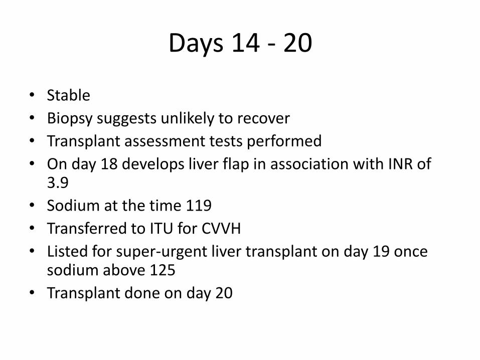
Days 14 - 20
• Stable
• Biopsy suggests unlikely to recover
• Transplant assessment tests performed
• On day 18 develops liver flap in association with INR of 3.9
• Sodium at the time 119
• Transferred to ITU for CVVH
• Listed for super-urgent liver transplant on day 19 once sodium above 125
• Transplant done on day 20

Transplant

Transplant

Case 1
Hepatectomy Specimen
Macroscopy

Case 1. Macroscopic Appearances
Shrunken liver, weight 640 g. Wrinkled/knobbly capsular surface

Case 1 - Macroscopic Appearances

Case 1 - Macroscopic Appearances
Right Lobe

Case 1 - Macroscopic Appearances
Left lobe

Case 1
Hepatectomy Specimen
Microscopy - Brown Areas

Multiacinar Necrosis

Panacinar Necrosis

Ductular Reaction – Keratin 7

Hepatic Vein Endophlebitis

HVG
Hepatic Vein Endophlebitis

Case 1
Hepatectomy Specimen
Microscopy – Yellow Nodules

Fatty Change (Mediovesicular)
No Inflammation or Necrosis
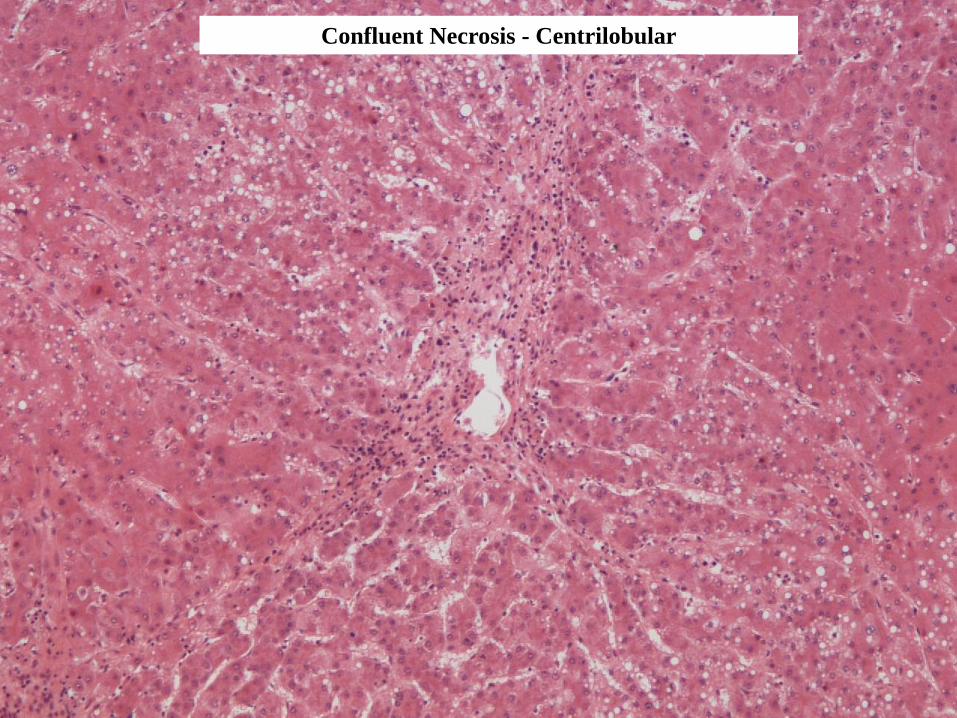
Confluent Necrosis - Centrilobular
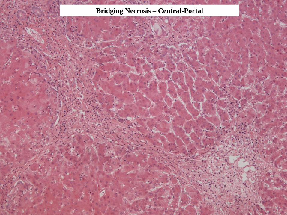
Bridging Necrosis – Central-Portal

Could this be cirrhotic?

Recent Post-Necrotic Collapse versus Longstanding Fibrosis
Use Of Connective Tissue Stains
Stain Material
Demonstrated
Distribution In
Normal Liver
Changes In Liver Disease
Reticulin Type III collagen
fibres
Portal tracts,
hepatic sinusoids
Collapse of reticulin
framework in areas of
recent liver cell necrosis.
(few days)
Haematoxylin
Van Gieson
(or Trichrome)
Type I collagen fibres Portal tracts, walls
of hepatic veins
Increased in hepatic fibrosis
(weeks/months)
Orcein Elastic fibres Portal tracts,
walls of hepatic
veins
Found in long-standing
fibrosis/cirrhosis
(months/years)

Retic

HVG

Orcein

Orcein

Ceroid Pigment Laden Macrophages
CD 68PAS-diastase

Rhodanine

Hepatectomy Specimen – Case 1
Histological Findings
• Large areas of panacinar necrosis (multi-acinar necrosis)
– Periportal ductular reaction
– Inflammation (including ceroid-laden macrophages)
– Inflammation of hepatic veins
• Surviving nodules of liver parenchyma
– Fatty change
– Confluent /bridging necrosis
– Little inflammation

Hepatectomy Specimen – Case 1
Diagnosis
• Severe acute hepatitis with multiacinar necrosis
(submassive hepatic necrosis)
• No strong aetiological pointers ( “seronegative
hepatitis”)

Post Transplant Progress
• Spent 3 days on intensive care
• Spent another 5 days on the ward
• On standard immunosuppresion
• No episodes of rejection
• Doing very well
• LFTs all normal
• Renal function normal
Final Diagnosis
Seronegative hepatitis associated with fulminant liver failure requiring
transplantation

1237 patients presenting to QEH, Bham with ALF (Jan 1992 – May 2008)
• Paracetamol accounts for 759/843 (90%) of drug –induced ALF cases
• Excluding paracetamol/Budd-Chiari/Wilson’s - seronegative hepatitis accounts for
186/452 (41%) of cases of severe acute hepatitis
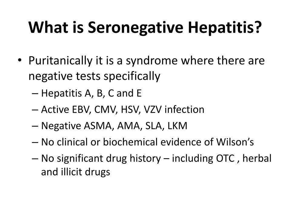
What is Seronegative Hepatitis?
• Puritanically it is a syndrome where there are negative tests specifically
– Hepatitis A, B, C and E
– Active EBV, CMV, HSV, VZV infection
– Negative ASMA, AMA, SLA, LKM
– No clinical or biochemical evidence of Wilson’s
– No significant drug history – including OTC , herbal and illicit drugs

Paracetamol DILI Viral Cryptogenic
Number 20 16 21 16
Anti-SLA 0 2 (13%) 9 (43%) 5 (31%)
ANA/ASM/LKM 0 4 (25%) 5 (25%) 2 (13%)
Any Autoantibody 0 5 (31%) 11 (53%) 7 (44%)
But…...
Bernal et al 2007
Case 2

Case 2
• 45 year old lady• Admitted to QE on 7th September 2016• Transfer from her local hospital – concern about
PT prolongation (44 at the time of transfer)• 2 week history of jaundice, pruritus and lethargy• 6 pounds weight loss• Background HT, OA and previous umbilical hernia
repair• Meds – omeprazole, lisinopril, movicol, naproxen
(regular use for 12 months), ondansteron, cyclizine, paracetamol

Investigations at Local Hospital
• ANA 1 in 400• IgG 28• dsDNA positive. ASMA, LKM, GPC, ENA negative• USS – mild intrahepatic biliary dilatation in the left
lobe. GB normal.• MRCP – normal• ALT 2157• ALP 177• Bili 173• PT 16 seconds. AFP 16. • Hep A-C negative• Hep E awaited

QE Admission Investigations
• INR 3.1• ALT 759• Bili 241• Na 131• Creat 65• USS - The gallbladder appears to have a thickened, slightly
oedematous wall measuring up to 5.4mm, and contains a tiny amount of echogenic debris. No obvious gallstones. No biliary duct dilatation.
• The liver appears normal in size, outline and echogenicity. No obvious focal liver lesions identified.
• The pancreas and spleen demonstrate normal US appearances.
• There is a left sided pleural effusion noted.

Our Assessment
• ALF– ? Autoimmune
– ? Infiltrative (weight loss and left pleural effusion)
– ? Naproxen DILI
• Plan– CT TAP -No evidence of malignancy. No cause for
liver dysfunction is identified. There are some changes suggestive of pulmonary hypertension - a cardiology opinion is recommended, with view to echo.
– Biopsy

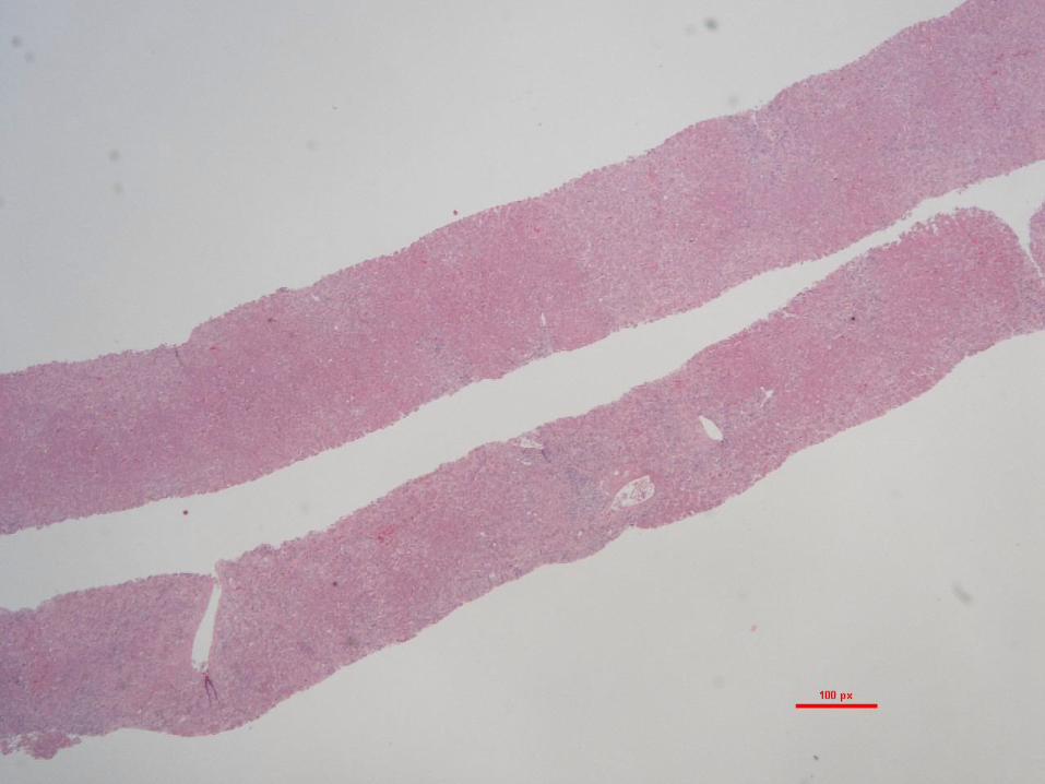
Case 2 – Liver Biopsy

Diffuse Lobular Inflammation and “Lobular Disarray”

Centrilobular Inflammation (Plasma Cells) and Confluent Necrosis

Hepatocyte Ballooning and Rosetting

Emperipolesis
Cholestatic
Rosette

Hepatitic Rosettes - Reticulin

Portal Inflammation (+ Interface Hepatitis ?)

Portal Plasma Cells

HVG
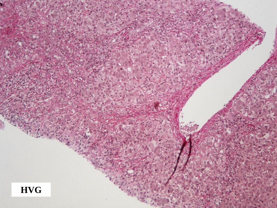
HVG

HVG

Orcein

Case 2 – Liver Biopsy
Histological Findings
• Portal inflammation (plasma cell rich)
• Interface hepatitis (?)
• Spotty lobular inflammation with lobular disarray
• Small foci of confluent necrosis (plasma cell rich)
• Moderate cholestasis
• Periportal and centrilobular fibrosis with early bridging

Case 2 – Liver Biopsy
Diagnosis
1. Acute/subacute hepatitis. Some features suggest possible transition to
chronicity
2. Moderately severe inflammatory activity, including foci of confluent
necrosis
3. Overall appearances in keeping with autoimmune hepatitis
– plasma cells, interface hepatitis, hepatitic rosettes, emperipolesis
1. Is this acute or chronic damage?
2. How severe is the damage?
3. What is the cause?

Treatment Started
PrednisoloneStarted

Progress
• Discharged after 14 days on the ward
• LFTs normalised in clinic and IgG fell to 11.49
• SLA came back as weakly positive
• Echo excluded significant PHT
• Azathioprine started 4 weeks after steroids
• Doing well

Final Diagnosis –
Steroid Responsive Acute Autoimmune Hepatitis

Autoimmune Hepatitis - Acute Presentation
Incidence & Diagnostic Criteria
30- 40% of cases present as acute hepatitis /acute liver failure ( Manns 2010, Lohse 2011, Gleeson 2012, EASL Guidelines 2015)
Reported prevalence of acute presentation ranges from 8.7% - 75%(Nguyen Canh 2017)
Autoantibodies unreliable in the diagnosis of acute AIH
• Autoantibodies and hypergammaglobulinaemia may not be present at the time
of presentation with acute AIH (EASL Guidelines 2015, Fujiwara 2016)
• Autoantibodies present in up to 40% of patients with other causes of acute liver
failure - e.g viral or drug-induced (Bernal 2007)

Acute Presentation of Autoimmune Hepatitis - Histological Features
1. Acute presentation of chronic liver disease
• 14-35% have features of chronic hepatitis (Fujiwara 2011, Yasui 2011, Fujiwara
2016, Dohmen 2017)
• 2-95% have bridging fibrosis/cirrhosis (Nikias 1994, Burgart 1995, Miyake 2010,
Fujiwara 2011, Nguyen Canh 2017)
2. Acute hepatitis (with no signs of chronic liver disease)
• Classical features of acute lobular hepatitis (resembling viral or drugs)
Histological Features Favouring Autoimmune Hepatitis As Likely Cause of Hepatitis
• Published criteria focus mainly on patients presenting with chronic (portal) hepatitis
˗ Lymphoplasmacytic portal inflammation
˗ Interface hepatitis, hepatitic rosettes, emperipolesis
• Criteria for diagnosing autoimmune hepatitis less clearly established in patients
presenting with acute (lobular) hepatitis

Acute Lobular Hepatitis - Histological Features Favouring a Diagnosis of AIH(Abe 2007, Fujiwara 2008, Stravitz 2011, Yasui 2011, Fujiwara 2016, Dohmen 2017, Nguyen Canh 2017)
• Portal inflammation / interface hepatitis (resembling chronic AIH)
• Plasma-cell rich inflammatory infiltrate
• Lymphoid follicles
• Centrilobular necrosis / central perivenulitis
• Hepatocyte rosettes
• Emperipolesis
BUT:
1. Many of the above features are frequently seen in other causes of acute hepatitis
• Portal inflammation seen in all types of acute hepatitis
• Rosettes and emperipolesis also common in non-autoimmune acute hepatitis (Balitzer, Modern Pathology 2017)
2. Interface hepatitis difficult to assess in the presence of diffuse lobular hepatitis

Case 3

Case 3• Complex
• Stage IVc melanoma with metastases in skin, lungs, hila and pleura, adrenal gland.
• Progressed on standard management.
• Started on Nivolumab 2-weekly in July 2015 with disease control Oct 2016.
• Bone metastasis repaired left femur Sept 2015.
• Immune related adverse events / complications of treatment
• Nivolumab suspended twice in 2016 for grade 1-2 diarrhoea treated with high dose steroids with rapid taper
• Evidence secondary immune deficiency.
• Significant weight loss (20kg Sept 2015 – Nov 2016)
• PMH – HT, HC, OA, angioplasties, hypothyroidism
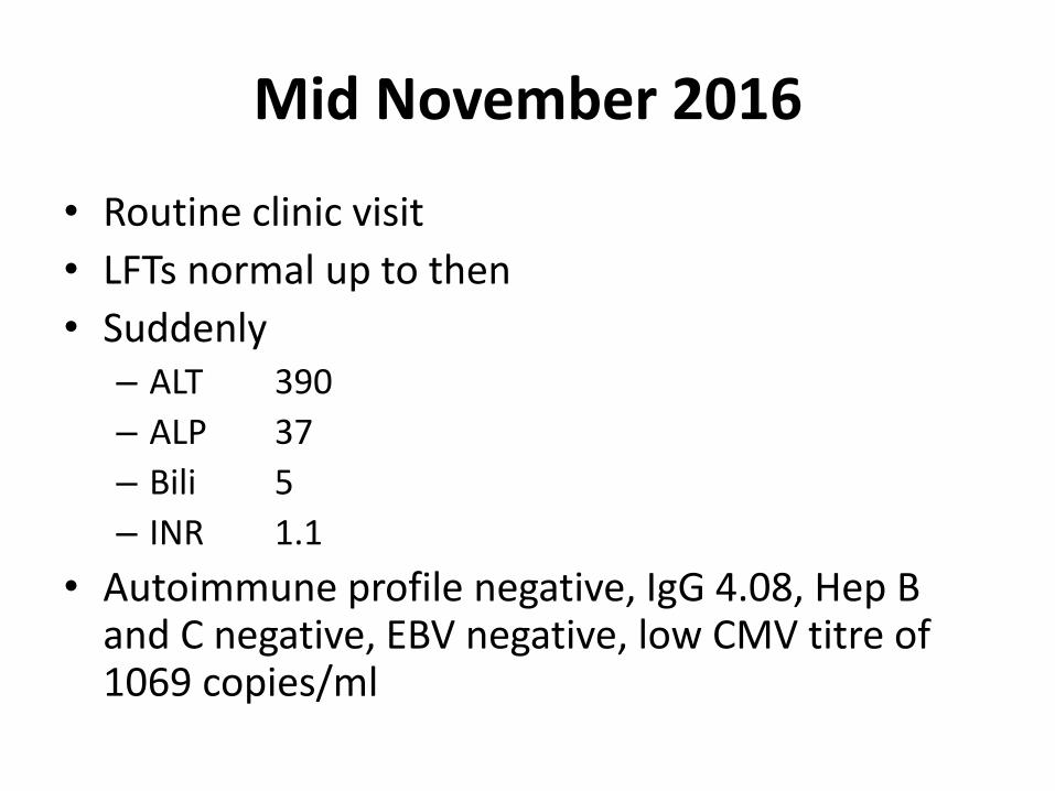
Mid November 2016
• Routine clinic visit
• LFTs normal up to then
• Suddenly– ALT 390
– ALP 37
– Bili 5
– INR 1.1
• Autoimmune profile negative, IgG 4.08, Hep B and C negative, EBV negative, low CMV titre of 1069 copies/ml

Differential Diagnosis
• CMV hepatitis
• DILI
• Autoimmune hepatitis triggered by Nivolumab
• Metastatic disease
Liver biopsy performed on the 17th of November 2016

Case 3 – Liver Biopsy


PAS -diastase

CD68

CD8

Reticulin

HVG

Case 3 – Liver Biopsy
Histological Findings
• Perivenular inflammation and necrosis (“central perivenulitis”)
• Little/no inflammation elsewhere in liver parenchyma or in
portal tracts

Case 3 – Liver Biopsy
Diagnosis
1. Acute injury with centrilobular inflammation + necrosis (“central
perivenulitis”)
2. Confluent zone 3 necrosis
3. In keeping with drug-induced liver injury (DILI)
1. Is this acute or chronic damage?
2. How severe is the damage?
3. What is the cause?

Drug-induced Acute Hepatitis
• Drugs account for approximately 10% of cases of acute hepatitis and acute liver failure (Ramachandran & Kakar 2009, Reuben 2010)
• Acute hepatitis/cholestatic hepatitis are two commonest pattern of DILI
– Present in 50% of 249 cases reviewed by DILI Network (Kleiner 2014)
• Many agents implicated – antimicrobial drugs commonest
Histological features
• Frequently indistinguishable from other causes of acute hepatitis (e.g. viral hepatitis, autoimmune hepatitis)
• Features favouring a drug aetiology:
– Predominantly centrilobular (zone 3) inflammation
– Disproportionately severe / well-circumscribed necrosis (relatively little inflammation – lobular and/or portal)
– Unusual patterns of necrosis - e.g periportal (zone 1) necrosis
– Unusually prominent cholestasis
– Eosinophils, granulomas


Histological Findings in Immune Checkpoint Inhibitor Induced Hepatitis(Doherty - EMSO 2017, Zen - Modern Pathology 2018, De Martin – J Hepatol 2018, in press)
• 26 cases studied
• Drugs implicated include inhibitors of PD-1/PD-L1 (nivolumab, pembrolizumab) and CTLA4 (ipilimumab) – individually or in combination
Main histological findings:
• Diffuse lobular hepatitis, variable confluent centrilobular necrosis
• Lobular inflammatory cells mainly CD8+ T lymphocytes (esp anti-CTLA-4 cases)
• Mild portal inflammation
Other Findings:
• Bile duct inflammation +/- bile duct loss (Doherty 2017)
• Lobular granulomas/microgranulomas
• Fibrin ring granulomas - anti-CTLA4 cases only (de Martin 2018)


Progress
• Started on IV methylprednisolone on the day before the biopsy
• Switched to prednisolone on day 3 after the biopsy
• Discharged
• Been in since for symptom control of diarrhoea
• No issues with LFTs


Nivolumab in MM
Scott LJ, Drugs 2015

Evidence for Efficacy
Larkin et al NEJM 2015

Incidence of SAEs to Nivolumab
Scott LJ, Drugs 2015

Final Diagnosis –
Steroid Responsive Nivolumabinduced liver injury

Changing Role of Liver Biopsy in Acute Hepatitis
• Many of the classical morphological studies of acute hepatitis were carried out before the main causes had been discovered
• Most cases of acute hepatitis now diagnosed on the basis of clinical, biochemical and serological findings and liver biopsy is rarely indicated
• Liver biopsy may still be carried out in cases where the clinical presentation is atypical or the cause is uncertain
– Confirm diagnosis of acute hepatitis
– Determine disease severity
– Identify possible aetiological factors (including cases of acute liver injury not related to hepatitis)

Liver Biopsy in Acute Hepatitis
Histological Approach
1. Is this acute or chronic damage?
2. How severe is the damage?
3. What is the cause?

Patterns of Inflammation in the Liver
• Portal Inflammation
– Most chronic liver diseases (e.g. viral, autoimmune)
– Also seen in acute hepatitis
• Lobular Inflammation
– Main pattern in acute hepatitis
– Varying degrees of lobular inflammation also commonly present in chronic viral and autoimmune hepatitis
• Mixed portal and lobular inflammation
Pattern of inflammation alone cannot reliably distinguish chronic from acute hepatitis
• Clinical context
• Assessment of fibrosis (progressive fibrosis versus post-necrotic collapse)

Severe Acute Hepatitis (e.g. case 1)
Acute versus Chronic Damage – Diagnostic Problems
Clinical
Severe acute hepatitis versus:
• decompensated chronic liver disease (e.g. Wilson disease)
• acute exacerbation of chronic liver disease (e.g. autoimmune hepatitis,
hepatitis A/E superimposed on underlying cirrhosis)
Histological
• Areas of bridging necrosis & nodular regeneration can resemble changes occurring in cirrhosis
• Areas of multiacinar necrosis can resemble inflamed fibrous septa in cirrhosis

Multiacinar Necrosis in Severe Acute Hepatitis (e.g. case 1)
Acute versus Chronic Damage - Helpful pointers
• Clinical context
• Identification of normal vascular relationships
• Use of connective tissue stains to determine age of lesions

Liver Biopsy in Acute Hepatitis
Histological Approach
1. Is this acute or chronic damage?
2. How severe is the damage?
3. What is the cause?

Liver Cell Death in Acute Hepatitis
Pattern of Cell Death Histological Features
Spotty necrosis Apoptosis of individual hepatocytes (acidophil bodies)
Confluent necrosis
(zone 3)
Loss of groups of adjacent liver cells
Bridging necrosis Confluent necrosis linking vascular structures
(central-central or central-portal bridging)
Panacinar necrosis Loss of hepatocytes in an entire acinus
Multiacinar necrosis Panacinar necrosis involving several adjacent acini
• Apoptosis > necrosis (in mild forms)
• Severe necro-inflammatory lesions uneven in distribution (e.g case 1)
Sampling variability in liver biopsies
Extent of hepatocyte necrosis predictive of poor outcome in some studies (Katoonizadeh
2006, Miraglia 2006, Rastogi 2011)

Liver Biopsy in Acute Hepatitis
Histological Approach
1. Is this acute or chronic damage?
2. How severe is the damage?
3. What is the cause?

Acute Hepatitis - Common Causes
1. Viral• Hepatitis viruses – A,B,C,D, E
• Other viruses – e.g. CMV, EBV, HSV
2. Drugs
3. Autoimmune
4. Unknown• Seronegative hepatitis (“non-A, non-B, non-C hepatitis”)
• Accounts for 40% of patients in the U.K presenting with severe
acute hepatitis leading to acute liver failure (Ichai 2008, Bernal
2010)
Histological Findings
• Viral hepatitis (A-E), drugs and AIH have overlapping histological features
Viral serology, drug history, auto-antibody serology required to identify the cause
• Other viruses rare, but have distinctive features

Liver biopsy rarely identifies a previously unsuspected aetiology
• Biopsies mostly obtained from people in whom main recognised causes
have been excluded (“seronegative hepatitis”)
• Biopsy sometimes provides aetiological pointers, including cases
presenting with acute liver injury not due to acute hepatitis
– Decompensated chronic liver disease (e.g. Wilson disease)
– Another cause of acute liver damage (e.g. ischaemic necrosis, severe
alcoholic hepatitis, paracetamol toxicity)
– Hepatic infiltration (usually lymphoma, rarely carcinoma)
• Liver usually enlarged
Acute Hepatitis - Aetiological Considerations

The End