acute chest syndrome (acs) - children's hospital colorado · clinical pathway page 2 of 10...
TRANSCRIPT

CLINICAL PATHWAY
Page 1 of 10
PEDIATRIC ACUTE CHEST SYNDROME (ACS) Patients with sickle cell disease presenting with 1) a new pulmonary infiltrate on chest radiography AND 2) evidence of lower airway disease (e.g. cough, shortness of breath, retractions, rales, etc.)
TABLE OF CONTENTS
Algorithm- N/A
Target Population
Background | Definitions
Initial Evaluation-N/A
Clinical Management
Diagnostic Tests
Fluids | Nutrition
Respiratory Therapy
Table 1. Pediatric Asthma Score
Treatment
Table 2. Antimicrobial Medication
Table 3. Pain Medication
Table 4. Respiratory Medication
Discharge Criteria
References
Clinical Improvement Team
TARGET POPULATION
Inclusion Criteria
• Patients with sickle cell disease (SS, SC, Sβ0 thalassemia, Sβ+ thalassemia)
• Patients of all ages
• Patients treated year round
Exclusion Criteria
• Patients without infiltrate on chest radiograph (e.g. asthma exacerbation)
• Patients with severe pulmonary hypertension
• Patients post bone marrow transplant
• Well children with pneumonia

CLINICAL PATHWAY
Page 2 of 10
BACKGROUND | DEFINITIONS
Acute chest syndrome (ACS) is the second most common reason for hospitalization in children with sickle cell disease and a leading cause of mortality. ACS is defined as a new pulmonary infiltrate on chest radiograph in the presence of evidence of lower respiratory tract disease (e.g. some combination of cough, shortness of breath, retractions, rales, etc.). In the majority of cases of ACS, an etiology is unable to be identified. The most common identified etiology of ACS is infection but it may also result from pulmonary vaso-occlusion, pulmonary infarction or fat embolism. The primary infectious agents implicated in ACS include: Chlamydia pneumoniae, Mycoplasma pneumoniae, Streptococcus pneumoniae, and viruses. Risk factors for ACS include vaso-occlusive pain crisis, anesthesia, and surgery. Patients are at increased risk for stroke in the two weeks immediately following an episode of ACS.
CLINICAL MANAGEMENT
• Hospitalize on hematology service
• Vital signs q 2-4 hours depending upon degree of respiratory compromise
• Record pain score every 4 hours
• Continuous cardiorespiratory monitor and pulse oximetry
• Encourage ambulation: out of bed to chair or ambulating at least 2-3 times per day
• Droplet precautions
• Continue medication for reactive airway disease if applicable
DIAGNOSTIC TESTS
• CBC with differential, platelet count, and reticulocyte count initially and daily until improving (compare with patient's baseline values)
• CXR initially, repeat for clinical deterioration (be aware that the x-ray often underestimates the degree of involvement and may appear worse when the child is clinically improving)
• Consider:
o Type and crossmatch for severe illness or if Hb is greater than 1 g/dL below baseline. Request minor-antigen-matched, sickle-negative, leukocyte-depleted RBC
o Blood cultures if febrile (greater than or equal to 38.3˚C) or history of recent fever (do not need to repeat daily)
o Arterial blood gas per clinical discretion
o Renal (BUN, creatinine) and liver (fractionated bilirubin, ALT) function tests for severe illness or if diffuse encephalopathy present (rule out acute multi-organ failure syndrome)
o If severe abdominal pain consider an ultrasound for gallstones
o Influenza A& B screening during the appropriate season
o We do not recommend routinely sending a respiratory pathogen PCR
• Echocardiography is not recommended routinely in patients with acute chest syndrome. Consult cardiology if concern for pulmonary hypertension arises (prolonged hypoxemia, fixed split S2 or pronounced pulmonary component of S2, hepatomegaly, persistent peripheral edema, or persistent pulmonary edema despite adequate fluid status)
FLUIDS | NUTRITION
• Daily weight
• Record intake and output strictly

CLINICAL PATHWAY
Page 3 of 10
• Maintain "euvolemia". IV + P.O. = 1 x maintenance. More fluid is appropriate only if patient is dehydrated or if insensible losses are increased (e.g. persistent fever). IV fluid should be D5 1/4 NS to avoid exacerbating the sickling process.
RESPIRATORY THERAPY
• Consult pulmonology
• Clinical features suggestive of asthma or acute bronchospasms make an initial assessment using the Pediatric Asthma Score (PAS); trial Albuterol 4 puffs with a spacer or 2.5 mg nebulized once. Reassess using the PAS, a positive response is a decrease of 2 or more in the PAS. If a positive response is noted, order Albuterol 2-4 puffs or Albuterol 2.5mg nebulized Q4 and PRN. If at any time PAS worsens by 2 or more increase frequency of the Albuterol and notify the provider
• Lung expansion strategies: EzPAP (along with IS) Q4 hours for 72 hours. After 72 hours of the initiation of the therapy, the patient will be evaluated by the respiratory therapist for pulmonary stability. If the chest x-ray (if done) remains stable, there are no signs of pulmonary infection, there is good aeration throughout all lung fields upon auscultation, and the patient can consistently achieve 14ml/kg with the IS, the EzPAP will then be discontinued. The patient will then receive IS Q2 hours while awake and Q4 hours at night by their RN
• Transfer to the ICU if patient is requiring Heated High Flow Nasal Cannula (HHFNC), invasive or non-inavsive (CPAP, BiPAP) mechanical ventilation is being considered
TABLE 1. PEDIATRIC ASTHMA SCORE
Inspiratory and expiratory wheezing to diminished breath sounds or poor
aeration
Expiratory wheezingNormal breath sounds to
end-expiratory wheeze only
Three or more sitesTwo sitesZero to one site
Less than 85% on room air85% to 90% on room airGreater than 90% on room
air
Respiratory Rate2 to 3 years4 to 5 years
6 to 12 yearsOlder than 12 years
34 or less30 or less26 or less23 or less
Speaks in partial sentences, short cry
Speaks in sentences, coos and babbles
321
Speaks in single words/short phrases/grunting
Score
35 to 3931 to 3527 to 3024 to 27
40 or greater36 or greater31 or greater28 or greater
Oxygen Requirements
Auscultation
Retractions
Dyspnea
Note: Use PAS Score to guide intervention & response to treatment. Older pediatric

CLINICAL PATHWAY
Page 4 of 10
TREATMENT
• Antipyretics
o Acetaminophen dose according to manufacturer’s recommendations PRN temperature greater than or equal to 38.3oC after blood cultures have been obtained on at least one occasion
• Antibiotics (see Table 2 for choices and doses)
o Ceftriaxone and azithromycin is the regimen of choice for initial inpatient management
o If a respiratory pathogen PCR is sent for another reason and is negative for Mycoplasma and Chlamydophila, azithromycin may be discontinued
o If there is a known or suspected cephalosporin allergy, levofloxacin is the regimen of choice and azithromycin can be omitted because levofloxacin has adequate coverage of atypical organisms
o Strongly consider adding vancomycin for severe illness, or if large infiltrate with pleural effusion present and S. aureus is suspected
o Prophylactic penicillin should be discontinued while patient is receiving antibiotics
o If antibiotics are continued upon discharge, an appropriate oral antibiotic should be continued to complete a course of 7-10 days (including inpatient IV therapy received): Amoxicillin-clavulanate is recommended as first line, cefpodoxime is the drug of choice in the presence of a penicillin allergy, and levofloxacin is the drug of choice in the presence of a cephalosporin allergy.
• Analgesia (if indicated)
o If pain is present patients should be started on scheduled anti-inflammatories and opiates if there are no contraindications
o Start ketorolac, but limit to 48 hour maximum duration then start ibuprofen PO q6h (not PRN) if no contraindication present (i.e. gastritis, ulcer, coagulopathy, renal impairment).
o If pain does not respond to anti-inflammatory alone, consult the sickle cell vaso-occlusion guidelines. Be aware narcotic administration may further suppress respiration
• Transfusions
o Give simple blood transfusion (10 mL/kg red blood cells) to improve oxygen carrying capacity to people with symptomatic ACS whose hemoglobin concentration is greater than 1 g/dL below baseline and hemoglobin would not rise to more than 10 g/dL. If baseline hemoglobin is 9 g/dL or higher, may require red cell exchange transfusion. Do not transfuse acutely to Hb greater than 10 g/dL, Hct greater than 30 percent if percent sickle hemoglobin is or is presumed to be greater than 30 percent since it is associated with inducing pain and stroke
o Urgent exchange transfusion may be indicated after consultation from hematology, critical care, and apheresis specialists – when there is rapid progression of ACS as manifested by oxygen saturation below 90 percent despite supplemental oxygen, increasing respiratory distress, progressive pulmonary infiltrates, and/or decline in hemoglobin concentration despite simple transfusion and/or inability to transfuse due to high baseline hemoglobin. May require transfer to ICU and transfusion medicine consult for erythrocytapheresis. Remove femoral or central venous catheters as soon as possible after exchange transfusion to reduce risk of thrombosis

CLINICAL PATHWAY
Page 5 of 10
TABLE 2. ANTIMICROBIAL MEDICATIONS
Medication Dosing Indication/Notes
Amoxicillin-clavulanate (PO)
90 mg/kg/day (maximum 3,000 mg/day if using suspension and 2,625 mg/day if using tablet) PO
divided TID
Antimicrobial – Outpatient 1st Choice
Use either amoxicillin-clavulanate ES suspension 600-42.9mg/5mL or 875-125 mg
tablet formulations
Azithromycin (IV/PO) *Highly Bioavailable*
10 mg/kg/day (maximum 500 mg/day) PO x 1 dose, followed by 5 mg/kg/day (maximum 250
mg/day) PO once daily x 4 doses
Antimicrobial – Inpatient/Atypical Coverage
Discontinue if RPP results negative for Chlamydophila and Mycoplasma
Not necessary to use azithromycin with
levofloxacin as levofloxacin covers atypical bacteria
Cefpodoxime (PO) 10 mg/kg/day (maximum 400 mg/day) PO
divided BID
Antimicrobial – Outpatient for penicillin allergy
*Ensure prescription sent in advance
(variable stock at some outpatient pharmacies)
Ceftriaxone (IV) 50 mg/kg/day (maximum 2,000 mg/day) IV q24h
Antimicrobial – Inpatient 1st Choice
Discontinue prophylactic penicillin while patient is receiving broad-spectrum
antimicrobials
Levofloxacin (IV/PO) *Highly Bioavailable*
Less than 5 years: Levofloxacin 20 mg/kg/day (maximum 750 mg/day) IV or PO divided q12h
Greater than or equal to 5 years: 10 mg/kg/day
(maximum 750 mg/day) IV or PO q24h
Antimicrobial – Inpatient and/or Outpatient for cephalosporin allergy
Discontinue prophylactic penicillin while
patient is receiving broad-spectrum antimicrobials
Not necessary to use azithromycin with
levofloxacin as levofloxacin covers atypical bacteria
Vancomycin (IV) Contact pharmacy for recommended dosing
Dosing interval based on age/renal function)
Antimicrobial – Inpatient for patients with severe illness or with large infiltrate with pleural effusion present and S. aureus
suspected
*Must monitor renal function (SCr, BUN, urine output) at baseline and minimum twice
weekly thereafter

CLINICAL PATHWAY
Page 6 of 10
TABLE 3. RESPIRATORY MEDICATION TABLE
Albuterol Increased work of breathing
4 puffs OR 2.5 mg nebulized x 1 dose. If effective (improved working of breathing, respiratory rate,
wheezing, aeration) order scheduled albuterol 2-4 puffs with spacer or 2.5 mg nebulized q4h
IndicationDosingMedication
0.5 – 1 mg/kg (maximum 40 mg/dose)
GI prophylaxis for steroid use
1 mg/kg (maximum 150 mg/dose) PO q12h
OR
1 mg/kg (maximum 50 mg/dose) IVq8h
Ranitidine
Furosemide Consider if signs of fluid overload present
Prednisone or Methylprednisolone
1 mg/kg (maximum 40 mg/dose) PO/IV q12h x 5 days
Followed by steroid wean to prevent rebound:
0.5 mg/kg (maximum 20 mg/dose) PO/IV q12h x 3 days
0.5 mg/kg (maximum 20 mg/dose) PO/IV q24h x 3 days
0.25 mg/kg (maximum 10 mg/dose) PO/IV q24h x 3 days
Consider in patients if wheezing/crackles/rales present or history of asthma
CLINICAL PATHWAY
Page 7 of 10
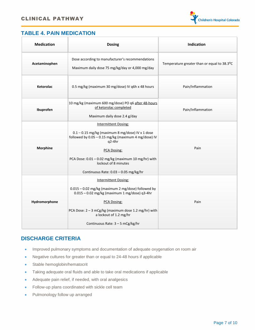
TABLE 4. PAIN MEDICATION
Acetaminophen
10 mg/kg (maximum 600 mg/dose) PO q6 after 48-hours of ketorolac completed
Maximum daily dose 2.4 g/day
Temperature greater than or equal to 38.3⁰C
Pain/Inflammation0.5 mg/kg (maximum 30 mg/dose) IV q6h x 48 hours
Dose according to manufacturer’s recommendations
Maximum daily dose 75 mg/kg/day or 4,000 mg/day
IndicationDosing
Ketorolac
Ibuprofen Pain/Inflammation
Medication
Morphine
Intermittent Dosing:
0.1 – 0.15 mg/kg (maximum 8 mg/dose) IV x 1 dose followed by 0.05 – 0.15 mg/kg (maximum 4 mg/dose) IV
q2-4hr
PCA Dosing:
PCA Dose: 0.01 – 0.02 mg/kg (maximum 10 mg/hr) with lockout of 8 minutes
Continuous Rate: 0.03 – 0.05 mg/kg/hr
Pain
Hydromorphone
Intermittent Dosing:
0.015 – 0.02 mg/kg (maximum 2 mg/dose) followed by 0.015 – 0.02 mg/kg (maximum 1 mg/dose) q3-4hr
PCA Dosing:
PCA Dose: 2 – 3 mCg/kg (maximum dose 1.2 mg/hr) with a lockout of 1.2 mg/hr
Continuous Rate: 3 – 5 mCg/kg/hr
Pain
DISCHARGE CRITERIA
• Improved pulmonary symptoms and documentation of adequate oxygenation on room air
• Negative cultures for greater than or equal to 24-48 hours if applicable
• Stable hemoglobin/hematocrit
• Taking adequate oral fluids and able to take oral medications if applicable
• Adequate pain relief, if needed, with oral analgesics
• Follow-up plans coordinated with sickle cell team
• Pulmonology follow up arranged
CLINICAL PATHWAY
Page 8 of 10
REFERENCES
1. Vichinsky E et al. Causes and outcomes of the acute chest syndrome in sickle cell disease. National Acute Chest Syndrome Study Group. NEJM 2000. 342: 1855-1865.
2. Yawn B et al. Management of sickle cell disease: Summary of the 2014 evidence-based report by expert panel members. JAMA 2014. 312: 1033-1048.
3. Howard J et al. Guidelines on the management of acute chest syndrome in sickle cell disease. BJH 2015. 169: 492-505.
4. Sobota A et al. Corticosteroids for acute chest syndrome in children with sickle cell disease: Variation in use and association with length of stay and readmission. Am J Hemat 2010; 85(1): 24-28.
5. Ahmad F et al. The use of incentive spirometry in pediatric patients with sickle cell disease to reduce the incidence of acute chest syndrome. J Pedatr Hematol Oncol 2011. 33: 415-420.
6. Bellet PS, Kalinyak KA, Shukla R et al. Incentive spirometry to prevent acute pulmonary complications in sickle cell diseases. NEJM 1995; 333(11): 699-703.
7. Saylors R et al. Comparison of automated red cell exchange transfusion and simple transfusion for the treatment of children with sickle cell disease acute chest syndrome. Pedatr Blood Cancer 2013. 60: 1952-1956.
8. Reagan M et al. Multi-modal intervention for the inpatient management of sickle cell pain significantly decreases the rate of acute chest syndrome. Pediatr Blood Cancer 2011; 56:262-266.
9. Saylors R et al. Comparison of automated red cell exchange transfusion and simple transfusion for the treatment of children with sickle cell disease acute chest syndrome. Pediatr Blood Cancer 2013; 60: 1952-1956.
10. Turner J et al. Exchange versus simple transfusion for acute chest syndrome in sickle cell anemia adults. Transfusion 2009; 49: 863-868.
11. Velasquez M et al. Erythrocytapheresis in children with sickle cell disease and acute chest syndrome. Pediatr Blood Cancer 2009; 53: 1060-1063.

CLINICAL PATHWAY
Page 9 of 10
Clinical pathways are intended for informational purposes only. They are current at the date of publication and are reviewed on a regular basis to align with the best available evidence. Some information and links may not be available to external viewers. External viewers are encouraged to consult other available sources if needed to confirm and supplement the content presented in the clinical pathways. Clinical pathways are not intended to take the place of a physician’s or other health care provider’s advice, and is not intended to diagnose, treat, cure or prevent any disease or other medical condition. The information should not be used in place of a visit, call, consultation or advice of a physician or other health care provider. Furthermore, the information is provided for use solely at your own risk. CHCO accepts no liability for the content, or for the consequences of any actions taken on the basis of the information provided. The information provided to you and the actions taken thereof are provided on an “as is” basis without any warranty of any kind, express or implied, from CHCO. CHCO declares no affiliation, sponsorship, nor any partnerships with any listed organization, or its respective directors, officers, employees, agents, contractors, affiliates, and representatives.
CLINICAL IMPROVEMENT TEAM MEMBERS
Chris McKinney, MD | Hematology Oren Kupfer, MD | Pulmonology Rachelle Nuss, MD | Hematology Joyce Baker, RT | Respiratory Therapist Abby Kim, PharmD | Clinical Pharmacist Paige Krack, MBA, MS | Process Improvement Lead
APPROVED BY
Clinical Care Guideline and Measures Review Committee – April 18, 2016 Antimicrobial Stewardship Committee – January 22, 2016 Pharmacy & Therapeutics Committee – February 10, 2016; minor revision March 2, 2017
MANUAL/DEPARTMENT Clinical Care Guidelines/Quality
ORIGINATION DATE November 24, 2015
LAST DATE OF REVIEW OR REVISION April 18, 2016
APPROVED BY
Lalit Bajaj, MD, MPH Medical Director, Clinical Effectiveness
REVIEW/REVISION SCHEDULE
Scheduled for full review on April 18, 2020

CLINICAL PATHWAY
Page 10 of 10