acute and sub-acute oral toxicity of dracaena cinnabari ... · research article open access acute...
TRANSCRIPT

RESEARCH ARTICLE Open Access
Acute and sub-acute oral toxicity ofDracaena cinnabari resin methanolextract in ratsNashwan Abdullah Al-Afifi1, Aied Mohammed Alabsi2*, Marina Mohd Bakri1 and Anand Ramanathan3,4
Abstract
Background: Dracaena cinnabari (DC) is a perennial tree that located on the Southern coast of Yemen native tothe Socotra Island. This tree produces a deep red resin known as the Dragon’s blood, the Twobrother’s Blood orDamm Alakhwain. The current study performed to evaluate the safety of the DC resin methanol extract after asingle or 28 consecutive daily oral administrations.
Methods: In assessing the safety of DC resin methanol extract, acute and sub-acute oral toxicity tests performedfollowing OECD guidelines 423 and 407, respectively, with slight modifications. In acute oral toxicity test, DC resinmethanol extract administered to female Sprague Dawley rats by oral gavage at a single dose of 300 and 2000 mg/kgbody weight. Rats observed for toxic signs for 14 days. In sub-acute oral toxicity test, DC resin methanol extractadministered to the rats by oral gavage at 500, 1000, and 1500 mg/kg body weight daily up to 28 days to male andfemale Spradgue Dawley rats. The control and high dose in satellite groups were also maintained and handled as theprevious groups to determine the late onset toxicity of DC resin methanol extract. At the end of each test, hematologicaland biochemical analysis of the collected blood were performed as well as gross and microscopic pathology.
Results: In acute oral toxicity, no treatment-related death or toxic signs were observed. It revealed that the DC resinmethanol extract could be well tolerated up to the dose 2000 mg/kg body weight and could be classified as Category 5.The sub-acute test observations indicated that there are no treatment-related changes up to the high dose levelcompared to the control. Food consumption, body weight, organ weight, hematological parameters, biochemicalparameters and histopathological examination (liver, kidney, heart, spleen and lung) revealed no abnormalities.Water intake was significantly higher in the DC resin methanol extract treated groups compared to the control.
Conclusion: This study demonstrates tolerability of DC resin methanol extract administered daily for 28 days up to1500 mg/kg dose.
Keywords: DC resin methanol extract, Acute oral toxicity, Sub-acute oral toxicity, Histopathology,Hematological parameters, Biochemical parameters
BackgroundMedicinal plants are used worldwide to treat many dis-eases, and new drugs continue to be developed throughresearch from these plants [1]. Medicinal plant prepara-tions can be formulated into many forms including liq-uids, and they have been used worldwide for many years[2]. The use of a medicinal plant for the treatment of
disease without the scientific foundation to adequatelysupport conclusions of safety and efficacy can be poten-tially dangerous as well as useless. In addition, misuse ofthese medicinal plants may cause serious toxicity forhumans [3]. As chemical compositions of the medicinalplant are complex, some moderate to severe side effectsmay arise from the use of herbal medicines. Therefore, itis important to establish medicinal plant safety throughthe use of well-controlled and validated scientific toxicitystudies or protocols [4].* Correspondence: [email protected]
2Department of Oral Biology and Biomedical Sciences, Faculty of Dentistry,MAHSA University, 42610 Jenjarom, Selangor, MalaysiaFull list of author information is available at the end of the article
© The Author(s). 2018 Open Access This article is distributed under the terms of the Creative Commons Attribution 4.0International License (http://creativecommons.org/licenses/by/4.0/), which permits unrestricted use, distribution, andreproduction in any medium, provided you give appropriate credit to the original author(s) and the source, provide a link tothe Creative Commons license, and indicate if changes were made. The Creative Commons Public Domain Dedication waiver(http://creativecommons.org/publicdomain/zero/1.0/) applies to the data made available in this article, unless otherwise stated.
Al-Afifi et al. BMC Complementary and Alternative Medicine (2018) 18:50 DOI 10.1186/s12906-018-2110-3

Toxicological studies subjected to make a decisionabout whether a new drug should be accepted for clin-ical use or not [5]. Toxicity study gives information ontoxic doses and therapeutic indices of drugs and xeno-biotic [6]. The outcome of the toxicity study in animalsis vitally needed to determine the safety of medicinalplants if they are found to be suitable for developmentinto pharmacological products. In addition, to determinethe appropriate dose for long-term toxicity tests and todetermine the affected organs at the end of the treatment,the toxicity test is achieved [7]. It is a scientific, ethicaland regulatory requirement that before any potential newmedicine administered to humans, its safety must beinvestigated in animals to define safe human doses [8].Dracaena cinnabari (DC) is a perennial tree located
on the Southern coast of Yemen native to the SocotraIsland. This tree produces a deep red resin which calledthe Dragon’s blood or the Twobrother’s Blood [9]. DCbelongs to Agavaceae family, which is commonly knownas Damm Alakhwain in Yemen [10]. It is a large, single-trunked tree with height up to 10 m and smooth greybark. Branches with sausage-shaped sections form anumbrella-shaped crown [11]. DC tree populations onSocotra do not regenerate to a significant extent, andtheir age structure indicates overmaturity [12]. The red“dragon’s blood” resin of the DC tree exudes from fis-sures and wounds in the bark or branches [1]. People inSocotra used the resin from DC for dying wool, gluepottery, breath freshener, to decorate a pottery andhouses and even as lipstick [13]. In addition, DC resinhas been a famous traditional medicine since ancienttimes in many cultures. It is used as an astringent fortreating diarrhea and dysentery as well as an antiseptic,haemostatic and as an antiulcer remedy [1, 10, 14, 15].Phytochemical studies of DC resin have led to theisolation of several active compounds belonging to theflavanoids, homoisoflavonoids, chalcones, sterols andterpenoids. Some homoisoflavonoids and chalcones iso-lated from the DC resin exhibited a strong antioxidantactivity [16]. The presence of flavonoids reported con-tributing in anti-inflammatory activities of methanol ex-tract of DC resin [17]. The chemical constituents of DCresin have been reviewed by D Gupta, B Bleakley andRK Gupta [1]. The methanolic extract of DC resin hasbeen reported to exert an antiviral activity againstHerpes simplex and Human influenza viruses [18]. DCresin is showing enormous potential interaction with anti-microbial [19] and antioxidants activities [10, 20] and con-sidered as good source of food preservative due to itsinhibitory effect on various foodborne pathogens [20]. Amethanol extract of DC resin exhibited a non-specific in-hibition of the parasites related to high cytotoxicity [21].Despite its wide uses, no study has been done to know
about its oral toxicity which in turn will provide proper
safety information regarding DC resin and to define safehuman doses. Thus, the aim of the current study is toevaluate the safety of the DC resin methanol extract aftera single or 28 consecutive daily oral administrations.
MethodsPreparation of Dracaena cinnabari (DC) resin extractThe resin of DC collected from Socotra Island (Yemen)in May 2013. The plant samples were identified and au-thenticated by Environmental Protection Authority ofYemen. A voucher specimen of the resin (DC/2013-8/122) has been deposited at the herbarium of departmentof Pharmacognosy, Faculty of Pharmacy, University ofSana’a, Yemen.DC resin was ground into powder form using an elec-
trical blender to ease the extraction process. 50 g of thepowdered resin of DC placed into a 1 L conical flaskthen macerated with methanol (500 ml methanol wasadded in, where 1 g of dried and ground DC is to 10 mlmethanol) in the ratio of (1:10). Methanol has high po-larity and thus greater efficacy towards the extraction ofpolar phytochemicals such as phenolics and flavonoids[22]. The conical flask was left at room temperature forapproximately 3 days on a shaker at 100 rpm. The re-sultant extract filtered through a fine muslin cloth, andthen a filter paper Whatman (Grade 1-Circles, 150 mm)was used to remove the crude part. For separating themethanol from the extract, Eyela rotary evaporator usedunder reduced pressure at 40 °C which produced agummy red resin extract. After that, a freeze dryer wasused to provide 28.0 g of a dry powder extract. The ex-tract was then wrapped with aluminum foil to prevent aphoto-oxidation that might be occurred and was storedat 4 °C until used. This methanol extract dissolved in(10% DMSO) before use.
Experimental animalsFemale and male Sprague Dawley (SD) rats were usedfor the acute and sub-acute oral toxicology tests. Ninerats (females) were used in an acute oral toxicity test,whereas 60 rats (30 males and 30 females) used in sub-acute oral toxicity test (the repeated dose 28 day). Thetoxicity tests were carried out according to Organizationfor Economic Cooperation and Development (OECD)test guideline, i.e., OECD Guideline 423 for the acuteoral toxicity test [23] and OECD Guideline 407 for thesub-acute oral toxicity test [24] with slight modifications.The rats were obtained from the Animal ExperimentalUnit, Faculty of Medicine, University of Malaya, KualaLumpur. SD rats (8-10 weeks) weigh between 235 ± 15 gwere used. Prior to the start of the experiment, bodyweight of animals was recorded individually for calculat-ing proper treatment dosage. The volume was adjusteddepending on the body weight of the rat using 10 ml/kg
Al-Afifi et al. BMC Complementary and Alternative Medicine (2018) 18:50 Page 2 of 14

as this is the normal volume to be used in the rat asmentioned elsewhere [25, 26]. The female SD rats werenulliparous and non-pregnant. The SD rats were givenstandard rat pellets and reverse osmosis (RO) water adlibitum. They were acclimatised to laboratory conditionsfor 7 days before the experiments and housed in groupsof three for acute oral toxicity and in groups of five forsub-acute oral toxicity. The rats were maintained at aroom temperature of 24 °C, with a 12 h light/darkcycle. Protocols approved by the Institutional AnimalCare and Use Committee (IACUC), Faculty of Medi-cine, University of Malaya, Malaysia (Ethics No. 2014-02-14/OBBS/R/NAA). The endpoint of all rats consid-ered when around 20% of body weight loss has beenshown.
Acute oral toxicityThe DC resin methanol extract was administered to thefemale rats under overnight fasting by using oral gavagein a volume of 10 ml/kg body weight.Nine female SD rats were randomly divided into 3
groups of 3 rats each as mentioned previously [27].The started dose at 300 mg/kg body weight of DCresin methanol extract that dissolved in 10% DMSOadministered to the group 1. The rats were observedfor general behavioural changes; symptoms of toxicityand mortality after treatment for the first 4 h, thenover a period of 48 h. Group 2 was administrated se-quentially at 48-h intervals with the next higher dose2000 mg/kg body weight of DC resin methanol ex-tract that dissolved in 10% DMSO when there wereno signs of toxicity or mortality showed in group 1after 48 h of treatment. In parallel, group 3 addedand treated with vehicle (10% DMSO) to establish acomparative negative control group according to theOECD guideline [23]. All animals observed at leastonce during the first 30 min in the first 24 h withgreat consideration given for the first 4 h followingvehicle or DC resin methanol extract administrationand then once a day for 14 days. This observationwas done to check the onset of clinical or toxico-logical symptoms according to the OECD guideline[23]. All observations included changes in skin andfur, eyes and mucous membranes and behavioral pat-tern were systematically recorded and maintainedwith an individual record. In addition, considerationwas given for observations of convulsions, tremors,diarrhea, salivation, lethargy, sleep, coma and mortal-ity. The food consumption and water intake recordeddaily. The body weights of animals recorded shortlybefore the administration of the tested substance andat the end of each week. The percentage of bodyweight change calculated according to the followingequation:
Body weight at the end of each week‐initial body weightInitial body weight
� 100
Sub-acute oral toxicity (repeated dose 28-day oral toxicity)The SD rats were randomly divided into six groups of10 rats each (n = 10/group, 5 males and 5 females) asmentioned elsewhere [28]. Four groups were adminis-tered daily with DC resin methanol extract at differentconcentration dissolved in 10% DMSO and two groupsadministered with the vehicle by using oral gavage.Group 1 received vehicle (10% DMSO) and served ascontrol. Groups 2, 3 and 4 received doses of DC resinmethanol extract at 500, 1000 and 1500 mg/kg bodyweight, respectively. The 5th and 6th groups namely sat-ellite groups added to determine the reversibility or re-covery from toxic effects of the test material and giventhe vehicle (10% DMSO) and the top dose of DC resinmethanol extract 1500 mg/kg body weight, respectively.They were handled as the previous groups. The test ma-terial was administered orally (gavage) once daily for 28consecutive days. The satellite groups were scheduledfor follow-up observations for the next 14 days withouta vehicle or DC resin methanol extract administration.Mortality, food consumption and water intake, as well
as observation for general toxicity signs of the animals,were monitored and recorded daily throughout thestudy. The initial body weight of all the groups has beenrecorded before the tested material is administered andat the end of each week.
Hematology and serum biochemistryAt the end of each experiment (at 15th day for acute oraltoxicity and at 29th day for sub-acute oral toxicity testsexcept the satellite groups which were at 43rd day), therats generally anesthetized by intraperitoneal injection of80 mg/kg of ketamine 100 mg/ml + 7 mg/kg of xylazine100 mg/ml (Troy Laboratories PTY. Limited, Smithfield,Australia). Blood sample (5 ml) by cardiac puncture wascollected using a disposable syringe. The blood kept inK2EDTA tube for analysing hematological parameters[haemoglobin (HGB), white blood cell (WBC), Neutro-phil, Lymphocyte, Monocyte, Eosinophil and Basophil]and in Plain tubes for biochemical parameters [Urea,Creatinine, Albumin, Globulin, Total bilirubin, Conju-gate Bilirubin, Alkaline phosphatase (ALP), Alaninetransaminase (ALT) and Aspartate aminotransferase(AST)]. The blood sample that placed in a plain tube leftfor 15 - 20 min at room temperature to promote bloodcoagulation. The blood sample was centrifuged at 5000rpm for 20 min at 4 °C; the serum obtained then ana-lysed. Immediately, after blood collection, rats weresacrificed by cervical dislocation.
Al-Afifi et al. BMC Complementary and Alternative Medicine (2018) 18:50 Page 3 of 14

Histopathological observationNecropsy was done in acute and sub-acute oral toxicitytests groups of animals on day 15 and 29 respectively,and for the satellite groups, on day 43. After the bloodcollection, rats were sacrificed by cervical dislocation,and the vital organs (liver, kidney, heart, spleen andlung) removed through a midline incision in the Rat’sabdomen. The organs were cleaned of fat and blottedwith clean tissue paper, and then weighed on balance.The relative organ’s weight (ROW) were calculated andrecorded in proportion to the body weight according tothe following equation:
RWO ¼ Absolute organ weightBody weight at sacrifice
� 100
Samples from the vital organs (liver, kidney, heart,lung and spleen) of both acute and sub-acute oral tox-icity tests were subjected to histopathological evaluation.They fixed in 10% buffered formalin, routinely processedand embedded in paraffin wax. Paraffin sections (5 μm)was cut on glass slides and stained with hematoxylin andeosin. An experienced pathologist who was unaware ofthe experimental groups to which each section belongedconducted the analysis. The slides were examined undera light microscope (Nikon E50i, Nikon Corporation,Japan) as mentioned elsewhere [29].
Statistical analysisResults expressed as a mean ± standard deviation. Thedifferences between groups of acute and sub-acute tox-icity tests determined by one-way analysis of variance(ANOVA) followed by Tukey multiple comparison test,and Student t test for satellite groups comparisons.Differences were considered significant at p < 0.05.
ResultsGeneral sign and behavioral analysisOral administration of DC resin methanol extractshowed no treatment-related mortality in both sexes ofrats for both acute and sub-acute oral toxicity teststhroughout the study. Physical observation of the DCtreated rats for both acute and sub-acute oral toxicitytests throughout this study indicated that none of themshowed signs of toxic effects such as changes in skin andfur, eyes and mucous membrane, behavior pattern,tremors, salivation, diarrhea and coma. No gross ormicroscopic pathological abnormalities in all groups(acute and sub-acute oral toxicity tests) observed in anyanimals. Concerning the Globally Harmonized Classifi-cation System, DC resin methanol extract can be classi-fied as Category 5 and this provides direct relevance forprotecting animal and human health up to the high doselevel that used in this study.
Effect of DC extract on body weight, organ’s weight, foodconsumption and water intake in acute and sub-acuteoral toxicity testsA raw data of body weight, organ’s weight, food con-sumption and water intake, is available in theAdditional file 1.The body weight of the control and DC treated rats
were as shown in Table 1 (A and B). There was agradual increase in body weight of the control andDC treated groups in both acute and sub-acute oraltoxicity tests. The percentage changes in body weightof the DC treated groups were not significantly differ-ent compared to the control rats as p > 0.05 (Table 1A and B). In addition, there is no significant differenthas been shown in Satellite groups in both sexes withp > 0.05 (Table 2).There was no statistically significant difference in
ROW between control and DC treated groups of bothtests as p > 0.05 (Table 3 A and B). No significant differ-ent has been shown in Satellite group in both sexes asp > 0.05 (Table 4).The food consumption of the DC treated groups in
both tests was not significantly different compared tothe control group measured throughout the study asp > 0.05 (Table 5 A and B) as well as in the satellitegroups in both sexes as p > 0.05 (Table 6).The water intake of the control and DC treated
groups in acute oral toxicity test showed no significantdifference as p > 0.05 (Table 7 A) while in the sub-acuteoral toxicity test a significant difference has been dem-onstrated between the control and DC treated groupsas p < 0.05 (Table 7 B). A post hoc Tukey test showedsignificant differences between the DC treated (maleand female) and control groups as shown in Table 8 asp < 0.05. Similarly, a significant difference has beenshown in Satellite groups between the DC treatedgroup and the control group in both sexes as p < 0.05(Table 9).
Effect of DC extract on hematological parameters in acuteand sub-acute oral toxicity testsA raw data of hematological parameters, is available inthe Additional file 1.The hematological profile of control and DC treated
groups summarised in Table 10 (A and B). The re-sults concluded that all hematological parameterssuch as hemoglobin (HGB) and total white blood cellcount are within the normal range in both controland DC treated groups. In ANOVA test, there is nosignificant association between the groups in bothacute and sub-acute toxicity tests as p > 0.05. In thesatellite groups, there was no significant differenceshowed in both sexes as p > 0.05 (Table 11).
Al-Afifi et al. BMC Complementary and Alternative Medicine (2018) 18:50 Page 4 of 14

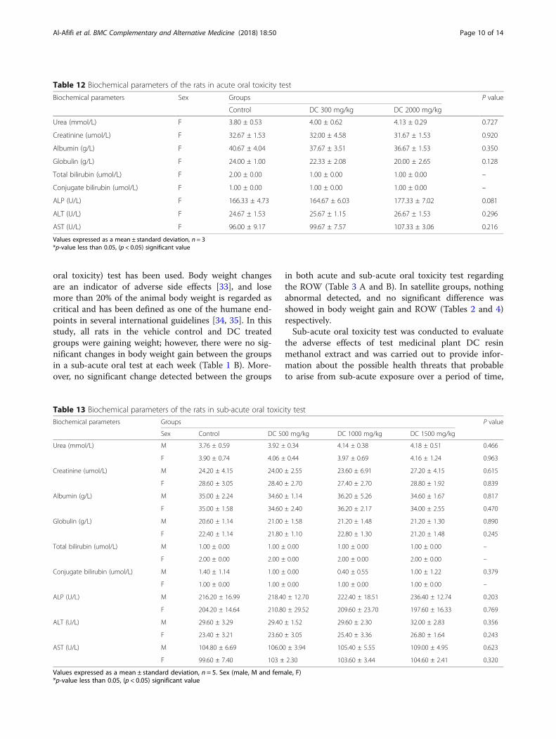
Effect of DC extract on serum biochemical parameters inacute and sub-acute oral toxicity testsA raw data of biochemical parameters, is available in theAdditional file 1.The data on biochemical parameters in control and
DC treated groups of the acute oral toxicity test
presented in Table 12. There was no significant differ-ence shown in the biochemical parameters betweengroups as p > 0.05.The data on biochemical parameters in control and
DC treated groups of sub-acute oral toxicity presentedin Table 13. There was no significant difference shownin the biochemical parameters between the groups inboth sexes (male and female rat) as well as in the satel-lite groups as p > 0.05 (Table 14).
Histopathological observationA histopathological study carried to confirm biochemicalfindings and to identify any structural changes. Lightmicroscopic examination of the vital organs includingliver, kidney, heart, lung and spleen of the rats in all theDC treated and control groups for acute oral toxicity(Fig. 1) and sub-acute oral toxicity (Fig. 2) did not revealany gross pathological lesions.The photomicrographs of the liver and kidney of the
control and DC treated groups as well as of satellitegroup, both male and female, showed with normal mor-phological architecture. Under microscopic examination,the liver of DC treated animals showed with normal cel-lular architecture and binucleation and was without anydistortions similar to the control groups. Furthermore,signs of injury, necrosis, congestion, fatty acid accumula-tion, or hemorrhagic regions around the central vein orsinusoids of the liver not observed. The hepatocytes ar-ranged in cords and clearly visible. The cross-section ofthe liver showed no lyses in the blood cells, or
Table 1 (A and B) Percentage of body weight gain of rats in acute and sub-acute oral toxicity tests at each week
A Acute oral toxicity
Sex Control DC 300 mg/kg DC 2000 mg/kg P value
0 day (g) F (n = 3) 235.33 ± 5.03 250.00 ± 8.54 249.67 ± 9.61
Week 1 (%) F (n = 3) 5.82 ± 1.44 4.92 ± 1.62 4.15 ± 0.37 0.340
Week 2 (%) F (n = 3) 8.65 ± 1.96 8.53 ± 1.59 7.37 ± 1.17 0.586
B Sub-acute oral toxicity
Sex Control DC 500 mg/kg DC 1000 mg/kg DC 1500 mg P value
0 day (g) M (n = 5) 232.00 ± 4.70 223.40 ± 5.22 226.40 ± 5.13 229.80 ± 3.96
F (n = 5) 247.20 ± 6.14 233.40 ± 2.70 228.60 ± 8.02 232.20 ± 2.56
Week 1 (%) M (n = 5) 8.28 ± 0.91 8.56 ± 0.79 8.11 ± 1.15 8.61 ± 1.28 0.853
F (n = 5) 5.03 ± 2.44 4.71 ± 1.09 5.44 ± 1.71 5.50 ± 2.21 0.907
Week 2 (%) M (n = 5) 16.73 ± 1.03 16.37 ± 1.19 16.78 ± 0.83 16.09 ± 1.87 0.811
F (n = 5) 7.53 ± 3.21 7.62 ± 2.09 7.27 ± 2.59 8.49 ± 2.30 0.889
Week 3 (%) M (n = 5) 24.25 ± 2.13 24.17 ± 1.09 23.69 ± 1.64 23.48 ± 2.00 0.881
F (n = 5) 11.42 ± 2.77 10.44 ± 2.82 10.55 ± 2.64 12.30 ± 3.20 .0715
Week 4 (%) M (n = 5) 29.34 ± 2.70 28.55 ± 1.60 29.71 ± 2.08 29.75 ± 4.94 0.920
F (n = 5) 13.83 ± 2.76 13.18 ± 2.24 13.36 ± 3.05 15.23 ± 2.94 0.649
Values expressed as a mean ± standard deviation. Sex (male, M and female, F)*p-value less than 0.05, (p < 0.05) significant value
Table 2 Satellite group/ Percentage of body weight change ofrats in sub-acute oral toxicity test
Satellite group/ Percentage of body weight change of rats in sub-acuteoral toxicity test
Sex Control DC 1500 mg/kg P value
0 day (g) M (n = 5) 229.20 ± 3.70 235.40 ± 3.85
F (n = 5) 222.20 ± 5.12 221.00 ± 6.67
Week 1 (%) M (n = 5) 8.38 ± 0.72 8.09 ± 1.21 0.657
F (n = 5) 4.86 ± 0.59 4.90 ± 1.54 0.960
Week 2 (%) M (n = 5) 15.36 ± 1.57 15.13 ± 0.77 0.782
F (n = 5) 7.57 ± 0.66 7.56 ± 1.67 0.994
Week 3 (%) M (n = 5) 19.90 ± 1.34 19.89 ± 1.62 0.988
F (n = 5) 10.09 ± 1.12 9.80 ± 1.68 0.756
Week 4 (%) M (n = 5) 24.45 ± 1.99 24.30 ± 0.83 0.883
F (n = 5) 12.96 ± 0.96 12.77 ± 1.35 0.807
Week 5 (%) M (n = 5) 28.81 ± 2.02 28.48 ± 1.68 0.788
F (n = 5) 15.22 ± 0.71 14.78 ± 1.81 0.631
Week 6 (%) M (n = 5) 34.03 ± 2.50 33.16 ± 2.69 0.612
F (n = 5) 17.28 ± 0.75 16.59 ± 1.65 0.415
Values expressed as a mean ± standard deviation. Sex (male, M and female, F)*p-value less than 0.05, (p < 0.05) significant value
Al-Afifi et al. BMC Complementary and Alternative Medicine (2018) 18:50 Page 5 of 14

infiltration of neutrophil, lymphocyte, or macrophage inthe acute oral toxicity group and the sub-acute oral tox-icity. For the kidneys, histologically there was no mor-phological change for all DC treated groups. Theappearance of the glomerular architecture showed nor-mal similar to the control groups. The glomeruli, distal,and proximal tubules in the kidney appeared normal inboth male and female rats. In addition, there was nointerstitial and intraglomerular congestion or tubular
atrophies. All the nephron cells showed normal andclearly visible nucleoli with no degeneration, bleeding, ornecrosis infiltration in acute oral toxicity group as wellas in the sub-acute oral toxicity. In both the control andDC treated female and male rats, the heart shows nor-mal cardiac muscle fibers and lungs show a normal al-veolar structure with no treatment-related inflammatoryresponse in acute oral toxicity group as well as in thesub-acute oral toxicity. Similarly, normal structure andhistology of the spleen also observed in all the rats ofacute and sub-acute oral toxicity tests. There is mildcongestion seen in the lung, liver, and kidney of the con-trol and DC treated groups of both sexes which were in-cidental or spontaneous with no relation to DC resinmethanol extraction treatment.Thus, the histopathological evaluations of the selected
organs did not reveal any morphological abnormalitiesthat could be attributed to the oral administration of DCresin methanol extract to the rats.
DiscussionHerbal medicines have acquired greater importance as asubstitute to conventional therapy [49]. As the use ofmedicinal plants increases, screening plant products toassess and evaluate the toxic characteristics of a naturalproduct extract, fraction, or compound consider an ini-tial step [30].
Table 3 (A and B) Relative organs weight (g%) of rats in acute and sub-acute oral toxicity tests
A Acute oral toxicity
Sex Control DC 300 mg/kg DC 2000 mg/kg P value
Heart F (n = 3) 0.37 ± 0.02 0.35 ± 0.01 0.35 ± 0.03 0.422
Liver F (n = 3) 2.58 ± 0.14 2.60 ± 0.12 2.50 ± 0.13 0.601
Kidney F (n = 3) 0.70 ± 0.02 0.67 ± 0.03 0.68 ± 0.02 0.484
Spleen F (n = 3) 0.18 ± 0.02 0.18 ± 0.02 0.18 ± 0.01 0.952
Lung F (n = 3) 0.49 ± 0.01 0.48 ± 0.02 0.48 ± 0.02 0.874
B Sub-acute oral toxicity
Sex Control DC 500 mg/kg DC 1000 mg/kg DC 1500 mg/kg P value
Heart M (n = 5) 0.36 ± 0.02 0.35 ± 0.02 0.34 ± 0.02 0.34 ± 0.03 0.757
F (n = 5) 0.37 ± 0.02 0.37 ± 0.03 0.37 ± 0.02 0.38 ± 0.02 0.938
Liver M (n = 5) 2.92 ± 0.15 2.81 ± 0.10 2.87 ± 0.17 2.92 ± 0.14 0.593
F (n = 5) 2.45 ± 0.09 2.38 ± 0.07 2.35 ± 0.05 2.43 ± 0.07 0.114
Kidney M (n = 5) 0.75 ± 0.02 0.75 ± 0.03 0.76 ± 0.02 0.76 ± 0.03 0.930
F (n = 5) 0.69 ± 0.03 0.70 ± 0.02 0.70 ± 0.05 0.72 ± 0.04 0.639
Spleen M (n = 5) 0.19 ± 0.02 0.19 ± 0.02 0.18 ± 0.03 0.19 ± 0.02 0.937
F (n = 5) 0.18 ± 0.02 0.18 ± 0.02 0.18 ± 0.02 0.19 ± 0.02 0.835
Lung M (n = 5) 0.40 ± 0.02 0.40 ± 0.02 0.39 ± 0.02 0.42 ± 0.01 0.241
F (n = 5) 0.49 ± 0.02 0.48 ± 0.02 0.49 ± 0.01 0.50 ± 0.02 0.536
Values expressed as a mean ± standard deviation. Sex (male, M and female, F)*p-value less than 0.05, (p < 0.05) significant value
Table 4 Satellite group/ Relative organs weight (g%) of rats inthe sub-acute oral toxicity test
Satellite group/ Relative organs weight (g) of rats in the sub-acute oraltoxicity test
Sex Control DC 1500 mg/kg P value
Heart M 0.34 ± 0.03 0.34 ± 0.02 0.797
F 0.38 ± 0.01 0.38 ± 0.01 0.493
Liver M 2.73 ± 0.09 2.76 ± 0.08 0.508
F 2.59 ± 0.13 2.64 ± 0.12 0.524
Kidney M 0.76 ± 0.02 0.75 ± 0.02 0.655
F 0.72 ± 0.03 0.72 ± 0.03 0.696
Spleen M 0.19 ± 0.01 0.19 ± 0.01 0.370
F 0.20 ± 0.02 0.20 ± 0.01 0.832
Lung M 0.40 ± 0.02 0.38 ± 0.02 0.308
F 0.50 ± 0.02 0.50 ± 0.01 0.722
Values expressed as a mean ± standard deviation. n= 5. Sex (male, M and female, F)*p-value less than 0.05, (p < 0.05) significant value
Al-Afifi et al. BMC Complementary and Alternative Medicine (2018) 18:50 Page 6 of 14

During the evaluation of the toxic characteristics ofmedicinal plants, an initial assessment of toxic manifes-tations is one of the initial screening experiments per-formed with all compounds. In addition, Data from theacute toxicity study may serve as the basis for classifica-tion and labelling of the test material [31]. Thus, thecurrent study was assumed to evaluate and focus on theacute and sub-acute toxicity of DC resin methanol ex-tract in an animal model.The oral route administration is the most useful and
normally used one while doing toxicity study. The ab-sorption may be slow; however, this methodology ex-penses less and is painless to the animals. As the crude
extracts administered orally, the animals need to fast be-fore administering the material because food and otherchemicals within the digestive system may have an effecton the reaction(s) of the tested materials. All the proce-dures were performed based on the appropriate OECDguideline [32].Test method with a starting dose of 300 mg/kg body
weight primarily used in situations where the investiga-tor has no information indicating that the test materialis likely to be toxic [23]. In this study, the rats in controland DC treated groups administrated with the vehicleand crude extracts, respectively. From the experimentperformed, the starting dose of 300 mg/kg body weighthas revealed no mortality in the experimental animals.Thus, the next higher dose of 2000 mg/kg body weightselected as described in the OECD Guidelines 423. Therats monitored daily until the last day of the experiment(day 14th) for any toxic signs and mortality. The clinicalsymptom is one amongst the most important observa-tions to indicate the toxicity effects on organs within thetreated groups [7]. During the 14 days of acute toxicityassessment period, all rats orally administrated with DCresin methanol extract at a single dose of 300 mg/kg and2000 mg/kg showed no obvious signs of distress, andthere were no noticeable symptoms of either toxicity ordeaths. All of the rats showed no significant changes inwellness parameters. Physical appearance features suchas skin, fur, eyes, mucous membrane, salivation, behav-ioural pattern, the sleep of the animals in control andDC treated groups (300 mg and 2000 mg) of DC extractwere found to be normal. Lethargy, tremors, diarrhoeaand coma did not occur in any of the animals. Moreover,the body weight of the rats showed an increase in bothcontrol and DC treated groups without significant
Table 5 (A and B) Food consumption (g) of rats in acute and sub-acute oral toxicity tests
A Acute oral toxicity
Sex Control DC 300 mg/kg DC 2000 mg/kg P value
Week 1 F (n = 3) 64.68 ± 2.80 63.43 ± 2.87 59.14 ± 14.67 0.089
Week 2 F (n = 3) 66.16 ± 2.89 67.05 ± 5.14 63.74 ± 2.15 0.236
B Sub-acute oral toxicity
Sex Control DC 500 mg/kg DC 1000 mg/kg DC 1500 mg/kg P value
Week 1 M (n = 5) 81.75 ± 1.21 81.15 ± 2.17 80.99 ± 2.55 79.26 ± 2.42 0.189
F (n = 5) 74.87 ± 2.59 72.99 ± 1.63 72.47 ± 2.28 71.55 ± 2.80 0.089
Week 2 M (n = 5) 84.52 ± 1.13 83.93 ± 0.93 83.80 ± 1.77 82.59 ± 1.11 0.061
F (n = 5) 78.69 ± 3.26 77.56 ± 3.21 75.99 ± 3.58 74.66 ± 4.64 0.220
Week 3 M (n = 5) 88.05 ± 2.54 87.92 ± 2.07 86.10 ± 1.94 85.19 ± 2.58 0.075
F (n = 5) 86.55 ± 3.64 86.15 ± 3.11 85.17 ± 3.75 83.21 ± 3.68 0.320
Week 4 M (n = 5) 91.84 ± 1.60 91.08 ± 0.79 90.30 ± 1.26 90.23 ± 1.11 0.069
F (n = 5) 88.69 ± 3.66 87.35 ± 3.26 87.75 ± 3.32 87.04 ± 2.77 0.798
Values expressed as a mean ± standard deviation. Sex (male, M and female, F)*p-value less than 0.05, (p < 0.05) significant value
Table 6 Satellite group/ Food consumption (g) of rats in thesub-acute oral toxicity test
Satellite group/ food consumption in oral sub-acute toxicity test
Sex Control DC 1500 mg/kg P value
Week 1 M (n = 5) 81.92 ± 1.44 79.30 ± 3.81 0.113
F (n = 5) 73.05 ± 3.11 71.26 ± 1.96 0.221
Week 2 M (n = 5) 84.60 ± 2.38 82.68 ± 1.56 0.099
F (n = 5) 73.68 ± 2.27 72.77 ± 1.93 0.435
Week 3 M (n = 5) 89.75 ± 1.96 87.72 ± 1.69 0.059
F (n = 5) 81.89 ± 3.76 78.62 ± 4.60 0.170
Week 4 M (n = 5) 91.96 ± 0.66 91.07 ± 0.93 0.061
F (n = 5) 83.55 ± 2.00 82.43 ± 1.59 0.265
Week 5 M (n = 5) 92.96 ± 1.63 91.87 ± 1.80 0.256
F (n = 5) 85.81 ± 1.98 83.92 ± 3.91 0.179
Week 6 M (n = 5) 101.90 ± 3.70 100.25 ± 2.85 0.366
F (n = 5) 86.55 ± 3.82 83.07 ± 4.73 0.155
Values expressed as a mean ± standard deviation. Sex (male, M and female, F)*p-value less than 0.05, (p < 0.05) significant value
Al-Afifi et al. BMC Complementary and Alternative Medicine (2018) 18:50 Page 7 of 14

difference seen (Table 1 A); this indicates that the DCresin methanol extract has no adverse effect on thegrowth of the animals.This study estimated that DC resin methanol extract
does not cause acute toxicity effects and no rat has died.Based on OECD guidelines 423 (Annex 2), the results ofthis test allow the substance to be ranked and classifiedaccording to the Globally Harmonized System of Classi-fication and Labelling of Chemicals. Thus, the DC resinmethanol extract can be classified as category 5 with lowacute toxicity hazard, which was the lowest toxicity class[23]. Therefore, it can be concluded that DC resinmethanol extract is tolerated up to 2000 mg/kg bodyweight when administered at a single dose. In a likemanner, a study performed by R Ramaswamy, N
Prathyusha, R Saranya, H Sumathy, K Mohanavalli, RPriya, J Venkhatesh, C Babu, K Manickavasakam and SThanikachalam [28] using Nuna Kadugu (a Siddha medi-cine prepared from leaves and fruits of MorindaPubescens) revealed that Nuna Kadugu can be classifiedunder category-5 when administered at single dose2000 mg/kg in accordance with Globally HarmonisedSystem of Classification and Labelling of Chemicals, andthis provides a direct relevance for protecting humanand animal health.Acute toxicity information is of limited clinical appli-
cation because cumulative toxic effects do occur even atvery low doses. Consequently, multiple dose studies aretypically useful in evaluating the safety profile of phyto-medicines. Therefore, sub-acute (Repeated dose 28-day
Table 7 (A and B) Water intake (ml) of rats in the acute and sub-acute oral toxicity tests
A Acute oral toxicity
Sex Control DC 300 mg/kg DC 2000 mg/kg P value
Week 1 F (n = 3) 102.56 ± 1.27 104.03 ± 1.78 104.34 ± 2.12 0.157
Week 2 F (n = 3) 102.54 ± 0.97 104.00 ± 1.77 103.93 ± 2.01 0.199
B Sub-acute oral toxicity
Sex Control DC 500 mg/kg DC 1000 mg/kg DC 1500 mg/kg P value
Week 1 M (n = 5) 123.18 ± 1.39 128.09 ± 2.34 128.03 ± 1.91 127.40 ± 2.11 0.000*
F (n = 5) 122.39 ± 0.42 125.51 ± 0.50 125.48 ± 0.37 125.46 ± 0.53 0.000*
Week 2 M (n = 5) 124.06 ± 1.17 127.31 ± 1.50 127.44 ± 1.48 127.97 ± 1.85 0.000*
F (n = 5) 123.33 ± 0.55 125.68 ± 0.45 125.99 ± 0.41 125.87 ± 0.41 0.000*
Week 3 M (n = 5) 125.23 ± 0.96 127.99 ± 1.33 127.98 ± 1.80 127.86 ± 1.69 0.004*
F (n = 5) 124.44 ± 0.49 127.29 ± 0.66 127.12 ± 0.61 127.16 ± 0.77 0.000*
Week 4 M (n = 5) 125.89 ± 0.78 128.29 ± 1.95 128.54 ± 1.22 128.34 ± 1.61 0.007*
F (n = 5) 124.99 ± 0.50 127.28 ± 0.86 127.36 ± 0.63 127.56 ± 0.61 0.000*
Values expressed as a mean ± standard deviation. Sex (male, M and female, F)*p-value less than 0.05, (p < 0.05) significant value. Numbers in bold indicate a statistically significant difference
Table 8 Tukey test of water intake (ml) of male and female rats in sub-acute oral toxicity test
Dependent variable (I) water intake (J) water intake Mean difference (I-J) in male rat P value Mean difference (I-J) in female rat P value
Week 1 Control DC 500 mg/kg −4.91 ± 1.05 0.001* −3.12 ± 0.24 0.000*
DC 1000 mg/kg −4.86 ± 1.05 0.001* −3.09 ± 0.24 0.000*
DC 1500 mg/kg −4.23 ± 1.05 0.003* −3.07 ± 0.24 0.000*
Week 2 Control DC 500 mg/kg −3.25 ± 0.81 0.003* −2.35 ± 0.25 0.000*
DC 1000 mg/kg −3.38 ± 0.81 0.002* −2.67 ± 0.25 0.000*
DC 1500 mg/kg −3.91 ± 0.81 0.000* −2.54 ± 0.25 0.000*
Week 3 Control DC 500 mg/kg −2.77 ± 0.79 0.009* −2.85 ± 0.34 0.000*
DC 1000 mg/kg −2.75 ± 0.79 0.010* −2.68 ± 0.34 0.000*
DC 1500 mg/kg −2.63 ± 0.79 0.014* −2.72 ± 0.34 0.000*
Week 4 Control DC 500 mg/kg −2.40 ± 0.78 0.025* −2.29 ± 0.35 0.000*
DC 1000 mg/kg −2.65 ± 0.78 0.012* −2.37 ± 0.35 0.000*
DC 1500 mg/kg −2.45 ± 0.78 0.021* −2.57 ± 0.35 0.000*
Values expressed as a mean ± Standard Error. n = 5*p-value less than 0.05, (p < 0.05) significant value. Numbers in bold indicate a statistically significant difference
Al-Afifi et al. BMC Complementary and Alternative Medicine (2018) 18:50 Page 8 of 14

Table 9 Satellite group/ Water intake (ml) of rats in thesub-acute oral toxicity test at each week
Satellite group/ Water intake (ml) of rats in the sub-acute oral toxicitytest at each week
Sex Control DC 1500 mg/kg P value
Week 1 M 124.39 ± 0.41 125.86 ± 0.85 0.001*
F 121.24 ± 1.79 123.59 ± 1.96 0.037*
Week 2 M 124.87 ± 0.64 127.14 ± 1.09 0.000*
F 123.33 ± 1.85 125.03 ± 0.76 0.044*
Week 3 M 126.40 ± 0.87 128.30 ± 1.37 0.009*
F 123.80 ± 0.95 125.80 ± 1.00 0.002*
Week 4 M 127.48 ± 0.95 128.89 ± 1.21 0.032*
F 124.53 ± 1.10 125.94 ± 1.01 0.027*
Week 5 M 128.38 ± 1.11 129.03 ± 0.86 0.244
F 125.68 ± 0.99 125.97 ± 0.68 0.538
Week 6 M 128.94 ± 1.28 129.30 ± 1.34 0.620
F 126.48 ± 0.90 126.58 ± 0.68 0.825
Values expressed as a mean± standard deviation. n= 5. Sex (male, M and female, F)*p-value less than 0.05, (p < 0.05) significant value. Numbers in bold indicate astatistically significant difference
Table 10 (A and B) Hematological parameters of the rats in acute and sub-acute oral toxicity tests
A Acute oral toxicity
Hematological Parameters Sex Control DC 300 mg/kg DC2000 mg/kg P value
HGB (g/L) F (n = 3) 153.00 ± 10.00 145.50 ± 9.50 149.33 ± 17.95 0.789
WBC (10^9/L) F (n = 3) 6.83 ± 0.23 7.77 ± 0.38 8.05 ± 0.75 0.057
Neutrophil (10^9/L) F (n = 3) 0.55 ± 0.13 0.56 ± 0.07 0.58 ± 0.14 0.961
Lymphocyte (10^9/L) F (n = 3) 6.39 ± 0.87 6.72 ± 1.15 7.29 ± 0.52 0.498
Monocyte (10^9/L) F (n = 3) 0.14 ± 0.03 0.15 ± 0.02 0.18 ± 0.3 0.228
Eosinophil (10^9/L) F (n = 3) 0.08 ± 0.02 0.10 ± 0.02 0.11 ± 0.02 0.125
Basophil (10^9/L) F (n = 3) 0.02 ± 0.01 0.03 ± 0.02 0.03 ± 0.02 0.531
B Sub-acute oral toxicity
Hematological Parameters Sex Control DC 500 mg/kg DC1000 mg/kg DC1500 mg/kg P value
HGB (g/L) M (n = 5) 149.40 ± 6.47 148.80 ± 5.40 150.40 ± 12.72 156.80 ± 12.91 0.575
F (n = 5) 156.40 ± 5.81 148.80 ± 7.82 150.60 ± 3.85 153.40 ± 5.81 0.243
WBC (10^9/L) M (n = 5) 9.22 ± 1.03 9.30 ± 2.45 9.46 ± 1.33 9.56 ± 3.67 0.996
F (n = 5) 6.86 ± 0.59 7.40 ± 1.15 7.38 ± 0.93 7.82 ± 0.81 0.431
Neutrophil (10^9/L) M (n = 5) 0.98 ± 0.12 1.01 ± 0.16 1.03 ± 0.18 1.14 ± 0.30 0.626
F (n = 5) 0.81 ± 0.21 0.77 ± 0.11 0.87 ± 0.17 0.89 ± 0.14 0.646
Lymphocyte (10^9/L) M (n = 5) 9.66 ± 2.12 9.64 ± 1.67 10.39 ± 1.99 10.77 ± 2.08 0.753
F (n = 5) 8.22 ± 1.07 8.44 ± 1.22 8.67 ± 1.00 9.20 ± 1.14 0.564
Monocyte (10^9/L) M (n = 5) 0.16 ± 0.02 0.15 ± 0.02 0.17 ± 0.04 0.19 ± 0.04 0.411
F (n = 5) 0.17 ± 0.03 0.16 ± 0.03 0.18 ± 0.02 0.18 ± 0.03 0.623
Eosinophil (10^9/L) M (n = 5) 0.08 ± 0.02 0.09 ± 0.02 0.10 ± 0.02 0.10 ± 0.03 0.388
F (n = 5) 0.08 ± 0.01 0.09 ± 0.02 0.07 ± 0.03 0.10 ± 0.02 0.215
Basophil (10^9/L) M (n = 5) 0.06 ± 0.2 0.04 ± 0.03 0.04 ± 0.03 0.04 ± 0.03 0.537
F (n = 5) 0.02 ± 0.02 0.03 ± 0.02 0.03 ± 0.02 0.04 ± 0.02 0.397
Values expressed as a mean ± standard deviation. Sex (male, M and female, F)*p-value less than 0.05, (p < 0.05) significant value
Table 11 Hematological parameters in sub-acute oral toxicitytest in Satellite group
Satellite group/ Hematological parameters in sub-acute oral toxicity test
Sex Control DC 1500 mg/kg P value
HGB (g/L) M 152.40 ± 6.80 153.20 ± 7.46 0.863
F 155.00 ± 6.82 154.80 ± 5.97 0.961
WBC (10^9/L) M 9.30 ± 1.36 10.76 ± 1.23 0.112
F 6.70 ± 0.72 6.74 ± 0.60 0.926
Neutrophil (10^9/L) M 1.08 ± 0.13 1.31 ± 0.23 0.084
F 1.25 ± 0.11 1.33 ± 0.11 0.295
Lymphocyte (10^9/L) M 9.77 ± 1.07 11.25 ± 1.41 0.098
F 9.29 ± 0.95 9.31 ± 0.70 0.977
Monocyte (10^9/L) M 0.21 ± 0.06 0.23 ± 0.07 0.649
F 0.22 ± 0.05 0.23 ± 0.03 0.773
Eosinophil (10^9/L) M 0.10 ± 0.01 0.10 ± 0.02 0.856
F 0.09 ± 0.02 0.10 ± 0.03 0.596
Basophil (10^9/L) M 0.03 ± 0.02 0.04 ± 0.03 0.636
F 0.02 ± 0.01 0.03 ± 0.01 0.545
Values expressed as a mean ± standard deviation. n = 5. Sex (male, M andfemale, F). *p-value less than 0.05, (p < 0.05) significant value
Al-Afifi et al. BMC Complementary and Alternative Medicine (2018) 18:50 Page 9 of 14
oral toxicity) test has been used. Body weight changesare an indicator of adverse side effects [33], and losemore than 20% of the animal body weight is regarded ascritical and has been defined as one of the humane end-points in several international guidelines [34, 35]. In thisstudy, all rats in the vehicle control and DC treatedgroups were gaining weight; however, there were no sig-nificant changes in body weight gain between the groupsin a sub-acute oral test at each week (Table 1 B). More-over, no significant change detected between the groups
in both acute and sub-acute oral toxicity test regardingthe ROW (Table 3 A and B). In satellite groups, nothingabnormal detected, and no significant difference wasshowed in body weight gain and ROW (Tables 2 and 4)respectively.Sub-acute oral toxicity test was conducted to evaluate
the adverse effects of test medicinal plant DC resinmethanol extract and was carried out to provide infor-mation about the possible health threats that probableto arise from sub-acute exposure over a period of time,
Table 12 Biochemical parameters of the rats in acute oral toxicity test
Biochemical parameters Sex Groups P value
Control DC 300 mg/kg DC 2000 mg/kg
Urea (mmol/L) F 3.80 ± 0.53 4.00 ± 0.62 4.13 ± 0.29 0.727
Creatinine (umol/L) F 32.67 ± 1.53 32.00 ± 4.58 31.67 ± 1.53 0.920
Albumin (g/L) F 40.67 ± 4.04 37.67 ± 3.51 36.67 ± 1.53 0.350
Globulin (g/L) F 24.00 ± 1.00 22.33 ± 2.08 20.00 ± 2.65 0.128
Total bilirubin (umol/L) F 2.00 ± 0.00 1.00 ± 0.00 1.00 ± 0.00 –
Conjugate bilirubin (umol/L) F 1.00 ± 0.00 1.00 ± 0.00 1.00 ± 0.00 –
ALP (U/L) F 166.33 ± 4.73 164.67 ± 6.03 177.33 ± 7.02 0.081
ALT (U/L) F 24.67 ± 1.53 25.67 ± 1.15 26.67 ± 1.53 0.296
AST (U/L) F 96.00 ± 9.17 99.67 ± 7.57 107.33 ± 3.06 0.216
Values expressed as a mean ± standard deviation, n = 3*p-value less than 0.05, (p < 0.05) significant value
Table 13 Biochemical parameters of the rats in sub-acute oral toxicity test
Biochemical parameters Groups P value
Sex Control DC 500 mg/kg DC 1000 mg/kg DC 1500 mg/kg
Urea (mmol/L) M 3.76 ± 0.59 3.92 ± 0.34 4.14 ± 0.38 4.18 ± 0.51 0.466
F 3.90 ± 0.74 4.06 ± 0.44 3.97 ± 0.69 4.16 ± 1.24 0.963
Creatinine (umol/L) M 24.20 ± 4.15 24.00 ± 2.55 23.60 ± 6.91 27.20 ± 4.15 0.615
F 28.60 ± 3.05 28.40 ± 2.70 27.40 ± 2.70 28.80 ± 1.92 0.839
Albumin (g/L) M 35.00 ± 2.24 34.60 ± 1.14 36.20 ± 5.26 34.60 ± 1.67 0.817
F 35.00 ± 1.58 34.60 ± 2.40 36.20 ± 2.17 34.00 ± 2.55 0.470
Globulin (g/L) M 20.60 ± 1.14 21.00 ± 1.58 21.20 ± 1.48 21.20 ± 1.30 0.890
F 22.40 ± 1.14 21.80 ± 1.10 22.80 ± 1.30 21.20 ± 1.48 0.245
Total bilirubin (umol/L) M 1.00 ± 0.00 1.00 ± 0.00 1.00 ± 0.00 1.00 ± 0.00 –
F 2.00 ± 0.00 2.00 ± 0.00 2.00 ± 0.00 2.00 ± 0.00 –
Conjugate bilirubin (umol/L) M 1.40 ± 1.14 1.00 ± 0.00 0.40 ± 0.55 1.00 ± 1.22 0.379
F 1.00 ± 0.00 1.00 ± 0.00 1.00 ± 0.00 1.00 ± 0.00 –
ALP (U/L) M 216.20 ± 16.99 218.40 ± 12.70 222.40 ± 18.51 236.40 ± 12.74 0.203
F 204.20 ± 14.64 210.80 ± 29.52 209.60 ± 23.70 197.60 ± 16.33 0.769
ALT (U/L) M 29.60 ± 3.29 29.40 ± 1.52 29.60 ± 2.30 32.00 ± 2.83 0.356
F 23.40 ± 3.21 23.60 ± 3.05 25.40 ± 3.36 26.80 ± 1.64 0.243
AST (U/L) M 104.80 ± 6.69 106.00 ± 3.94 105.40 ± 5.55 109.00 ± 4.95 0.623
F 99.60 ± 7.40 103 ± 2.30 103.60 ± 3.44 104.60 ± 2.41 0.320
Values expressed as a mean ± standard deviation, n = 5. Sex (male, M and female, F)*p-value less than 0.05, (p < 0.05) significant value
Al-Afifi et al. BMC Complementary and Alternative Medicine (2018) 18:50 Page 10 of 14

the possibilities of cumulative effects, and an estimate ofthe dose at which there is no observed adverse effect.Evaluation of safety margin between different dose levelthat produces the therapeutic effect and that which pro-duces the adverse effects is necessary. Evaluation of
safety is exactly to provide benefit to risk assessment.Animal experimental model is the only method that canassess this matter [36]. Determination of food consump-tion is an important to study the safety of a product withtherapeutic purpose as proper intake of nutrients is
Table 14 Biochemical parameters of the rats in satellite group for subacute oral toxicity test
Biochemical parameters Satellite group P value
Sex Control DC 1500 mg/kg
Urea (mmol/L) M 3.78 ± 0.34 3.92 ± 0.48 0.608
F 3.96 ± 0.70 4.06 ± 0.17 0.765
Creatinine (umol/L) M 25..00 ± 3.87 26.40 ± 3.78 0.579
F 29.40 ± 3.05 27.20 ± 3.96 0.354
Albumin (g/L) M 35.20 ± 1.30 34.40 ± 1.67 0.423
F 34.20 ± 2.77 32.40 ± 3.36 0.382
Globulin (g/L) M 20.20 ± 0.84 19.40 ± 1.34 0.290
F 22.00 ± 1.58 21.20 ± 1.30 0.408
Total bilirubin (umol/L) M 1.00 ± 0.00 1.00 ± 0.00 –
F 2.00 ± 0.00 2.00 ± 0.00 –
Conjugate bilirubin (umol/L) M 1.00 ± 0.00 1.00 ± 0.00 –
F 1.00 ± 0.00 1.00 ± 0.00 –
ALP (U/L) M 223.60 ± 29.98 226.20 ± 28.35 0.891
F 184.40 ± 29.11 190.40 ± 29.21 0.753
ALT (U/L) M 31.00 ± 3.39 30.00 ± 3.81 0.673
F 25.20 ± 1.79 25.60 ± 2.07 0.752
AST (U/L) M 107.00 ± 6.04 111.40 ± 5.94 0.279
F 103.40 ± 3.29 105.00 ± 2.74 0.427
Values expressed as a mean ± standard deviation, n = 5. Sex (male, M and female, F)*p-value less than 0.05, (p < 0.05) significant value
Liver Kidney Heart Lung Spleen
Control
DC 300 mg/kg
DC 2000 mg/kg
Fig. 1 Photomicrograph of vital organs in acute oral toxicity (H & E Stain, ×100). Liver: black arrow – portal vein; white arrow – portal triad.Kidney: black arrow – cortex; white arrow – medulla. Heart: black arrow – myocardium; white arrow – blood vessel. Lung: Black arrow – alveoli;white arrow – bronchiole. Spleen: Black arrow – white pulp, white arrow – red pulp
Al-Afifi et al. BMC Complementary and Alternative Medicine (2018) 18:50 Page 11 of 14

essential to the physiological status of the animal andgive a good impression of the appropriate response tothe treatment [37].For food consumption, no significant changes ob-
served in all groups (vehicle and DC extract treatedgroups) in both acute and subacute oral toxicity testsand this reveals that it did not adversely affect the basicmetabolic processes of the experimental animals. On theother hand, water intake showed more in the DC resinmethanol extract treated groups than the control, with asignificant difference showed in sub-acute oral toxicitytest (Table 8) and satellite group (Table 9) for both maleand female rats during the administration period. Thisresult could be refereed to that DC resin extract canproduce vasodilatation (hypotension) due to relaxationof smooth muscles of blood vessels [9] which in turnstimulate thirst and increase water intake [38].Hematological and biochemistry data play a major role
in determining the toxicity induced by drugs [39]. Bloodparameters analysis is appropriate to risk evaluation asthe hematological system has a higher prognostic valuefor toxicity [40]. Blood serves as the main medium oftransport for many drugs and xenobiotics in the bodyand for that reason components of the blood exposed tosubstantial concentrations of toxic compounds. Damageto and destruction of the blood cells are inimical to nor-mal functioning of the body both in humans and animals[41]. In the present study, the hematological parameters
data indicated that DC resin methanol extract did notaffect blood cells production as there was no significantdifference between the groups in acute and sub-acute oraltoxicity tests (Table 10 A and B). In the satellite group, nosignificant difference showed between the two groups(Table 11). The change in hematological parameters waswithin the normal range as showed elsewhere [39, 42].Evaluation of Kidney and Liver function is important
in the assessment toxicity of plant extracts as both ofthem are necessary for the survival of an organism [43].In animal model toxicity studies, the serum level of cre-atinine remains the most widely used laboratory test toestimate renal function. It kept within a relatively stablerange as daily production and renal excretion are con-tinuous in healthy mammals [44]. In the present study,for kidney function test, two serum renal biochemicalparameters, namely urea and creatinine were analyzed aspreviously mentioned [45]. There were no significantchanges observed in urea and creatinine levels betweenthe control and all doses of DC resin methanol extractgroups in both acute and sub-acute oral toxicity tests.The enzymatic activity of the liver such as alanine ami-
notransferase (ALT), aspartate aminotransferase (AST)and alkaline phosphatase (ALP) was studied to evaluateliver malfunctions. Liver enzymes levels are usuallyraised in acute hepatoxicity but tend to decrease withprolonged intoxication due to damage to the liver [46].In the present study, there were no significant
Fig. 2 Photomicrograph of vital organs in sub-acute oral toxicity (H & E Stain, × 100). Liver: black arrow – portal vein; white arrow – portal triad.Kidney: black arrow – cortex; white arrow – medulla. Heart: black arrow – myocardium; white arrow – blood vessel. Lung: Black arrow – alveoli;white arrow – bronchiole. Spleen: Black arrow – white pulp, white arrow – red pulp
Al-Afifi et al. BMC Complementary and Alternative Medicine (2018) 18:50 Page 12 of 14

differences shown in the biochemical analysis in acuteand sub-acute oral toxicity test. The level was withinnormal expected range for the rat species used in thisstudy. Bilirubin formed from the breakdown ofhemoglobin in the liver, spleen, and bone marrow. Anincrease in tissue or serum bilirubin level occurs throughincreased breakdown of RBC (hemolysis) or in the caseof hepatitis or bile duct obstruction (liver damage) [33].Reduction in serum albumin level may suggest infectionor continuous loss of albumin [47]. Thus, the insignifi-cant change in serum concentration of albumin andglobulin in control and DC treated groups at all dosesused in this study proposed that DC resin methanol ex-tract not do damage in hepatocellular or secretory func-tions of the liver which in turn indicated non-adverseeffects of the tested material. For biochemical analysis,in the satellite group, there was no significant differencehas been noted which concluded that the tested material(DC resin methanol extract) would not produce the de-layed onset of toxicity.The assessment of histopathological alterations in or-
gans considered as a basic test in the safety assessmentof tested materials [48]. No abnormality observed ongross or histopathological evaluations of organs exam-ined in this study. Histopathological findings of liver,kidney, heart, lung and spleen were normal in all ani-mals that given different doses of DC resin methanol ex-tract in both acute and sub-acute oral toxicity tests.However, to determine definitely the oral safety dose
and to detect any unanticipated variability of DC resinmethanol extract a sub-chronic toxicity and genotoxicitystudies might be required. Therefore, sub-chronic tox-icity should be proceeded based on the oral doses of DCresin methanol extract in sub-acute oral toxicity test.
ConclusionsIn conclusion, according to Globally Harmonised Classifi-cation System, DC resin methanol extract can be classifiedas Category 5. In addition, we may conclude that DC resinmethanol extract is well tolerated up to the dose of1500 mg/kg body weight administered daily for 28 days.DC resin methanol extract did not cause any lethality orproduce any serum chemical alteration or importanthistopathological signs. The present investigation demon-strates, at least in part, the safety of DC resin methanolextract suggesting its promising potential for pharmaceut-ical uses.
Additional file
Additional file 1: The raw data of body weight, organ’s weight, foodconsumption and water intake, and hematological and biochemicalparameters of each rat in the acute and subacute oral toxicity tests.(XLSX 43 kb)
AbbreviationsALP: Alkaline phosphatase; ALT: Alanine aminotransferase; ANOVA: One-wayanalysis of variance; AST: Aspartate aminotransferase; DC: Dracaena cinnabari;F: Female; HGB: Hemoglobin; kg: Kilogram; M: Male; mg: Milligram;OECD: Organization for economic cooperation and development;ROW: Relative organ’s weight; SD: Sprague-Dawley; WBC: White blood cell
AcknowledgementsThis study financially supported by UMRG Grant (RG422/12HTM), Universityof Malaya, Malaysia and FRGS Grant (FP030-2015A) from the Ministry ofEducation Malaysia. We would like to thank Dr. Mohammed Nasser forchecking English text in our manuscript.
FundingA University Malaya Research Grant (UMRG) (RG422/12HTM) and FRGS Grant(FP030-2015A) from the Ministry of Education Malaysia supported this study.
Availability of data and materialsAll data generated or analyzed during this study are included in thispublished article (and its supplementary information files).
Authors’ contributionsEach author participated sufficiently in taking public responsibility forappropriate portions of the content. Study conception and design: NA, AAand MB. Analysis and interpretation of data: NA, AR. Funding acquisition: AA.Drafting of manuscript: NA, AA and AR. Revision: NA, AA, MB and AR. Allauthors read and approved the final manuscript.
Ethics approvalThe toxicity tests were carried out according to Organization for EconomicCooperation and Development (OECD) test guideline, i.e., OECD Guideline423 for acute oral toxicity test and OECD Guideline 407 for the sub-acute oraltoxicity test, with slight modifications. Protocols of this study (Acute andSub-acute oral toxicity tests) approved by the Institutional Animal Care andUse Committee (IACUC), Faculty of Medicine, University of Malaya, Malaysia(Ethics No. 2014-02-14/OBBS/R/NAA).
Consent for publicationNot applicable.
Competing interestsThe authors declare that they have no competing interests.
Publisher’s NoteSpringer Nature remains neutral with regard to jurisdictional claims inpublished maps and institutional affiliations.
Author details1Department of Oral and Craniofacial Sciences, Faculty of Dentistry,University of Malaya, 50603 Kuala Lumpur, Malaysia. 2Department of OralBiology and Biomedical Sciences, Faculty of Dentistry, MAHSA University,42610 Jenjarom, Selangor, Malaysia. 3Department of Oral & MaxillofacialClinical Sciences, Faculty of Dentistry, University of Malaya, Kuala Lumpur50603, Malaysia. 4Oral Cancer Research and Coordinating Centre, Faculty ofDentistry, University of Malaya, Kuala Lumpur 50603, Malaysia.
Received: 7 April 2017 Accepted: 24 January 2018
References1. Gupta D, Bleakley B, Gupta RK. Dragon's blood: botany, chemistry and
therapeutic uses. J Ethnopharmacol. 2008;115(3):361–80.2. Kamal MSA, Ghazali AR, Yahya NA, Wasiman MI, Ismail Z. Acute toxicity
study of standardized Mitragyna speciosa korth aqueous extract in Spraguedawley rats. J Plant Stud. 2012;1(2):120.
3. Mir AH, Sexena M, Malla MY. An acute oral toxicity study of methanolicextract from Tridex procumbens in Sprague Dawley’s rats as per OECDguidelines 423. Asian J Plant Sc Res. 2013;3:16–20.
4. Bhushan B, Sardana S, Bansal G. Acute and sub-acute toxicity study ofClerodendrum Inerme, Jasminum Mesnyi Hance and Callistemon citrinus.J Acute Dis. 2014;324:327.
Al-Afifi et al. BMC Complementary and Alternative Medicine (2018) 18:50 Page 13 of 14

5. Aneela S, De S, Kanthal L, Choudhury N, Das B, Sagar K. Acute oral toxicitystudies of Pongamia Pinnata and Annona squamosa on albino wister rats.Int J Res Pharm Chem. 2011;1(4):820–4.
6. Rahman HS, Rasedee A, Othman HH, Chartrand MS, Namvar F, Yeap SK,Abdul Samad N, Andas RJ, Muhammad Nadzri N, Anasamy T. Acute toxicitystudy of zerumbone-loaded nanostructured lipid carrier on BALB/c micemodel. Bio Med Res Int. 2014;2014
7. Jothy SL, Zakaria Z, Chen Y, Lau YL, Latha LY, Sasidharan S. Acute oraltoxicity of methanolic seed extract of Cassia fistula in mice. Molecules. 2011;16(6):5268–82.
8. Robinson S, Chapman K, Hudson S, Sparrow S, Spencer-Briggs D, Danks A,Hill R, Everett D, Mulier B, Old S. Guidance on dose level selection forregulatory general toxicology studies for pharmaceuticals. London: NC3Rs/LASA; 2009.
9. Al-Awthan YS, Zarga MA, Abdalla S. Flavonoids content of Dracaenacinnabari resin and effects of the aqueous extract on isolated smoothmuscle preparations, Perfused heart, blood pressure and Diuresis in the rat.Jordan J Pharm Sci. 2010;3(1):8–16.
10. Yehia A-TA, Alzowahi FAM, Kadam TA, Shaikh RU. In vitro evaluation ofantimicrobial and antioxidant activity of Dragon’s blood tree (Dracaenacinnabari Balf. f.) of Socotra Island (Yemen). J Coastal Life Med. 2013;1(2):123–9.
11. Hubálková I, Maděra P, Volařík D. Growth dynamics of Dracaena cinnabariunder controlled conditions as the most effective way to protectendangered species. Saudi J Biol Sci. 2015;
12. Adolt R, Pavlis J. Age structure and growth of Dracaena cinnabaripopulations on Socotra. Trees. 2004;18(1):43–53.
13. Alexander D, Miller A. A, saving the spectacular flora of Socotra. Plant Talk.1996;7:19–22.
14. Mothana R, Gruenert R, Lindequist U, Bednarski P. Study of the anticancerpotential of Yemeni plants used in folk medicine. Die Pharmazie-An Int JPharm Sci. 2007;62(4):305–7.
15. Gupta D, Bleakley B, Gupta RK. Bioassay guided isolation of antibacterialhomoisoflavone from Dragon’s blood resin (Dammul-akhwain). Nat ProdRad. 2009;8(5):494–7.
16. Machala M, Kubínová R, Hořavová P, Suchý V. Chemoprotective potentialsof homoisoflavonoids and chalcones of Dracaena cinnabari: modulations ofdrug-metabolizing enzymes and antioxidant activity. Phytother Res. 2001;15(2):114–8.
17. Gupta D, Verma N, Das HR, Gupta RK. Evaluation of anti-inflammatory activityof Dracaena cinnabari Balf. f. resin. Indian J Natl Prod Res. 2014;5(3):215–22.
18. Mothana RA, Mentel R, Reiss C, Lindequist U. Phytochemical screening andantiviral activity of some medicinal plants from the island Soqotra.Phytother Res. 2006;20(4):298–302.
19. Mothana RA, Lindequist U. Antimicrobial activity of some medicinal plantsof the island Soqotra. J Ethnopharmacol. 2005;96(1):177–81.
20. Gupta D, Gupta RK: Bioprotective properties of Dragon's blood resin: in vitroevaluation of antioxidant activity and antimicrobial activity. BMCComplement Altern Med 2011, 11(1):1.
21. Mothana RA, Al-Musayeib NM, Matheeussen A, Cos P, Maes L. Assessment ofthe in vitro antiprotozoal and cytotoxic potential of 20 selected medicinalplants from the island of Soqotra. Molecules. 2012;17(12):14349–60.
22. Anwar F, Abdul Qayyum HM, Ijaz Hussain A, Iqbal S. Antioxidant activity of100% and 80% methanol extracts from barley seeds (Hordeum vulgare L.):stabilization of sunflower oil. Grasas Aceites. 2010;61(3):237–43.
23. OECD: OECD Guideline for testing of chemicals. Acute oral toxicity-acutetoxic class method, guideline no. 423. adopted 2001 Organisation forEconomic Cooperatio and Development, Rome 2001.
24. OECD. Test no. 407: repeated dose 28-day oral toxicity study in rodents.Adopted: 3 October 2008: OECD Publishing; 2008.
25. Perret-Gentil MI. RAT BIOMETHODOLOGY. In: Hands-on rat bio methodologyworkshop in the laboratory animal resources center (LARC) the University ofTexas, vol. 2005. San Antonio; 2005.
26. Turner PV, Brabb T, Pekow C, Vasbinder MA. Administration of substances tolaboratory animals: routes of administration and factors to consider. J AmAssoc Lab Anim Sci. 2011;50(5):600–13.
27. Shende P, Kulkarni YA, Gaud R, Deshmukh K, Cavalli R, Trotta F, Caldera F.Acute and repeated dose toxicity studies of different β-Cyclodextrin-basedNanosponge formulations. J Pharm Sci. 2015;104(5):1856–63.
28. Ramaswamy R, Prathyusha N, Saranya R, Sumathy H, Mohanavalli K, Priya R,Venkhatesh J, Babu C, Manickavasakam K, Thanikachalam S. Acute toxicity
and the 28-day repeated dose study of a Siddha medicine Nuna Kadugu inrats. BMC Complement Altern Med. 2012;12(1):190.
29. Xu J, Hu Z-Q, Wang C, Yin Z-Q, Wei Q, Zhou L-J, Li L, Du Y-H, Jia R-Y, Li M.Acute and subacute toxicity study of 1, 8-cineole in mice. Int J Clin ExpPathol. 2014;7(4):1495.
30. Ekor M: The growing use of herbal medicines: issues relating to adversereactions and challenges in monitoring safety. Frontiers in Pharmacology2014;4(177):1-10.
31. Ukwuani A, Abubakar M, Hassan S, Agaie B. Toxicological studies ofhydromethanolic leaves extract of Grewia crenata. Int J Pharm Sci Drug Res.2012;4(4):245–9.
32. Kumar VK, Lalitha K. Acute oral toxicity studies of Anacyclus pyrethrum dcroot in albino rats. Int J Pharm Pharm Sci. 2013;5(4):675–8.
33. Chitra B, Ramaswamy R, Suba V. Toxicity evaluation of Pũrṇa CantirotayaCentũram, a Siddha medicine in Wistar rats. Int Sch Res Notices. 2015;2015
34. CCAC: CCAC guidelines on: choosing an appropriate endpoint inexperiments using animals for research, teaching and testing 1998.
35. OECD: OECD guidance document on the recognition, assessment, and useof clinical signs as humane endpoints for experimental animals used insafety evaluation. 2000.
36. Prajapati P, Sarkar PK, Nayak SV, Joshi RD, Ravishankar B. Safety and toxicityprofile of some metallic preparations of Ayurveda. Ancient Sci Life. 2006;25(3-4):57.
37. Sathish R, Anbu J, Murgesan M, Ashwini A, Kumar A. Toxicity study onsiddha formulation mega sanjeevi mathirai in albino rats. Int J Pharm BioSci. 2012;3:121–30.
38. Thunhorst RL, Beltz TG, Johnson AK. Drinking and arterial blood pressureresponses to ANG II in young and old rats. Am J Physiol Reg Integr CompPhysiol. 2010;299(5):R1135–41.
39. Petterino C, Argentino-Storino A. Clinical chemistry and haematologyhistorical data in control Sprague-Dawley rats from pre-clinical toxicitystudies. Exp Toxicol Pathol. 2006;57(3):213–9.
40. Olson H, Betton G, Robinson D, Thomas K, Monro A, Kolaja G, Lilly P,Sanders J, Sipes G, Bracken W. Concordance of the toxicity ofpharmaceuticals in humans and in animals. Regul Toxicol Pharmacol. 2000;32(1):56–67.
41. Abotsi WK, Ainooson GK, Gyasi EB, Abotsi WKM. Acute and sub-acute toxicitystudies of the ethanolic extract of the aerial parts of Hilleria latifolia (lam.) H.Walt.(Phytolaccaceae) in rodents. West Afr J Pharma. 2011;22(1):27–35.
42. Abiola O, Said NM. Haematological profile shows that inbred SpragueDawley rats have exceptional promise for use in biomedical andpharmacological studies. Asian J Biomed Pharm Sci. 2014;4(37):33–7.
43. Olorunnisola O, Bradley G, Afolayan A. Acute and sub-chronic toxicitystudies of methanolic extract of Tulbaghia Violacea rhizomes in Wistar rats.Afr J Biotechnol. 2012;11(83):14934–40.
44. Yilmaz O, Yurt Lambrecht F, Soylu A, Durkan K, Kavukcu S. Biodistribution of99m technetium-labeled creatinine in healthy rats. Braz J Med Biol Res.2007;40(6):807–12.
45. P'ng XW, Akowuah GA, Chin JH. Evaluation of the sub–acute oral toxiceffect of methanol extract of Clinacanthus nutans leaves in rats. J Acute Dis.2013;2(1):29–32.
46. Imafidon K, Okunrobo L. Study on biochemical indices of liver function testsof albino rats supplemented with three sources of vegetable oils. Niger JBasic Appl Sci. 2012;20(2):105–10.
47. Yakubu M, Bilbis L, Lawal M, Akanji M. Effect of repeated administration ofsildenafil citrate on selected enzyme activities of liver and kidney of malealbino rats. Nig J Pure Appl Sci. 2003;18:1395–400.
48. Greaves P. Histopathology of preclinical toxicity studies: interpretation andrelevance in drug safety evaluation: Academic Press; 2011.
49. Martins Ekor, (2014) The growing use of herbal medicines: issues relating toadverse reactions and challenges in monitoring safety. Frontiers inPharmacology 4(177):1-10.
Al-Afifi et al. BMC Complementary and Alternative Medicine (2018) 18:50 Page 14 of 14