active mutants of the tcr-mediated p38α alternative
TRANSCRIPT

doi:10.1016/j.jmb.2010.11.023 J. Mol. Biol. (2011) 405, 1154–1169
Contents lists available at www.sciencedirect.com
Journal of Molecular Biologyj ourna l homepage: ht tp : / /ees .e lsev ie r.com. jmb
Active Mutants of the TCR-Mediated p38α AlternativeActivation Site Show Changes in the PhosphorylationLip and DEF Site Formation
Netanel Tzarum, Ron Diskin, David Engelberg and Oded Livnah⁎Department of Biological Chemistry, Alexander Silberman Institute of Life Sciences, Hebrew University of Jerusalem,Jerusalem 91904, IsraelWolfson Centre for Applied Structural Biology, Hebrew University of Jerusalem, Jerusalem 91904, Israel
Received 7 September 2010;received in revised form8 November 2010;accepted 10 November 2010Available online10 December 2010
Edited by I. Wilson
Keywords:MAP kinase;p38α;T-cells;signaling;alternative activation
*Corresponding author. DepartmenChemistry, Alexander Silberman InsHebrew University of Jerusalem, SaRam, Jerusalem 91904, Israel. [email protected] used: MAPK, mito
kinase; ERK, extracellular signal-regMAPK kinase; MKI, MAPK insert; Atranscription factor; β-OG, n-octyl-βESRF, European Synchrotron RadiaProtein Data Bank; ZAP-70, zeta-chkinase 70; DEF site, docking site for
0022-2836/$ - see front matter © 2010 E
The p38α mitogen-activated protein kinase is commonly activated by dual(Thr and Tyr) phosphorylation catalyzed by mitogen-activated proteinkinase kinases. However, in T-cells, upon stimulation of the T-cell receptor,p38α is activated via an alternative pathway, involving its phosphorylationby zeta-chain-associated protein kinase 70 on Tyr323, distal from thephosphorylation lip. Tyr323-phosphorylated p38α is autoactivated, result-ing in monophosphorylation of Thr180. The conformational changesinduced by pTyr323 mediating autoactivation are not known. The lack ofpTyr323 p38α for structural studies promoted the search for Tyr323mutations that may functionally emulate its effect when phosphorylated.Via a comprehensive mutagenesis of Tyr323, we identified mutations thatrendered the kinase intrinsically active and others that displayed noactivity. Crystallographic studies of selected active (p38αY323Q, p38αY323T,and p38αY323R) and inactive (p38αY323F) mutants revealed that substantialchanges in interlobe orientation, extended conformation of the activationloop, and formation of substrate docking DEF site (docking site forextracellular signal-regulated kinase FXF) interaction pocket are associatedwith p38α activation.
© 2010 Elsevier Ltd. All rights reserved.
Introduction
The p38 kinases are a subgroup of the mitogen-activated protein kinase (MAPK) enzymes1 that also
t of Biologicaltitute of Life Sciences,fra Campus, Givatl address:
gen-activated proteinulated kinase; MKK,TF, activating-D-glucopyranoside;tion Facility; PDB,ain-associated proteinERK FXF.
lsevier Ltd. All rights reserve
include extracellular signal-regulated kinases(ERKs), big MAPKs, and c-Jun N-terminal kinases.The p38 subfamily consists of four isoforms, α, β, γ,and δ, which share a high level of sequencesimilarity2 but differ in how they are recognizedby various MAPK kinases (MKKs)3 and in theirtissue expression pattern.4 These serine/threoninekinases participate in various cellular processesincluding inflammatory responses, differentiation,cell death, senescence, and tumor suppression.5–7
Abnormal activity of p38 is associated with variousdiseases including chronic inflammatory diseases,8,9
psoriasis, and cancer,10–13 making it a viable targetfor drug design.10–16
The p38 enzymes are catalytically activated whencells experience extracellular stimuli, commonlystress signals including osmotic shock and UVradiation and biological signals such as growth
d.

Fig. 1 (legend on next page)
1155Mutants of p38α Alternative Activation Site

1156 Mutants of p38α Alternative Activation Site
and inflammatory factors.8,9,17–21 These stimuliactivate specific sensors and receptors that trans-duce the signal to the cytoplasmic components.Many of the signals ultimately reach the MAPKsystem whose core comprises three kinases thatsuccessively phosphorylate and activate each other(MKK kinase, MKK, and MAPK)22 (Fig. 1a). Themost upstream kinase in this three-tiered hierarchyis a member of the MKK kinase group, which, inturn, phosphorylates and activates members of theMKK family. MKKs are dual-specificity Thr and Tyrkinases that phosphorylate their substrates, MAPKs,on a unique Thr-Xaa-Tyr motif located on theactivation loop (also termed phosphorylation lip).Once dually phosphorylated, MAPKs become cata-lytically active and are capable of phosphorylatingnumerous substrates in the cytosol and nucleus.24
MKK-dependent phosphorylation was consid-ered the sole activation mechanism of MAPKcatalysis,1 yet three alternative activation mecha-nisms have been described for p38: (1) Activatingp38α in response to transforming growth factor-βsignaling by a direct interaction with transforminggrowth factor-β-activated protein kinase 1 bindingprotein 1 (TAB1). This interaction leads to autopho-sphorylation, which activates p38α.25 (2) Activationvia binding to phosphatidylinositol analogues,which induces p38α autophosphorylation.26 (3)Activation via phosphorylation of Tyr323, distalfrom the canonic phosphorylation sites on theactivation loop.27 Tyr323 phosphorylation leads top38α autophosphorylation solely on Thr180 of theThr-Gly-Tyr motif and to subsequent activation ofp38α.28 This mechanism is exclusive to T-cells and isinduced following T-cell receptor activation andrecruitment of the tyrosine kinase Lck and zeta-chain-associated protein kinase 70 (ZAP-70) thatultimately phosphorylates p38α/p38β on Tyr32327
(Fig. 1a). The MKK-independent phospho-Tyr323-invoked pathway is essential for producing cyto-kines, such as interferon-γ, in response to T-cellreceptor activation,29 although the canonic MKK-dependent p38 activation pathway is also functionalin T-cells. Moreover, the Thr-monophosphorylated,Tyr323-activated p38 was shown to have a substrateselectivity different from that of the dually phos-phorylated p38.28
Fig. 1. p38 activation pathways and structure. (a) Schemapathway, which results in dual phosphorylation on neighborion the phosphorylation lip (left). An alternative p38 actiphosphorylation of p38α/p38β on Tyr323, by the ZAP-70 tyrop38α is autoactivated and monophosphorylated on Thr180. (bfor other kinases, the overall topology of p38 consists of N′- anphosphorylation lip located between the lobes is colored graextension, and the adjacent C-helix are shown in red and bllocated in the L16 loop region, is shown in black and indicatedfamily, is located at an external part of the C′-lobe consisting oAll molecular graphics figures were generated using PyMOL.
It is not known which conformational changesinduced by Tyr323 phosphorylation lead to autop-hosphorylation. Available structures of p38s andother MAPKs have an overall topology similar tothat of the canonical protein kinase structure, whichconsists of two lobes (N′ and C′) that form thecatalytic groove between them30 (Fig. 1b). Thephosphorylation lip bearing the phosphoacceptorsis also located between the lobes. The MAPK insert(MKI) region, unique in MAPKs, is located at theedge of the C′-lobe.31 The lobes are linked by twosegments (residues 107–113 and 305–320 in p38α)that allow flexibility upon activation and/or sub-strate binding. The latter segment bears the commondocking domain (residues 313–316), which dictatesmolecular recognition toward upstream activators,substrates, and phosphatases.32–34 The DEF site(docking site for ERK FXF) interaction pocket onthe C′-lobe of certain MAPKs is a secondary dockingsite.32,35,36 The DEF site interaction pocket of ERK2is formed upon phosphorylation37 and can accom-modate aromatic/hydrophobic residues proximal tothe substrate phosphoacceptor. Another distinctivefeature of MAPKs is the C-terminal extensionconsisting of a loop (L16) region (residues 321–334,p38α numbering) followed by helix 16. Here, the L16loop region is a continuation of the second kinaseinterlobe linker.Tyr323, located in the L16 loop region, is
conserved in p38α, p38β, and p38γ. However,p38δ and the yeast ortholog, Hog1, have a phenyl-alanine residue in the equivalent position (Phe324and Phe318, respectively) (Supplemental Fig. 1).Mutating this Phe in Hog1 or p38δ results inintrinsic (MKK-independent) autophosphorylationand catalytic activity.38,39 Autophosphorylation andautoactivation also occur in p38α when Phe327,adjacent to Tyr323, is mutated to Ser or Leu.40 Theircorresponding crystal structures displayed localconformational changes, suggesting that similarchanges may occur naturally when Tyr323 isphosphorylated.41
The pivotal role of pTyr323 in inducing autopho-sphorylation and autoactivation, combined with thestructural data of the intrinsically active p38αF327L
and p38αF327S, suggests that manipulating Tyr323could induce autophosphorylation of p38α and
tic diagram of the canonic three-stage MAPK activationng Thr180 and Tyr182 residues (p38α numbering) locatedvation pathway, exclusive in T-cells, which results insine kinase (right). Subsequent to Tyr323 phosphorylation,) A ribbon cartoon of wild-type p38α (PDB code 1P38). Asd C′-lobes forming the catalytic groove between them. They. The L16 loop region, which is part of the C-terminalue, respectively. Tyr323, the alternative phosphoacceptorby an arrow. The MKI, which is a signature feature of thef two helices connected by a short loop (shown in orange).23
1157Mutants of p38α Alternative Activation Site
render an intrinsically active enzyme. Since noamino acid can structurally mimic pTyr, we con-ducted a targeted and comprehensive search of the323 activation site. Previous results of Tyr323mutagenesis into Ser or Leu showed low intrinsicactivity of less than 1% of activated p38αwt.39
Additionally, Tyr323Phe in T-cells abolished p38αactivation via the alternative pathway.29
Comprehensive and targeted site-directedmutagen-esis of Tyr323 resulted in all but two mutants withintrinsic activity higher than the basal p38αwt state.The five most active mutants and the least activeTyr323Phe mutant were chosen for further studies.The active mutants may emulate the conformationalchanges induced by Tyr323 phosphorylation and thusreveal structural features responsible for the alterna-tive activation mechanism of p38α in T-cells. Thecrystal structures of selected active and inactivemutants were determined to evaluate thecorresponding structural changes. Dramatic confor-mational changes occurred in the mutated activemolecules that provide unique insight into the factorspromoting autoactivation. The active mutantsp38αY323Q, p38αY323R, and p38αY323T show substantialchanges in the kinase interlobe orientation while thisorientation remains similar to that of the wild-typestructure in the inactive mutant p38αY323F. Thesemutant-induced changes are part of the DEF siteinteraction pocket and make the active site receptivefor autophosphorylation, suggesting that similar con-formations may be also induced by Tyr323 phosphor-ylation. The residue-323 active mutants may be auseful tool for understanding the alternative activationpathway exclusive to T-cells and the differencesbetween MKK-dependent activation and ZAP-70activation in these cells. This would be an importantstage in understanding the versatile MAPK signalingpathways and their resulting phenotypes.
Results
Design, expression, and purification ofp38αY323 mutants
The crystal structures of the p38αF323L andp38αF323S mutants have shown local conformationalchanges in the L16 loop region.41 Phosphorylation ofthe proximal Tyr323 of p38αwt, which is also part ofL16, was hypothesized to induce a similar subset ofconformations resulting in a similar activationmechanism. The ability to functionally emulate theeffect of phosphorylated Tyr323 via mutagenesiswas tested by constructing a set of 17 p38αmutants,each with a different residue at position 323. Thetwo remaining mutants, p38αY323L and p38αY323S,have been described and assayed previously.39 Themutants were expressed in Escherichia coli as N-
terminal hexahistidine-tagged proteins and purifiedby Ni2+-CAM affinity chromatography. All mutantswere expressed as soluble proteins in E. coli, exceptfor p38αY323P, which was insoluble and was thus notincluded in further assays.
Biochemical analysis of the 323-site mutants
The purified mutant proteins were assayed for theirability to phosphorylate glutathione S-transferase-activating transcription factor (ATF) 2 in vitro. p38αwt
was used as a negative control while the activity ofMKK6-activated, dually phosphorylated p38αwas setto 100%. Activities were monitored quantitativelyusing paper-spotted assay and qualitatively by sepa-rating the reaction mixture on SDS-PAGE andexamining the radiolabeled, phosphorylated substrate.Of the 16 p38αY323 mutants examined, 5 mutants—p38αY323A, p38αY323D, p38αY323Q, p38αY323R, andp38αY323T—displayed an intrinsic activity of 1–3%relative to the activity manifested by MKK6-activatedp38αwt (Fig. 2a and b). These activities are relativelylow compared to those previously described for theintrinsically active mutants p38αD176A, p38αF327L, andp38αF327S, which had ∼10% activity40 but are compa-rable to that of Thr-monophosphorylated p38α.42
Mutating Tyr323 to the two other aromatic residues,p38αY323F and p38αY323W, resulted in proteins thatdisplayed lower intrinsic activities than inactivatedp38αwt (Fig. 2a and b). The activity levels of mostTyr323 mutants after activation by MKK6EE werecomparable to those of p38αwt, while phosphorylatedp38αY323F exhibited 76% of the level of activity ofphosphorylated p38αwt (Fig. 2c).In order to evaluatewhether the intrinsic activity of
the p38α Tyr323 mutants is a result of phosphoryla-tion,wemonitored their phosphorylation status via aWestern blot assay using anti-p-p38. Figure 2d showsthat the active Tyr323 mutants have higher phos-phorylation levels than p38αwt, whereas the inactivep38αY323F mutant is not phosphorylated (Fig. 2d).The levels of phospho-Thr and phospho-Tyr wereevaluated separately using anti-pTyr and anti-pThrantibodies, but these assays resulted in very weaksignals, probably indicating low specificities.A “kinase-dead” version of the active variant
p38αY323Q was constructed and expressed to deter-mine if the spontaneous phosphorylation of theexpressed mutants is a result of autophosphoryla-tion. Western blot analysis clearly indicated that thep38αY323Q+K53A mutant is not phosphorylated (Fig.2e), strongly suggesting that p38αY323Q is autopho-sphorylated and that the other active mutantsbecome active via a similar mode of activation.
The structures of p38αY323 mutants
The structural changes that occur upon mutatingTyr323 were determined by comparing the X-ray
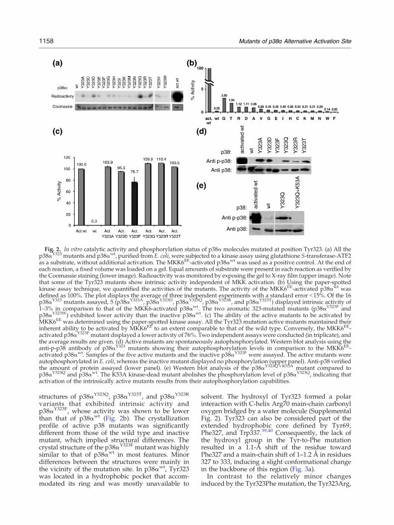
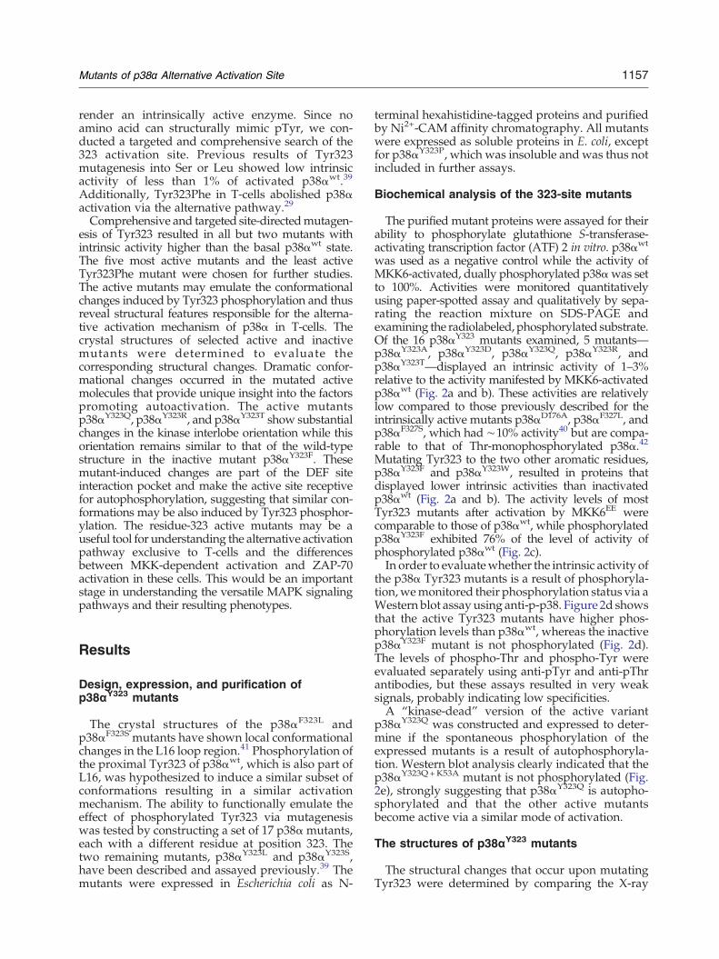
Fig. 2. In vitro catalytic activity and phosphorylation status of p38α molecules mutated at position Tyr323. (a) All thep38αY323 mutants and p38αwt, purified from E. coli, were subjected to a kinase assay using glutathione S-transferase-ATF2as a substrate, without additional activation. The MKK6EE-activated p38αwt was used as a positive control. At the end ofeach reaction, a fixed volume was loaded on a gel. Equal amounts of substrate were present in each reaction as verified bythe Coomassie staining (lower image). Radioactivity wasmonitored by exposing the gel to X-ray film (upper image). Notethat some of the Tyr323 mutants show intrinsic activity independent of MKK activation. (b) Using the paper-spottedkinase assay technique, we quantified the activities of the mutants. The activity of the MKK6EE-activated p38αwt wasdefined as 100%. The plot displays the average of three independent experiments with a standard error b15%. Of the 16p38αY323 mutants assayed, 5 (p38αY323A, p38αY323D, p38αY323Q, p38αY323R, and p38αY323T) displayed intrinsic activity of1–3% in comparison to that of the MKK6-activated p38αwt. The two aromatic 323-mutated mutants (p38αY323F andp38αY323W) exhibited lower activity than the inactive p38αwt. (c) The ability of the active mutants to be activated byMKK6EE was determined using the paper-spotted kinase assay. All the Tyr323 mutation active mutants maintained theirinherent ability to be activated by MKK6EE to an extent comparable to that of the wild type. Conversely, the MKK6EE-activated p38αY323F mutant displayed a lower activity of 76%. Two independent assays were conducted (in triplicate), andthe average results are given. (d) Active mutants are spontaneously autophosphorylated. Western blot analysis using theanti-p-p38 antibody of p38αY323 mutants showing their autophosphorylation levels in comparison to the MKK6EE-activated p38αwt. Samples of the five active mutants and the inactive p38αY323F were assayed. The active mutants wereautophosphorylated in E. coli, whereas the inactive mutant displayed no phosphorylation (upper panel). Anti-p38 verifiedthe amount of protein assayed (lower panel). (e) Western blot analysis of the p38αY323Q+K53A mutant compared top38αY323Q and p38αwt. The K53A kinase-dead mutant abolishes the phosphorylation level of p38αY323Q, indicating thatactivation of the intrinsically active mutants results from their autophosphorylation capabilities.
1158 Mutants of p38α Alternative Activation Site
structures of p38αY323Q, p38αY323T, and p38αY323R
variants that exhibited intrinsic activity andp38αY323F, whose activity was shown to be lowerthan that of p38αwt (Fig. 2b). The crystallizationprofile of active p38 mutants was significantlydifferent from those of the wild type and inactivemutant, which implied structural differences. Thecrystal structure of the p38αY323F mutant was highlysimilar to that of p38αwt in most features. Minordifferences between the structures were mainly inthe vicinity of the mutation site. In p38αwt, Tyr323was located in a hydrophobic pocket that accom-modated its ring and was mostly unavailable to
solvent. The hydroxyl of Tyr323 formed a polarinteraction with C-helix Arg70 main-chain carbonyloxygen bridged by a water molecule (SupplementalFig. 2). Tyr323 can also be considered part of theextended hydrophobic core defined by Tyr69,Phe327, and Trp337.39,40 Consequently, the lack ofthe hydroxyl group in the Tyr-to-Phe mutationresulted in a 1.1-Å shift of the residue towardPhe327 and a main-chain shift of 1–1.2 Å in residues327 to 333, inducing a slight conformational changein the backbone of this region (Fig. 3a).In contrast to the relatively minor changes
induced by the Tyr323Phe mutation, the Tyr323Arg,

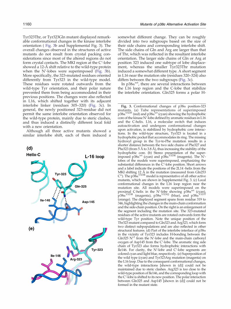
Fig. 3. Conformational changes of p38α position-323mutants. (a) Tube representations of superimposedp38αY323F (red) and p38αwt (cyan) showing the hydrophobiccore of the kinaseN′-lobe definedby aromatic residues in L16and the C-helix. L16, a molecular switch that inducesautoactivation and undergoes conformational changesupon activation, is stabilized by hydrophobic core interac-tions. In the wild-type structure, Tyr323 is located in ahydrophobic pocket that accommodates its ring. Themissing
1160 Mutants of p38α Alternative Activation Site
Tyr323Thr, or Tyr323Gln mutant displayed remark-able conformational changes in the kinase interlobeorientation ( Fig. 3b and Supplemental Fig. 3). Theoverall changes observed in the structures of activemutants do not result from crystal packing con-siderations since most of the altered regions do notform crystal contacts. The MKI region at the C′-lobeshowed a 12-Å shift relative to the wild-type proteinwhen the N′-lobes were superimposed (Fig. 3b).More specifically, the 323-mutated residues orienteddifferently from Tyr323 in the wild-type model.These residues were rotated outwards from thewild-type Tyr orientation, and their polar natureprevented them from being accommodated in theirprevious positions. The changes were also reflectedin L16, which shifted together with its adjacentinterlobe linker (residues 305–320) (Fig. 3c). Ingeneral, the newly positioned 323-residue did notpermit the same interlobe orientation observed forthe wild-type protein, mainly due to steric clashes,and thus induced a distinctly different local foldwith a new orientation.Although all three active mutants showed a
similar interlobe shift, each of them induced a
somewhat different change. They can be roughlydivided into two subgroups based on the size oftheir side chains and corresponding interlobe shift.The side chains of Gln and Arg are larger than thatof Thr, which was reflected in the resultant interlobeorientation. The larger side chains of Gln or Arg atposition 323 induced one subtype of lobe displace-ment, whereas the smaller Tyr323Thr mutationinduced a somewhat different type. A short segmentin L16 near the mutation site (residues 320–324) alsodiffers between the two subgroups (Fig. 3c).In p38αwt, there are several interactions between
the L16 loop region and the C-lobe that stabilizethe interlobe orientation. Gln325 forms a polar H-
hydroxyl group in the Tyr-to-Phe mutation results in ashorter distance between the two side chains of Phe327 andPhe323 (from 5Å to 3.8 Å), thus increasing the stability of thehydrophobic core. (b) Stereo presentation of the super-imposed p38αwt (cyan) and p38αY323R (magenta). The N′-lobes of the models were superimposed, emphasizing thesubstantial differences in the C′-lobe position. Short arrowsand a label indicate the positions of the 2L14 -helix from theMKI shifting 12 Å in the mutation (measured from Gln253Cα). The p38αY323R model is representative of all other activemutants, which are shown in Supplemental Fig. 3. (c) Localconformational changes in the L16 loop region near themutation site. All models were superimposed on theproximal C-helix in the N′-lobe showing p38αwt (cyan),p38αY323R (magenta), p38αY323Q (blue), and p38αY323T
(orange). The displayed segment spans from residue 319 to346, highlighting the changes in themain-chain conformationand the side-chain position. On the right is an enlargement ofthe segment including the mutation site. The 323-mutatedresidues of the active mutants are rotated outwards form thewild-type Tyr position. Note the unique position of theThr323mutant compared toGln323 andArg323,which formtwo distinct subpopulations and are also reflected in otherstructural features. (d) Part of the interlobe interface of p38αin the vicinity of Tyr323 includes H-bonding between theGln325 Nɛ2 from the N′-lobe and the main-chain carbonyloxygen of Asp145 from the C′-lobe. The aromatic ring sidechain of Tyr323 also forms hydrophobic interactions withIle146. For clarity, the N′-lobe and C′-lobe segments arecolored cyan and light blue, respectively. (e) Superposition ofthe wild type (cyan) and Tyr323Arg mutation (magenta) onthe L16 loop. Due to the consequent conformational changes,the wild-type interactions [shown in (d)] could not bemaintained due to steric clashes. Arg323 is too close to thewild-type position of Ile146, and the corresponding loopwiththeC′-lobe is shifted to its newposition. The polar interactionbetween Gln325 and Asp145 [shown in (d)] could not beformed in the mutant state.
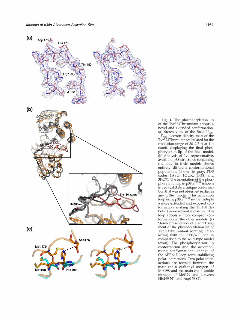
Fig. 4. The phosphorylation lipof the Tyr323Thr mutant adopts anovel and extended conformation.(a) Stereo view of the final 2Fobs−Fcalc electron density map of theTyr323Thrmutant calculated for theresolution range of 50–2.7 Å at 1 σcutoff, displaying the final phos-phorylation lip of the final model.(b) Analysis of five representative,available p38 structures containingthe loop in their models showsentirely different conformationalpopulations (shown in gray; PDBcodes 1A9U, 1OUK, 1P38, and3KQ7). The orientation of the phos-phorylation lip in p38αY323T (shownin red) exhibits a unique conforma-tion that was not observed earlier inany p38α model. The activationloop in the p38αY323Tmutant adoptsa more extended and exposed con-formation, making the Thr180 (la-beled) more solvent accessible. Thisloop adopts a more compact con-formation in the other models. (c)Stereo presentation of a short seg-ment of the phosphorylation lip ofTyr323Thr mutant (orange) inter-acting with the αEF/αF loop incomparison to the wild-type model(cyan). The phosphorylation lipconformation and the accompa-nying conformational change ofthe αEF/αF loop form stabilizingpolar interactions. Two polar inter-actions are formed between themain-chain carbonyl oxygen ofMet198 and the main-chain amidenitrogen of Met179 and betweenHis199 Nɛ1 and Asp176 Oδ.
1161Mutants of p38α Alternative Activation Site
1162 Mutants of p38α Alternative Activation Site
bond with Asp145 located in the loop connectingthe E-helix and β6 of the C′-lobe, residues 144–145. Additionally, Tyr323 forms a hydrophobicinteraction with Ile146 (from β6 of the C′-lobe)(Fig. 3d). These interactions are prevented by stericclashes due to the conformational changes in theL16 loop region upon mutating Tyr323 to Gln orArg (Fig. 3e). The shift in the interlobe orientationsatisfies steric requirements of the interlobe inter-face (Fig. 3e).The side chain of Thr323 in p38αY323T is too small
to induce the steric clashes described for the largerresidues; thus, the observed conformational changesprobably did not result solely from steric considera-tions. The structure of p38αY323T exhibited somelocal changes in the segment of residues 320–324(Fig. 3c). The distance between His77 Nɛ2 andAsp321 main-chain oxygen was too large (4.6 Å) tomaintain the hydrogen bond found in the wild typeand other position-323 active mutants, with anaverage distance of 2.8 Å (not shown). Since theL16 loop region is part of the segment connecting thelobes, the local change induced by Thr323 wasreflected in different interlobe orientations com-pared to that of p38wt.
The phosphorylation lip
The availability of the phosphorylation lip (resi-dues 171–183) also differs between the two 323active mutant subgroups in the model. The phos-phorylation lip region was not modeled in most ofthe available p38 structures due to its flexibility. Thecomplete loop segment of all but one of the 323active mutants could not be modeled since theelectron density map in this region was weak anddiscontinuous. Only p38αY323T had a clear andinterpretable electron density map in this regionand was modeled (Fig. 4a and b). However, theoverall trace of the loop of the p38αY323R/p38αY323Q
mutants resembled that of p38αY323T rather thanthat of available wild-type models. The differencesin the stabilization of the activation loops of the 323active mutants probably resulted from thecorresponding interlobe orientations. The shift ofthe lobe in p38αY323T was relatively larger than thatin p38αY323Q and p38αY323R. As a consequence, thenew and more exposed loop orientation inp38αY323T formed stabilizing interactions with theC-helix (residues 63–78), L16, and the short αEF/αFloop (residues 196–202), which followed the phos-phorylation lip (Fig. 4c). The phosphorylation lip ofp38αY323T adopted a unique exposed conformationthat has not previously been observed in any p38structure in which the loop was modeled. In thisconformation, the position of the Thr180 phos-phoacceptor is substantially shifted by 17 Å (be-tween Cα atoms) compared to the wild-type models(Fig. 4b).
The DEF site interaction pocket
The DEF site interaction pocket is located in the C-lobe and comprises residues from several segmentsincluding the loop connecting P+1 and F-helix(residues 203–217), which precedes the activationloop, the G-helix (residues 227–237), the MKI region(residues 240–262), and part of the phosphorylationlip (residues 180–182)36 (Fig. 5a). In the inactivep38α models, the DEF site interaction pocket isunavailable and thus unreceptive for substratebinding. The structures of the 323 active mutantsdisplayed conformational changes in the C′-lobe,which enabled the formation of the DEF siteinteraction pocket. More specifically, in thep38αY323T mutant, changes were observed in thephosphorylation lip where the Cα atoms of the TGYmotifs are shifted bymore than 17 Å to form the DEFsite rim close to the p38 active site (Fig. 5b). Inaddition, notable shifts are also observed in otherresidues contributing to the DEF site interactionpocket. In this regard, Leu195 and Trp197 move bymore than 5 Å (Cα distances) to form the pocketwhere other contributing residues are shifted bydistances between 1 and 3.5 Å (Fig. 5a and b). Thisformed a pocket with a hydrophobic cavity similarto that observed for the dually phosphorylatedERK237 (Fig. 5c). In the p38αY323Q and p38αY323R
active mutants, the changes and correspondingshifts in the C-lobe that form the DEF site interactionpocket were similar to those observed for thep38αY323T mutant (not shown). The general pocket-like shape of the DEF site lacked the rim formed bythe C-lobe due to the lack of the phosphorylation lipin the model.
Discussion
The canonic, well-characterized MAPK pathwayis one of the critical signaling cascades in alleukaryotic cells and is involved in most vital cellularprocesses. MAPK activation requires dual phos-phorylation on neighboring Thr and Tyr residues.This mode of activation is unique among kinasesthat commonly require monophosphorylation ofThr for activation.43 Most kinases are phosphory-lated and activated via autophosphorylation.43,44
Recent findings, primarily with p38, have sug-gested that MAPKs can also be activated viaautophosphorylation. The p38α alternative activa-tion pathways in T-cells may provide at least apartial explanation for the features promotingautoactivation. This study focused on the structuralchanges that occur upon mutating the alternativephosphorylation site Tyr323. The resulting struc-tures displayed remarkable details of the uniqueconformational changes that induce autoactivationof p38α and revealed the molecular basis of their

1163Mutants of p38α Alternative Activation Site
activity. Some of the mutants rendered the mole-cule intrinsically active at relatively low levels(1–3%) compared to the dually phosphorylatedform (normalized to 100%). These levels of activityare comparable to those of Thr180-monopho-sphorylated p38α.42,45 Interestingly, Tyr323 phos-phorylation leads to autoactivation in trans of p38and subsequent monophosphorylation of the acti-vation loop Thr180.28
Fig. 5 (legend
The X-ray structures of the mutants showed thatthe change from Tyr323 to Phe induced Phe327 andPhe323 to form hydrophobic interactions thatincreased the stability of the hydrophobic core. TheL16 loop region has been postulated to undergoconformational changes upon activation, and thishas been observed in ERK2 structures. In addition,we have previously shown that the L16 loop regionof p38α can be regarded as a molecular switch that
on next page)

Fig. 5. The DEF site interaction pocket in p38α and ERK2. (a) The segments defining the DEF site interaction pocket(highlighted in gray) indicated in the sequence alignment of p38α and ERK2. The individual residues that participate in theDEF site pocket are highlighted in gray and showa high level of similarity between the twoproteins. In p38α and ERK2, thepocket is defined by selected hydrophobic residues that came from the phosphorylation lip, a loop that follows thephosphorylation lip, the G-helix, and theMKI region. (b) Surface presentation of p38αwt (cyan, left; adopted from the 1P38model) and p38αY323T (orange, right) highlighting the residues forming the DEF site interaction pocket. The residuescontributing to the pocket are highlighted in gray, and TGY from the phosphorylation lip are in red. For clarity, only the C′-lobe of the molecular surface is displayed. Tyr323Thr mutation forms the DEF site interaction pocket, which canaccommodate hydrophobic residues to stabilize its substrate. In the p38αY323R and p38αY323Q mutants, thephosphorylation loop was not modeled, yet the cavity indicating the DEF site interaction pocket is also formed. Labelsindicate the positions of selected residues contributing to the DEF site. (c) The formation of the DEF site interaction pocketwas initially observed upon conformational changes consequent to activation of ERK2. The inactive form (left) and thedually phosphorylated form (right) indicate the formation of the hydrophobic pocket. The residues contributing to thepocket and the TEYphosphoacceptors are colored gray and red, respectively. Selected residues contributing to theDEF siteare indicated by labels. (d) Sequence alignment of the segments including the Thr phosphoacceptor in p38α and threeselected substrates (ATF2, SAP1, and Elk1). A gap of 6–12 residues between the Thr phosphoacceptor and an aromatic sidechain that could be accommodated in the DEF site interaction pocket is shown, although larger gaps are also allowed. (e)Cartoon of the conformational changes induced in the Tyr323 active mutants (pink, right) compared to p38αwt (gray, left).The active mutants induce substantial changes in the kinase interlobe orientation, which, together with the unique andextended conformation of the activation loop, contribute to the formation of the additional substrate DEF site interactionpocket. These changes may confer an appropriate conformation for autophosphorylation in trans as for pTyr323 in p38α.
1164 Mutants of p38α Alternative Activation Site
upon conformational changes induces auto-activation.46 The increased stability of the hydro-phobic core presumably hinders the requiredconformational changes upon activation in thisregion, which consequently results in lower activity.The increased stability of the L16 hydrophobic corewas also reflected in the lower activity (76%) of thedually phosphorylated form of p38αY323F (Fig. 2c).Unlike p38αY323F, the structures of intrinsically
active mutants p38αY323Q, p38αY323R, and p38αY323T
displayed dramatic conformational changes thatmay indicate factors promoting their activities.Despite differences in their chemical nature, their
induced molecular structural changes are similar.The most notable change is the unique interlobeorientation, probably making the kinase active sitemore substrate receptive. Our initial assumptionwasthat phospho-Tyr323 induces local conformationalchanges similar to those observed for L16 in thep38αF327L and p38αF327S active mutants, suggestinga similar mechanism.46 However, the 323 activemutants displayed substantially different changes,both locally (L16 loop) and globally, probablyindicating a different activation mechanism. Itcould be thus suggested that Tyr323 restrains L16in a conformation, maintaining the low basal activity
1165Mutants of p38α Alternative Activation Site
of p38α by preventing autophosphorylation. Suchextreme structural changes have been observed forphosphorylated MAPKs. Only two structures, ERK2and p38γ, have dually phosphorylated forms whereonly ERK2 is available in both the inactive and theactive modes.37,47 Upon phosphorylation, the twophosphoacceptors in ERK2 and p38γ form a networkof polar interactions, resulting in a new interlobeorientation that induces a conformational change ofthe activation loop and consequently exposes theMAPK active site to substrates.The conformation of the activation loop in
p38αY323T may provide another clue to its intrinsicactivity. In most p38α structures, the activation loophas no defined conformation. In p38αmodels wherethe loop is defined, all conformations differ sub-stantially from those observed in the p38αY323T
mutant described here (Fig. 4b). The conformationof the p38αY323T activation loop displayed higheravailability and accessibility of the p38α active site,which may account for its intrinsic activity. Similarconformational changes in L16 and resulting ex-posed activation loop have also been observed inthe structures of ERK2 complexed with shortdocking-site binding peptides. Upon peptide bind-ing, conformational changes occur in the L16 region,near the common docking motif, resulting in anextended conformation of the activation loop thatdiffers from both phosphorylated and non-phos-phorylated forms.48In p38αY323T, the unique conformation of the
phosphorylation lip also contributed to the DEF siteinteraction pocket, which has been identified as anadditional docking site in MAPKs.32,36 This site wasinitially identified in ERK2 and is formed as part ofthe conformational changes occurring uponactivation.37 This distinctive docking motif is as-sumed to accommodate large hydrophobic oraromatic side chains 6–20 residues downstream tothe phosphoacceptor with high selectivity towardtranscription factors.49–52 It has been recentlyshown, using substrate-derived peptides, thatp38α and p38β also contain a DEF site interactionpocket.36 Additionally, mutagenesis of SAP-1 tran-scription factor on the DEF site binding regionunequivocally verifies its significance for efficientphosphorylation by p38α35 (Fig. 5a).Structural analysis of the active mutants provided
the first substantiation for the formation of the DEFinteraction site in p38α. In the 323 active mutants,the DEF site interaction pocket, formed by changesin the C′-lobe, could promote autophosphorylationby accommodating part of the now extended andavailable phosphorylation lip of another p38αmolecule. The sequence of the phosphorylation lipof p38α includes aromatic side chains (Trp187 andTyr188) seven and eight residues away from theThr180 phosphoacceptor (Fig. 5a). These aromaticresidues from the lip may be anchored in the DEF
site pocket where, consequently, Thr180 becomesembedded in the active site near the catalyticresidues and consequently phosphorylated, render-ing the molecule active.It has been repeatedly shown that Asp and Glu
can at least partially functionally replace thephosphorylated state of Ser and Thr.53,54 However,there are no standard amino acids that are structur-ally and chemically similar to phospho-Tyr. Wewere able to functionally emulate pTyr323 in p38αby imposing conformational changes that promoteautoactivation. The combined features of the inter-lobe orientation, active-site components, phosphor-ylation lip conformation, and formation of the DEFsite interaction pocket all provided a groundbreak-ing view into the unique activation state of p38α thatis probably attained upon Tyr323 phosphorylationand leads to auto-monophosphorylation of Thr180(Fig. 5e). Differences between the monophosphory-lated and the dually phosphorylated forms presum-ably dictate substrate selectivity, resulting indifferent cellular phenotypes. These mutants areunique candidates for studying p38 activation in T-cells as well as in other cell systems.
Materials and Methods
Site-directed mutagenesis of the p38α mutants
Site-directed mutagenesis of the p38αY323 site wascarried out using a QuickChange kit (Stratagene). Muta-genesis was performed on the human p38αwt cDNA sub-cloned into pET-28a (Novagen) vector downstream andin-frame with the hexahistidine coding sequence. Theprocedure included polymerase chain reaction usingspecific complementary primers (Supplemental Table 1).Mutagenesis of the p38αY323Q+K53A was performed on thepET-28a vector containing p38αY323Q as a template usingspecific complementary primers (Supplemental Table 1).All mutated cDNAs were verified by sequencing of theentire p38α cDNA.
Paper-spotted kinase assay and Western blot
Cell cultures (0.5 L) were grown at 30–32 °C in order toobtain phosphorylated and active mutants. Proteinexpression and purification, as the in vitro kinase assay,were conducted in triplicate as previously described.40 Inparallel, for a quality assay, samples from the paper-spotted kinase reactions were mixed by applying Laemmlisample buffer and boiling at 100 °C for 5 min. The assaysamples were run on SDS-PAGE, stained with Coomassiestaining, and then exposed to X-ray film for ∼6 h withintensifier.For the Western blot assay, 0.3 μg of purified recombi-
nant protein was heated at 100 °C for 5–10 min, separatedby SDS-PAGE, and then transferred to a nitrocellulosemembrane. After incubating the membrane with theappropriate antibodies, we visualized specific proteins
1166 Mutants of p38α Alternative Activation Site
using an enhanced chemiluminescence detection reagentand thenmonitored themby exposingmembranes to X-rayfilm and by densitometry. Goat anti-p38 was obtainedfrom Santa Cruz Biotechnology and rabbit anti-phospho-p38 was obtained from Cell Signaling.
Crystallization of p38α mutants
For crystallization purposes, cell cultures (3–6 L) weregrown at 21 °C, resulting in homogeneous non-phosphor-ylated protein as previously described.46 Expression athigher temperatures (30–32 °C) yielded an increasedamount of (heterogeneously phosphorylated) protein,which was unsuitable for crystallization assays as oc-curred previously for other p38α active mutants.41,46
Expression and purification protocols for crystallizationpurposes were conducted as previously described.46 Theposition-323 mutants were crystallized by applyingconditions similar to those obtained for p38αwt.41,46
Subsequently, as previously described, the streak-seedingmethod was employed to induce crystallization usingwild-type crystals as the source of microseeds.55 Crystalsfor all mutants were obtained using the vapor diffusionsitting-drop method at 4 °C with a 6-μL crystallizationdroplet containing equal amounts of protein and reservoirsolution. Crystallization assays of the p38αY323F inactivemutant were conducted using 1 ml reservoir solutioncontaining 17% (w/v) polyethylene glycol 3350, 0.1 MHepes (pH 7.25), 0.2 M potassium fluoride, and 25 mM n-
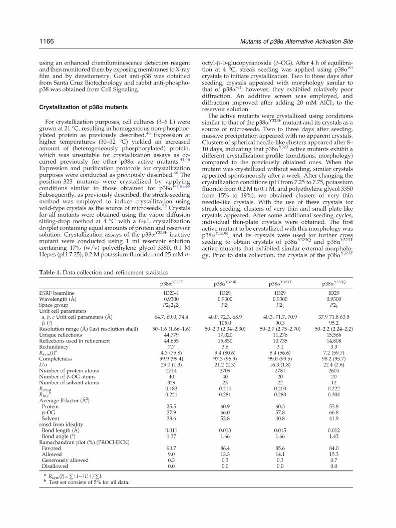
Table 1. Data collection and refinement statistics
p38αY323F
ESRF beamline ID23-1Wavelength (Å) 0.9300Space group P212121Unit cell parametersa, b, c Unit cell parameters (Å) 64.7, 69.0, 74.4β (°)Resolution range (Å) (last resolution shell) 50–1.6 (1.66–1.6)Unique reflections 44,779Reflections used in refinement 44,655Redundancy 7.7Rsym(I)
a 4.3 (75.8)Completeness 99.9 (99.4)I/σ 29.0 (1.3)Number of protein atoms 2714Number of β-OG atoms 40Number of solvent atoms 329Rcryst 0.183Rfree
b 0.221Average B-factor (Å2)Protein 25.5β-OG 27.9Solvent 38.6rmsd from idealityBond length (Å) 0.011Bond angle (°) 1.37Ramachandran plot (%) (PROCHECK)Favored 90.7Allowed 9.0Generously allowed 0.3Disallowed 0.0a Rsym(I)=∑|I− ⟨I⟩|/∑I.b Test set consists of 5% for all data.
octyl-β-D-glucopyranoside (β-OG). After 4 h of equilibra-tion at 4 °C, streak seeding was applied using p38αwt
crystals to initiate crystallization. Two to three days afterseeding, crystals appeared with morphology similar tothat of p38αwt; however, they exhibited relatively poordiffraction. An additive screen was employed, anddiffraction improved after adding 20 mM AlCl3 to thereservoir solution.The active mutants were crystallized using conditions
similar to that of the p38αY323F mutant and its crystals as asource of microseeds. Two to three days after seeding,massive precipitation appeared with no apparent crystals.Clusters of spherical needle-like clusters appeared after 8–10 days, indicating that p38αY323 active mutants exhibit adifferent crystallization profile (conditions, morphology)compared to the previously obtained ones. When themutant was crystallized without seeding, similar crystalsappeared spontaneously after a week. After changing thecrystallization conditions (pH from 7.25 to 7.75, potassiumfluoride from 0.2 M to 0.1 M, and polyethylene glycol 3350from 15% to 19%), we obtained clusters of very thinneedle-like crystals. With the use of these crystals forstreak seeding, clusters of very thin and small plate-likecrystals appeared. After some additional seeding cycles,individual thin-plate crystals were obtained. The firstactive mutant to be crystallized with this morphology wasp38αY323R, and its crystals were used for further crossseeding to obtain crystals of p38αY323Q and p38αY323T
active mutants that exhibited similar external morpholo-gy. Prior to data collection, the crystals of the p38αY323F
p38αY323R p38αY323T p38αY323Q
ID29 ID29 ID290.9300 0.9300 0.9300P21 P21 P21
40.0, 72.3, 68.9 40.3, 71.7, 70.9 37.9 71.8 63.5105.0 90.3 95.2
50–2.3 (2.34–2.30) 50–2.7 (2.75–2.70) 50–2.2 (2.24–2.2)17,020 11,276 15,56615,850 10,735 14,8083.6 3.1 3.3
9.4 (80.6) 8.4 (56.6) 7.2 (59.7)97.3 (96.9) 99.0 (99.5) 98.2 (95.7)21.2 (2.3) 16.3 (1.8) 22.4 (2.6)2709 2781 260440 20 2023 22 12
0.214 0.200 0.2220.281 0.283 0.304
60.9 60.3 55.866.0 57.8 66.852.8 40.8 41.9
0.013 0.015 0.0121.66 1.66 1.43
86.4 85.6 84.013.3 14.1 15.30.3 0.3 0.70.0 0.0 0.0
1167Mutants of p38α Alternative Activation Site
mutant were cryo-protected using Paratone-N oil andimmediately flash cooled in liquid nitrogen. Thep38αY323Q, p38αY323R, and p38αY323T crystals were cryo-protected using the corresponding reservoir solution with20% glycerol.
Crystallographic data collection and refinement
Crystallographic data for all p38αY323 mutants werecollected at the European Synchrotron Radiation Facility(ESRF) (see Table 1). Datawere integrated and scaled usingthe HKL suite.56 The crystals of the p38αY323F belonged tothe orthorhombic P212121 space group, and the crystals ofthe p38αY323Q, p38αY323R, and p38αY323T mutantsbelonged to the monoclinic P21 space group, with onep38αmolecule in the asymmetric unit for both symmetries(Table 1). The active p38 mutants exhibited some differ-ences in their corresponding cell parameters (Table 1).Most of the thin-plate crystals of the active mutantsdisplayed poor diffraction, and better-diffracting crystalscould be identified only via extensive screening afterwhichcomplete data sets were collected. The structure of thep38αY323F mutant was solved via molecular replacementmethods using Molrep57 implemented in CCP4i usingp38α [Protein Data Bank (PDB) code 2FSO46] as the searchmodel after removing all solvent and detergent molecules.The solution resulted in an R value of 0.46 and a score of0.56 in the resolution range of 50–4.0 Å. The initial Fobs−Fcalc and 2Fobs−Fcalc electron density maps were calcu-lated after 10 cycles of restrained refinement usingREFMAC5.58 The structure was further refined in theresolution range of 50–1.6 Å using PHENIX,59 and solventmolecules were added utilizing ARP/wARP.60 The struc-ture was fitted into electron density maps using thegraphics program Coot.61 The final p38αY323F model(Rcryst=19.7; Rfree=24.6) consists of residues 5–175, 185–263, and 267–352, with 380 solvent molecules and 2 β-OGmolecules (Table 1).The structure of the p38αY323R mutant was solved via
molecular replacement methods using Molrep with thestructure of p38αY323F as the search model. The solutionresulted in an Rcryst of 0.48 and a score of 0.42 in theresolution range of 66–4.0 Å. The solved structure wasthen refined by REFMAC558 using the rigid-body protocoldefining the two kinase lobes as different domains(domain 1 included residues 5–110 and 320–352; domain2 included residues 111–319) at the resolution range of 50–4.0 Å. Rcryst was reduced from 47.2% to 44.1% after 20cycles of refinement. Consequently, the model was refinedusing the REFMAC restrained refinement option in theresolution range of 50.0–2.3 Å, and Rcryst decreased from44.1% to 30.0%. The structure was further refined usingPHENIX,59 and solvent molecules were added utilizingARP/wARP. The structure was fitted into electron densitymaps using the graphics program Coot. The p38αY323R
model consists of residues 4–31, 37–172, and 183–353, with24 solvent molecules and 2 β-OG molecules (Table 1). Thestructures of the p38αY323Q and p38αY323T mutants weresolved via molecular replacement methods using modelp38αY323R as a search model and refined by a similarprotocol described above. For all 323 mutant structures,the initial electron density maps clearly indicated thepresence of the mutated residue and their conformation(not shown). The p38αY323T model was refined in the
resolution limits of 50–2.7 Å using PHENIX59 to a finalRcryst of 20.0% (Rfree=28.3%) and consists of residues 4–31and 37–352, with 24 solvent molecules and 1 β-OGmolecule (Table 1). The p38αY323Q model was refined inthe resolution limits of 50.0–2.2 Å to a final Rcryst of 22.2%(Rfree=30.4%) consisting of residues 4–31, 37–172, 183–242, and 257–353, with 14 solvent molecules and 1 β-OGmolecule (Table 1).
PDB accession codes
PDB coordinates and structure factors have beendeposited with accession codes 3OD6, 3ODY, 3ODZ,and 3OEF for the Y323T, Y323Q, Y323R, and Y323Fmutants, respectively.
Acknowledgements
This research was supported by the Israel ScienceFoundation grant 630/07 and Israel Science Foun-dation Research Center of Excellence 180/09awarded to O.L. and D.E. We would like to thankDr. Mario Lebendiker from the Protein PurificationUnit of the Wolfson Centre for Applied StructuralBiology for his continuous helpful advice andassistance and to Dr. Deborah E. Shalev for help inconstructing Fig. 5e and for insightful discussions.We would like to thank the staff of ESRF (Grenoble,France) for their outstanding help and for maintain-ing and upgrading the facility.
Supplementary Data
Supplementary data to this article can be foundonline at doi:10.1016/j.jmb.2010.11.023
References
1. Widmann, C., Gibson, S., Jarpe, M. B. & Johnson, G. L.(1999). Mitogen-activated protein kinase: conservationof a three-kinase module from yeast to human. Physiol.Rev. 79, 143–180.
2. Jiang, Y., Gram, H., Zhao, M., New, L., Gu, J., Feng, L.et al. (1997). Characterization of the structure andfunction of the fourth member of p38 group mitogen-activated protein kinases, p38delta. J. Biol. Chem. 272,30122–30128.
3. Enslen, H., Brancho, D. M. & Davis, R. J. (2000).Molecular determinants that mediate selective activa-tion of p38 MAP kinase isoforms. EMBO J. 19,1301–1311.
4. Wang, X. S., Diener, K., Manthey, C. L., Wang, S.,Rosenzweig, B., Bray, J. et al. (1997). Molecular cloningand characterization of a novel p38 mitogen-activatedprotein kinase. J. Biol. Chem. 272, 23668–23674.
5. Eckert, R. L., Efimova, T., Balasubramanian, S., Crish,J. F., Bone, F. &Dashti, S. (2003). p38mitogen-activated
1168 Mutants of p38α Alternative Activation Site
protein kinases on the body surface—a function forp38 delta. J. Invest. Dermatol. 120, 823–828.
6. Hu, M. C., Wang, Y. P., Mikhail, A., Qiu, W. R. & Tan,T. H. (1999). Murine p38-delta mitogen-activatedprotein kinase, a developmentally regulated proteinkinase that is activated by stress and proinflammatorycytokines. J. Biol. Chem. 274, 7095–7102.
7. Shi, Y. & Gaestel, M. (2002). In the cellular garden offorking paths: how p38 MAPKs signal for down-stream assistance. Biol. Chem. 383, 1519–1536.
8. Lee, J. C., Laydon, J. T., McDonnell, P. C., Gallagher,T. F., Kumar, S., Green, D. et al. (1994). A proteinkinase involved in the regulation of inflammatorycytokine biosynthesis. Nature, 372, 739–746.
9. Han, J., Lee, J. D., Bibbs, L. & Ulevitch, R. J. (1994). AMAP kinase targeted by endotoxin and hyperosmo-larity in mammalian cells. Science, 265, 808–811.
10. Haq, R. & Zanke, B. (1998). Inhibition of apoptoticsignaling pathways in cancer cells as a mechanism ofchemotherapy resistance. Cancer Metastasis Rev. 17,233–239.
11. Harkin, D. P., Bean, J. M., Miklos, D., Song, Y. H.,Truong, V. B., Englert, C. et al. (1999). Induction ofGADD45 and JNK/SAPK-dependent apoptosis fol-lowing inducible expression of BRCA1. Cell, 97,575–586.
12. Recio, J. A. & Merlino, G. (2002). Hepatocyte growthfactor/scatter factor activates proliferation in melano-ma cells through p38 MAPK, ATF-2 and cyclin D1.Oncogene, 21, 1000–1008.
13. Lee, J. C., Kumar, S., Griswold, D. E., Underwood, D.C., Votta, B. J. & Adams, J. L. (2000). Inhibition of p38MAP kinase as a therapeutic strategy. Immunopharma-cology, 47, 185–201.
14. English, J. M. & Cobb, M. H. (2002). Pharmacologicalinhibitors of MAPK pathways. Trends Pharmacol. Sci.23, 40–45.
15. Jackson, P. F. & Bullington, J. L. (2002). Pyridinylimi-dazole based p38 MAP kinase inhibitors. Curr. Top.Med. Chem. 2, 1011–1020.
16. Kumar, S., Boehm, J. & Lee, J. C. (2003). p38 MAPkinases: key signalling molecules as therapeutictargets for inflammatory diseases. Nat. Rev., DrugDiscov. 2, 717–726.
17. Clerk, A., Michael, A. & Sugden, P. H. (1998).Stimulation of multiple mitogen-activated proteinkinase sub-families by oxidative stress and phosphor-ylation of the small heat shock protein, HSP25/27, inneonatal ventricularmyocytes. Biochem. J. 333, 581–589.
18. Hazzalin, C. A., Cano, E., Cuenda, A., Barratt, M. J.,Cohen, P. & Mahadevan, L. C. (1996). p38/RK isessential for stress-induced nuclear responses: JNK/SAPKs and c-Jun/ATF-2 phosphorylation are insuffi-cient. Curr. Biol. 6, 1028–1031.
19. Ono, K. & Han, J. (2000). The p38 signal transductionpathway: activation and function. Cell Signalling, 12,1–13.
20. Pugazhenthi, S., Boras, T., O'Connor, D., Meintzer,M. K., Heidenreich, K. A. & Reusch, J. E. (1999).Insulin-like growth factor I-mediated activation of thetranscription factor cAMP response element-bindingprotein in PC12 cells. Involvement of p38 mitogen-activated protein kinase-mediated pathway. J. Biol.Chem. 274, 2829–2837.
21. Xing, J., Kornhauser, J. M., Xia, Z., Thiele, E. A. &Greenberg, M. E. (1998). Nerve growth factor activatesextracellular signal-regulated kinase and p38 mito-gen-activated protein kinase pathways to stimulateCREB serine 133 phosphorylation. Mol. Cell. Biol. 18,1946–1955.
22. Kyriakis, J. M. & Avruch, J. (2001). Mammalianmitogen-activated protein kinase signal transductionpathways activated by stress and inflammation.Physiol. Rev. 81, 807–869.
23. DeLano, W. L. (2002). The PyMOL Molecular GraphicsSystem. DeLano Scientific, San Carlos, CA.
24. Zarubin, T. & Han, J. (2005). Activation and signalingof the p38 MAP kinase pathway. Cell Res. 15, 11–18.
25. Ge, B., Gram, H., Di Padova, F., Huang, B., New, L.,Ulevitch, R. J. et al. (2002). MAPKK-independentactivation of p38alpha mediated by TAB1-dependentautophosphorylation of p38alpha. Science, 295,1291–1294.
26. Gills, J. J., Castillo, S. S., Zhang, C., Petukhov, P. A.,Memmott, R. M., Hollingshead, M. et al. (2007).Phosphatidylinositol ether lipid analogues that inhibitAKT also independently activate the stress kinase,p38alpha, through MKK3/6-independent and-dependent mechanisms. J. Biol. Chem. 282,27020–27029.
27. Salvador, J. M., Mittelstadt, P. R., Guszczynski, T.,Copeland, T. D., Yamaguchi, H., Appella, E. et al.(2005). Alternative p38 activation pathway mediatedby T cell receptor-proximal tyrosine kinases. Nat.Immunol. 6, 390–395.
28. Mittelstadt, P. R., Yamaguchi, H., Appella, E. &Ashwell, J. D. (2009). T cell receptor-mediatedactivation of p38α by mono-phosphorylation of theactivation loop results in altered substrate specificity.J. Biol. Chem. 284, 15469–15474.
29. Jirmanova, L., Sarma, D. N., Jankovic, D., Mittelstadt,P. R. & Ashwell, J. D. (2009). Genetic disruption ofp38alpha Tyr323 phosphorylation prevents T-cellreceptor-mediated p38alpha activation and impairsinterferon-gamma production. Blood, 113, 2229–2237.
30. Wilson, K. P., Fitzgibbon, M. J., Caron, P. R.,Griffith, J. P., Chen, W., McCaffrey, P. G. et al.(1996). Crystal structure of p38 mitogen-activatedprotein kinase. J. Biol. Chem. 271, 27696–27700.
31. Zhang, F., Strand, A., Robbins, D., Cobb, M. H. &Goldsmith, E. J. (1994). Atomic structure of the MAPkinase ERK2 at 2.3 Å resolution. Nature, 367, 704–711.
32. Lee, T., Hoofnagle, A. N., Kabuyama, Y., Stroud, J.,Min, X., Goldsmith, E. J. et al. (2004). Docking motifinteractions in MAP kinases revealed by hydrogenexchange mass spectrometry. Mol. Cell, 14, 43–55.
33. Tanoue, T., Adachi, M., Moriguchi, T. & Nishida, E.(2000). A conserved docking motif in MAP kinasescommon to substrates, activators and regulators. Nat.Cell Biol. 2, 110–116.
34. Chang, C. I., Xu, B. E., Akella, R., Cobb, M. H. &Goldsmith, E. J. (2002). Crystal structures of MAPkinase p38 complexed to the docking sites on itsnuclear substrate MEF2A and activator MKK3b. Mol.Cell, 9, 1241–1249.
35. Galanis, A., Yang, S. H. & Sharrocks, A. D. (2001).Selective targeting of MAPKs to the ETS domaintranscription factor SAP-1. J. Biol. Chem. 276, 965–973.
1169Mutants of p38α Alternative Activation Site
36. Sheridan, D. L., Kong, Y., Parker, S. A., Dalby, K. N.& Turk, B. E. (2008). Substrate discriminationamong mitogen-activated protein kinases throughdistinct docking sequence motifs. J. Biol. Chem. 283,19511–19520.
37. Canagarajah, B. J., Khokhlatchev, A., Cobb, M. H. &Goldsmith, E. J. (1997). Activation mechanism of theMAP kinase ERK2 by dual phosphorylation. Cell, 90,859–869.
38. Bell, M., Capone, R., Pashtan, I., Levitzki, A. &Engelberg, D. (2001). Isolation of hyperactivemutants of the MAPK p38/Hog1 that are indepen-dent of MAPK kinase activation. J. Biol. Chem. 276,25351–25358.
39. Avitzour, M., Diskin, R., Raboy, B., Askari, N.,Engelberg, D. & Livnah, O. (2007). Intrinsically activevariants of all human p38 isoforms. FEBS J. 274,963–975.
40. Diskin, R., Askari, N., Capone, R., Engelberg, D. &Livnah, O. (2004). Active mutants of the humanp38alpha mitogen-activated protein kinase. J. Biol.Chem. 279, 47040–47049.
41. Diskin, R., Engelberg, D. & Livnah, O. (2007). High-resolution diffracting crystals of intrinsically activep38alpha MAP kinase: a case study for low-through-put approaches. Acta Crystallogr., Sect. D: Biol. Crystal-logr. 63, 260–265.
42. Askari, N., Beenstock, J., Livnah, O. & Engelberg, D.(2009). p38alpha is active in vitro and in vivo whenmonophosphorylated at threonine 180. Biochemistry,48, 2497–2504.
43. Huse, M. & Kuriyan, J. (2002). The conformationalplasticity of protein kinases. Cell, 109, 275–282.
44. Taylor, S. J. & Shalloway, D. (1994). An RNA-bindingprotein associated with Src through its SH2 and SH3domains in mitosis. Nature, 368, 867–871.
45. Zhang, Y. Y., Mei, Z. Q., Wu, J. W. & Wang, Z. X.(2008). Enzymatic activity and substrate specificity ofmitogen-activated protein kinase p38alpha in differ-ent phosphorylation states. J. Biol. Chem. 283,26591–26601.
46. Diskin, R., Lebendiker, M., Engelberg, D. & Livnah, O.(2007). Structures of p38alpha active mutants revealconformational changes in L16 loop that induceautophosphorylation and activation. J. Mol. Biol. 365,66–76.
47. Bellon, S., Fitzgibbon, M. J., Fox, T., Hsiao, H. M. &Wilson, K. P. (1999). The structure of phosphory-lated p38gamma is monomeric and reveals aconserved activation-loop conformation. Structure,7, 1057–1065.
48. Zhou, T., Sun, L., Humphreys, J. & Goldsmith, E. J.(2006). Docking interactions induce exposure ofactivation loop in the MAP kinase ERK2. Structure,14, 1011–1019.
49. Jacobs, D., Glossip, D., Xing, H., Muslin, A. J. &Kornfeld, K. (1999). Multiple docking sites on sub-strate proteins form a modular system that mediatesrecognition by ERK MAP kinase. Genes Dev. 13,163–175.
50. Murphy, L. O., Smith, S., Chen, R. H., Fingar, D. C. &Blenis, J. (2002). Molecular interpretation of ERKsignal duration by immediate early gene products.Nat. Cell Biol. 4, 556–564.
51. Murphy, L. O., MacKeigan, J. P. & Blenis, J. (2004). Anetwork of immediate early gene products propagatessubtle differences in mitogen-activated protein kinasesignal amplitude and duration. Mol. Cell. Biol. 24,144–153.
52. Vinciguerra, M., Vivacqua, A., Fasanella, G., Gallo, A.,Cuozzo, C., Morano, A. et al. (2004). Differentialphosphorylation of c-Jun and JunD in response to theepidermal growth factor is determined by thestructure of MAPK targeting sequences. J. Biol.Chem. 279, 9634–9641.
53. Cowley, S., Paterson, H., Kemp, P. & Marshall, C. J.(1994). Activation of MAP kinase kinase is necessaryand sufficient for PC12 differentiation and fortransformation of NIH 3T3 cells. Cell, 77, 841–852.
54. Dean, A. M., Lee, M. H. & Koshland, D. E., Jr (1989).Phosphorylation inactivates Escherichia coli isocitratedehydrogenase by preventing isocitrate binding. J.Biol. Chem. 264, 20482–20486.
55. Stura, E. A., Chen, P., Wilmot, C. M., Arevalo, J. H. &Wilson, I. A. (1992). Crystallization studies of glyco-sylated and unglycosylated human recombinantinterleukin-2. Proteins, 12, 24–30.
56. Otwinowski, Z. & Minor, W. (1997). Processing of X-ray diffraction data collected in oscillation mode.Methods Enzymol. 276, 307–326.
57. Vagin, A. A. & Isupov, M. N. (2001). Sphericallyaveraged phased translation function and its applica-tion to the search for molecules and fragments inelectron-density maps. Acta Crystallogr., Sect. D: Biol.Crystallogr. 57, 1451–1456.
58. Murshudov, G. N., Vagin, A. A. & Dodson, E. J.(1997). Refinement of macromolecular structures bythe maximum-likelihood method. Acta Crystallogr.,Sect. D: Biol. Crystallogr. 53, 240–255.
59. Adams, P. D., Afonine, P. V., Bunkoczi, G., Chen,V. B., Davis, I. W., Echols, N. et al. (2010). PHENIX:a comprehensive Python-based system for macromo-lecular structure solution. Acta Crystallogr., Sect. D:Biol. Crystallogr. 66, 213–221.
60. Morris, R. J., Perrakis, A. & Lamzin, V. S. (2003). ARP/wARP and automatic interpretation of protein elec-tron density maps. Methods Enzymol. 374, 229–244.
61. Emsley, P. & Cowtan, K. (2004). Coot: model-buildingtools for molecular graphics. Acta Crystallogr., Sect. D:Biol. Crystallogr. 60, 2126–2132.