activation of oxygen nucleophiles in enzyme catalysis · nucleophilicity. the size and direction of...
TRANSCRIPT

Activation of Oxygen Nucleophiles in Enzyme Catalysis
Vernon E. Anderson,*,† Mark W. Ruszczycky,† and Michael E. Harris†,‡
Department of Biochemistry and the Center for RNA Molecular Biology, Case Western Reserve University, School of Medicine,Cleveland, Ohio 44106
Received January 17, 2006
Contents1. Enhancing Alcohol Nucleophilicity 3236
1.1. Introduction 32361.2. Effect of Variation of Electron Density on
Alcohol Nucleophilicity3236
1.3. Desolvation 32371.4. H-Bonding to a General Base 32381.5. Ionization 3239
2. Nucleophilic Activation of Alcohols by Enzymes 32402.1. Cyclic-AMP-Dependent Protein Kinase 32402.2. Serine O-Acetyltransferase 32412.3. Hexokinase 32422.4. Serine Proteases 32432.5. Catechol O-Methyltransferase 32442.6. Ribozyme Catalysis 32442.7. Group I Intron 32452.8. HDV Ribozyme 3247
3. Spectroscopic Characterization of Nucleophiles atEnzyme Active Sites
3248
3.1. Vibrational Spectroscopic Characterization ofAlcohols
3248
3.2. NMR Characterization of AlcoholNucleophiles
3248
3.3. Isotope Effects on Alcohol Nucleophiles 32484. Acknowledgments 32495. References 3249
1. Enhancing Alcohol Nucleophilicity
1.1. IntroductionGroup transfer reactions, as shown in eq 1, represent a
large class of enzyme catalyzed reactions.
In these reactions, the acceptor forms a new bond to thetransferred group. When the bonding electrons are derivedfrom the acceptor, it functions as a nucleophile in thereaction, while the atom of the transferred group is theelectrophile, and the atom whose bond is cleaved constitutesthe leaving group. Physical organic chemists have developednumerous tools that can be used to dissect the contributionsof each of these three functionalities to the reactivity of the
system. This review focuses on the application of thesemethods to enzyme reactions, to ascertain how the environ-ment of the enzyme active site affects the reactivity ofoxygen nucleophiles, with an emphasis on alcohols.
On the simplest level, there should be minimal variationin the nucleophilicity of different oxygen atoms; in everybiologically relevant structure, every oxygen will have twoaccessible lone pairs of electrons, one of which would serveas the nucleophilic lone pair. This assertion, however, isdemonstrably false, as the second-order rate constants forthe reaction of oxygen nucleophiles can vary over 7 ordersof magnitude in the standard displacement of halides fromCH3X.1,2 Qualitatively, the fundamental mechanism ofenhancing the nucleophilic power of any given oxygen atomis to enhance the electron density of the nucleophilic loneelectron pair. This leads to three molecular mechanisms ofgenerating enhanced electron density in the reactant state ofthe nucleophile by altering the coordination and/or bondingof the nucleophile: (1) desolvation, or more precisely thestripping of H-bond donors to the nucleophilic lone pair, (2)coordination of the alcohol proton, i.e., H-bonding to ageneral base, and (3) ionization, as shown in Figure 1. Inaddition to these three mechanisms, an electric field generatedat the active site can polarize a bond to the nucleophile,enhancing its nucleophilicity. Moreover, enzymes reduce theorder of the substitution reaction by simultaneously bindingboth the nucleophile and electrophile and can stabilize thenucleophilic transition state by general base catalysis. Thepotential importance of nucleophilic catalysis has beenhighlighted by Houk and co-workers.3 In section 2, examplesfrom several protein and RNA enzymes will be reviewed,and in section 3, methods of experimentally determining theelectron density on nucleophilic alcohol oxygen atoms willbe considered.
1.2. Effect of Variation of Electron Density onAlcohol Nucleophilicity
The electron density on alcohol oxygens can be variedmolecularly by inductive or resonance effects. This variationin electron density is experimentally characterized by thepKa of the neutral hydroxyl oxygen in aqueous solution;lower pKa’s correlate with lower electron density. The effectsof the reduced electron density on the rate constants fornucleophilic reactions are often characterized by linear freeenergy correlations, plotting logknuc vs pKa, as shown inFigure 2.
The slope of these lines is the Bronstedâ, and if the baseis functioning as a nucleophile, it is referred to asânuc. Theinterpretation of the data in Figure 2 is nontrivial, since therate constants include contributions from both general base
* To whom correspondence should be addressed. E-mail: [email protected]: (216) 368-2599. Fax: (216) 368-3419.† Department of Biochemistry.‡ Center for RNA Molecular Biology.
RO-H + X-RLG a RO-X + H-RLG (1)
3236 Chem. Rev. 2006, 106, 3236−3251
10.1021/cr050281z CCC: $59.00 © 2006 American Chemical SocietyPublished on Web 07/26/2006

and nucleophilic catalysis as well as changes in the ratedetermining step from decomposition to formation of atetrahedral intermediate. Nonetheless, the variation in rateconstants of 6 to 8 orders of magnitude amply demonstratesthe potential for catalysis achieved by altering the electrondensity on the nucleophilic oxygen atom.
In biological nucleophilic reactions, the intrinsic nucleo-philicity of the substrate cannot be altered, as the physi-ological role of the enzymatic transformation and thesubstrate specificity of most enzymes define the covalentstructure of the alcohol nucleophile. It is possible that electricfields at an enzyme active site can alter the electron densityand distribution on the nucleophilic oxygen to enhance itsnucleophilicity. The size and direction of electric fields canbe calculated,5 and their effect on specific bonds within asubstrate bound at an active site can be spectroscopicallydetected in favorable cases6,7 (see also the Carey contributionto this thematic issue). An illustrative example is the variationin electron density on the nucleophilic O induced byphosphate binding to the active site of nucleoside phospho-rylase observed by vibrational spectroscopy.8 The vibrationalspectra reveal that one of the three resonance equivalent P-Obonds of HPO42-, presumably the bond to the nucleophilicO, becomes so polarized in the active site that its bond orderis decreased from 1.31 to 1.23 with a concomitant increasein electron density of the nucleophilic O.
1.3. DesolvationOxygen nucleophiles are H-bond acceptors in protic
solvents, and such interactions have a profound influenceon their reactivity. Figure 1A emphasizes the potentialdeactivation of oxygen nucleophiles by H-bonding to aqueoussolvent. A lone electron pair that is the acceptor of an H-bond
Vernon E. Anderson received a B.S. in Chemical Engineering from theUniversity of MissourisColumbia and a Ph.D. in Biochemistry studyingisotope effects with W. W. Cleland. Vernon’s interests, in addition todetermination of isotope effects by mass spectrometry, expanded to includethe use of stable isotopes to characterize enzyme−substrate complexesby spectroscopic means. These interests were developed while hepretended to be an organic chemist for eight years at Brown University.With the development of electrospray and MALDI mass spectrometry,Vernon has found a wider scope for the application of stable isotopes asProfessor of Biochemistry and Chemistry at Case Western ReserveUniversity.
Mark W. Ruszczycky received his B.S. in Biochemistry and his M.S. inChemistry from the University of California at Riverside. He then movedto Cleveland to earn his M.D. and Ph.D. degrees in the Medical ScientistTraining Program at Case Western Reserve University. Currently, Markis finishing his thesis work for the Ph.D. phase of his training by studyingtransition states in enzyme catalyzed nucleophilic displacements.
Figure 1. Three molecular mechanisms of activating methanol as a nucleophile are shown in the transitions: Af B shows transfer ofH-bond donating solvent water molecules (A) to a desolvated molecule (B), Bf C shows H-bond donation to form a complex with ageneral base, NH3 in panel C, and Cf D shows ionization where proton transfer results in methoxide and a protonated base.
Michael E. Harris attended Florida State University, where he received aB.S. degree in Chemistry in 1986, and he received a Ph.D. in Biochemistryfrom the University of Alabama in 1992 with Dr. Stephen Hajduk, workingon RNA processing. Dr. Harris’ interest in the chemistry of nucleic acidswas further developed during postdoctoral work in the laboratory of NormPace at Indiana University. He is currently an Associate Professor atCWRU, where his laboratory focuses on mechanisms of RNA catalysisand the interactions of RNA with metal ions and protein. In addition toenzyme and RNA research, Dr. Harris spends his energy raising histeenage children and restoring their 100 year old home.
Activation of Oxygen Nucleophiles in Enzyme Catalysis Chemical Reviews, 2006, Vol. 106, No. 8 3237

cannot be nucleophilic for both electronic and steric con-siderations. Consequently, prior to reaching the transitionstate for a nucleophilic displacement, the nucleophilic lonepair must lose any interaction with an H-bond donor. Inaddition to this primary requirement, H-bonding to a secondlone pair (or third for oxyanions) can reduce the electrondensity on the nucleophilic lone pair.
To quantify the contribution of the specific solvation ofoxygen nucleophiles by protic solvents, the rate constantsfor nucleophilic displacements in dipolar aprotic solventshave been compared to those in protic solvents. Increasesbetween 104- and 107-fold, depending both on the nucleophileand on the electrophile, are routinely observed.9 These verylarge rate enhancements are attributed to preferential solva-tion of the reactants relative to the transition states of thereactions. These effects will be greatest when the specificH-bond solvation of the nucleophile is greatest, i.e., alkoxidesand other very basic anionic oxygen nucleophiles.
It has become possible to examine solvation effects byquantum chemical calculations.10-13 To reproduce the effectsof aqueous solvation, specific water molecules must beincluded in a supermolecule approach. Recent developments,generically termed QM/MM methods, have permitted thesolvent molecules to be treated with molecular mechanicswhile the chemically reactive species are considered at theavailable quantum chemical level. A computational studythat specifically analyzed the contribution of desolvation tothe rate enhancement provided by DMSO relative to waterconcluded that removing the H-bond donating solventmolecules played a significant role in contributing to the rateenhancement.11
The second indication of the importance of desolvationin activating oxygen nucleophiles in aqueous solution comes
from cases of a Bronsted linear free energy relationship(LFER) such as that shown in Figure 2. As the bases becomestronger in Figure 2, the slope of the plot, i.e.,ânuc, decreases.As the anions of these bases are more strongly H-bonded tosolvent, there is a greater energetic cost to desolvation priorto forming the transition state. This increased energeticdemand offsets the increase in nucleophilicity projected forthe stronger base and will consequently result in a decreasein ânuc.4,14 This effect has been demonstrated to apply toneutral nucleophiles as well as anionic nucleophiles.15 Thisinterpretation of curved Bronsted LFERs remains a prominentmethod to implicate a role for nucleophile desolvation inphysical organic studies of reactions with biological rel-evance.16,17
Desolvation as a mechanism of nucleophile activation,however, remains contentious. As Warshel’s group concludesfrom computational studies, it is not desolvation that enzymesuse to effect catalysis but specific solvation of the transitionstate.18,19 Warshel does not focus on the contribution of thesolvent interaction with the nucleophile, as he adopts a moreholistic approach, defining the solvation energy as thebinding energy of the entire nucleophile and electrophile.This “solvation energy” is greater for transfer to the enzymeactive site than for transfer to aqueous solution. This reactantstate approach contrasts with the focus on the individualinteractions with the nucleophilic oxygen atom. Experimen-tally or computationally, dissecting the specific energeticcontribution from H-bonding to the nucleophilic water posestechnical problems and will require greater care in definitionthan the too general term “desolvation”. Nonetheless, thechemical imperative of removing the specific H-bond donorsfor nucleophilic reactions to occur, coupled with the crystal-lographic evidence that the nucleophilic lone pairs do notserve as H-bond acceptors in ternary enzyme structures, willcontinue to support the concept that this is one of thefundamental mechanisms employed by enzymes to promotecatalysis.
1.4. H-Bonding to a General BaseGeneral base catalysis, according to Jencks’ libido rule,
occurs “in complex reactions in aqueous solution only at sitesthat undergo a large change in pKa in the course of thereaction, and when the pKa of the catalyst is intermediatebetween the initial and final pKa values of the substratesite.”20 This is shown in Figure 3 for an alcohol as thenucleophile. As the pKa of the alcohol would decrease from∼16 to <0 for a protonated ether, nucleophilic substitutionby alcohols satisfies the libido rule if the pKa of the generalbase lies anywhere between these two extremes. Somewhatsurprisingly, there are few examples of complete Bronstedanalyses of general base catalysis of alcohols as nucleophiles,in part because the alcohols compete poorly with water and/or hydroxide in aqueous solution. In those cases where theBronstedâ has been determined, the values range between0.2 and 0.3.21-23
The Bronstedâ value corresponds roughly to the fractionof proton transfer that has occurred in the transition state,i.e., the distance along thex-axis in the More O’Ferrall plotof Figure 3. Low values of Bronstedâ are consistent withan early transition state and only partial transfer of the protonto the general base. However, all of the reactions character-ized have very good leaving groups. In reactions with poorerleaving groups, later transition states and greater protontransfer to the general base would be anticipated. In
Figure 2. Second-order rate constants vs pKa for the reaction offour different esters, phenyl acetate (PA),p-nitrophenylacetate(PNPA), dinitrophenyl acetate (DNPA), and 1-acetoxy-4-methoxy-pyridinium perchlorate (AMPP) vs the pKa of three classes ofionized oxygen nucleophiles, substituted acetates (pKa’s 3-5),phenoxides (pKa’s 6-10), and halogenated alkoxides (pKa’s 12-16). Reproduced with permission from ref 4. Copyright 1968American Chemical Society.
3238 Chemical Reviews, 2006, Vol. 106, No. 8 Anderson et al.

computational studies of the ammonia and water promotedadditions of methanol to formamide, effectively completeproton transfer from the alcohol to the general base waspredicted.24
Independent of the position of the proton in the transitionstate, the donation of an H-bond by the alcohol O-H willenhance the electron density on the nucleophilic oxygen. Thisis most easily demonstrated computationally. In Figure 4,the presence of the electrons of the H-bond acceptor attractsthe proton from the alcohol, decreasing the O-H σ-bondorder. The result is an increased partial positive charge onthe proton and an increase in negative charge on thenucleophilic oxygen, as suggested by the Mulliken chargescalculated for methanol H-bonded to NH3. This increase inelectron density will increase the nucleophilicity of theoxygen and provides a mechanism of catalysis independentof whether there is complete proton transfer, i.e. general basecatalysis, in the reaction. Experimental evidence for thiseffect can be found by NMR and vibrational spectroscopy(section 3).
The correlation of the geometry of the RO-H‚‚‚acceptorH-bond in the reactant and transition states is an issue of
significant interest. H-bond strength between an alcoholdonor and a general base acceptor has been correlated withthe distance between the heteroatoms in small moleculecrystal structures25 or by decreases in the O-H stretchingfrequency.26 These spectroscopic/crystallographic parametersare technically problematic to obtain from enzyme-substratecomplexes. The large background contributed by O-H bondsfrom solvent water and other functional groups in the proteinmakes spectroscopy difficult. Even high resolution crystalstructures find it problematic to correctly place H-bondedprotons of active site serine nucleophiles (cf. the 0.78 and0.81 Å structures of serine proteases27,28). These problemscombine to make general base promoted nucleophilicityperhaps the most highly cited but poorly characterizedmechanism of enzyme catalysis. The problem of spectro-scopically characterizing these H-bonds is the topic of section3.
1.5. Ionization
Full deprotonation, or ionization, of an oxygen nucleophilein aqueous solution is identified as specific base catalysisand is readily recognized by determining a pH rate profilethat breaks at the pKa of the nucleophile. The presumptionis held that carboxylates will always be bound in the ionizedform when functioning as nucleophiles; for phenols, bindingof the phenoxide is probable, and for alcohols, the neutralform will be bound and, subsequently, will require depro-tonation. As shown in Figure 3, this deprotonation may occurafter binding, as a result of specific base catalysis, or duringthe reaction, as general base catalysis.
Figure 3 emphasizes that the difference between generaland specific base catalysis is in the timing of the protontransfer. This difference should be detectable by primarysolvent deuterium kinetic isotope effects. A proton transferprior to the nucleophilic substitution/addition step will beaccompanied only by an equilibrium isotope effect that wasinitially estimated to be minimal for proton transfers from
Figure 3. More O’Ferrall diagram comparing the extent of nucleophile-electrophile bond formation on they-axis with proton transfer onthex-axis. General base catalysis, according to Jencks’ libido rule, can occur when the pKa of the base is between the pKa of the nucleophile(∼16 for an alcohol) and that of the immediate product of the displacement reaction (the protonated ether, pKa ∼ 0) shown on they-axis.General base mechanisms have transition states in the interior of the More O’Ferrall diagram with the proton transfer concerted withformation of the nucleophilic O-X bond and cleavage of the O-Rlg bond. Specific base catalysis follows the perimeter along the bottomand right sides with the rate determining step corresponding to the nucleophilic displacement.
Figure 4. Mulliken charges on the methanol oxygen and hydrogenin the H-bond to ammonia determined at the B3LYP/6-31+G(d,p)level compared. The values for methanol alone in the gas phaseare shown in parentheses, emphasizing the polarization of the O-Hbond and the increase in electron density on the oxygen resultingfrom the formation of the H-bond.
Activation of Oxygen Nucleophiles in Enzyme Catalysis Chemical Reviews, 2006, Vol. 106, No. 8 3239

O-H to O or N acceptors.29 However, if the proton transferoccurs by general base catalysis during the nucleophilic step,there would be a comparatively large primary deuteriumkinetic isotope effect. Primary deuterium kinetic isotopeeffects are routinely in the range of 4-6 and in the presenceof tunneling can be significantly larger. A large number ofD2O kinetic isotope effects have been determined forenzymes catalyzing nucleophilic reactions of alcohols, andsurprisingly, the observed effects are rarely larger than 2-3.To account for the small solvent kinetic isotope effectsobserved, Schowen and Limbach, elaborating on proposalsof Swain et al.21 as well as Kreevoy and Cordes,30,31 haveproposed that, while proton transfer occurs during thenucleophilic reaction, the proton remains in a normalpotential well and proton transfer is not coupled to thereaction coordinate.32 Additional complications that contrib-ute to the difficulty in interpreting solvent kinetic isotopeeffects derive from the large equilibrium isotope effects of∼2.5 that accompany the formation of strong low barrierH-bonds.33 Thus, the modest D2O kinetic isotope effects thatare routinely observed are consistent with the formation ofa strong low barrier H-bond in the transition state rather thanactual proton transfer.
In hydrolysis reactions, where water is the nucleophile,coordination to metal ions which promotes ionization andlocalizes the nucleophilic oxygen is common. To promoteionization of alcohols, however, there are few examples ofmetal ion coordination; the examples of pyruvate kinasecoordinating the enolate of pyruvate34,35 and catecholO-methyltransferase (COMT) where the ionized catecholsubstrate is stabilized by the Mg2+ present at the active site(see section 2.5) are notable exceptions.36
In summary, physical organic chemistry provides clearevidence that small increases in electron density on nucleo-philic oxygen atoms can result in dramatic rate enhancementsfor nucleophilic displacements. The process of increasingthe electron density of an oxygen nucleophile, particularlyof an alcohol, can be viewed as the sequential removal ofprotons. The initial step is removing solvent waters thatdonate H-bonds, thus generating the requisite “desolvated”nucleophilic lone pair of electrons. The second step is tocoordinate the O-H proton, and the final step is completetransfer of the proton to an acceptor. Crystal structures ofenzymes have provided strong evidence for the first twosteps, but kinetic, spectroscopic, and computational methodswill be required to quantify the strength of the interactionsand to determine their timing relative to nucleophilic bondformation.
2. Nucleophilic Activation of Alcohols byEnzymes
Nucleophilic displacements, as shown in eq 1, constitutean important class of reactions in biological systems and arecatalyzed by protein and RNA enzymes. They play anintegral role in signal transduction pathways mediated byprotein kinases; they are prevalent in nearly all metabolicpathways and are necessary for the timely degradation andturnover of cellular components. Primary alcohols representone type of nucleophile in these reactions and are exemplifiedby the hydroxymethylenes of sugars and the amino acidserine. Structural characterization has demonstrated that thebound nucleophiles have been stripped of solvent water whilebiochemical investigations of the enzymatic propertiesresponsible for the activation of primary alcohols have
focused primarily on pH and mutational analyses along withsolvent isotope effects, establishing the requirement for aproton acceptor in most of these reactions. Studies of cyclic-AMP-dependent protein kinase (PKA), serineO-acetyltrans-ferase (SAT), hexokinase, the serine proteases, and catecholO-methyltransferase (COMT) are presented in approximateorder of increasing apparent activation of the nucleophilicoxygen. Additionally, RNA enzymes or ribozymes in biologyalso catalyze nucleophilic displacement involving nucleo-philic activation of ribose hydroxyl groups, and the mech-anisms of two exemplary systems, the GI self-splicing intronand the HDV ribozyme, are discussed and contrasted withprotein catalysis.
2.1. Cyclic-AMP-Dependent Protein KinaseProtein kinases are a pervasive group of enzymes that
catalyze reactions particularly important in various signaltransduction pathways. The prototypical reaction catalyzedby the protein kinases is transfer of theγ-phosphate of ATPto the hydroxyl group of a serine, threonine, or tyrosineresidue in a target substrate protein. Thus, the enzymes mayfunction, at least in part, to increase the nucleophilicity ofthe hydroxyl group in order to effect catalysis. Due to itsrole in a large number of biologically important processes,one of the most thoroughly studied enzymes in this group isPKA, which has received an extensive review by Adams.37
PKA catalyzes the transfer of theγ-phosphate of ATP toa serine or threonine hydroxyl in the consensus sequenceArg-Arg-X-Ser/Thr-Y, where X is a small residue and Y isa hydrophobic residue.37-39 Catalysis is in part attributableto the removal of solvent H-bonds from the nucleophilic Oof the serine as well as its H-bonding to the conserved activesite aspartate, Asp166,37,40-42 as shown in Figure 5.
While the Asp166 residue is known to enhance thecatalytic rate of PKA, it is not essential, since mutation ofthe aspartate residue to alanine reduces thekcat valueapproximately 300-fold.45 Similar rate reductions have alsobeen seen with aspartate mutations to alanine and asparaginein phosphorylase kinase.46 Changes in theKM values for thepeptide substrates were either unaffected or increased by nomore than 4-fold with these mutations.45,46 Based on theseresults, the role of the Asp166 is primarily to enhance thecatalytic power of the kinase with only a minor contributionto substrate binding.37,45,46 This role as a putative generalbase has thus been evaluated along a continuum of possibletransition state structures where at one end it is responsiblefor nearly complete ionization of the nucleophilic hydroxylin the transition state, while at the other end it aids inorienting the hydroxyl for attack, accepting the proton latealong the reaction coordinate.37,47
Determination of the pH dependence of the steady-stateparameters for PKA demonstrated reduction inV/Kpeptideatlow and high pH, indicating the presence of residuesnecessary to catalysis with pKa values of 6.5 and 8.5.48 Thisfurther implicated Asp166 as a general base involved inabstracting the hydroxyl proton during in-line attack on theγ-phosphate.48-50 However, subsequent studies have indi-cated that it is not the active site aspartate which is beingtitrated but rather other ionizable groups necessary forsubstrate recognition.51 While this decreased the importanceof the active site aspartate as a general base necessary forionizing the nucleophilic hydroxyl in the transition state, itdoes not rule it out, since the steady-state kinetics of PKAare limited by the ADP dissociation step rather thanphosphoryl transfer.52,53
3240 Chemical Reviews, 2006, Vol. 106, No. 8 Anderson et al.

Attempts to circumvent this problem have focused on thepre-steady-state kinetics of PKA.47,53 Grant and Adamsobserved an initial burst of activity attributable to the initialphosphoryl transfer with an intrinsic rate constant of 500s-1 followed by a slow linear reaction rate-limited by productrelease.53 It was then discovered by Zhou and Adams thatthe burst phase was independent of both pH and solventisotopic composition even when the phosphoryl transfer stepwas slowed by replacing Mg2+ with Mn2+.47 The lack of asolvent deuterium isotope effect suggests that the transferredproton resides in a stable bonding interaction with either thenucleophilic hydroxyl or some other atom in the transitionstate complex.21,47,54The pH independence indicated eithera lack of general base catalysis or a base with a pKa below5.47 Such a residue would be unable to deprotonate thenucleophilic hydroxymethylene prior to considerable Oserine-Pγ-phosbond formation.47 The independence of burst kineticson solvent D2O and pH is thus indicative of proton transferlikely taking place late in the formation of the Oserine-Pγ-phos
bond.47
The role of Asp166 has been investigated computationally.In the absence of Asp166, DFT computations identified atransition state stabilized by substrate assisted catalysis. Theidentified transition state was consistent with prior protontransfer, as the Oserine-H bond distance of 1.40 Å wassignificantly longer than the Oγ-phos-H distance of 1.07 Å,55
which corroborated earlier semiempirical calculations.56,57
Inclusion of Asp166 in postminimization refinement dem-onstrated a reduction in activation energy compared to thecase of its absence.55 These two results suggest that proton
transfer from the serine hydroxyl is an important facet ofthe catalytic mechanism.
Two more recentab initio calculations of the transitionstate for phosphoryl transfer in the active site at the DFTlevel of theory that included Asp166 both showed a dis-sociative transition state with Ser O-H bond lengths of 1.0Å and H-bond distances of 2.5-2.6 Å to Asp166 and productstructures where Asp166 was protonated.58,59Based on theseresults, both groups concluded that the active site aspartateis necessary for helping substrate bind, accepting the protonlate during nucleophilic attack, and acting as a mediator inthe transfer of the proton from the hydroxyl group to thephosphate following nucleophilic attack.58,59 A third com-putational study using DFT QM/MM methods to model theentire enzyme complex rather than cluster models limitedto just the active site residues further supports the role ofthe catalytic aspartate as a late proton acceptor.60 It was alsoshown in this calculation that removal of the aspartate hadno effect on the orientation of the nucleophilic hydroxylgroup, indicating it functioned primarily as a proton trap.60
Current results for the reaction catalyzed by PKA supportthe hypothesis that the transition state for nucleophilic attackby serine involves minimal Oserine-H bond cleavage. Theprimary source of nucleophile activation remains removingany solvent water H-bonds. The very late proton transferwould correspond to a smallânuc, indicating that increasingthe proton affinity of Asp166 would not contribute signifi-cantly to the enzyme activity. Nevertheless, the active siteaspartate contributes to nucleophilic activation as a generalbase primarily to accept the proton that is being releasedlate along the reaction coordinate and to act as an intermedi-ary in the protonation of the phosphoserine product.
2.2. Serine O-AcetyltransferaseSAT is a mechanistically well studied member of the left-
handedâ-helix family of acyltransferases which catalyze thetransfer of the acetyl moiety of acetyl-CoA to a nucleophilicalcohol.61 SAT catalyzes the transfer to the hydroxymeth-ylene of L-serine to formO-acetyl-L-serine as part of thebacterial cysteine biosynthetic pathway.62,63 As in the caseof the protein kinases, studies on the catalytic properties ofSAT suggest that nucleophilic activation involves a generalbase; however, the detailed role of the general base is stillpoorly understood.
The most direct evidence for the presence of a catalyticbase comes from a combination of crystallographic and pHstudies. Structural studies on the enzymes fromHaemophilusinfluenzaeandEscherichia colicomplexed with the competi-tive inhibitor cysteine place two histidyl residues, His154Aand His189B inH. influenzae, within H-bonding distanceof the cysteine thiol.64,65 As shown in Figure 6, His154Anot only forms an H-bond to the thiol via its Nε2 but also ispart of a catalytic dyad formed by an H-bond between itsNδ1 and the active site aspartate, Asp139B,64,65 similar tothat seen in the serine proteases (see below).66,67 His154Ahas thus been proposed to serve as an active site generalbase to activate serine as a nucleophile for attack at thecarbonyl carbon of acetyl-CoA.
His189B is believed to stabilize the resulting tetrahedralintermediate. Its H-bonding to the thiol of cysteine is likelya result of the larger S-H bond distance compared to thatof O-H. It is this extra H-bonding that is believed to beresponsible for the lower dissociation constant of cysteineversus serine and the lack of nucleophilic activation of the
Figure 5. Crystal structure of cAMP-dependent protein kinase witha substrate peptide, and ADP with AlF3 bound as a transition stateanalogue, adapted from protein database structure 1L3R43 usingRasTop.44 The atoms of the substrates and key active site residuesare shown as balls-and-sticks, carbon is light green, Mg2+ is darkgreen, and crystallographic waters are white spheres. The proteinbackbone is represented as a ribbon cartoon colored cyan. Theactivation of the nucleophilic serine is shown where the only H-bonddonor/acceptor closer than 4.0 Å is Asp166, which is inferred toaccept an H-bond from the nucleophilic Ser. The Câ-O-Al angleis 109°, consistent with an in-line displacement by an oxygen lonepair.
Activation of Oxygen Nucleophiles in Enzyme Catalysis Chemical Reviews, 2006, Vol. 106, No. 8 3241

cysteine thiol as an acetyl acceptor, explaining why cysteineis an effective competitive inhibitor, and not an alternativesubstrate, of the enzyme.64,65,68,69Evidence for two differentH-bonding geometries has been obtained from Ramancrystallography of [2-2H2]Cys bound at the active site, asthere are at least four C-D stretching frequencies observed,all red-shifted from the frequencies observed in solution(Maiti, Roderick, Carey, and Anderson, unpublished).
Evaluation of the pH dependence of the steady-stateparameters of SAT fromH. influenzaealso is indicative ofgeneral base catalysis. The enzyme demonstrates re-duced activity at low pH but not in alkaline solution withacidic pKa values of approximately 7 forV, V/Kserine, andV/Kacetyl-CoA.70 Furthermore, other acyltransferases such asUDP-N-acetylglucosamine acyltransferase71 and chloram-phenicol acetyltransferase72 show a reduction of catalyticactivity upon mutation of the homologous histidines toalanines. However, mutation of the histidine in chloram-phenicol acetyltransferase to glutamate produced an enzymethat retained some activity as measured bykcat and almostno change in theKM values for either chloramphenicol oracetyl-CoA, suggesting a catalytic requirement for an activesite base.72,73These observations indicate that the active siteHis154A residue of SAT is necessary for full enzymaticactivity and implicates it as a general base.61
Despite the apparent necessity of the unprotonated His154Aresidue for catalytic activity in SAT, its role in the activationof the nucleophilicγ-hydroxyl of serine is still uncertain.For theH. influenzaeenzyme, proton inventory experimentsindicate a single proton transfer during nucleophilic attack;however, theD2OV and D2O(V/Kserine) solvent isotope effectswere measured as 1.9( 0.2 and 2.5( 0.4, respectively.70
The authors suggest that theV/K isotope effect reflects theintrinsic isotope effect for proton transfer from the serinehydroxyl to His154A, whereas theV isotope effect isdiminished due to a commitment caused by slow dissociationof CoA, the second product released.70 This being the case,the intrinsic isotope effect of 2.5 is small when compared tokinetic isotope effects in the range 6-10 that reflect proton
transfers that are unbonded in the transition state.21,54 Thus,the measuredD2O(V/Kserine) suggests that proton transfer isasynchronous with tetrahedral intermediate formation. Asnoted in section 1.5, modest solvent isotope effects such asthat observed here can arise from the transferring protonremaining in a stable vibrational well.
Secondary deuterium isotope effects onV/Kserinehave alsobeen measured for both thepro-R andpro-Sâ-hydrogens inthe serine nucleophile using isotope ratio mass spectrom-etry.74,75Both effects, found to be nearly unity, are interpretedas being reflective of the nucleophilic transition state basedon the absence of commitments determined in the solventisotope effect study.70 Since these effects are not significantlynormal, any decrease in the serine O-H bond order mustbe compensated for by an increase in the incipient O-C bondorder. His154A appears to act as a general base duringnucleophilic attack; however, the modest solvent isotopeeffect suggests that the proton is transferred in a stablevibrational potential with a fractionation factor of∼2.5 (seesection 1.5) and that the nucleophilic serine hydroxyl is notionized prior to attack at the carbonyl carbon.
2.3. HexokinaseHexokinase catalyzes the transfer of theγ-phosphate of
ATP to the C6-hydroxyl of glucose, initiating the glycolyticpathway. Due to its central role in metabolism, it has alsoreceived considerable attention with regard to its catalyticmechanism. Like PKA and other kinases, it possesses anactive site aspartyl residue, Asp189 in yeast hexokinase B,within H-bonding distance of the nucleophilic hydroxyl ofglucose that is likewise believed to function as a generalbase.76
The steady-state parameters of hexokinase exhibit thedecreases inV and V/K at acidic pH expected for generalbase catalysis.77,78 Additionally, the measured acidic pKa of6.15 was found to increase upon addition of the organicsolvent DMF to cationic-acid buffered solution, helping toassign the active site aspartate to the catalytic role.77 Mutationof the active site aspartyl residue to alanine in humanhexokinase likewise leads to reductions inV andV/K to lessthan 1% of the corresponding parameters of the wild-typeenzyme.79 The corresponding mutation in rat liver glucoki-nase yielded an enzyme with a 500-fold reduction inkcat,but with little change in theKM values for either glucose orATP.80 These results help to confirm the catalytic role ofthe active site aspartyl residue as a general base.
As in the case of PKA, measurements of solvent deuteriumisotope effects on hexokinase are complicated by thephosphoryl transfer step not being as rate determining asthose corresponding to product dissociation and conformationrearrangement,81 which also exhibit solvent deuterium isotopeeffects.82,83 Some headway has been made, however, by themeasurement of secondary tritium equilibrium bindingisotope effects at C6 of the glucose substrate using themethod of ultrafiltration and human brain hexokinase.84-86
These measurements have demonstrated a normal equilibriumisotope effect of 6.5% for the binding of [6,6-3H2]glucoseto the enzyme alone85 and 3.4% for forming the ternarycomplex in the presence ofâ,γ-CH2-ATP, which correspondto deuterium effects of 4.6% and 2.4%, respectively.86 Thereduction in the isotope effect in the presence of the ATPanalogue was proposed to result from coordination of thehydroxyl lone pair electrons with the phosphorus center ofthe γ-phosphate in the ternary Michaelis complex.86 These
Figure 6. Heavily hydrated active site of SAT adapted from1SSQ62 using RasTop44 with the atom representation defined inFigure 5 and the protein backbone appearing as a ribbon coloredcyan for coil, red for helix, and yellow forâ-strands. The H-bonddistances between the substrate cysteine (Cys) and the two activesite His residues are shown. Even in the absence of the secondsubstrate, acetyl-CoA, the only H-bonds to the nucleophile are fromthe two His residues, as all of the water molecules are in excess of4.0 Å from the sulfur.
3242 Chemical Reviews, 2006, Vol. 106, No. 8 Anderson et al.

binding effects are of the same magnitude as the measureddeuterium equilibrium isotope effect between theR- andâ-anomers of glucose at the C1 position of 4.6%.87 Themolecular origin of these effects is discussed in section 3.3.
These binding isotope effects are still small when com-pared to calculated effects on ionization and/or strong H-bondformation. Calculations on isopropanol in the gas phasesuggest normalâ-secondary tritium isotope effects up to 60%for complete ionization and 20% for H-bonding with formate,depending on how the HC6-OH torsion angle changes uponH-bond formation.84,87,88 Furthermore, these equilibriumeffects do not indicate the nature of the interaction betweenthe active site base and the nucleophilic hydroxyl group inthe transition state. Therefore, while there does appear to bean H-bond between the active site aspartate of hexokinaseand the C6 hydroxyl group, the nucleophilic hydroxyl is notsignificantly ionized in the Michaelis complex and the roleof this interaction in the transition state remains to beclarified.
2.4. Serine ProteasesThe serine proteases constitute a broad class of enzymes
that cleave peptide bonds by way of an acyl-enzymeintermediate, and a comprehensive review of these enzymeshas been written by Hedstrom.67 The nucleophile in thesereactions is an active site serine which attacks at the carbonylcarbon of the peptide bond to be cleaved, generating atetrahedral intermediate which subsequently collapses to forman acyl-enzyme, thereby expelling the C-terminal fragmentof the original peptide.66,67,89,90 The acyl-enzyme is thenhydrolyzed, expelling the N-terminal fragment of the peptideand regenerating the active deacylated form of the pro-tease.66,67,89,90One of the most extensively studied membersof this class of enzymes isR-chymotrypsin, which isdiscussed below as a representative example.
The active site nucleophile ofR-chymotrypsin responsiblefor initial attack at the peptide bond is Ser195,66 shown inFigure 7. In the presence of an inhibitor peptide, the serineparticipates in a single H-bond to His57, with a desolvatedlone electron pair directed at an angle of∼109° toward thecarbonyl carbon of the inhibitor. The rate constant forenzyme acylation decreases below a pKa of approximately7, which was attributed to His57 following its identification
in the first crystal structure of a serine protease. A require-ment for a deprotonated His that is H-bonded to thenucleophile in the crystal structure suggested general basecatalysis.91-93 Further support for assigning this pKa to His57came from the observation of a solvent deuterium equilibriumisotope effect on this pKa.91 In addition to the interactionwith the nucleophilic serine hydroxyl, His57 interacted viaan H-bond to Asp102, forming the much studied “catalytictriad”.66
The precise mechanism of how the catalytic triad activatesSer195 is the subject of a still unresolved debate. However,nearly all investigations to this end have focused primarilyon the role of proton motion between His57 and Asp102during catalysis rather than the transition state structure ofSer195 during the initial attack. Nevertheless, attempts todiscriminate between one proton and two proton transfermechanisms have provided some insight into how the serineis being activated.
Unlike the cases of SAT and hexokinase, isotopic labelingof the Ser195 residue ofR-chymotrypsin and subsequentlymeasuring secondary deuterium kinetic isotope effects is atechnically daunting task. For this reason, most work hasfocused on solvent deuterium isotope effects. The first setof such experiments used the non-natural ester substratesp-nitrophenyl acetate andp-nitrophenyl trimethylacetate todetermine the rate constants for serine acylation in both H2Oand D2O using steady-state and pre-steady-state tech-niques.91,95 The results indicated an intrinsic solvent deute-rium isotope effect on acylation of 2.291,95and aD2O(kacylation/KM) isotope effect of 1.68.95 These results indicated thatgeneral base catalysis was likely to be operant as opposedto unassisted nucleophile catalysis95,96 and that there was asignificant inverse isotope effect onKM, interpreted as anequilibrium binding isotope effect of approximately 0.76 dueto solvent-solvent interactions.95 While the magnitudes ofthese effects are comparable to those of other nonenzymaticgeneral base-catalyzed nucleophilic attacks, they once againare small compared to those of reactions involving nonen-zymatic direct proton abstraction.54,96
It is also of interest that solvent deuterium isotope effectsof similar magnitude were also found for other aspects ofthe R-chymotrypsin reaction. The intrinsic solvent isotopeeffect for deacylation oftrans-cinnamoyl-R-chymotrypsinwas measured as 2.5 while that for trimethylacetyl-R-chymotrypsin was 3.0.95 Likewise,D2OV for the ester substrateN-acetyl-L-tryptophan methyl ester was 2.8 for deacylation.95
These values again suggest that, compared to the case ofpurely nucleophilic reactions, proton abstraction influencesthe reaction coordinate but that either proton abstraction fromserine is asynchronous with the transition state for acylationor it is being transferred in a stable vibrational well (seesection 1.5).
Later work on the serine proteases focused primarily oninvestigating the role of the catalytic triad. These studiesutilized proton inventory measurements as a means todiscriminate between one and two proton transfer mecha-nisms and in the process determined the solvent deuteriumisotope effects for these transfers. One of the main discover-ies of this work was that with substrates that made minimalcontacts with the active site, e.g. nitrophenylacetate, a singleproton transfer was found, while with more natural amidesubstrates simultaneous two proton transfer catalysis wassuggested.97 Measurements of the acylation reaction with thesubstrateN-acetyl-L-tryptophanamide demonstrated a proton
Figure 7. Catalytic triad ofR-chymotrypsin adapted from theprotein database coordinates for 1ACB94 using RasTop44 withdefaults defined in Figure 5. Ser195 donates an H-bond to His57and approaches a carbonyl carbon (purple sphere) of the inhibitorpeptide (purple).
Activation of Oxygen Nucleophiles in Enzyme Catalysis Chemical Reviews, 2006, Vol. 106, No. 8 3243
inventory consistent with two proton transfer and aD2OVeffect of∼2.0 with intrinsic effects of 1.69 and 1.14 for eachof the two transfers.91,97
Interpretation of these effects is difficult due to thecomplexity of the reaction catalyzed byR-chymotrypsin.Although acylation is generally believed to be rate limitingfor the hydrolysis of amides,98 this assumption has beenquestioned.67 Furthermore, acylation itself exhibits twotransition states: nucleophilic attack forming a tetrahedralintermediate followed by collapse to form the acyl-enzyme.Transient kinetic and pH variation experiments on elastaseandR-lytic protease have implied that the latter may be therate-limiting step99 or that His57 may be acylated prior toan Nf O acyl shift to Ser195.100 While many mechanisticpossibilities exist, isotope effects of similar magnitude havebeen found for other serine proteases.101-103 Thus, the generalconclusion has been that mobilization of the catalytic triadfor either single or double proton transfer depends on boththe enzyme and the substrate but that the primary deuteriumisotope effect on proton transfer from Ser195 to His57 duringnucleophilic attack is small compared to values observed fordirect nonenzymatic proton transfers.
Recently, QM/MM calculations by Ishida and Kato haveanalyzed the acylation reaction using the serine proteasetrypsin as the model system.104 Their results using a peptidesubstrate indicate that a single proton transfer mechanism isfavored with the Asp102 functioning to stabilize the His57cation, rather than to deprotonate it, and orient it forinteraction with Ser195. They also found that proton transferbetween Ser195 and His57 is nearly complete in the transitionstate. This result is in contrast to the results for the enzymesdescribed above, where theab initio calculations suggestedthat deprotonation trailed formation of the covalent bondbetween the nucleophile and electrophile. Nevertheless, theearly deprotonation is still consistent with the small solventdeuterium isotope effects. It is notable, however, that whenthe Asp102 is mutated to asparagine, the molecular dynamicsQM/MM calculations resulted in minimal deprotonation inthe acylation transition state of trypsin and, consequently,the proton transfer followed nucleophile bond formation inthis mutant.105
2.5. Catechol O-MethyltransferaseCOMT catalyzes the transfer of a methyl group from the
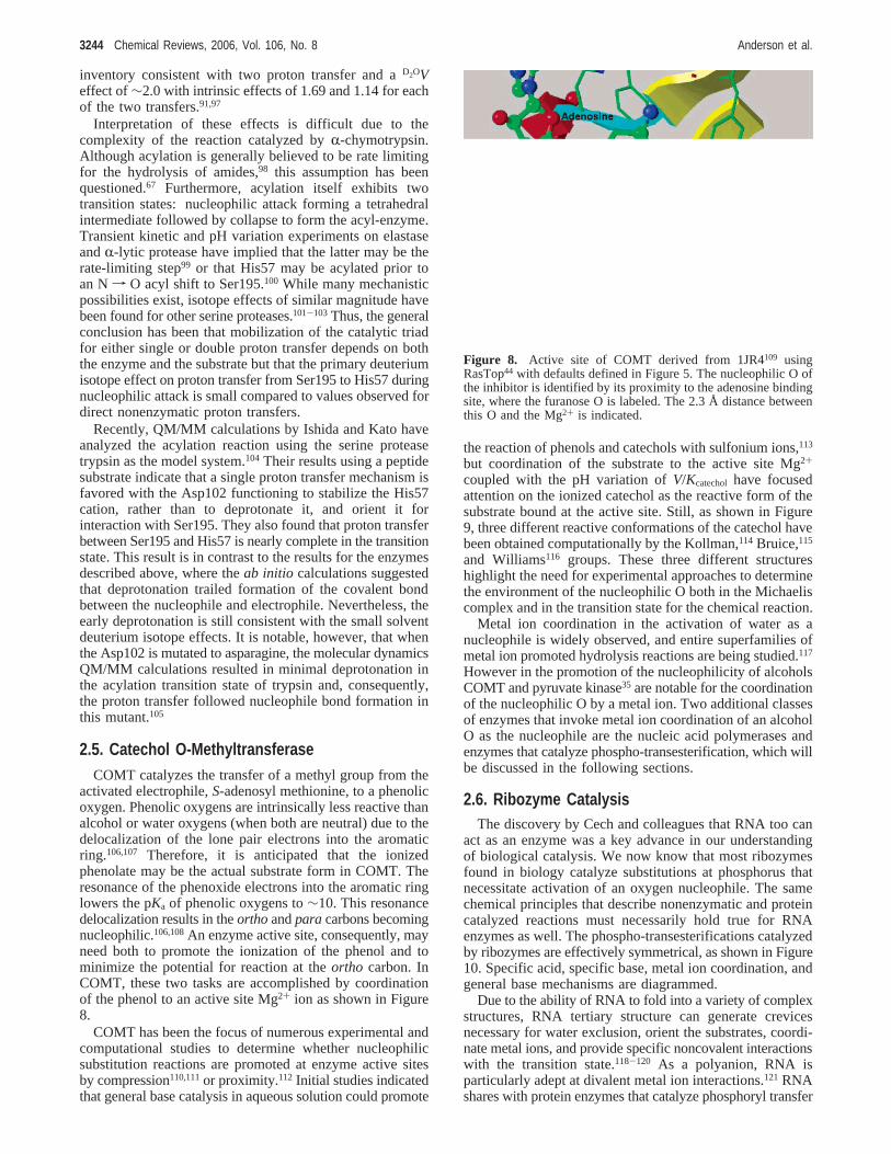
activated electrophile,S-adenosyl methionine, to a phenolicoxygen. Phenolic oxygens are intrinsically less reactive thanalcohol or water oxygens (when both are neutral) due to thedelocalization of the lone pair electrons into the aromaticring.106,107 Therefore, it is anticipated that the ionizedphenolate may be the actual substrate form in COMT. Theresonance of the phenoxide electrons into the aromatic ringlowers the pKa of phenolic oxygens to∼10. This resonancedelocalization results in theorthoandparacarbons becomingnucleophilic.106,108An enzyme active site, consequently, mayneed both to promote the ionization of the phenol and tominimize the potential for reaction at theortho carbon. InCOMT, these two tasks are accomplished by coordinationof the phenol to an active site Mg2+ ion as shown in Figure8.
COMT has been the focus of numerous experimental andcomputational studies to determine whether nucleophilicsubstitution reactions are promoted at enzyme active sitesby compression110,111or proximity.112 Initial studies indicatedthat general base catalysis in aqueous solution could promote
the reaction of phenols and catechols with sulfonium ions,113
but coordination of the substrate to the active site Mg2+
coupled with the pH variation ofV/Kcatechol have focusedattention on the ionized catechol as the reactive form of thesubstrate bound at the active site. Still, as shown in Figure9, three different reactive conformations of the catechol havebeen obtained computationally by the Kollman,114 Bruice,115
and Williams116 groups. These three different structureshighlight the need for experimental approaches to determinethe environment of the nucleophilic O both in the Michaeliscomplex and in the transition state for the chemical reaction.
Metal ion coordination in the activation of water as anucleophile is widely observed, and entire superfamilies ofmetal ion promoted hydrolysis reactions are being studied.117
However in the promotion of the nucleophilicity of alcoholsCOMT and pyruvate kinase35 are notable for the coordinationof the nucleophilic O by a metal ion. Two additional classesof enzymes that invoke metal ion coordination of an alcoholO as the nucleophile are the nucleic acid polymerases andenzymes that catalyze phospho-transesterification, which willbe discussed in the following sections.
2.6. Ribozyme CatalysisThe discovery by Cech and colleagues that RNA too can
act as an enzyme was a key advance in our understandingof biological catalysis. We now know that most ribozymesfound in biology catalyze substitutions at phosphorus thatnecessitate activation of an oxygen nucleophile. The samechemical principles that describe nonenzymatic and proteincatalyzed reactions must necessarily hold true for RNAenzymes as well. The phospho-transesterifications catalyzedby ribozymes are effectively symmetrical, as shown in Figure10. Specific acid, specific base, metal ion coordination, andgeneral base mechanisms are diagrammed.
Due to the ability of RNA to fold into a variety of complexstructures, RNA tertiary structure can generate crevicesnecessary for water exclusion, orient the substrates, coordi-nate metal ions, and provide specific noncovalent interactionswith the transition state.118-120 As a polyanion, RNA isparticularly adept at divalent metal ion interactions.121 RNAshares with protein enzymes that catalyze phosphoryl transfer
Figure 8. Active site of COMT derived from 1JR4109 usingRasTop44 with defaults defined in Figure 5. The nucleophilic O ofthe inhibitor is identified by its proximity to the adenosine bindingsite, where the furanose O is labeled. The 2.3 Å distance betweenthis O and the Mg2+ is indicated.
3244 Chemical Reviews, 2006, Vol. 106, No. 8 Anderson et al.

the common, but not necessarily ubiquitous, use of metalions as a component of nucleophile activation.122-125 Generalbase catalysis, commonly invoked in protein phosphodi-esterases, was first thought to be difficult for RNA, havingfunctional groups with pKa’s far from neutral. However,application of Jencks’ libido rule20 (section 1.4) suggests thatthis is not a significant limitation, and extensive experimentalwork has resulted in a greater appreciation of the ability ofRNA to employ general base catalysis.
Ribozymes fall into two general classes distinguished bydifferences in structural complexity and in reaction mecha-nism (Figure 11). Self-splicing introns, e.g. group I and groupII introns, catalyze phosphotransesterification reactions (Fig-ure 11A) using either the 2′-O or 3′-O; and RNase Pconstitutes a class of large ribozymes. Like their proteincounterparts that catalyze similar reactions, such as endo-nucleases, polymerases, phosphodiesterases, etc., divalentmetal ions appear to play a dominant role. The second class,small ribozymes consisting of 70-100 nucleotide structures,catalyzes intramolecular attack of a 2′-OH on the adjacentphosphodiester bond similar to protein ribonucleases suchas RNase A (Figure 11B). Small ribozymes are involved inreplication of RNA phages as well as several newlydiscovered classes that are involved in gene regulation.Members of this class can function in the absence of divalentmetal ions, and exploration of their mechanisms has shed
new light on the capabilities of RNA to effect general baseactivation of the substrate nucleophile.
2.7. Group I Intron
GI intron splicing involves two successive transesterifi-cation reactions. In the first reaction, the active site positionsthe 3′-O of a free guanosine substrate for nucleophilic attackat the phosphodiester bond of the 5′ splicing site; the 3′-Oof the last exon nucleotide is the leaving group. In the secondreaction, the 3′-O product from the first reaction attacks the3′ splice site, generating the spliced exons, and the 3′-O ofthe last intron nucleotide (gammaG) is the leaving group.An important feature is that the 3′-O leaving group in thefirst step is the nucleophile for the second step. Similarly,the interactions with the leaving group in the second stepwill replicate interactions involved in nucleophilic activationin the first step.
Figure 9. Three computational results of nucleophile activation by coordination to the active site Mg2+: (A) The Kollman group’s transitionstate structure with only the nucleophilic O of the catechol coordinated to the Mg2+.114 (B) The Bruice group’s structure with both O atomscoordinated to the Mg2+.115 (C) The Williams group’s structure with the nucleophilic O ionized but only the non-nucleophilic O coordinatedto the Mg2+.110
Figure 10. Four symmetrical transition states for phospho-transesterification that correspond to the different mechanisms forpromoting nucleophilicity of the ribose OH, R1-OH. Specific acidcatalysis of the leaving group requires only desolvation of thenucleophile, general acid/base catalysis results in greater nucleophileactivation, metal coordination in the two-metal ion mechanismresults in pre-equilibrium deprotonation of the nucleophile, andspecific base catalysis generates an alkoxide nucleophile.
Figure 11. Reactions catalyzed by naturally occurring ribozymesrequire nucleophilic activation that may be effected by general basechemistry (denoted as B:). Neutralization of negatively chargedintermediates (purple arcs) by metal ion interactions is alsoimportant. (A) Large ribozymes such as the group I intron catalyzeattack of an extrinsic nucleophile at a specific phosphodiester bond.(B) Small ribozymes exemplified by the HDV ribozyme catalyzeintramolecular phosphoryl transfer. Reprinted with permission fromref 121. Copyright 2002 Cell Press.
Activation of Oxygen Nucleophiles in Enzyme Catalysis Chemical Reviews, 2006, Vol. 106, No. 8 3245

Biochemical and structural analysis shows that a conservedhelix in the highly structured catalytic core of the GI intronbinds the exogenous G nucleophile, as well as the gammaG,for the second step of splicing. The Hoogsteen face of thebound G forms a base triple with a conserved G-C pairwhere its 3′-O is positioned for nucleophilic attack in thefirst step. In the second step of splicing, a similar Hoogsteenbase pair with gammaG positions the 3′ splice site phosphatefor the reverse reaction, i.e., the 3′-O in this step is the leavinggroup. A recent high-resolution structure of an intermediate,prior to the second step, provides a detailed model of the 3′splice site substrate interactions that position the appropriatehydroxyl nucleophile and phosphate for the second step.126-128
Extensive base pairing and tertiary interactions align theexons for the reaction. Notably, the substrate is in a highlyconstrained conformation with complete reversal of stranddirection at the 3′ splice site. The network of hydrogenbonding interactions positions the 3′-OH of the 5′ exonproximal to the scissile phosphate, and the angle of approachis consistent with the geometry expected for inline nucleo-philic attack shown in Figure 12.
With respect to active site interactions with the nucleo-phile/leaving group oxygens, the majority of attention hasfocused on divalent metal ion interactions, and there is nowstrong evidence that nucleophilic activation at the 3′-Oinvolves direct coordination to active site Mg2+ ions as shownin Figure 12. However, studying metal ion interactions inRNA raises some special problems.120,124,125First, structure-function analyses of RNA-metal ion interactions involvedin nucleophile activation, or any other aspect of catalysis,are complicated by the necessity of Mg2+ ion interactionsfor folding. Also, RNAs can often fold into more than oneenergetically favorable ground state conformation and un-dergo conformational changes. These complications makemechanistic interpretations of structural studies problematic.Importantly, modifications necessary to trap a crystal-lographic complex prior to catalysis for structural studiesmay perturb native metal ion interactions. Indeed, in anotherparallel with protein enzymes, different ribozyme structures
can have significant differences in the number and locationof metal ions.130,131 These issues clearly underscore theimportance of achieving both high-resolution structuralmodels of these enzymes and extensive correlations withfunctional studies.
Strong functional evidence for active site metal interactionswith the nucleophile comes from pioneering biochemicalanalyses involving phosphorothioate modifications to test formetal ion interactions.132-135 In this analysis, functionallydeleterious sulfur or nitrogen substitutions that exhibit rescueupon increasing cation softness (e.g., replacing Mg2+ withCd2+ or Mn2+) provide evidence for direct metal ioncoordination during catalysis.135,136 The development andmost extensive applications of this approach have involveda version of theTetrahymenaribozyme engineered tocatalyze the attack of guanosine on a short oligonucleotidesubstrate. Substrate phosphorothioate-metal ion rescue ex-periments identified four atoms, including the 3′-O leavinggroup/nucleophile for both steps. For example, to probe forinteraction with the 3′-O nucleophile of the 5′ exon in thesecond step, a substrate resembling the ligated exons wasgenerated and the enzyme was engineered to catalyze thereverse of the second splicing step. There is a significantrescue for the reaction; thus, by the principle of microscopicreversibility, this metal must interact in the forward reactionwith nucleophilic oxygen in the second step.137
To gain a more detailed understanding of the relationshipsbetween metal ion interactions with the nucleophile and thosewith other atoms of the reactive phosphate, a thermodynamicfingerprint analysis has been used.132-135 This extension ofthe phosphorothioate-rescue approach involves quantitativelyanalyzing the reactivity of modified substrates relative tounmodified substrates over a range of rescuing metal ionconcentrations. The concentration dependence of the relativerates can be interpreted in terms of apparent affinities withmagnitudes that serve as distinctive “fingerprints” for therescuing metal ion(s). Thus, by comparison of these apparentaffinities for different modified substrates, information isgained as to whether the same or distinct metal ions interactwith the identified substrate ligands. These studies providedfunctional evidence for a network of three distinct metal ionswithin the Tetrahymenaribozyme active site: Metal ionscoordinate to the 3′-oxygen leaving group, the 3′-oxygen onthe G nucleophile in the first step (MA and MB, respectively),and a third oxygen interacting with the 2′-hydroxyl of the Gnucleophile (MC) (Figure 13). Two metal ions (MA and MC)coordinate thepro-SP oxygen of the scissile phosphate, anda third interacts with the nucleophilic 3′O. Extension of thisanalysis to encompass phosphate oxygens within the intronitself provides evidence that these metal ions are positionedby coordination interactions with nonbridging oxygens thatconstitute, in part, the active site of the ribozyme.138,139
Recently, a structure including the 2′-O of the substrateG, a key metal ion ligand, has been reported, providing themost consistent model to date of active site interactionsincluding metal ion coordination to the 3′-O’s involved innucleophilic attack (Figure 12). In this structure, two metalions coordinate to nonbridging phosphate oxygens in theintron and substrate. This result seemingly contrasts with theresults from biochemical studies that indicate three activesite metal ions. However, the two Mg2+ ions observed inthe three-dimensional structure are able to satisfy all of thebiochemically identified interactions, including Mg2+ interac-tions with both 3′-O’s. Their geometry is consistent with the
Figure 12. Structure of group I intron adapted from 1ZZN129 usingRasTop.42 The distance between the nucleophilic 3′-O and thetransferred phosphate is depicted. Both the nucleophile and elec-trophile are within inner sphere distances of the two Mg2+ ions(dark green spheres) located at the active site.
3246 Chemical Reviews, 2006, Vol. 106, No. 8 Anderson et al.

Steitz two metal ion mechanism from DNA polymerase.140,141
Importantly, one Mg2+ ion makes inner sphere contacts withboth the 2′-O and the 3′-O nucleophile/leaving groups ofthe gammaG. This geometry is consistent with the biochemi-cal data that both positions are ligands for metal ions, but inthe crystal structure only a single metal ion is seencoordinating to both ligands. This difference from themechanism drawn from biochemical data could formallyarise from differences in ground state versus transition stateinteractions or, potentially, differences in the electrostaticenvironments of the active site due to different functionalgroup modifications. Nonetheless, it is abundantly clear thatdirect metal coordination to both 3′-O’s is a key feature ofthe catalytic mechanism.
2.8. HDV RibozymeIn biology, small RNA motifs that catalyze RNA self-
cleavage via attack of a ribose 2′-O play essential roles inRNA virus replication, as well as regulation of geneexpression. With respect to nucleophilic activation, it hasvariously been proposed, as with protein enzymes, to involvemetal catalysis or general base catalysis via an RNAnucleobase functional group or the water molecule in theinner hydration sphere of Mg2+.119,120However, some smallribozymes can function in the absence of divalent metalions.142-144 Thus, intensive research has focused on identify-ing potential general acid/base mechanisms. However, themost detailed models of the catalytic mechanism are builtlargely on structural rather than functional data. Additionally,there is some conflict between structural studies and theresults of detailed functional analyses. Nonetheless, there issignificant progress in outlining the distinct geometries fornucleophilic activation in some examples exemplified hereby the HDV ribozyme.
The HDV ribozyme is a small ca. 70 nucleotide RNA that,like all small ribozymes, folds to form a compact structurethat results in cleavage of a specific phosphodiester bondvia attack on the adjacent 2′-O nucleophile.145 As is the casefor protein enzymes such as RNase A that catalyze the samechemistry, the reaction is inhibited by acidic pH, consistentwith a deprotonation, potentially of the nucleophile, preced-ing the rate-limiting step under these conditions.146,147
Importantly, the HDV ribozyme is able to function in theabsence of divalent metal ions, albeit more slowly. Highermonovalent ion concentrations are required for folding,presumably to screen electrostatic repulsion. This resultimplied that nucleobases could fulfill the role of amino acidside chains in functioning as general acids and bases. Indeed,a growing body of structural and biochemical analysessupport that this is in fact the case.
Initial insights into the overall architecture of the ri-bozymes came from a structure of the 3′ product complex.148
While this structure did not provide evidence for theinteractions with the nucleophile, it showed a nucleobase(C76) potentially positioned for acid-base catalysis. Furtherphysical and mutagenesis studies provided important evi-dence that the protonation state of C76 is indeed importantfor catalysis.149-152 These analyses gave rise to a model inwhich C76, in its deprotonated form, functions as a generalbase catalyst to accept a proton from the nucleophilic 2′-O.
Alternatively, C76 could function as a general acid, whilea different base catalyst activates the 2′-O nucleophile.Support for this second model came from the observationthat the pH dependence of the activity varied with thedivalent metal ion concentration, which suggested that theionization of metal-bound water might contribute to the pHdependence.147 These results were consistent with the ideathat either hydroxide or a metal coordinated hydroxideaccepts a proton from the 2′-OH to activate the nucleophile.Additionally, recent experiments investigating the effects of5′-O leaving group substitutions by Das and Piccirilli furtherimplicate C76 as a general acid catalyst.153 Recent structuresin which C76 has been mutated to U show this residue inposition for an interaction with the leaving group, but thereis no metal ion in the vicinity of the 2′-O. Such resultsprovide important insights into HDV active site function,but they leave in doubt the precise mechanism of nucleophilicactivation in the absence of metal ions. A potential rationalefor the slower catalytic activity of the ribozyme under theseconditions is that nucleophile deprotonation is not assisted.
It is clear that RNA does not invent new mechanisms fornucleophilic activation or catalysis but is constrained, justas protein enzymes are, to employ those kinds of interactionsdictated by the intrinsic chemistry of the reaction. Thedifference lies in the unique geometries that position RNAfunctional groups relative to the nucleophile and reactivephosphate in order to stabilize the transition state. Thesekinds of interactions include employing networks of non-covalent interactions to position the nucleophile in theoptimal geometry for in-line attack. RNA is particularly adeptat interacting with divalent metal ions, and there is very goodevidence for a direct role for active site metal ion coordina-tion in nucleophile activation. Unlike protein enzymes,however, RNA needs metal ions to fold, and the couplingof structure stabilization and catalysis makes achieving aclear picture of the roles of individual interactions challeng-ing. Thus, it is with hindsight that it becomes clear that RNA,despite having a limited repertoire of functional groups, canform an active site environment in which a significant pKa
shift occurs, enabling general acid/base catalysis. Yet, veryfew examples have been described in any depth, and evenfor the best characterized examples, there remain importantquestions concerning the sum total of catalytic interactionsand the ultimate effect of active site nucleophile interactionson bonding in the ground state and transition state.
Figure 13. Group I intron splicing reaction. The last nucleotideof the 5′ intron (U-1) is shown at the top. The 3′-O leaving groupof this nucleotide serves as the nucleophile for the second splicingstep. At the bottom is the exogenous G whose 3′-O serves as thenucleophile. The last nucleotide of the intron (gammaG) occupiesthis position in the second step of splicing, where its corresponding3′-O is the leaving group. Metal ion interactions demonstrated byquantitative metal rescue of phosphorothioate modifications areshown in red. Reprinted with permission from ref 138. CopyrightHougland et al.
Activation of Oxygen Nucleophiles in Enzyme Catalysis Chemical Reviews, 2006, Vol. 106, No. 8 3247

3. Spectroscopic Characterization of Nucleophilesat Enzyme Active Sites
The previous sections have established that, in the processof binding to enzyme active sites, every alcohol nucleophilehas the solvent H-bond donors removed and that the alcoholO-H is either coordinated to an active site general base or,less commonly, deprotonated by coordination to a metal ion.This section focuses on the emerging use of spectroscopicmethods to characterize the environment of the nucleophilein enzyme-substrate complexes. In the application of NMRand vibrational spectroscopy, the requisite model studies havenow been completed, but the number of applications to thecharacterization of O nucleophiles in enzyme active sites isvery limited.
3.1. Vibrational Spectroscopic Characterization ofAlcohols
H-bond strength from alcohols to Bronsted base acceptorshas been studied extensively by vibrational spectroscopy. Asthe H-bond becomes stronger, based on thermodynamicmeasurements, the O-H stretching frequency (νstr) decreasesby several 100 cm-1.154 While this approach works well inaprotic organic solvents, in aqueous solution, the broadintense band from the solvent O-H stretching modesprevents the identification of the single substrate O-H stretchof interest. This bond cannot be labeled with D, as itexchanges rapidly with solvent and18O substitution has aminimal effect on the O-H νstr.
To characterize nucleophilicity, the electron density of thelone pairs is more important than the strength of the H-bond.Studies of negative hyperconjugation of C-H bonds vicinalto lone pairs was prompted by the observation that the C-Hbondanti to a lone pair has aνstr ∼30-60 cm-1 lower thana C-H bondanti to an O-H bond, as shown in Figure 14.H-bond donation by an alcohol increases the electron densityaround the O, increasing the extent of negative hypercon-jugation and decreasing the C-H νstr.155,156Ionization of thealcohol to the alkoxide results in a large (>100 cm-1)decrease in the C-H νstr. Unlike the case of the O-H stretch,it is possible to uniquely label a single C-H bond in asubstrate by D-substitution. The C-D νstr of 2100-2250cm-1 appears in a spectral window free of both water andprotein vibrations. Single C-D bonds can be observed byvibrational spectroscopy in proteins (Maiti, Carey, andAnderson, unpublished), allowing the electron density of alone pair on a geminal O to be interrogated. The applicationof vibrational spectroscopy to characterize changes inelectron density of substrates bound at active sites has beenfurther reviewed by Carey in this issue.
3.2. NMR Characterization of AlcoholNucleophiles
NMR has proven to be an extremely valuable tool incharacterizing H-bonds between N-H donors and N accep-tors. These measurements include characterization of the1Hchemical shift of the H-bonded proton, its fractionationfactor, solvent exchange kinetics, and the scalar couplingconstants1JNH and 1JNN. In application to alcohol nucleo-philes, many of these NMR observations have not beenpossible due to the rapid solvent exchange of the H-bondedproton. Even in cases where solvent exchange may be slow,the chemical shift has proven too small to be identifiablefrom bulk water and the scalar coupling constants,1JOH to17O or 2JCH, have not been investigated.
However, in favorable cases, O‚‚‚H‚‚‚N H-bonded systemscan be characterized by the same parameters as N‚‚‚H‚‚‚Nsystems, i.e., the1H chemical shift, the fractionation factor,and the1JNH scalar coupling constantsall of which correlatewith the strength of the H-bond.157 The model experimentalsystems have focused on aprotic solvents and low temper-atures to minimize chemical exchange, and in most systemsthey have favored zwitterionic complexes with the protonresiding closer to the N, i.e., greater N-H than O-H bondorder.158,159 The best characterized O‚‚‚H‚‚‚N H-bondedsystem comes from NMR studies of serine proteases, wherethe observed1H resides between the Asp and the imidazoleof the catalytic triad, and is not directly an H-bond donatedby the serine nucleophile (reviewed in ref 160). The stronglydeshielded proton resonance has a low fractionation factorcharacteristic of short strong H-bonds.33 The NMR charac-terization of this H-bonded system demonstrates the potentialfor characterizing H-bonds at enzyme active sites, whenexchange with solvent has been minimized. Theoreticalcalculations extend the correlation of chemical shift, frac-tionation factor, and1JNH to systems with greater O-H bondorder, as anticipated for SerγO-H‚‚‚imidazole H-bonds.161
An alternative approach, based on the observation that theadjacent C-H bond was weakened in H-bond complexes,is to observe the1JCH of the vicinal methylene protons inalcohol H-bonded systems. A correlation between1JCH forhexafluoroisopropanol and the H-bond strength formed withamines of varying basicities has been observed.162
For O‚‚‚H‚‚‚O H-bonded complexes, as observed withcarboxylate H-bond acceptors to alcohol nucleophiles inkinases and acyl transferases, the only model NMR studyof H-bond parameters comes from fast magic angle spinningdeterminations of the1H chemical shift in crystals wherethe exchange of the H-bonded protons is minimized andneutron diffraction has characterized the H-bond geom-etry.160,163The chemical shift correlates with the displacementof the1H from the middle of the H-bonded system.164 Exceptfor very strong H-bonds, these methods have not been appliedto enzyme systems.
3.3. Isotope Effects on Alcohol NucleophilesIn characterizing nucleophile activation by isotope effect
studies, the most obvious substitution is to label the nucleo-philic O with 18O. The effect of removing the H-bond donorscan be examined by determining the18O equilibrium isotopeeffect on transfer to the gas phase, and this is determined tobe small, 1.008.165 There have been no experimental deter-minations of the equilibrium isotope effect on alcoholsfunctioning as a donor in forming an H-bonded complex.
Figure 14. Variation of C-D νstr with H-bonding and ionization.The anti (left) andgauche(right) C-D bonds of an alcohol havedifferentνstr values. The approximate values given are (in order ofdecreasing frequency) for the alcohol in CHCl3, i.e., with no H-bonddonor or acceptor, complexed to a strong amine base, and as thealkoxide in water.
3248 Chemical Reviews, 2006, Vol. 106, No. 8 Anderson et al.

As the O-H stretching frequency can be decreased signifi-cantly, it is anticipated that forming an H-bond could resultin a significant normal equilibrium isotope effect, butcomputational studies suggest that there is a compensatingincrease in the C-O bond order.156 Deprotonation of O-Hbonds, however, results in a large normal equilibrium isotopeeffect; the18O effect on ionization of water is 1.04.166 Metalion coordination, by introducing a new bond to the alcoholO, results in an inverse18O equilibrium isotope effect of 0.98for coordination to a divalent metal ion.167 The presence ofan inverse18O isotope effect on binding of an alcohol to anenzyme active site would be strongly suggestive of coordina-tion to a metal ion. These small18O isotope effects on bindingcan be measured with sufficient precision by whole moleculemass spectrometry.168-170 Thus, both ionization and metalion coordination of an oxygen nucleophile should give riseto a readily interpretable18O isotope effect on binding.
Since equilibrium isotope effects are generated by changesin vibrational frequencies between the substrate and productstates, the measurement of these effects can be considered asubsidiary of vibrational spectroscopy. The advantages ofdetermining the isotope effects are that they can be measuredwith a trace label and with minimal material employed atlow concentrations and thatkinetic isotope effects provideinsight into the transition state of a reaction, a capabilityeffectively unavailable to any other spectroscopic method.The disadvantage of isotope effect determinations is that allof the vibrational modes of the isotopically substituted atomcan contribute to the measured effect,171not just the stretchingvibration of major interest.
The decrease in theνstr of a C-D bond of a substrate onformation of the Michaelis complex, as predicted to occuron formation of a strong H-bond, will contribute to a normalequilibrium isotope effect on association of the substrate.These equilibrium binding isotope effects have been pre-dicted87,156 and observed.84-86,172 For hexokinase, there is asignificant isotope effect of 1.06 measured with [6-3H]-glucose.85 This normal effect is interpreted as suggesting thatthe active site Asp657 forms a significant H-bond to promotethe nucleophilicity of the C6 hydroxyl group even in thebinary enzyme-glucose complex. The value is inconsistentwith ionization of the C6 hydroxyl, as ionization of alcoholsresults in significantly larger equilibrium isotope effects,>1.15.88,156,173
Unexpectedly large 5′-3H kinetic isotope effects have beenobserved in enzymes catalyzing the cleavage of the glyco-sidic bond in nucleosides.174,175These results, while attributedto interactions present in the Michaelis complex,176 areindicative of the ability to observe changes in the electronicdistribution about an alcohol in the transition state. Preequi-librium ionization will result in large secondary kineticisotope effects and should provide a diagnostic for thismechanism. In the serine transacetylase reaction, [3-2H]-serine kinetic isotope effects have been determined to be nearunity, indicating that in this reaction the serine has not beendeprotonated in the rate-determining transition state.74
4. Acknowledgments
This review was supported in part by NIH Grant GM56740,NSF Grant MCB-0110610, and the Medical Scientist Train-ing Program at Case Western Reserve University.
5. References(1) Swain, C. G.; Scott, C. B.J. Am. Chem. Soc.1953, 75, 141.(2) Pearson, R. G.; Sobel, H. R.; Songstad, J.J. Am. Chem. Soc.1968,
90, 319.(3) Zhang, X.; Houk, K. N.Acc. Chem. Res.2005, 38, 379.(4) Jencks, W. P.; Gilchrist, M.J. Am. Chem. Soc.1968, 90, 2622.(5) Honig, B.; Nicholls, A.Science1995, 268, 1144.(6) Belasco, J. G.; Knowles, J. R.Biochemistry1980, 19, 472.(7) D’Ordine, R. L.; Pawlak, J.; Bahnson, B. J.; Anderson, V. E.
Biochemistry2002, 41, 2630.(8) Deng, H.; Lewandowicz, A.; Schramm, V. L.; Callender, R.J. Am.
Chem. Soc.2004, 126, 9516.(9) Alexander, R.; Ko, E. C. F.; Parker, A. J.; Broxton, T. J.J. Am.
Chem. Soc.1968, 90, 5049.(10) Adamovic, I.; Gordon, M. S.J. Phys. Chem. A2005, 109, 1629.(11) Acevedo, O.; Jorgensen, W. L.Org. Lett.2004, 6, 2881.(12) Freccero, M.; Di Valentin, C.; Sarzi-Amade, M.J. Am. Chem. Soc.
2003, 125, 3544.(13) Re, S.; Morokuma, K.J. Phys. Chem. A2001, 105, 7185.(14) Jencks, W. P.AdV. Chem. Ser.1987, 215 (Nucleophilicity), 155.(15) Jencks, W. P.; Haber, M. T.; Herschlag, D.; Nazaretian, K. L.J.
Am. Chem. Soc.1986, 108, 479.(16) Rucker, V. C.; Byers, L. D.J. Am. Chem. Soc.2000, 122, 8365.(17) Onyido, I.; Swierczek, K.; Purcell, J.; Hengge, A. C.J. Am. Chem.
Soc.2005, 127, 7703.(18) Olsson, M. H. M.; Warshel, A.J. Am. Chem. Soc.2004, 126, 15167.(19) Warshel, A.; Aqvist, J.; Creighton, S.Proc. Natl. Acad. Sci., U.S.A.
1989, 86, 5820.(20) Jencks, W. P.J. Am. Chem. Soc.1972, 94, 4731.(21) Swain, C. G.; Kuhn, D. A.; Schowen, R. L.J. Am. Chem. Soc.1965,
87, 1553.(22) Knipe, J.; Coward, J. K.J. Am. Chem. Soc.1979, 101, 4339.(23) Dietze, P. E.; Jencks, W. P.J. Am. Chem. Soc.1989, 111, 340.(24) Strajbl, M.; Florian, J.; Warshel, A.J. Am. Chem. Soc.2000, 122,
5354.(25) Hibbert, F.; Emsley, J.AdVances in Physical Organic Chemistry;
Bethell, D., Ed.; Academic Press: New York, 1990; Vol. 26.(26) Del Bene, J. E.; Jordan, M. J. T.Int. ReV. Phys. Chem.1999, 18,
119.(27) Kuhn, P.; Knapp, M.; Soltis, S. M.; Ganshaw, G.; Thoene, M.; Bott,
R. Biochemistry1998, 37, 13446.(28) Rypniewski, W. R.; Ostergaard, P. R.; Norregaard-Madsen, M.;
Dauter, M.; Wilson, K. S.Acta Crystallogr., Sect. D: Biol.Crystallogr.2001, 57, 8.
(29) Quinn, D. M.; Sutton, L. D. InEnzyme Mechanism from IsotopeEffects; Cook, P. F., Ed.; CRC Press: Boca Raton, FL, 1991.
(30) Cordes, E. H.Prog. Phys. Org. Chem.1967, 4, 1.(31) Eliason, R.; Kreevoy, M. M.J. Am. Chem. Soc.1978, 100, 7037.(32) Schowen, K. B.; Limbach, H. H.; Denisov, G. S.; Schowen, R. L.
Biochim. Biophys. Acta2000, 1458, 43.(33) Lin, J.; Westler, W. M.; Cleland, W. W.; Markley, J. L.; Frey, P. A.
Proc. Natl. Acad. Sci., U.S.A.1998, 95, 14664.(34) Dougherty, T. M.; Cleland, W. W.Biochemistry1985, 24, 5875.(35) Larsen, T. M.; Benning, M. M.; Rayment, I.; Reed, G. H.Biochem-
istry 1998, 37, 6247.(36) Lerner, C.; Masjost, B.; Ruf, A.; Gramlich, V.; Jakob-Roetne, R.;
Zurcher, G.; Borroni, E.; Diederich, F.Org. Biomol. Chem.2003, 1,42.
(37) Adams, J. A.Chem. ReV. 2001, 101, 2271.(38) Kemp, B. E.; Benjamini, E.; Krebs, E. G.Proc. Natl. Acad. Sci.,
U.S.A.1976, 73, 1038.(39) Kemp, B. E.; Graves, D. J.; Benjamini, E.; Krebs, E. G.J. Biol.
Chem.1977, 252, 4888.(40) Hanks, S. K.; Quinn, A. M.; Hunter, T.Science1988, 241, 42.(41) Madhusudan; Trafny, E. A.; Xuong, N. H.; Adams, J. A.; Ten Eyck,
L. F.; Taylor, S. S.; Sowadski, J. M.Protein Sci.1994, 3, 176.(42) Zheng, J.; Knighton, D. R.; Ten Eyck, L. F.; Karlsson, R.; Xuong,
N.; Taylor, S. S.; Sowadski, J. M.Biochemistry1993, 32, 2154.(43) Biology, N. S.; Madhusudan; Akamine, P.; Xuong, N. H.; Taylor,
S. S.Nat. Struct. Biol.2002, 9, 273.(44) Valadon, P.; Bernstein, H. J.RasTop, Version 1.3.1; 2001.(45) Gibbs, C. S.; Zoller, M. J.J. Biol. Chem.1991, 266, 8923.(46) Skamnaki, V. T.; Owen, D. J.; Noble, M. E. M.; Lowe, E. D.; Lowe,
G.; Oikonomakos, N. G.; Johnson, L. N.Biochemistry1999, 38,14718.
(47) Zhou, J.; Adams, J. A.Biochemistry1997, 36, 2977.(48) Yoon, M. Y.; Cook, P. F.Biochemistry1987, 26, 4118.(49) Ho, M.; Bramson, H. N.; Hansen, D. E.; Knowles, J. R.; Kaiser, E.
T. J. Am. Chem. Soc.1988, 110, 2680.(50) Knighton, D. R.; Zheng, J.; Ten Eyck, L. F.; Ashford, V. A.; Xuong,
N.; Taylor, S. S.; Sowadski, J. M.Science1991, 253, 407.(51) Adams, J. A.; Taylor, S. S.J. Biol. Chem.1993, 268, 7747.
Activation of Oxygen Nucleophiles in Enzyme Catalysis Chemical Reviews, 2006, Vol. 106, No. 8 3249

(52) Adams, J. A.; Taylor, S. S.Biochemistry1992, 31, 8516.(53) Grant, B. D.; Adams, J. A.Biochemistry1996, 35, 2022.(54) Jencks, W. F.Catalysis in Chemistry and Enzymology; McGraw-
Hill: New York, 1969.(55) Hirano, Y.; Masayuki, H.; Hoshino, T.; Tsuda, M.J. Phys. Chem. B
2002, 106, 5788.(56) Hart, J. C.; Sheppard, D. W.; Hillier, I. H.; Burton, N. A.Chem.
Commun.1999, 1999, 79.(57) Hutter, M. C.; Helms, V.Protein Sci.1999, 8, 2728.(58) Valiev, M.; Kawai, R.; Adams, J. A.; Weare, J. H.J. Am. Chem.
Soc.2003, 125, 9926.(59) Diaz, N.; Field, M. J.J. Am. Chem. Soc.2004, 126, 529.(60) Cheng, Y.; Zhang, Y.; McCammon, J. A.J. Am. Chem. Soc.2005,
127, 1553.(61) Johnson, C. M.; Roderick, S. L.; Cook, P. F.Arch. Biochem. Biophys.
2005, 433, 85.(62) Kredich, N. M.; Tomkins, G. M.J. Biol. Chem.1966, 241, 4955.(63) Kredich, N. M.; Becker, M. A.; Tomkins, G. M.J. Biol. Chem.1969,
244, 2428.(64) Olsen, L. R.; Huang, B.; Vetting, M. W.; Roderick, S. L.Biochemistry
2004, 43, 6013.(65) Pye, V. E.; Tingey, A. P.; Robson, R. L.; Moody, P. C. E.J. Biol.
Chem.2004, 279, 40729.(66) Blow, D. M.; Birktoft, J. J.; Hartley, B. S.Nature1969, 221, 337.(67) Hedstrom, L.Chem. ReV. 2002, 102, 4501.(68) Hindson, V. J.; Shaw, W. V.Biochemistry2003, 42, 3113.(69) Hindson, V. J.Biochem. J.2003, 375, 745.(70) Johnson, C. M.; Huang, B.; Roderick, S. L.; Cook, P. F.Biochemistry
2004, 43, 15534.(71) Wyckoff, T. J. O.; Raetz, C. R. H.J. Biol. Chem.1999, 274, 27047.(72) Lewendon, A.; Murray, I. A.; Shaw, W. V.Biochemistry1994, 33,
1944.(73) Murray, I. A.; Shaw, W. V.Antimicrob. Agents Chemother.1997,
41, 1.(74) Ruszczycky, M. W. Ph.D. Thesis, Case Western Reserve University,
2006.(75) Ruszczycky, M. W.; Anderson, V. E. Unpublished work.(76) Anderson, C. M.; Stenkamp, R. E.; McDonald, R. C.; Steitz, T. A.
J. Mol. Biol. 1978, 123, 207.(77) Viola, R. E.; Cleland, W. W.Biochemistry1978, 17, 4111.(78) Bohnensack, R.; Hofmann, E.Eur. J. Biochem.1969, 9, 534.(79) Arora, K. K.; Filburn, C. R.; Pedersen, P. L.J. Biol. Chem.1991,
266, 5359.(80) Lange, A. J.; Xu, L. Z.; van Poelwijk, F.; Lin, K.; Granner, D. K.;
Pilkis, S. J.Biochem. J.1991, 277, 159.(81) Wilkinson, K. D.; Rose, I. A.J. Biol. Chem.1979, 254, 12567.(82) Taylor, K. B.; Cook, P. F.; Cleland, W. W.Eur. J. Biochem.1983,
134, 571.(83) Cornish-Bowden, A.; Pollard-Knight, D.Arch. Biol. Med. Exp.
(Santiago)1985, 18, 293.(84) Lewis, B. E.; Schramm, V. L. InIsotope Effects in Chemistry and
Biology; Kohen, A., Limbach, H.-H., Eds.; CRC Press: Boca Raton,FL, 2006.
(85) Lewis, B. E.; Schramm, V. L.J. Am. Chem. Soc.2003, 125, 4785.(86) Lewis, B. E.; Schramm, V. L.J. Am. Chem. Soc.2003, 125, 4672.(87) Lewis, B. E.; Schramm, V. L.J. Am. Chem. Soc.2001, 123, 1327.(88) Lewis, B. E.; Schramm, V. L.J. Am. Chem. Soc.2003, 125, 7872.(89) Bender, M. L.J. Am. Chem. Soc.1962, 84, 2582.(90) Voet, D.; Voet, J. G.Biochemistry, 2nd ed.; John Wiley & Sons:
New York, 1995.(91) Bender, M. L.; Clement, C. E.; Kezdy, F. J.; Heck, H. D. A.J. Am.
Chem. Soc.1964, 86, 3680.(92) Kezdy, F. J.; Bender, M. L.Biochemistry1962, 1, 1097.(93) Gutfreund, H.; Sturtevant, J. M.Proc. Natl. Acad. Sci., U.S.A.1956,
42, 719.(94) Frigerio, F.; Coda, A.; Pugliese, L.; Lionetti, C.; Menegatti, E.;
Amiconi, G.; Schnebli, H. P.; Ascenzi, P.; Bolognesi, M.J. Mol.Biol. 1992, 225, 107.
(95) Bender, M. L.; Hamilton, G. A.J. Am. Chem. Soc.1962, 84, 2570.(96) Bender, M. L.; Pollock, E. J.; Neveu, M. C.J. Am. Chem. Soc.1962,
84, 595.(97) Elrod, J. P.; Hogg, J. L.; Quinn, D. M.; Venkatasubban, K. S.;
Schowen, R. L.J. Am. Chem. Soc.1980, 102, 3917.(98) Zerner, B.; Bond, R. P. M.; Bender, M. L.J. Am. Chem. Soc.1964,
86, 3674.(99) Hunkapiller, M. W.; Forgac, M. D.; Richards, J. H.Biochemistry
1976, 15, 5581.(100) Quinn, D. M.; Elrod, J. P.; Ardis, R.; Friesen, P.; Schowen, R. L.J.
Am. Chem. Soc.1980, 102, 5358.(101) Stein, R. L.J. Am. Chem. Soc.1983, 105, 5111.(102) Enyedy, E. J.; Kovach, I. M.J. Am. Chem. Soc.2004, 126, 6017.(103) Zhang, D.; Kovach, I. M.J. Am. Chem. Soc.2005, 127, 3760.(104) Ishida, T.; Kato, S.J. Am. Chem. Soc.2003, 125, 12035.
(105) Ishida, T.; Kato, S.J. Am. Chem. Soc.2004, 126, 7111.(106) Lu, Y.; Handoo, K. L.; Parker, V. D.Org. Biomol. Chem.2003, 1,
36.(107) Kevill, D. N.; Kim, C. B.Bull. Soc. Chim. Fr.1988, 383.(108) Breslow, R.; Groves, K.; Mayer, M. U.J. Am. Chem. Soc.2002,
124, 3622.(109) Lerner, C.; Ruf, A.; Gramlich, V.; Masjost, B.; Zurcher, G.; Jakob-
Roetne, R.; Borroni, E.; Diederich, F.Angew. Chem., Int. Ed. Engl.2001, 40, 4040.
(110) Ruggiero, G. D.; Williams, I. H.; Roca, M.; Moliner, V.; Tunon, I.J. Am. Chem. Soc.2004, 126, 8634.
(111) Rodgers, J.; Femec, D. A.; Schowen, R. L.J. Am. Chem. Soc.1982,104, 3263.
(112) Lau, E. Y.; Bruice, T. C.J. Am. Chem. Soc.1998, 120, 12387.(113) Knipe, J. O.; Vasquez, P. J.; Coward, J. K.J. Am. Chem. Soc.1982,
104, 3202.(114) Kuhn, B.; Kollman, P. A.J. Am. Chem. Soc.2000, 122, 2586.(115) Lau, E. Y.; Bruice, T. C.J. Am. Chem. Soc.2000, 122, 7165.(116) Roca, M.; Marti, S.; Andres, J.; Moliner, V.; Tunon, I.; Bertran, J.;
Williams, I. H. J. Am. Chem. Soc.2003, 125, 7726.(117) Seibert, C. M.; Raushel, F. M.Biochemistry2005, 44, 6383.(118) Herschlag, D.Nature1998, 395, 548.(119) Doudna, J. A.; Lorsch, J. R.Nat. Struct. Mol. Biol.2005, 12, 395.(120) Fedor, M. J.; Williamson, J. R.Nat. ReV. Mol. Cell Biol. 2005, 6,
399.(121) DeRose, V. J.Chem. Biol.2002, 9, 961.(122) Doudna, J. A.; Cech, T. R.Nature2002, 418, 222.(123) Hanna, R.; Doudna, J. A.Curr. Opin. Chem. Biol.2000, 4, 166.(124) Pyle, A. M.J. Biol. Inorg. Chem.2002, 7, 679.(125) DeRose, V. J.Curr. Opin. Struct. Biol.2003, 13, 317.(126) Stahley, M. R.; Strobel, S. A.Science2005, 309, 1587.(127) Adams, P. L.; Stahley, M. R.; Gill, M. L.; Kosek, A. B.; Wang, J.;
Strobel, S. A.RNA2004, 10, 1867.(128) Adams, P. L.; Stahley, M. R.; Kosek, A. B.; Wang, J.; Strobel, S. A.
Nature2004, 430, 45.(129) Stahley, M. R.; Strobel, S. A.Science2005, 309, 1587.(130) Vicens, Q.; Cech, T. R.Trends Biochem. Sci.2005, 12, 12.(131) Guo, F.; Gooding, A. R.; Cech, T. R.Mol. Cell 2004, 16, 351.(132) Shan, S.; Kravchuk, A. V.; Piccirilli, J. A.; Herschlag, D.Biochemistry
2001, 40, 5161.(133) Yoshida, A.; Sun, S.; Piccirilli, J. A.Nat. Struct. Biol.1999, 6, 318.(134) Shan, S.; Yoshida, A.; Sun, S.; Piccirilli, J. A.; Herschlag, D.Proc.
Natl. Acad. Sci., U.S.A.1999, 96, 12299.(135) Piccirilli, J. A.; Vyle, J. S.; Caruthers, M. H.; Cech, T. R.Nature
1993, 361, 85.(136) Jaffe, E. K.; Markham, G. D.Biochemistry1987, 26, 4258.(137) Weinstein, L. B.; Jones, B. C.; Cosstick, R.; Cech, T. R.Nature1997,
388, 805.(138) Hougland, J. L.; Kravchuk, A. V.; Herschlag, D.; Piccirilli, J. A.
PLoS Biol.2005, 3, e277. Epub 2005 Aug 16.(139) Szewczak, A. A.; Kosek, A. B.; Piccirilli, J. A.; Strobel, S. A.
Biochemistry2002, 41, 2516.(140) Beese, L. S.; Steitz, T. A.EMBO J.1991, 10, 25.(141) Steitz, T. A.; Steitz, J. A.Proc. Natl. Acad. Sci., U.S.A.1993, 90,
6498.(142) Murray, J. B.; Seyhan, A. A.; Walter, N. G.; Burke, J. M.; Scott, W.
G. Chem. Biol.1998, 5, 587.(143) Curtis, E. A.; Bartel, D. P.RNA2001, 7, 546.(144) Nakano, S.; Proctor, D. J.; Bevilacqua, P. C.Biochemistry2001, 40,
12022.(145) Shih, I. H.; Been, M. D.Annu. ReV. Biochem.2002, 71, 887.(146) Bevilacqua, P. C.; Brown, T. S.; Nakano, S.; Yajima, R.Biopolymers
2004, 73, 90.(147) Nakano, S.; Chadalavada, D. M.; Bevilacqua, P. C.Science2000,
287, 1493.(148) Ferre-D’Amare, A. R.; Zhou, K.; Doudna, J. A.Nature1998, 395,
567.(149) Kumar, P. K.; Suh, Y. A.; Miyashiro, H.; Nishikawa, F.; Kawakami,
J.; Taira, K.; Nishikawa, S.Nucleic Acids Res.1992, 20, 3919.(150) Perrotta, A. T.; Shih, I.; Been, M. D.Science1999, 286, 123.(151) Shih, I. H.; Been, M. D.Proc. Natl. Acad. Sci., U.S.A.2001, 98,
1489.(152) Suh, Y. A.; Kumar, P. K.; Kawakami, J.; Nishikawa, F.; Taira, K.;
Nishikawa, S.FEBS Lett.1993, 326, 158.(153) Das, S. R.; Fong, R.; Piccirilli, J. A.Curr. Opin. Chem. Biol.2005,
9, 585.(154) Piaggio, P.; Tubino, R.; Dellepiane, C.J. Mol. Struct.1983, 96, 277.(155) Maiti, N. C.; Carey, P. R.; Anderson, V. E.J. Phys. Chem. A2003,
107, 9910.(156) Gawlita, E.; Lantz, M.; Paneth, P.; Bell, A.; Tonge, P.; Anderson,
V. E. J. Am. Chem. Soc.2000, 122, 11660.
3250 Chemical Reviews, 2006, Vol. 106, No. 8 Anderson et al.

(157) Limbach, H. H.; Pietrzak, M.; Sharif, S.; Tolstoy, P. M.; Shendero-vich, I. G.; Smirnov, S. N.; Golubev, N. S.; Denisov, G. S.Chemistry2004, 10, 5195.
(158) Tolstoy, P. M.; Smirnov, S. N.; Shenderovich, I. G.; Golubev, N.S.; Denisov, G. S.; Limbach, H.-H.J. Mol. Struct.2004, 700, 19.
(159) Janke, E. M. B.; Limbach, H.-H.; Weisz, K.J. Am. Chem. Soc.2004,126, 2135.
(160) Harris, T. K.; Mildvan, A. S.Proteins1999, 35, 275.(161) Limbach, H.-H.; Pietrzak, M.; Sharif, S.; Tolstoy, P. M.; Shendero-
vich, I. G.; Smirnov, S. N.; Golubev, N. S.; Denisov, G. S.Chem.-Eur. J. 2004, 10, 5195.
(162) Maiti, N. C.; Zhu, Y.; Carmichael, I.; Serianni, A. S.; Anderson, V.E. J. Org. Chem.2006, 71, 2878.
(163) McDermott, A.; Ridenour, C. F.Encyclopedia of NMR; Wiley:Sussex, U.K., 1996; pp 3820-3825.
(164) Emmler, T.; Gieschler, S.; Limbach, H. H.; Buntkowsky, G.J. Mol.Struct.2004, 700, 29.
(165) Jansco, G.; Van Hook, W. A.Chem. ReV. 1974, 74, 689.(166) Green, M.; Taube, H.J. Phys. Chem.1963, 67, 1565.
(167) Feder, H. M.; Taube, H.J. Chem. Phys.1952, 20, 1335.(168) Gawlita, E.; Anderson, V. E.; Paneth, P.Biochemistry1995, 34, 6050.(169) Cassano, A. G.; Anderson, V. E.; Harris, M. E.J. Am. Chem. Soc.
2002, 124, 10964.(170) Cassano, A. G.; Anderson, V. E.; Harris, M. E.Biochemistry2004,
43, 10547.(171) Bigeleisen, J.; Wolfsberg, M.AdV. Chem. Phys.1958, 1, 15.(172) LaReau, R. D.; Wan, W.; Anderson, V. E.Biochemistry1989, 28,
3619.(173) DeFrees, D. J.; Taagepera, M.; Levi, B. A.; Pollack, S. K.;
Summerhays, K. D.; Taft, R. W.; Wolfsberg, M.; Hehre, W. J.J.Am. Chem. Soc.1979, 101, 5532.
(174) Birck, M. R.; Schramm, V. L.J. Am. Chem. Soc.2004, 126, 2447.(175) Horenstein, B. A.; Parkin, D. W.; Estupinan, B.; Schramm, V. L.
Biochemistry1991, 30, 10788.(176) Birck, M. R.; Schramm, V. L.J. Am. Chem. Soc.2004, 126, 6882.
CR050281Z
Activation of Oxygen Nucleophiles in Enzyme Catalysis Chemical Reviews, 2006, Vol. 106, No. 8 3251