activation and inhibition of erythropoietin receptor function: role of
TRANSCRIPT

MOLECULAR AND CELLULAR BIOLOGY, June 1994, p. 3535-35490270-7306/94/$04.00+0Copyright X) 1994, American Society for Microbiology
Activation and Inhibition of Erythropoietin Receptor Function:Role of Receptor Dimerization
STEPHANIE S. WATOWICH,1 DOUGLAS J. HILTON,1t AND HARVEY F. LODISHl 2*
Whitehead Institute for Biomedical Research, Cambridge, Massachusetts 02142,1 and Department of Biology,Massachusetts Institute of Technology, Cambridge, Massachusetts 021392
Received 22 December 1993/Returned for modification 10 February 1994/Accepted 24 February 1994
Members of the cytokine receptor superfamily have structurally similar extracellular ligand-bindingdomains yet diverse cytoplasmic regions lacking any obvious catalytic domains. Many of these receptors formligand-induced oligomers which are likely to participate in transmembrane signaling. A constitutively active(factor-independent) mutant of the erythropoietin receptor (EPO-R), R129C in the exoplasmic domain, formsdisulfide-linked homodimers, suggesting that the wild-type EPO-R is activated by ligand-induced homodimer-ization. Here, we have taken two approaches to probe the role EPO-R dimerization plays in signaltransduction. First, on the basis of the crystal structure of the ligand-bound, homodimeric growth hormonereceptor (GH-R) and sequence alignment between the GH-R and EPO-R, we identified residues of the EPO-Rwhich may be involved in intersubunit contacts in an EPO-R homodimer. Residue 129 of the EPO-Rcorresponds to a residue localized to the GH-R dimer interface region. Alanine or cysteine substitutions were
introduced at four other residues of the EPO-R predicted to be in the dimer interface region. Substitution ofresidue E-132 or E-133 with cysteine renders the EPO-R constitutively active. Like the arginine-to-cysteinemutation at position 129 in the exoplasmic domain (R129C), E132C and E133C form disulfide-linkedhomodimers, suggesting that constitutive activity is due to covalent dimerization. In the second approach, wehave coexpressed the wild-type EPO-R with inactive mutants of the receptor missing all or part of the cytosolicdomain. These truncated receptors have a dominant inhibitory effect on the proliferative action of the wild-typereceptor. Taken together, these results strengthen the hypothesis that an initial step in EPO- andEPO-R-mediated signal transduction is ligand-induced receptor dimerization.
Ligand-induced oligomerization of growth factor receptorsplays a crucial role in signal transduction by bringing receptorcytoplasmic domains into proximity such that they are ren-
dered competent to bind and activate downstream signalingmolecules. This mechanism of transmembrane signaling hasbeen found for members of the tyrosine kinase receptor family,such as c-Kit and the epidermal growth factor receptor (EGF-R); ligand-induced oligomerization activates their cytoplasmickinase domains, leading to receptor phosphorylation and in-teraction with intracellular signaling proteins (29, 50). Recep-tors for the hematopoietic growth factors compose a new
family and are defined by regions of similarity in their exoplas-mic domains and which lack kinase-related sequences in theircytoplasmic domains (2, 6). Although phosphorylation ofcytosolic proteins is a consequence of receptor-ligand binding(24, 27, 35), the precise modes of signal transduction formembers of the cytokine receptor family remain to be defined.Oligomerization of cytokine receptors in response to ligandbinding is likely to be an essential first step in the signalingpathway (10, 26, 36), generating complexes which bind andactivate intracellular, nonreceptor tyrosine kinases (1, 44, 52).The oligomeric structures of the members of the cytokine
receptor family are varied and complex. The interleukin-2(IL-2) receptor is composed of three membrane-bound polypep-tides, a, ,B, and -y (48); mutation of the latter subunit has recentlybeen implicated as the genetic defect in human X-linked severe
* Corresponding author. Mailing address: The Whitehead Institutefor Biomedical Research, 9 Cambridge Center, Cambridge, MA 02142.Phone: (617) 258-5216. Fax: (617) 258-9872. Electronic mail address:[email protected].
t Present address: The Walter and Eliza Hall Institute for Medical
Research, Parkville 3050, Victoria, Australia.
combined immunodeficiency (39). The high-affinity forms of theIL-3, IL-5, and granulocyte-macrophage colony-stimulating factorreceptors are heterooligomers composed of a ligand-specifica-subunit and a common 13-subunit (12, 18, 19, 26, 28, 46, 49).Ligand-induced heterooligomerization of a- and ,3-subunits isrequired for proliferation in physiological concentrations of hor-mone (26, 27). Similarly, the high-affinity receptors for leukemia-inhibitory factor, IL-6, ciliary neutrotrophic factor, and oncostatinM have distinct, as well as common, components which oligomer-ize in response to ligand binding to initiate the signaling cascade(10, 16, 20, 23, 45).
In contrast, some other members of the cytokine receptorfamily form homodimers following ligand binding, such as thereceptors for growth hormone (GH), prolactin, and granulo-cyte colony-stimulating factor (8, 13-15, 22, 43). Homodimer-ization of the erythropoietin receptor (EPO-R) also appears tobe critical for signal transduction. A constitutively active(hormone-independent) EPO-R, containing an arginine-to-cysteine mutation at position 129 in the exoplasmic domain(R129C), was isolated following retroviral transduction (55).Since substitution of position 129 with Glu, Pro, or Sergenerates receptors that require EPO for activation, the pres-ence of cysteine and not the loss of arginine at position 129 isnecessary for hormone-independent proliferation. The R129Cmutant forms disulfide-linked homodimers in the absence ofEPO; these are found both in internal membranes and on theplasma membrane (51). We hypothesized that the disulfide-linked dimers might mimic the structure of the hormone-bound form of the wild-type EPO-R, which may be activatedby ligand-induced dimerization, and thus the covalently dimer-ized receptors are able to transmit a constitutive proliferativesignal (51).The structure of the GH-GH receptor (GH-R) complex
3535
Vol. 14, No. 6
Dow
nloa
ded
from
http
s://j
ourn
als.
asm
.org
/jour
nal/m
cb o
n 30
Jan
uary
202
2 by
119
.237
.156
.199
.

3536 WATOWICH ET AL.
provides a paradigm for the structure of other homodimericcytokine receptors (11). The hormone-receptor complex formsan unusual structure, since two receptor molecules are boundto a monomeric, nonsymmetrical ligand molecule (8, 11).Similar binding determinants in each receptor monomer inter-act with distinct sites (site I and site II) on GH (11). Receptordimerization is essential for signal transduction and occurs bysequential binding; one GH-R molecule binds to site I on GH,and then a second binds to site 11 (8, 14). Residues in themembrane-proximal domains of the two GH-R monomers
(termed here the dimer interface) interact with each other andstabilize the ligand-bound dimer (11). Ligand-induced ho-modimerization stimulates interaction with the tyrosine kinaseJAK2, which may initiate a signaling cascade, culminating incell proliferation (1).
In this study we have utilized two distinct approaches to testthe hypothesis that the EPO-R is activated by homodimeriza-tion. In the first, we introduced cysteine point mutations intothe putative EPO-R dimer interface region, identified bycomparison with the GH-R structure, and identified new
constitutively active, disulfide-linked homodimeric forms ofthe EPO-1. The extracellular domains of the cytokine recep-
tors share a similar genomic organization, have approximately20% amino acid identity, and are thought to form similartertiary structures (2, 6). The GH-R crystal structure demon-strates that many of the highly conserved residues maintain thethree-dimensional structure of the molecule, making it likelythat other members of the family adopt a similar conformation(11). Here we show by alignment of the exoplasmic domainsequences of the GH-R and the EPO-R that R-129 of theEPO-R falls in the region corresponding to the dimer interfaceof the GH-R and that other residues of the EPO-R inproximity to R-129 are also predicted to lie in this region.Mutation of residues flanking R-129 to cysteine generates two
novel, hormone-independent forms of the EPO-R. Similar to
the R129C receptors, these mutant EPO-Rs form disulfide-linked homodimers.
In the second approach, we generated and assayed mutantsof the EPO-R deleted in all or part of their cytoplasmicdomains. Unlike the wild-type EPO-R, these mutant receptors
cannot stimulate EPO-dependent cell proliferation when ex-
pressed in IL-3-dependent hematopoietic cells. When thesemutants were coexpressed with the wild-type EPO-R in an
EPO-dependent cell line, they inhibited the proliferative ac-
tivity of the receptor. Coexpression of the truncated receptors
did not affect the biosynthesis of the wild-type EPO-R, and celllines containing both wild-type and truncated receptors exhib-ited a 2- to 50-fold increase in the number of surface EPObinding sites. These results suggest that the inhibitory activityof the truncated receptors is due to ligand-induced oligomer-ization between wild-type and truncated receptors at the cellsurface.
MATERIALS AND METHODS
Mutagenesis techniques. The dimer interface mutants were
constructed by a cassette mutagenesis approach. Briefly,pBluescript, containing the murine EPO-R cDNA (9) cloned
into the KpnI site, was partially digested with SacII andcompletely with BsaAI, and then the DNA was dephosphory-lated. The partially complementary oligonucleotides RRAEE1 and RRAEE 2 were annealed to each other and then ligatedto the plasmid DNA. RRAEE 1 is 5'-471-GGGCCTGCTGGCGCCCGCAGCAGCAGCGATATCACAT-507-3', and RRA
EE 2 is 5'-507-ATGTGATATCGCTGCTGCTGCGGGCGCCAGCAGGCCCGC-468-3', by the numbering system in ref-
erence 9. Plasmid minipreparations were screened by restric-tion enzyme digestions, and a clone which had the oligonucle-otides ligated at the desired position (substituting nucleotides471 to 507 of the EPO-R cDNA) was isolated. This cassetteconstruct has a unique Narl site at position 481 and a uniqueEcoRV site at position 500 of the EPO-R cDNA, and thesequences 5' to the Narl site and 3' to the EcoRV site encodeamino acids corresponding to the wild-type EPO-R. ThecDNA insert encoding the cassette version of the EPO-R wasthen subcloned into expression vector pXM (53) that had beenaltered to remove its EcoRV site. This construct was digestedwith Narl and EcoRV and dephosphorylated, and pairs ofoligonucleotides corresponding to each point mutant werethen annealed and ligated into the cassette DNA. The oligo-nucleotide pairs utilized are as follows: R129C 1, 5'-482-CGTGTCGGGCAGAAGAAGG-500-3', and R129C 2, 5'-500-CCTrCTTlCTGCCCGACA-484-3'; R130A 1, 5'-482-CGCGCGCTGCAGAAGAAGG-500-3', and R130A 2, 5'-500-CCTTCTTCTGCAGCGCG-484-3'; R130C 1, 5'-482-CGCGCTGTGCAGAAGAAGG-500-3', and R130C 2,5'-500-CCTTCTTCTGCACAGCG-484-3'; A131C 1, 5'-482-CGCGCCGGTGTGAAGAAGG-500-3', and A131C 2, 5'-500-CCTTCTTCACACCGGCG-484-3'; E132A 1, 5'-482-CGCGCCGGGCAGCTGAAGG-500-3', and E132A 2, 5'-500-CCTTCAGCTGCCCGGCG-484-3'; E132C 1, 5'-482-CGCGCCGGGCATGTGAAGG-500-3', and E132C 2, 5'-500-CCYICACATGCCCGGCG-484-3'; E133A 1, 5'-482-CGCGCCGGGCAGAAGCTGG-500-3', and E133A 2, 5'-500-CCAGCY1CTGCCCGGCG-484-3'; E133C 1, 5'-482-CGCGCCGGGCAGAATGTGG-500-3', and E133C 2, 5'-500-CCACATTCTGCCCGGCG-484-3'; and AAAAA 1, 5'-482-CGGCAGCCGCTGCGGCAGG-500-3', and AAAAA 2, 5'-500-CCTGCCGCAGCGGCTGC-484-3'. The nucleotide positions of the EPO-R cDNAsequence to which the oligonucleotides correspond are indi-cated (see reference 9), and the sequences which encodeamino acid changes are underlined. Plasmid minipreparationswere prepared, and their sequences were determined to verifythat only the desired mutations had been introduced, prior tothe mammalian cell transfection experiments.
Construction of the EPO-R cytoplasmic deletion mutantswas accomplished by insertion of premature terminationcodons into the EPO-R coding sequence. Mutants EPO-R/1-306 and EPO-R/1-257 were constructed by Escherichia coliexonuclease III digestion of the EPO-R cDNA, as describedelsewhere (37a). The precise endpoints of these mutants weredetermined by dideoxy sequence analysis. EPO-R/1-306 en-codes amino acids 1 to 306 of the mature EPO-R plus anadditional two amino acids, NK, encoded by the oligonucleo-tide used to introduce the stop codons. EPO-R/1-257 containsa stop codon immediately following residue 257 of the matureEPO-R. Mutant 1-256/R129C was generated from the EPO-RR129C mutant in the pXM vector by partial digestion withBglII and insertion of an oligonucleotide encoding a stopcodon following residue 256. The truncation mutants weresubcloned into the unique KpnI sites in the pXM and pMEXvectors.
Cell culture conditions, transfections, and cell line selectionconditions. The untransfected myeloid 32D cell line and thepro-B BA/F3 cell line utilized in this study are strictly IL-3dependent. Both cell lines were maintained in RPMI 1640medium supplemented with 10% heat-inactivated fetal calfserum (FCS) and 5% conditioned medium from the WEHI 3Bcell line (WEHI CM) as a source of IL-3. Cell lines expressingthe wild-type EPO-R cDNA or the mutants R130A, R130C,A131C, E132A, E133A, and AAAAA were maintained inRPMI 1640 medium supplemented with 10% FCS and 1 U of
MOL. CELL. BIOL.
Dow
nloa
ded
from
http
s://j
ourn
als.
asm
.org
/jour
nal/m
cb o
n 30
Jan
uary
202
2 by
119
.237
.156
.199
.

DIMERIZATION OF THE EPO-R 3537
EPO per ml. Cell lines expressing mutants R129C, E132C, andE133C were maintained in RPMI 1640 medium supplementedonly with 10% FCS. The cell lines expressing 1-256/R129C orcoexpressing the wild-type EPO-R and EPO-R/1-306 or EPO-R/1-257 were maintained in RPMI 1640 medium supple-mented with 10% FCS and 5% WEHI CM.
Transfection of 32D and BA/F3 cells was accomplished byelectroporation with a Bio-Rad Gene Pulser. Cells were placedin phosphate-buffered saline at a density of 107/0.8 ml, linear-ized DNAs (20 ,ug) were added and mixed, and the cells wereelectroporated at 960 ,uF and 0.25 V. Cells were placed inRPMI 1640 medium supplemented with 10% FCS and 5%WEHI CM for 48 h at 37°C to recover.32D cells were cotransfected with cDNAs encoding the
dimer interface mutants in the vector pXM (20 jig of testDNA) and pSV2neo (1 ,ug) and were initially selected either inmedium containing IL-3 and G418 (500 ,ug/ml) or in mediumcontaining 1 U of EPO per ml. All of the cell lines expressingthe dimer interface mutants proliferated in response to EPO,and therefore clonal cell lines were isolated by limiting dilutionand growth in medium containing 1 U of EPO per ml. BA/F3cell lines expressing the dimer interface mutants were estab-lished by electroporation and selection in medium containing 1U of EPO per ml.The 32Dn20 cell line was established by electroporation of
32D cells with the wild-type EPO-R cDNA in pXM andselection in medium containing 1 U of EPO per ml. Clonallines were isolated by limiting dilution and assayed for EPO-Rexpression by immunoprecipitation. The 32Dn20 line ex-presses relatively low levels of the EPO-R (data not shown).This line was then electroporated with either the vector pMEXalone or pMEX containing EPO-R/1-306 or EPO-RI1-257 andtransfected cells were selected by growth in medium containing5% WEHI CM and 500 ,ug of G418 per ml. Clonal lines wereisolated by limiting dilution.Growth factor-dependent proliferation assays. The ability of
the EPO-R dimer interface mutants to confer growth factor-independent proliferation was tested by transferring the celllines to medium lacking added growth factors. EPO wasremoved from the medium two different ways, either by 10-folddilutions of the cells into RPMI 1640 medium containing 10%FCS when they reached a density of _ 106/ml or by washing thecells three times with RPMI 1640 medium containing 10%FCS and plating directly into RPMI 1640 medium containing10% FCS and lacking any added EPO.
Cell lines were assayed for EPO-responsive growth bywashing the cells three times in RPMI 1640 medium containing10% FCS to remove EPO or IL-3 and then plating them at104/ml in RPMI 1640 medium containing 10% FCS and theindicated concentrations of EPO, in RPMI 1640 mediumcontaining 10% FCS and 5% WEHI CM, or in RPMI 1640medium plus 10% FCS. After 3 days in culture, viable cellswere counted.The doubling times of clonal cell lines were determined by
passage in RPMI 1640 medium containing 10% FCS andsupplemented with 1 to 10 U of EPO per ml (as indicated inthe legends), 5% WEHI CM, or no growth factor. Cells werewashed three times in RPMI 1640 medium containing 10%FCS prior to plating in the different growth factor conditionsand were maintained (by dilution) under a density of 106/ml forthe duration of the assay. Viable cells were counted each day,for up to 7 days.
Iodination of EPO and equilibrium binding studies. EPOwas radioiodinated by the iodine monochloride method (21,21a) and was routinely labeled to a specific activity of 4 x 10⁶cpm/pmol. Clonal cell lines expressing the wild-type EPO-R or
EPO-R mutants were used for binding studies as describedelsewhere (21, 21a).
Immunoprecipitation, SDS-PAGE, and immunoblot analy-sis. Cells were metabolically labeled with [35S]methionine and[35S]cysteine (35S Express; NEN) for the indicated times, andsome samples then were incubated in medium containingexcess unlabeled amino acids for a 2-h chase period. Immuno-precipitations, with antipeptide antibodies specific for theamino- or carboxy-terminal residues of the murine EPO-R(54), were performed as described previously (51). Immuno-precipitated proteins were separated by one- or two-dimen-sional sodium dodecyl sulfate (SDS)-polyacrylamide gel elec-trophoresis (PAGE) (31). For two-dimensional SDS-PAGE,proteins were separated under nonreducing conditions in thefirst dimension and under reducing (5% ,-mercaptoethanol)conditions in the second. The gels were subjected to fluorog-raphy and were visualized by autoradiography. Immunoblot-ting was performed as previously described (51).
RESULTS
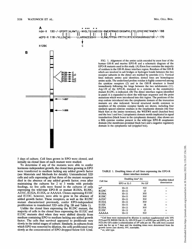
The intron-exon boundaries of the cytokine receptor genesare highly conserved (for an overview, see reference 2). Wealigned the amino acid sequences of the human GH-R andmurine EPO-R exoplasmic domains exon by exon. Motifswhich are conserved in the cytokine receptor superfamily, suchas the four cysteine residues (exons 2 and 3 [not shown]), theproline residue following the GH-R hinge region (exon 4 [Fig.1A]) and the WSXWS homology sequence (exon 5 [notshown]) were aligned initially, and then identical and homol-ogous amino acids were aligned. Gaps were introduced in eachsequence to align identical or conserved residues. Figure 1Ashows the portions encoded by exon 4, containing the majorityof residues of the GH-R at the dimer interface (11). Residue129 of the EPO-R, which is mutated to cysteine in theconstitutively active EPO-R R129C, aligns with residues of theGH-R which participate in intersubunit interactions. Thisalignment suggested residues in proximity to R-129 (R-130,A-131, E-132, and E-133) that might also be involved inintersubunit contacts in a ligand-induced homodimer (Fig.1A).Generation of mutations in the EPO-R dimer interface. To
test this hypothesis, we constructed several mutants in theputative EPO-R dimer interface region (Fig. 1B). Positions 130through 133 of the EPO-R were altered individually to encodealanine (R130A, E132A, and E133A) or cysteine (R129C,R130C, A131C, E132C, and E133C), and in addition, all fiveresidues (129 to 133) were changed to alanine (mutantAAAAA). As analyzed by transient transfection in COS cells,all mutant receptors are expressed on the cell surface and bind125I-EPO. In addition, transfected cells expressing the mutantreceptors internalized 125I-EPO at rates similar to those ofcells expressing the wild-type receptor, as judged by the ratio ofinternal to cell surface'1 I-EPO after a 5-h incubation at 37°C(data not shown).
Proliferative ability of the EPO-R dimer interface mutants.The mutant receptors were assayed for their ability to conferEPO-dependent or hormone-independent proliferation to theIL-3-dependent cell lines 32D or BA/F3. Stable expression ofthe wild-type EPO-R in these cell lines enables them toproliferate in the presence of EPO as the sole supplementalgrowth factor (33, 34). Preliminary studies of 32D cells trans-fected with the EPO-R mutants indicated that all of themutants enabled cells to proliferate in the presence of 1 U ofEPO per ml. Untransfected 32D cells maintained in mediumcontaining either G418 and IL-3 or EPO alone died after 3 to
VOL. 14, 1994
Dow
nloa
ded
from
http
s://j
ourn
als.
asm
.org
/jour
nal/m
cb o
n 30
Jan
uary
202
2 by
119
.237
.156
.199
.
3538 WATOWICH ET AL.
A **** **
hGH-R 130-/-QPDPPIALNWTLL-NVSLTGIHADIQV-RWEAPRNADIQKGWMVLEYELQYKEVNETKWKM/-188I 1 11 I I I 11 I I
mEPO-R 118-/VLLDAPAG----LLARRAEEGSHV---VLRWLPPPGA-PMTTHIRYEVDVSAGNRAGGTQR-/-170
R129C C
B
/R129_-- C-//R130-+- A,CA131+--- CE132-+- A,C
\El133_P A,C
-4--- 1-257
5 days of culture. Cell lines grown in EPO were cloned, andinitially six clonal lines of each mutant were studied.To determine if any of the mutants were able to confer
hormone-independent growth, the clonal lines growing in EPOwere transferred to medium lacking any added growth factor(see Materials and Methods for details). Untransfected 32Dcells and cells expressing all but three of the mutant receptorsdied in the absence of any added growth factor; even aftermaintaining the cultures for 2 to 3 weeks with periodicfeedings, no live cells were found in the cultures of cellsexpressing the wild-type EPO-R or mutant R130A, R130C,A131C, E132A, E133A, or AAAAA. Clones expressing E132Cand E133C, however, were able to grow in the absence ofadded growth factor. These receptors, as well as the R129Cmutant characterized previously, confer EPO-independentproliferation to transfected 32D cells (Fig. 2B and Table 1).
Unlike the clonal lines expressing the R129C mutant, themajority of cells in the clonal lines expressing the E132C andE133C mutants died when they were shifted directly frommedium containing EPO to medium lacking any added growthfactor. The cells that survived appeared to proliferate veryslowly in the initial stages of culture. Similarly, in cultures fromwhich EPO was removed by dilution, the cells proliferated veryslowly as the concentration of EPO dropped below 0.01 U/ml.
FIG. 1. Alignment of the amino acids encoded by exon four of thehuman GH-R and murine EPO-R and a schematic diagram of theEPO-R mutants used in this study. (A) Exon four contains the majorityof residues in the GH-R dimer interface region. Residues of the GH-Rwhich are involved in salt bridges or hydrogen bonds between the tworeceptor subunits in the dimer are marked by asterisks (11). Verticallines indicate amino acid identities; dotted lines are homologousamino acids. The underlined proline residue is highly conserved amongthe cytokine receptors (2) and in the GH-R structure is foundimmediately following the hinge between the two subdomains (11).Arg-129 of the EPO-R, mutated to a cysteine in the constitutivemutant R129C, is indicated. (B) The dimer interface region identifiedin panel A is expanded to show the wild-type sequence and the pointmutations which were introduced into this region. The positions of thestop codons introduced into the cytoplasmic domain of the truncationmutants are also indicated. Several structural motifs common tomembers of the cytokine receptor family are shown, including foursimilarly spaced cysteine residues in the exoplasmic domain (the fourblack bars at the amino terminus), the WSXWS motif (hatched box)and the box 1 and box 2 cytoplasmic domain motifs implicated in signaltransduction (black boxes in the cytoplasmic domain). Also shown area fifth cysteine residue present in the wild-type EPO-R exoplasmicdomain (the membrane-proximal black bar) and a negative regulatorydomain in the cytoplasmic tail (stippled box).
TABLE 1. Doubling times of cell lines expressing the EPO-Rdimer interface mutants
Doubling timea (h) Disulfide-linkedCell line dmrztoEPO or IL-3 No GF dimerization
wt" 10-11 NV -R129C 10-11 15 +R130A 10-11 NV -R130C 10-11 NV -A131C 10-11 NV -E132A 10-11 NV -E132C 10-11 18 +E133A 10-11 NV -E133C 10-11 15 +AAAAA 10-11 NV -
a Cell lines were maintained by dilution in medium supplemented with 10%FCS and 5% WEHI CM (IL-3), 10% FCS and 1 U ofEPO per ml (EPO), or 10%FCS (No GF) under a concentration of 106 cells per ml. The cells were countedeach day for up to 7 days and the doubling times were determined from thegrowth curves (not shown). NV, nonviable.
b wt, wild type.
22
MOL. CELL. BIOL.
-4* 1-306
Dow
nloa
ded
from
http
s://j
ourn
als.
asm
.org
/jour
nal/m
cb o
n 30
Jan
uary
202
2 by
119
.237
.156
.199
.

DIMERIZATION OF THE EPO-R 3539
0
B.
100
3 80E._x
2 600-
.0E 40
o 20
0
FIG. 2mutants iithe wild-tmediummedium N
Materialsand the n
a parallelrepresent(closed sq
(closed timonds)(vR129C (cltriangles)
The factor-independent clonal cell lines were maintainedwithout supplemental growth factor in the medium for 4 to 8weeks prior to testing in dose-response or proliferation assays;at this point in culture, the cell lines were well established andgrowing at a consistent rate. This adaptation phase to growthin medium lacking added growth factors may reflect selectionof cells expressing higher numbers of receptors if there is aminimal number of mutant surface receptors necessary forhormone-independent proliferation.The level of EPO-R expression varied among the 32D clonal
cell lines, as assayed by immunoprecipitation of metabolicallylabeled receptors (data not shown). However, all six clonallines expressing a single mutant were identical with respect togrowth factor requirements, and one clone of each mutant waschosen for further study.
Cells expressing all of the EPO-dependent mutants (R130A,R130C, A131C, E132A, E133A, and AAAAA) proliferated, inthe presence of EPO, at a rate similar to that of cells expressingthe wild-type receptor (Fig. 2A and Table 1). The half-maximalgrowth response was at a concentration of approximately 0.1 Uof EPO per ml (-23 pM), and the cells did not proliferate atconcentrations of less than 0.001 U/ml (Fig. 2A). Thus, indi-vidual substitutions of amino acids 130 to 133 with alanine,
0 0O.0001 0.001 0.01 0.1 1 substitution of residue 130 or 131 with cysteine, or replacementof all five residues (129 to 133) with alanine did not interfere
EPO u/ml with EPO-induced activation of the receptor. All three consti-tutive mutants, R129C, E132C, and E133C, proliferated in lowconcentrations of EPO or in the absence of EPO. However,the cells grew at a faster rate at concentrations of EPO above0.1 U/ml (Fig. 2B). To quantify factor-dependent and -inde-pendent proliferation, the doubling times of the 32D cellsexpressing the mutant EPO-Rs were measured. In the pres-ence of either IL-3 or EPO, cell lines expressing the wild-typeEPO-R or any mutant EPO-R grew at approximately the samerate: -10-h doubling time. Cell lines expressing the constitu-tive mutants R129C, E132C, and E133C grew at a slower rate(15- to 18-h doubling times) in the absence of growth factor,suggesting that in cells expressing these mutants either afraction of EPO-Rs are responsive to EPO and a fraction areconstitutively active or the covalently linked receptors retainsome EPO responsiveness (Table 1).
Covalent dimerization of EPO-R cysteine point mutants. Allmutant EPO-Rs containing substitutions to cysteine weretested for their abilities to form disulfide-linked homodimers.Preliminary studies indicated that, when expressed in 32Dcells, R129C, E132C, and E133C formed disulfide-linkeddimers; the level of expression of the EPO-R in these lines was,
10 t p @ + ..-+ . ,,,,,,,however,very low (data not shown). Thus, BA/F3 cells were
0 0.0001 0.001 0.01 0.1 1 transfected with cDNAs encoding the cysteine mutants andwere selected for growth in EPO. Growth factor-independentEPO u/mi proliferation was tested as described in Materials and Meth-
Proliferation of 32D cells expressing the dimer interface ods. All mutants tested behaved identically in BA/F3 and 32Dn various concentrations of EPO. Clonal cell lines expressing cells with respect to growth factor requirements.type and mutant receptors were tested for proliferation in BA/F3 cells expressing the wild-type EPO-R and mutantssupplemented with EPO (from 0.0001 to I U/ml) or in R129C, R130C, A131C, E132C, and E133C were metabolicallywithout additional growth factor (0 U/ml), as described in . 35 3Sand Methods. After 3 days in culture the cells were counted lael ed with[edi[ o]thiningexfo 1mmandumbers are expressed as a percentage of the cells growing in then were incubated with medium containing excess unlabeled
I culture in the presence of 5% WEHI CM. The symbols amino acids at 18°C for a 2-h chase period. The 18°C chasecells expressing the wild-type EPO-R (closed circles), R130A allows the normally unstable EPO-R polypeptides to accumu-juares), R130C (hatched squares), A131C (crosses), AAAAA late intracellularly. Proteins were immunoprecipitated withriangles), E132A (lined squares), and E133A (closed dia- antiserum raised against a carboxy-terminal peptide of theA) and cells expressing the wild-type EPO-R (closed circles), EPO-R and separated in two-dimensional SDS-polyacrylamidelosed squares), E132C (closed diamonds), and El33C (closed gels in which the first dimension was nonreducing and the(B). second was reducing. As evidenced by an -160-kDa species
under nonreducing conditions that converted to a single -64-kDa species after reduction (Fig. 3, arrows), the constitutively
A.
100
E
xm
0
Ez0
80
60
40
20
VOL. 14, 1994
Dow
nloa
ded
from
http
s://j
ourn
als.
asm
.org
/jour
nal/m
cb o
n 30
Jan
uary
202
2 by
119
.237
.156
.199
.

3540 WATOWICH ET AL.
Non-Reducing
_.C
0.
wild-type-
R13OC-
A131C-
R129C-
E132C-
E133C- *
FIG. 3. Homodimerization of the constitutively active EPO-Rs. BA/F3 cells expressing the wild-type EPO-R or mutants R129C, R130C,A131C, E132C, and E133C were pulse-labeled for 15 min and chased for 2 h at 18°C in the presence of excess unlabeled amino acids.Immunoprecipitated proteins were separated by two-dimensional SDS-PAGE, and the fluorographed gels were visualized by autoradiography. Thedisulfide-linked dimers of R129C, E132C, and E133C (arrows) migrate identically under nonreducing conditions (-160 kDa [data not shown]);the differences detected in this experiment are due to differential swelling of the gels during fluorography.
active mutants R129C, E132C, and E133C formed disulfide-linked dimers, whereas the wild-type EPO-R and the EPO-dependent mutants R130C and A131C did not (Fig. 3). Thedisulfide-linked dimers migrated more slowly than expectedunder nonreducing conditions (-160 versus -128 kDa); it ispossible that the intermolecular disulfide linkage inhibitedSDS from efficiently binding and denaturing the receptors. Aminor, faster migrating form of the disulfide-linked dimers wasoften detected, particularly in cells expressing R129C; theidentity of this species is unknown, although it may be adifferentially phosphorylated or glycosylated form of the re-ceptor.
Disulfide-linked dimers and non-disulfide-linked forms ofR129C are detected on the cell surface at a ratio of approxi-mately 1:1 (51). Cells expressing R129C display a single affinityfor 125I-EPO (Kd = 700 pM), indicating that both monomericand dimeric forms of the receptor bind EPO with the same
affinity (51). Preliminary evidence obtained by cross-linking125I-EPO to intact cells indicates that both monomeric anddimeric forms of E132C and E133C are also found on the cellsurface, although we could not reproducibly determine theratio of monomeric to dimeric forms. BA/F3 cells expressingR129C, E132C, and E133C receptors display a single affinityfor EPO, similar to that of the wild-type receptor (Kd - 700 to1,000 pM; -1,000 binding sites per cell) (data not shown),demonstrating that covalent dimerization of E132C and E133Cis not likely to significantly alter the affinity of the receptor. Asfound previously for the R129C mutant, cell surface expressionof the E132C and E133C mutant receptors is slightly lowerthan that for the wild-type receptor (data not shown).
Generation of cytoplasmic truncations of the EPO-R andanalysis of their biological activity. Since covalent dimeriza-tion of the EPO-R is correlated with constitutive activity, it islikely that disruption of ligand-induced receptor dimerization
MOL. CELL. BIOL.
Dow
nloa
ded
from
http
s://j
ourn
als.
asm
.org
/jour
nal/m
cb o
n 30
Jan
uary
202
2 by
119
.237
.156
.199
.

DIMERIZATION OF THE EPO-R 3541
Non-Reducing Reducing
- 200
97
-69
-46
-30
1 2 3 4FIG. 4. Dimerization of the 1-256/R129C mutant EPO-R. Lysates
from untransfected 32D cells (lanes 1 and 3) or cells expressing the1-256/R129C mutant (lanes 2 and 4) were separated by SDS-PAGEunder nonreducing (lanes 1 and 2) or reducing (lanes 3 and 4)conditions. The proteins were transferred to nitrocellulose, and theblot was incubated with an antiserum specific for the amino terminusof the EPO-R. The migration positions of the disulfide-linked dimers(-64 kDa; black arrowhead) and the monomers of 1-256/R129C (-32kDa; striped arrowhead), as well as the migration positions of molec-ular mass standards (in kilodaltons), are indicated.
will repress EPO-R signal transduction. To test this hypothesis,truncation mutants of the EPO-R cytoplasmic domain (Fig.1B) were assayed for their ability to inhibit the function of thewild-type EPO-R when coexpressed in hematopoietic cells.
Initially, plasmid DNAs encoding the truncated EPO-Rswere transfected into 32D or BA/F3 cells, and cell lines wereselected for growth in medium containing IL-3 and G418.Three mutants were tested. Mutant 1-256/R129C contains nineamino acids of the EPO-R cytoplasmic tail and the R129Cmutation in the exoplasmic domain. Mutants EPO-R/1-257and EPO-R/1-306 have wild-type sequences in the exoplasmicdomain and 10 and 59 residues of the cytoplasmic domain,respectively. Cell lines that synthesized EPO-R polypeptides ofthe predicted size (data not shown) were tested for their abilityto proliferate in the presence of EPO as the sole supplementalgrowth factor or in the absence of supplemented growth factor.None of the mutants was able to confer either EPO-dependentor factor-independent proliferation to the transfected cells,suggesting that portions of the cytoplasmic domain requiredfor EPO-induced proliferation had been deleted (data notshown).
Deletion of the last 40 amino acids from the cytoplasmicdomain of the R129C mutant does not have an effect ondisulfide-linked dimerization (51). However, the possibilitythat membrane-proximal cytoplasmic residues are critical forthe formation of stable EPO-R dimers exists. To determinewhether the membrane-proximal region of the cytoplasmicdomain is required for dimerization, the mutant 1-256/R129Cwas tested for its ability to form disulfide-linked dimers.Disulfide-linked dimers of the 1-256/R129C mutant expressed
in 32D cells were detected following electrophoresis undernonreducing conditions but not under reducing conditions(Fig. 4), demonstrating that deletion of all but nine aminoacids of the EPO-R cytoplasmic domain does not have asignificant effect on covalent dimerization. These results indi-cate that the cytoplasmically truncated EPO-Rs are likely toform ligand-induced homodimers or heterodimers if coex-pressed with the full-length EPO-R.
Coexpression of the wild-type EPO-R and truncated recep-tors. To test the EPO-R truncation mutants for inhibitoryactivity, an EPO-dependent 32D cell line expressing low levelsof the wild-type EPO-R was established. 32D cells weretransfected with the wild-type EPO-R cDNA under the controlof the adenovirus major late promoter in vector pXM, whichwe had previously found to be a poor promoter in these cells.Cell lines were selected by growth in EPO, and several clonallines were isolated. One of these lines, 32Dn20, was found tohave relatively low levels of EPO-R expression (data notshown) and was chosen for use in subsequent studies.
This cell line was transfected with plasmids encoding thetruncated receptors, whose synthesis was driven by a strongpromoter, in order to approximate a 10:1 ratio of truncatedrelative to full-length EPO-Rs. Such a ratio favored thedetection of receptors with dominant negative activity, sincethe level of wild-type to truncated receptor heterodimerswould exceed the level of wild-type receptor homodimers. Toaccomplish this, the EPO-R truncation mutants were sub-cloned into vector pMEX, which contains the Moloney sar-coma virus long terminal repeat as the promoter, as well as theneomycin resistance gene. 32Dn20 cells were transfected witheither the vector alone or the vector encoding the EPO-Rtruncations, and cell lines were selected for growth in IL-3 andG418. Clonal cell lines were isolated and assayed for expres-sion of wild-type and truncated EPO-Rs by immunoblotting ofwhole-cell lysates. Three clonal cell lines from each transfec-tion were chosen for further study.
Biosynthesis of EPO-Rs in the cotransfected cell lines. Toestimate the amount of truncated EPO-Rs and wild-typeEPO-Rs, the coexpressing cell lines were metabolically labeledfor 2.25 h and the EPO-Rs were immunoprecipitated withantiserum specific for the amino or carboxy terminus of themurine EPO-R. A portion of the immunoprecipitated polypep-tides was also treated with endoglycosidase H (endo H) toassess the level of biosynthetic processing of the receptors. Theresults of immunoprecipitations with the carboxy-terminalspecific antiserum, which reacts only with the full-lengthreceptor, are shown in Fig. 5A. The expression level of thewild-type EPO-R in all cell lines is similar, with a maximumtwofold variation. The extent of biosynthetic processing of thereceptor is also similar in all cell lines (Fig. 5A). Figure 5Bshows the results of immunoprecipitations with the amino-terminal antiserum, which has a higher background but whichreacts with both the full-length and truncated EPO-Rs. Thetruncated receptors are synthesized at 5- to 10-fold-higherlevels than the full-length receptor (Fig. 5B). Since the wild-type EPO-R has a half-life of -40 min, in this experiment thecells were labeled for 2.25 h. Although we have not measuredthe half-life of EPO-R/1-306 and EPO-R/1-257, these labelingconditions approximate steady-state conditions, as similar ra-tios of full-length to truncated receptors were obtained byimmunoblot analysis (data not shown).As judged by metabolic pulse-chase experiments, the wild-
type EPO-R is inefficiently transported from the endoplasmicreticulum (ER) to the medial Golgi apparatus in transfected32D cells, with approximately 10 to 30% of the newly synthe-sized receptors acquiring resistance to endo H (37a). Coex-
VOL. 14, 1994
Dow
nloa
ded
from
http
s://j
ourn
als.
asm
.org
/jour
nal/m
cb o
n 30
Jan
uary
202
2 by
119
.237
.156
.199
.

3542 WATOWICH ET AL.
A.
Vector alone EPO-R/1 -306 EPO-R/1 -257
- 1 2 3 1 2 3 1 2 3
+ _ + _ + _ + _ + _ + _ + -I-
1 2 3 4 5 6 7 8 9 10 11 12 13 14 15 16 17 18 19 20
B.
Vector alone EPO-R/1 -306 EPO-R/1 -257
- 1 2 3 1 2 3 1 2 3
+ -+ - + - + - + + - + -+ - + - +
1 2 3 4 5 6 7 8 9 10 11 12 13 14 15 16 17 18 19 20
FIG. 5. Coexpression of the wild-type EPO-R and cytoplasmic truncation mutants in 32D cells. 32Dn2O cells, expressing the wild-type EPO-R,and cotransfected with the vector alone, EPO-R/1-306, or EPO-R/1-257 were metabolically labeled with [35S]methionine and [35S]cysteine for 2.25h, and then EPO-Rs were immunoprecipitated with antiserum specific for the carboxy (A) or amino (B) terminus of the EPO-R. Threeindependently derived clonal lines expressing each mutant were tested in this experiment. Immunoprecipitated polypeptides were left untreated(-) or were digested with endo H (+) and resolved by SDS-PAGE. An autoradiogram of the results is shown. The migration positions of the endoH-resistant form of the wild-type EPO-R (black arrowhead) and deglycosylated, endo H-sensitive form (striped arrowhead) are indicated. Alsomarked are the migration positions of the deglycosylated, endo H-sensitive forms of EPO-R/1-306 and EPO-R/1-257 (bars) and the endoH-resistant species (brackets) (B). The migration positions of molecular mass standards are indicated on the left in kilodaltons.
pression of truncated EPO-Rs with the wild-type receptor in32D cells does not have a significant effect on the amount ofwild-type receptor which acquires endo H resistance (Fig.5A), demonstrating that although the wild-type receptor isinefficiently transported out of the ER, coexpression of a
vast excess of truncated receptor does not impair this trans-port. While there are multiple endo H-resistant species ofthe truncated receptors, the majority of newly synthesizedmolecules retain sensitivity to endo H, indicating that theseforms are also poorly transported from the ER to the Golgi
+ - +
69-
46-
69 -
46_4
30-
J-1l-306
J- 1-2571-306
- 1-257
MOL. CELL. BIOL.
7, 7
Alm.
Zs: ..s:--f- ssp.
JAWow,
Dow
nloa
ded
from
http
s://j
ourn
als.
asm
.org
/jour
nal/m
cb o
n 30
Jan
uary
202
2 by
119
.237
.156
.199
.

DIMERIZATION OF THE EPO-R 3543
TABLE 2. Cell growth and EPO-R surface expression of cell linescoexpressing the wild-type and truncated receptorse
Cell line and % Growth No. of EPO Affinityclone in EPO binding sites/cell (pM)
Vector alone1 80 500 6702 90 360 6203 100 380 520
EPO-R/1-3061 60 670 7802 33 860 5903 45 540 700
EPO-R/1-2571 22 15,500 1,5002 2 27,300 1,0003 16 8,500 900
a 32Dn2O cells, expressing the wild-type EPO-R, cotransfected with the vectoralone, EPO-R/1-306, or EPO-R/1-257 were used in 12'I-EPO equilibriumbinding experiments. Three clonal lines, expressing each mutant, were tested forbinding as described in the legend to Fig. 6, and the results were analyzed by theScatchard procedure. EPO-dependent proliferation (in 100 U of EPO per ml)over a 3-day period, relative to proliferation in IL-3, was determined from thegraphs presented in Fig. 7.
apparatus (Fig. 5B). The identity of the various endo H-resistant species is unclear; they may reflect forms with partialglycosylation or which differ in other posttranslational modifi-cations.
Equilibrium binding experiments. Equilibrium bindingstudies with iodinated EPO were performed on cell linescoexpressing the full-length and truncated receptors to deter-mine the effect of coexpression on receptor affinity and cellsurface levels. BA/F3 or 32D cells expressing the wild-typeEPO-R have very low levels of surface receptors (300 to 2,000sites per cell). These receptors display a single affinity for EPO,with a dissociation constant in the range of 200 to 1,000 pM(51, 54). Table 2 summarizes the results obtained from bindingstudies on the cell lines coexpressing the full-length andtruncated receptors, as analyzed by the Scatchard procedure.Representative Scatchard plots and binding curves are shownin Fig. 6. As seen in Table 2, coexpression of truncatedEPO-Rs does not have a significant effect on receptor Kdvalues. However, surface receptor numbers are increased 2- to50-fold in cells expressing the truncated receptors, comparedwith that in cells expressing only the wild-type EPO-R, sug-gesting that the truncated receptors are expressed at anequivalent (EPO-R/1-306 cell lines) or increased (EPO-RI1-257 cell lines) level relative to that of the wild-type receptor.
Proliferation of cotransfected cell lines in EPO reveals thedominant inhibitory activity ofEPO-R truncation mutants. Todetermine the effect of the truncated EPO-Rs on EPO-responsive proliferation, the transfected 32Dn20 cell lines weretested for their ability to grow in concentrations of EPOranging from 0.001 to 100 U/ml (approximately 230 fM to 23nM) (Fig. 7). 32Dn20 cells transfected with the vector alonehave an EPO response profile identical to that of 32D cellsexpressing the wild-type EPO-R, with half-maximal prolifera-tion (relative to IL-3) at -0.1 U of EPO per ml (cf. Fig. 2A andFig. 7A). In contrast, cells expressing EPO-R/1-306 have areduced level of proliferation in EPO, relative to their prolif-eration in IL-3 (Fig. 7B). The proliferative response of the cellsis not stimulated significantly above -1 to 5 U of EPO per ml,and at concentrations of EPO above 50 U/ml, over 90% of thereceptor binding sites are occupied with hormone, given the Kd
of the EPO-Rs (Table 2). These results indicate that a largeportion of the surface full-length receptors are unable to senda proliferative signal in response to bound ligand. In addition,half-maximal stimulation is seen at a slightly higher EPOconcentration, -0.3 U of EPO per ml, than found in 32Dn20cells transfected with vector alone (Fig. 7A and B).
Expression of EPO-R/1-257 has an even more severe effecton EPO-stimulated proliferation (Fig. 7C). The half-maximalresponse is at - 1 U of EPO per ml, and no significant increasein the proliferative rate is found above EPO concentrations of10 U/ml, indicating that even above 90% receptor occupancyno additional proliferative signals are transmitted. These dataalso suggest that a large portion of the ligand-occupied wild-type surface EPO-Rs are unable to respond to EPO.While the doubling times of the cells in EPO can be
estimated directly from the data in Fig. 7, we chose to measurethe growth rate of the cell lines directly. This independentmeasurement was done to determine if EPO-dependent pro-liferation is exponential or if an initial burst is followed bycessation of growth. The cell lines doubled at a slower rate inEPO than in IL-3 (Table 3), as predicted from Fig. 7, andfollowed an exponential mode of growth (data not shown). Allthe cell lines grew at rates similar to that of untransfected 32Dcells in IL-3 (-10-h doubling time), indicating that the reduc-tion in EPO-responsive proliferation in cells coexpressingfull-length and truncated receptors is specific to EPO-medi-ated signal transduction and is not due to a general defect insignal transduction, DNA synthesis, or cell division.
DISCUSSION
Our results, derived from two independent experimentalapproaches, support the hypothesis that ligand-inducedEPO-R dimerization is an essential step in signal transduction.The first series of experiments provides biochemical evidencefor the role of EPO-R dimerization: EPO-Rs which formcovalent disulfide-linked homodimers through cysteine resi-dues introduced into the predicted receptor dimer interfaceregion are constitutively active for cell proliferation in theabsence of added EPO. The second series of experiments,which demonstrates that the activity of the wild-type receptorcan be inhibited by coexpression with inactive, truncatedEPO-R mutants, provides additional evidence for the func-tional role of receptor dimerization.
Disulfide-linked dimerization of EPO-R mutants correlateswith their constitutive activity: mutants R129C, E132C, andE133C are all constitutively active and form disulfide-linkeddimers, whereas mutants R130C and A131C are wild type withrespect to EPO-dependent proliferation and fail to formdetectable disulfide-linked dimers. The R129C, E132C, andE133C disulfide-linked dimers form in the ER shortly aftersynthesis, as judged by pulse-chase experiments (25a, 51),indicating that residues 129, 132, and 133 in neighboringreceptor molecules are in proximity during receptor foldingand assembly. Although residues 130 and 131 are also pre-dicted to be in the dimer interface region (Fig. 1A), thedistance between the C. atoms in neighboring receptor mole-cules may be greater than 4 to 9 A (0.4 to 0.9 nm), the distancerequired for disulfide bond formation, or the orientation of theside chains may not allow disulfide linkage (7).On the basis of analogy with the GH-R, residues 129, 132,
and 133 of the EPO-R, and perhaps others in proximity, maystabilize the ligand-induced homodimer (11). However, at theGH-R dimer interface, amino acids from one receptor subunitcontact different amino acids in the other subunit (11), raisingthe possibility that either the EPO-R dimer interface region is
VOL. 14, 1994
Dow
nloa
ded
from
http
s://j
ourn
als.
asm
.org
/jour
nal/m
cb o
n 30
Jan
uary
202
2 by
119
.237
.156
.199
.

3544 WATOWICH ET AL.
A.
0.028-110
0.024 8
CL
0 0.00.028 ~ *\0
0.01 20 200 400 600 800100012001400Bound Free (pM)
0.012
0.008
0.004
0 2 4 6 8 1 0 1 2 1 4 1 6
Bound (pM)FIG. 6. Equilibrium 125I-EPO binding and Scatchard analysis. 32Dn20 cells, expressing the wild-type EPO-R, cotransfected with the vector
alone (A), EPO-R/1-306 (B), or EPO-R/1-257 (C) were incubated with various concentrations of '25I-EPO in the presence or absence of 50 nMunlabeled EPO for 14 to 16 h at 4°C. The free and cell-bound radioactivity was counted in a gamma counter, and specific binding was determinedas the difference between radioactivity bound in the absence or presence of 50 nM unlabeled competitor EPO. Scatchard plots of the data as wellas the binding profiles (insets) are shown.
more symmetrical than that of the GH-R or the disulfide-linked EPO-R dimers are structurally different from ligand-induced dimers of the wild-type receptor. Since the R129Cmutant can support EPO-independent proliferation and dif-ferentiation of murine fetal liver erythroid progenitor cells(41), it is unlikely that the disulfide-linked homodimers differsignificantly in structure from the activated wild-type receptor.
Substitution of alanine for any individual residue in positions130 to 133 or for all five residues (129 to 133) did not abolishEPO-dependent proliferation. The corresponding region inthe GH-GH-R structure stabilizes the interaction betweenreceptor subunits, but dimerization of the receptor is driven byligand binding to a different region of the molecule (11). Thereare several possible explanations for why the alanine mutantsare wild type in EPO responsiveness. If the EPO-R dimerinterface region is slightly assymetrical, then the substitutedresidues may not be interacting sufficiently to destabilizeligand-driven dimerization. Since only three of the five resi-dues we have mutagenized are close enough or in the correctorientation to form intermolecular disulfide bonds, it appearsthat their substitution to alanine does not significantly desta-bilize the dimer interface. R129C, E132C, and E133C stimu-late hormone-independent proliferation; thus, it is likely theregion we have mutated forms part of the dimer interface inthe wild-type receptor. It is likely that we have not mapped the
entire dimer interface region, since there are several otherresidues of the EPO-R which align with residues in the GH-Rdimer interface (Fig. IA). Identification of the residues in-volved in intersubunit contacts in the EPO-R dimer will be animportant step towards the development of receptor antago-nists and agonists, as this surface of the EPO-R moleculeprovides a potential target site for their binding. Saturationmutagenesis of the putative dimer interface region by cysteinesubstitutions provides one approach to mapping residues in-volved in dimerization.Our results, as well as previously published results, suggest
that disulfide-linked dimerization of the constitutive mutantsmimics EPO-driven dimerization of the wild-type receptor.However, we have been unable to detect, by biochemicalmeans, ligand-induced dimers of the wild-type receptor. Ourinability to detect cell surface molecules may be due to severaltechnical problems, including an inability to radioiodinate thelow numbers of cell surface receptors (1,000 to 2,000 moleculesper cell) and inefficient chemical cross-linking of receptor-receptor complexes. Even the disulfide-linked dimeric R129Creceptors are difficult to cross-link with N-hydroxysuccinimide-based cross-linkers; this is probably related to the fact that themature, exoplasmic domain of the receptor contains only threelysine residues (9).
In order to bypass these problems, we have pursued an
MOL. CELL. BIOL.
Dow
nloa
ded
from
http
s://j
ourn
als.
asm
.org
/jour
nal/m
cb o
n 30
Jan
uary
202
2 by
119
.237
.156
.199
.

DIMERIZATION OF THE EPO-R 3545
B.
0.04
0.035
0.03
0.025U-m 0.02
0.015'
0.01
0.005
0
0 4 8 12Bound (pM)
1 6 20 24
40 60Bound (pM)
FIG. 6-Continued.
C.
0.072
0.054
U.
m 0.036
0.018
0120
VOL. 14, 1994
Dow
nloa
ded
from
http
s://j
ourn
als.
asm
.org
/jour
nal/m
cb o
n 30
Jan
uary
202
2 by
119
.237
.156
.199
.
3546 WATOWICH ET AL.
A.
EEx
0
.0Ez
i0
0 t-0.001
B.
E 1Ex
02
.0Ez
0
0.01 0.1 1 1 0 100
Epo (u/ml)
0.1 1Epo (u/ml)
C.
120 -
E loo -
EX 80-
60
E 402
-
() 20-0
0.001...r .. ..T
0.01 0.1 1Epo (u/ml)
1 0
alternative strategy. Mutant EPO-Rs, deleted for all or part oftheir cytoplasmic domain, were coexpressed with the wild-typeEPO-R and assayed for the ability to inhibit EPO-driven prolif-eration. Dominant negative mutants have been reported for theEGF-R and c-Kit (3, 25, 32, 42), which are members of thetyrosine kinase receptor family, but not previously for members ofthe cytokine receptor family. Several independent, spontaneousmutations in the murine W (c-kit) locus have a dominant,inhibitory phenotype; these mutant c-Kit receptors have reducedkinase function (17, 38, 40, 47). Their dominant, inhibitoryphenotype in heterozygous animals has been attributed to theformation of inactive complexes due to ligand-induced dimeriza-tion of wild-type and mutant receptors (32, 40). Similarly, domi-nant negative mutants have been generated in the EGF-R bydeleting functional portions of the cytoplasmic tail. Coexpressionof these deleted receptors with the full-length receptor has an
inhibitory effect on signaling by the wild-type EGF-R, because of
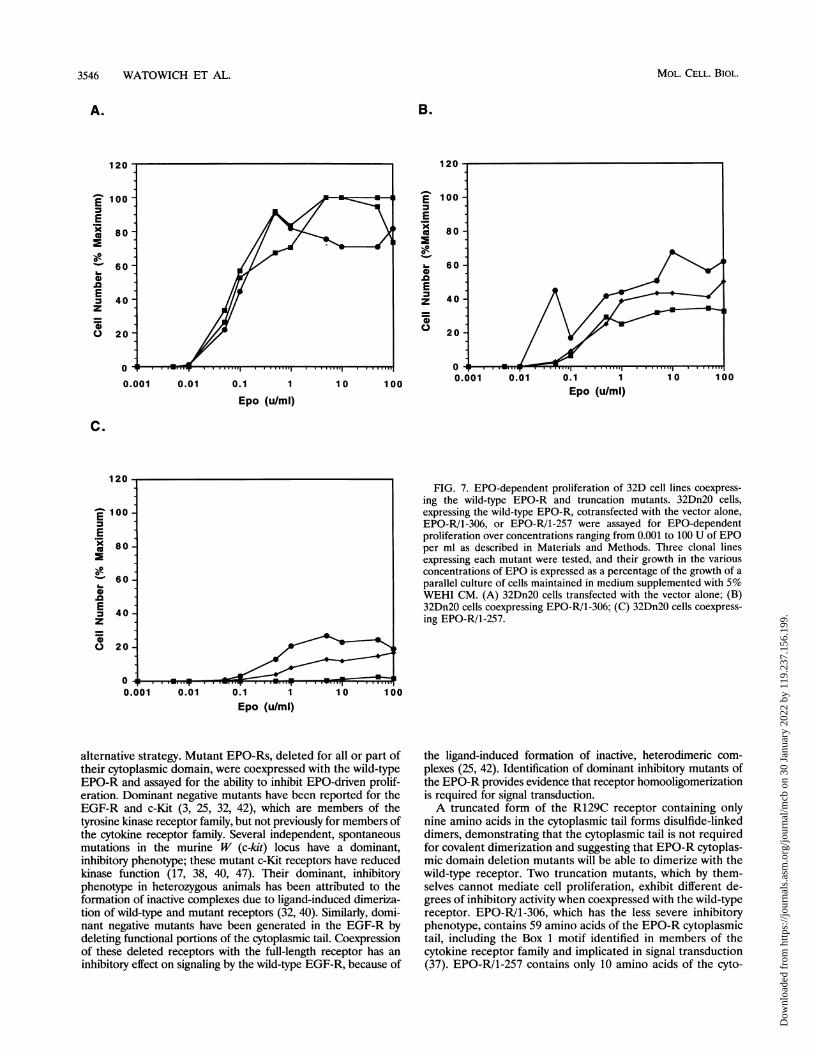
FIG. 7. EPO-dependent proliferation of 32D cell lines coexpress-ing the wild-type EPO-R and truncation mutants. 32Dn20 cells,expressing the wild-type EPO-R, cotransfected with the vector alone,EPO-R/1-306, or EPO-R/1-257 were assayed for EPO-dependentproliferation over concentrations ranging from 0.001 to 100 U of EPOper ml as described in Materials and Methods. Three clonal linesexpressing each mutant were tested, and their growth in the variousconcentrations of EPO is expressed as a percentage of the growth of aparallel culture of cells maintained in medium supplemented with 5%WEHI CM. (A) 32Dn20 cells transfected with the vector alone; (B)32Dn20 cells coexpressing EPO-R/1-306; (C) 32Dn20 cells coexpress-ing EPO-R/1-257.
100
the ligand-induced formation of inactive, heterodimeric com-plexes (25, 42). Identification of dominant inhibitory mutants ofthe EPO-R provides evidence that receptor homooligomerizationis required for signal transduction.A truncated form of the R129C receptor containing only
nine amino acids in the cytoplasmic tail forms disulfide-linkeddimers, demonstrating that the cytoplasmic tail is not requiredfor covalent dimerization and suggesting that EPO-R cytoplas-mic domain deletion mutants will be able to dimerize with thewild-type receptor. Two truncation mutants, which by them-selves cannot mediate cell proliferation, exhibit different de-grees of inhibitory activity when coexpressed with the wild-typereceptor. EPO-R/1-306, which has the less severe inhibitoryphenotype, contains 59 amino acids of the EPO-R cytoplasmictail, including the Box 1 motif identified in members of thecytokine receptor family and implicated in signal transduction(37). EPO-R/1-257 contains only 10 amino acids of the cyto-
MOL. CELL. BIOL.
Dow
nloa
ded
from
http
s://j
ourn
als.
asm
.org
/jour
nal/m
cb o
n 30
Jan
uary
202
2 by
119
.237
.156
.199
.
DIMERIZATION OF THE EPO-R 3547
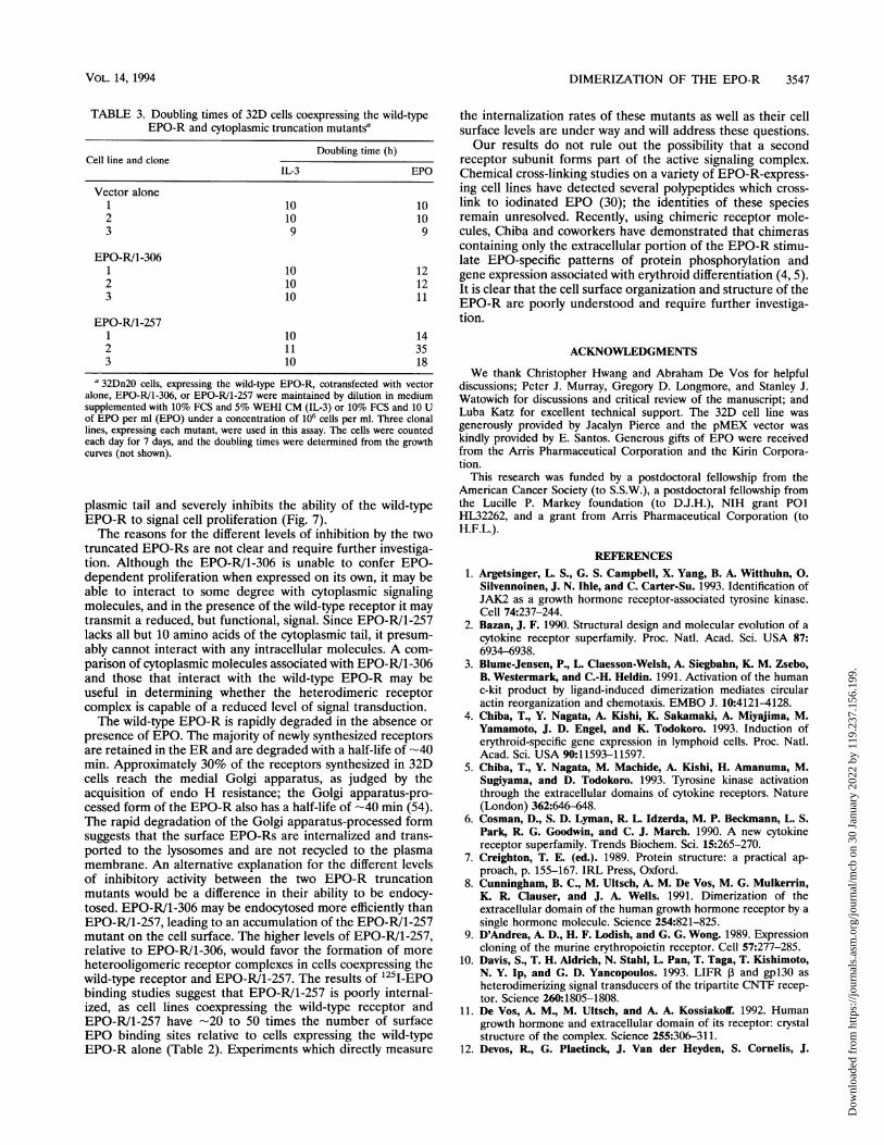
TABLE 3. Doubling times of 32D cells coexpressing the wild-typeEPO-R and cytoplasmic truncation mutantsa
Doubling time (h)Cell line and clone
IL-3 EPO
Vector alone1 10 102 10 103 9 9
EPO-R/1-3061 10 122 10 123 10 11
EPO-R/1-2571 10 142 11 353 10 18
a 32Dn20 cells, expressing the wild-type EPO-R, cotransfected with vectoralone, EPO-R/1-306, or EPO-R/1-257 were maintained by dilution in mediumsupplemented with 10% FCS and 5% WEHI CM (IL-3) or 10% FCS and 10 Uof EPO per ml (EPO) under a concentration of 106 cells per ml. Three clonallines, expressing each mutant, were used in this assay. The cells were countedeach day for 7 days, and the doubling times were determined from the growthcurves (not shown).
plasmic tail and severely inhibits the ability of the wild-typeEPO-R to signal cell proliferation (Fig. 7).The reasons for the different levels of inhibition by the two
truncated EPO-Rs are not clear and require further investiga-tion. Although the EPO-R/1-306 is unable to confer EPO-dependent proliferation when expressed on its own, it may beable to interact to some degree with cytoplasmic signalingmolecules, and in the presence of the wild-type receptor it maytransmit a reduced, but functional, signal. Since EPO-R/1-257lacks all but 10 amino acids of the cytoplasmic tail, it presum-ably cannot interact with any intracellular molecules. A com-parison of cytoplasmic molecules associated with EPO-RI1-306and those that interact with the wild-type EPO-R may beuseful in determining whether the heterodimeric receptorcomplex is capable of a reduced level of signal transduction.The wild-type EPO-R is rapidly degraded in the absence or
presence of EPO. The majority of newly synthesized receptorsare retained in the ER and are degraded with a half-life of -40min. Approximately 30% of the receptors synthesized in 32Dcells reach the medial Golgi apparatus, as judged by theacquisition of endo H resistance; the Golgi apparatus-pro-cessed form of the EPO-R also has a half-life of -40 min (54).The rapid degradation of the Golgi apparatus-processed formsuggests that the surface EPO-Rs are internalized and trans-ported to the lysosomes and are not recycled to the plasmamembrane. An alternative explanation for the different levelsof inhibitory activity between the two EPO-R truncationmutants would be a difference in their ability to be endocy-tosed. EPO-R/1-306 may be endocytosed more efficiently thanEPO-R/1-257, leading to an accumulation of the EPO-R/1-257mutant on the cell surface. The higher levels of EPO-R/1-257,relative to EPO-RI1-306, would favor the formation of moreheterooligomeric receptor complexes in cells coexpressing thewild-type receptor and EPO-R/1-257. The results of 125I-EPObinding studies suggest that EPO-R/1-257 is poorly internal-ized, as cell lines coexpressing the wild-type receptor andEPO-R/1-257 have -20 to 50 times the number of surfaceEPO binding sites relative to cells expressing the wild-typeEPO-R alone (Table 2). Experiments which directly measure
the internalization rates of these mutants as well as their cellsurface levels are under way and will address these questions.Our results do not rule out the possibility that a second
receptor subunit forms part of the active signaling complex.Chemical cross-linking studies on a variety of EPO-R-express-ing cell lines have detected several polypeptides which cross-link to iodinated EPO (30); the identities of these speciesremain unresolved. Recently, using chimeric receptor mole-cules, Chiba and coworkers have demonstrated that chimerascontaining only the extracellular portion of the EPO-R stimu-late EPO-specific patterns of protein phosphorylation andgene expression associated with erythroid differentiation (4, 5).It is clear that the cell surface organization and structure of theEPO-R are poorly understood and require further investiga-tion.
ACKNOWLEDGMENTS
We thank Christopher Hwang and Abraham De Vos for helpfuldiscussions; Peter J. Murray, Gregory D. Longmore, and Stanley J.Watowich for discussions and critical review of the manuscript; andLuba Katz for excellent technical support. The 32D cell line wasgenerously provided by Jacalyn Pierce and the pMEX vector waskindly provided by E. Santos. Generous gifts of EPO were receivedfrom the Arris Pharmaceutical Corporation and the Kirin Corpora-tion.
This research was funded by a postdoctoral fellowship from theAmerican Cancer Society (to S.S.W.), a postdoctoral fellowship fromthe Lucille P. Markey foundation (to D.J.H.), NIH grant P01HL32262, and a grant from Arris Pharmaceutical Corporation (toH.F.L.).
REFERENCES1. Argetsinger, L. S., G. S. Campbell, X. Yang, B. A. Witthuhn, 0.
Silvennoinen, J. N. Ihle, and C. Carter-Su. 1993. Identification ofJAK2 as a growth hormone receptor-associated tyrosine kinase.Cell 74:237-244.
2. Bazan, J. F. 1990. Structural design and molecular evolution of acytokine receptor superfamily. Proc. Natl. Acad. Sci. USA 87:6934-6938.
3. Blume-Jensen, P., L. Claesson-Welsh, A. Siegbahn, K. M. Zsebo,B. Westermark, and C.-H. Heldin. 1991. Activation of the humanc-kit product by ligand-induced dimerization mediates circularactin reorganization and chemotaxis. EMBO J. 10:4121-4128.
4. Chiba, T., Y. Nagata, A. Kishi, K. Sakamaki, A. Miyajima, M.Yamamoto, J. D. Engel, and K. Todokoro. 1993. Induction oferythroid-specific gene expression in lymphoid cells. Proc. Natl.Acad. Sci. USA 90:11593-11597.
5. Chiba, T., Y. Nagata, M. Machide, A. Kishi, H. Amanuma, M.Sugiyama, and D. Todokoro. 1993. Tyrosine kinase activationthrough the extracellular domains of cytokine receptors. Nature(London) 362:646-648.
6. Cosman, D., S. D. Lyman, R. L. Idzerda, M. P. Beckmann, L. S.Park, R. G. Goodwin, and C. J. March. 1990. A new cytokinereceptor superfamily. Trends Biochem. Sci. 15:265-270.
7. Creighton, T. E. (ed.). 1989. Protein structure: a practical ap-proach, p. 155-167. IRL Press, Oxford.
8. Cunningham, B. C., M. Ultsch, A. M. De Vos, M. G. Mulkerrin,K. R. Clauser, and J. A. Wells. 1991. Dimerization of theextracellular domain of the human growth hormone receptor by asingle hormone molecule. Science 254:821-825.
9. D'Andrea, A. D., H. F. Lodish, and G. G. Wong. 1989. Expressioncloning of the murine erythropoietin receptor. Cell 57:277-285.
10. Davis, S., T. H. Aldrich, N. Stahl, L. Pan, T. Taga, T. Kishimoto,N. Y. Ip, and G. D. Yancopoulos. 1993. LIFR P and gpl30 asheterodimerizing signal transducers of the tripartite CNTF recep-tor. Science 260:1805-1808.
11. De Vos, A. M., M. Ultsch, and A. A. Kossiakoff. 1992. Humangrowth hormone and extracellular domain of its receptor: crystalstructure of the complex. Science 255:306-311.
12. Devos, R., G. Plaetinck, J. Van der Heyden, S. Cornelis, J.
VOL. 14, 1994
Dow
nloa
ded
from
http
s://j
ourn
als.
asm
.org
/jour
nal/m
cb o
n 30
Jan
uary
202
2 by
119
.237
.156
.199
.

3548 WATOWICH ET AL.
Vandekerckhove, W. Fiers, and J. Tavernier. 1991. Molecularbasis of a high affinity murine interleukin-5 receptor. EMBO J.10:2133-2137.
13. Elberg, G., P. A. Kelly, J. Djiane, L. Binder, and A. Gertler. 1990.Mitogenic and binding properties of monoclonal antibodies to theprolactin receptor in Nb2 rat lymphoma cells. Selective enhance-ment by anti-mouse IgG. J. Biol. Chem. 265:14770-14776.
14. Fuh, G., B. C. Cunningham, R. Fukunaga, S. Nagata, D. V.Goeddel, and J. A. Wells. 1992. Rational design of potent antag-onists to the human growth hormone receptor. Science 256:1677-1680.
15. Fukunaga, R., E. Ishizaka-Ikeda, and S. Nagata. 1990. Purificationand characterization of the receptor for murine granulocytecolony-stimulating factor. J. Biol. Chem. 265:14008-14015.
16. Gearing, D. P., M. R. Comeau, D. J. Friend, S. D. Gimpel, C. J.Thut, J. McGourty, K. K. Brasher, J. A. King, S. Gillis, B. Mosley,S. F. Ziegler, and D. Cosman. 1992. The IL-6 signal transducer,gpl3O: an oncostatin M receptor and affinity converter for the LIFreceptor. Science 255:1434-1437.
17. Geissler, E. N., M. A. Ryan, and D. E. Housman. 1988. Thedominant-white spotting (W) locus of the mouse encodes the c-kitproto-oncogene. Cell 55:185-192.
18. Hara, T., and A. Miyajima. 1992. Two distinct functional highaffinity receptors for mouse interleukin-3 (IL-3). EMBO J. 11:1875-1884.
19. Hayashida, K., T. Kitamura, D. M. Gorman, K.-I. Arai, T. Yokota,and A. Miyajima. 1990. Molecular cloning of a second subunit ofthe receptor for human granulocyte-macrophage colony-stimulat-ing factor (GM-CSF): reconstitution of a high-affinity GM-CSFreceptor. Proc. Natl. Acad. Sci. USA 87:9655-9659.
20. Hibi, M., M. Murakami, M. Saito, T. Hirano, T. Taga, and T.Kishimoto. 1990. Molecular cloning and expression of an IL-6signal transducer, gpl3O. Cell 63:1149-1157.
21. Hilton, D. J., N. A. Nicola, and D. Metcalf. 1988. Specific bindingof murine leukemia inhibitory factor to normal and leukemicmonocytic cells. Proc. Natl. Acad. Sci. USA 85:5971-5975.
21a.Hilton, D. J., S. S. Watowich, L. Katz, and H. F. Lodish. Submittedfor publication.
22. Hooper, K. P., R. Padmanabhan, and K. E. Ebner. 1993. Expres-sion of the extracellular domain of the rat liver prolactin receptorand its interaction with ovine prolactin. J. Biol. Chem. 268:22347-22352.
23. Ip, N. Y., S. H. Nye, T. G. Boulton, S. Davis, T. Taga, Y. Li, S. J.Birren, K. Yasukawa, T. Kishimoto, D. J. Anderson, N. Stahl, andG. D. Yancopoulos. 1992. CNTF and LIF act on neuronal cells viashared signaling pathways that involve the IL-6 signal transducingreceptor component gpl3O. Cell 69:1121-1132.
24. Kanakura, Y., B. Druker, S. A. Cannistra, Y. Furukawa, Y.Torimoto, and J. D. Griffin. 1990. Signal transduction of thehuman granulocyte-macrophage colony-stimulating factor and in-terleukin-3 receptors involves tyrosine phosphorylation of a com-mon set of cytoplasmic proteins. Blood 76:706-715.
25. Kashles, O., Y. Yarden, R. Fischer, A. Ullrich, and J. Schlessinger.1991. A dominant negative mutation suppresses the function ofnormal epidermal growth factor receptors by heterodimerization.Mol. Cell. Biol. 11:1454-1463.
25a.Katz, L., and S. S. Watowich. Unpublished data.26. Kitamura, T., K. Hayashida, K. Sakamaki, T. Yokota, K.-I. Arai,
and A. Miyajima. 1991. Reconstitution of functional receptors forhuman granulocyte/macrophage colony-stimulating factor (GM-CSF): evidence that the protein encoded by the AIC2B cDNA isa subunit of the murine GM-CSF receptor. Proc. Natl. Acad. Sci.USA 88:5082-5086.
27. Kitamura, T., and A. Miyajima. 1992. Functional reconstitution ofthe human interleukin-3 receptor. Blood 80:84-90.
28. Kitamura, T., N. Sato, K.-I. Arai, and A. Miyajima. 1991. Expres-sion cloning of the human IL-3 receptor cDNA reveals a shared Psubunit for the human IL-3 and GM-CSF receptors. Cell 66:1165-1174.
29. Koch, C. A., D. Anderson, M. F. Moran, C. Ellis, and T. Pawson.1991. SH2 and SH3 domains: elements that control interactions ofcytoplasmic signaling proteins. Science 252:668-674.
30. Krantz, S. B. 1991. Erythropoietin. Blood 77:419-434.
31. Laemmli, U. K. 1970. Cleavage of structural proteins during theassembly of the head of bacteriophage T4. Nature (London)227:680-685.
32. Lev, S., Y. Yarden, and D. Givol. 1992. Dimerization and activationof the kit receptor by monovalent and bivalent binding of the stemcell factor. J. Biol. Chem. 267:15970-15977.
33. Li, J.-P., A. D. D'Andrea, H. F. Lodish, and D. Baltimore. 1990.Activation of cell growth by binding of Friend spleen focus-forming virus gp55 glycoprotein to the erythropoietin receptor.Nature (London) 343:762-764.
34. Migliaccio, A. R., G. Migliaccio, A. D. D'Andrea, M. Baiocchi, S.Crotta, S. Nicolis, S. Ottolenghi, and J. W. Adamson. 1991.Response to erythropoietin in erythroid subclones of the factor-dependent cell line 32D is determined by translocation of theerythropoietin receptor to the cell surface. Proc. Natl. Acad. Sci.USA 88:11086-11090.
35. Miura, O., A. D. D'Andrea, D. Kabat, and J. N. IhIe. 1991.Induction of tyrosine phosphorylation by the erythropoietin recep-tor correlates with mitogenesis. Mol. Cell. Biol. 11:4895-4902.
36. Murakami, M., M. Hibi, N. Nakagawa, T. Nakagawa, K. Ya-sukawa, K. Yamanishi, T. Taga, and T. Kishimoto. 1993. IL-6-induced homodimerization of gpl3O and associated activation of atyrosine kinase. Science 260:1808-1810.
37. Murakami, M., M. Narazaki, M. Hibi, H. Yawata, K. Yasukawa,M. Hamaguchi, T. Taga, and T. Kishimoto. 1991. Critical cyto-plasmic region of the interleukin 6 signal transducer gpl30 isconserved in the cytokine receptor family. Proc. Natl. Acad. Sci.USA 88:11349-11353.
37a.Murray, P. J., S. S. Watowich, H. F. Lodish, R. A. Young, and D. J.Hilton. Unpublished data.
38. Nocka, K., J. C. Tan, E. Chiu, T. Y. Chu, P. Ray, P. Traktman, andP. Besmer. 1990. Molecular bases of dominant negative and loss offunction mutations at the murine c-kit/white spotting locus: W37,Wv, W4' and W. EMBO J. 9:1805-1813.
39. Noguchi, M., H. Yi, H. M. Rosenblatt, A. H. Filipovich, S.Adelstein, W. S. Modi, 0. W. McBride, and W. J. Leonard. 1993.Interleukin-2 receptor gamma chain mutation results in X-linkedsevere combined immunodeficiency in humans. Cell 73:147-157.
40. Pawson, T., and A. Bernstein. 1990. Receptor tyrosine kinases:genetic evidence for their role in Drosophila and mouse develop-ment. Trends Genet. 6:350-356.
41. Pharr, P. N., D. Hankins, A. Hofbauer, H. F. Lodish, and G. D.Longmore. 1993. Expression of a constitutively active erythropoi-etin receptor in primary hematopoietic progenitors abrogateserythropoietin dependence and enhances erythroid colony-form-ing unit, erythroid burst-forming unit, and granulocyte/macro-phage progenitor growth. Proc. Natl. Acad. Sci. USA 90:938-942.
42. Redemann, N., B. Holzmann, T. von Ruden, E. F. Wagner, J.Schiessinger, and A. Ulirich. 1992. Anti-oncogenic activity ofsignalling-defective epidermal growth factor receptor mutants.Mol. Cell. Biol. 12:491-498.
43. Rozakis-Adcock, M., and P. A. Kelly. 1991. Mutational analysis ofthe ligand-binding domain of the prolactin receptor. J. Biol. Chem.266:16472-16477.
44. Silvennoinen, O., B. A. Witthuhn, F. W. Quelle, J. L. Cleveland, T.Yi, and J. N. Ihle. 1993. Structure of the murine Jak2 protein-tyrosine kinase and its role in interleukin 3 signal transduction.Proc. Natl. Acad. Sci. USA 90:8429-8433.
45. Taga, T., M. Hibi, Y. Hirata, K. Yamasaki, K. Yasukawa, T.Matsuda, T. Hirano, and T. Kishimoto. 1989. Interleukin-6 trig-gers the association of its receptor with a possible signal trans-ducer, gpl3O. Cell 58:573-581.
46. Takaki, S., S. Mita, T. Kitamura, S. Yonehara, N. Yamaguchi, A.Tominaga, A. Miyajima, and K. Takatsu. 1991. Identification ofthe second subunit of the murine interleukin-5 receptor: interleu-kin-3 receptor-like protein, AIC2B is a component of the highaffinity interleukin-5 receptor. EMBO J. 10:2833-2838.
47. Tan, J. C., K. Nocka, P. Ray, P. Traktman, and P. Besmer. 1990.The dominant W42 spotting phenotype results from a missensemutation in the c-kit receptor kinase. Science 247:209-212.
48. Taniguchi, T., and Y. Minami. 1993. The IL-2/IL-2 receptorsystem: a current overview. Cell 73:5-8.
MOL. CELL. BIOL.
Dow
nloa
ded
from
http
s://j
ourn
als.
asm
.org
/jour
nal/m
cb o
n 30
Jan
uary
202
2 by
119
.237
.156
.199
.
DIMERIZATION OF THE EPO-R 3549
49. Tavernier, J., R. Devos, S. Cornelis, T. Tuypens, J. Van derHeyden, W. Fiers, and G. Plaetinck 1991. A human high affinityinterleukin-5 receptor (ILSR) is composed of an IL5-specific achain and a ,B chain shared with the receptor for GM-CSF. Cell66:1175-1184.
50. Ullrich, A., and J. Schlessinger. 1990. Signal transduction byreceptors with tyrosine kinase activity. Cell 61:203-212.
51. Watowich, S. S., A. Yoshimura, G. D. Longmore, D. J. Hilton, Y.Yoshimura, and H. F. Lodish. 1992. Homodimerization andconstitutive activation of the erythropoietin receptor. Proc. Natl.Acad. Sci. USA 89:2140-2144.
52. Witthuhn, B. A., F. W. Quelle, 0. Silvennoinen, T. Yi, B. Tang, 0.
Miura, and J. N. Ihle. 1993. JAK2 associates with the erythropoi-etin receptor and is tyrosine phosphorylated and activated follow-ing stimulation with erythropoietin. Cell 74:227-236.
53. Yang, Y. C., A. B. Ciarletta, P. A. Temple, M. P. Chung, S. Kovacic,J. S. Witek-Giannotti, A. C. Leary, R. Kriz, R. E. Donahue, G. G.Wong, and S. C. ClarL 1986. Human IL-3 (multi-CSF): identifi-cation by expression cloning of a novel hematopoietic growthfactor related to murine IL-3. Cell 47:3-10.
54. Yoshimura, A., A. D. D'Andrea, and H. F. Lodish. 1990. Friendspleen focus-forming virus glycoprotein gp55 interacts with theerythropoietin receptor in the endoplasmic reticulum and af-fects receptor metabolism. Proc. Natl. Acad. Sci. USA 87:4139-4143.
55. Yoshimura, A., G. Longmore, and H. F. Lodish. 1990. Pointmutation in the exoplasmic domain of the erythropoietin receptorresulting in hormone-independent activation and tumorigenicity.Nature (London) 348:647-649.
VOL. 14, 1994
Dow
nloa
ded
from
http
s://j
ourn
als.
asm
.org
/jour
nal/m
cb o
n 30
Jan
uary
202
2 by
119
.237
.156
.199
.