activated irrigation by gentlefile during root canal … · activated irrigation by gentlefile...
TRANSCRIPT

34
ACTIVATED IRRIGATION BY GENTLEFILE DURING ROOT CANAL THERAPY
Agata Koprowicz, Halina Pawlicka, Joanna B. Grącka-Mańkowska
Department of Endodontics, Medical University of Łódź, Poland
A B S T R A C T
Introduction: The development of modern dentistry has changed the nature of endodontic therapy. Formerly arduous and strenuous root canal treatment has become a sequence of precise procedures, usually with no, or negligible, side-effects. The introduction of rotary nickel-titanium (NiTi) files onto the market has considerably shortened the time of canal preparation. Unchangeably, the mechanical preparation is aimed at shaping the canal, having preserved its original curvature, removing the infected tissue from the root canal system, and allowing dense and homogeneous obturation of the canal. Regardless of the cross-section of traditional rotary files, it has been confirmed that contact zones of rotary NiTi instruments never negotiate the canal walls in their entirety. The remaining wall surface of the canal is intact and therefore remains untreated. To improve the root canal dis-infection, the preparation stage should include a chemical phase with the use of rinsing agents. Current published research findings indicate that activation of the irrigation solutions improves their cleansing properties. As a con-sequence, many researchers constantly develop the new methods and aim for new equipment to allow efficient irrigation of the root canal system. Case description: The article presents four clinical cases managed with the use of a new instrument called the Gentlefile (MedicNRG, Israel), which serves not only for mechanical preparation but also for the activation of rinsing agents during endodontic treatment. Conclusions: After completing the mechanical preparation of the root canals, the application of the appropriate protocol of rinsing seems to be an elementary stage for successful endodontic treatment. Activation of rinsing agents with the Gentlefile, by improving the effectiveness of cleaning, enables an impermeable, fluid tight seal within the en-tire root canal, increasing the success rate in endodontic treatment.
Key words: irrigation, Gentlefile, root canal therapy.
J Stoma 2019; 72, 1: 34–42DOI: https://doi.org/10.5114/jos.2019.86495
INTRODUCTION
The main goal of endodontic treatment is to remove the irreversibly inflamed pulp, disinfect the root canals, and shape them, which will enable three-dimensional obturation, healing of periapical lesions, and protection against reinfection [5]. Both manual and mechanical canal preparation results in the creation of the smear
layer (of different thickness) on the walls of the ca-nal [17]. Studies confirm that removal of the smear layer by activation of rinsing agents increases the adhe-sion of the filling material and reduces the occurrence of micro-leakage along the canal wall [6]. Various meth-ods of activation of the rinsing agents are described in the literature, some of which are as simple as mixing the solutions using a gutta-percha cone or an irrigation
C A S E R E P O R T © 2019 Polish Dental Association
Address for correspondence: Agata Koprowicz, Department of Endodontics, Medical University of Łódź, 251 Pomorska St., 92-213 Łódź, Poland, e-mail: [email protected]
Received: 27.01.2019 • Accepted: 26.05.2019 • Published: 28.06.2019OFFICIAL JOURNAL OF THE POLISH DENTAL ASSOCIATION ORGAN POLSKIEGO TOWARZYSTWA STOMATOLOGICZNEGO
Vol. 71
Bimonthly ISSN 0011-4553Vol. 71 Issue 3 May-June 2018 p. 249-314
20183
The relationship between temporomandibular disorder and work stress in type C private hospital nursesFadhilah Nur Amalina, Ira Tanti, David Maxwell
The relationship between interleukin-18 level in smokers and chronic periodontitis: radiographic overview of posterior mandibular teeth
F.X. Andi Wiyanto, Sri Lelyati C. Masulili, Elza Ibrahim Auerkari, Fatimah Maria Tadjoedin
Antifungal effectivity of virgin coconut oil mousse against Candida albicans biofilm in children with early childhood caries
Monica Monica, Eva Fauziah, Sarworini Bagio Budiardjo, Margaretha Suharsini, Heriandi Sutadi, Ike Siti Indiarti, Mochamad Fahlevi Rizal
In vitro efficacy of garlic extract against Candida albicans biofilms from children with early childhood caries Mochamad Rizal, Sarworini Budiardjo, Vidya Tjokrosetio, Eva Fauziah, Ike Indiarti, Heriandi Sutadi, Margaretha Suharsini
Dental health of five-year-old children in Mazowieckie province as revealed by monitoring of dental health and its determinants in 2011 and 2016
Małgorzata Dudek, Iwona Soika, Weronika Jończyk, Anna Turska-Szybka, Dariusz Gozdowski, Dorota Olczak-Kowalczyk
The use of polymerase chain reaction in patients with periodontal disease before prosthetic treatmentKatarzyna Taraszkiewicz-Sulik, Gabriela Pękała, Łukasz Magnuszewski, Maria Gołębiewska
Cognitive functioning and myofascial pain in masticatory organ dysfunctionEwa Ferendiuk, Józef Gierowski, Małgorzata Pihut, Joanna Biegańska-Banaś
Orthodontic and surgical treatment of a patient with an impacted upper central incisor with dilacerations – systematic review of the literature with the presentation of a case
Magdalena Rudnik, Bartłomiej Loster
Comparison of five deep caries management methods and their use in contemporary dentistryLidia Postek-Stefańska, Alicja Leś-Smolarczyk, Anna Jodłowska
The C-shaped second mandibular molar and intentional replantationElżbieta Bołtacz-Rzepkowska, Agnieszka Żęcin, Michał Łęski
35
Gentlefile in root canal therapy
J Stoma 2019, 72, 1
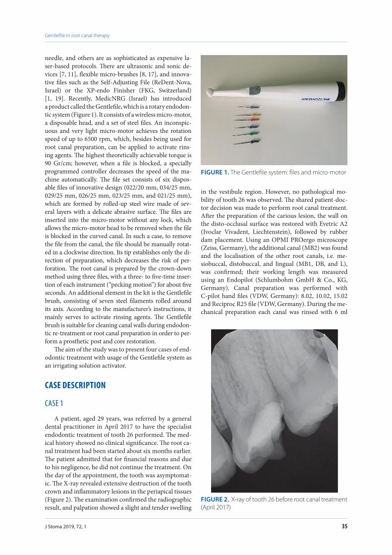
needle, and others are as sophisticated as expensive la-ser-based protocols. There are ultrasonic and sonic de-vices [7, 11], flexible micro-brushes [8, 17], and innova-tive files such as the Self-Adjusting File (ReDent-Nova, Israel) or the XP-endo Finisher (FKG, Switzerland) [1, 19]. Recently, MedicNRG (Israel) has introduced a product called the Gentlefile, which is a rotary endodon-tic system (Figure 1). It consists of a wireless micro-motor, a disposable head, and a set of steel files. An inconspic-uous and very light micro-motor achieves the rotation speed of up to 6500 rpm, which, besides being used for root canal preparation, can be applied to activate rins-ing agents. The highest theoretically achievable torque is 90 Gr/cm; however, when a file is blocked, a specially programmed controller decreases the speed of the ma-chine automatically. The file set consists of six dispos-able files of innovative design (022/20 mm, 034/25 mm, 029/25 mm, 026/25 mm, 023/25 mm, and 021/25 mm), which are formed by rolled-up steel wire made of sev-eral layers with a delicate abrasive surface. The files are inserted into the micro-motor without any lock, which allows the micro-motor head to be removed when the file is blocked in the curved canal. In such a case, to remove the file from the canal, the file should be manually rotat-ed in a clockwise direction. Its tip establishes only the di-rection of preparation, which decreases the risk of per-foration. The root canal is prepared by the crown-down method using three files, with a three- to five-time inser-tion of each instrument (“pecking motion”) for about five seconds. An additional element in the kit is the Gentlefile brush, consisting of seven steel filaments rolled around its axis. According to the manufacturer’s instructions, it mainly serves to activate rinsing agents. The Gentlefile brush is suitable for cleaning canal walls during endodon-tic re-treatment or root canal preparation in order to per-form a prosthetic post and core restoration.
The aim of the study was to present four cases of end-odontic treatment with usage of the Gentlefile system as an irrigating solution activator.
CASE DESCRIPTION
CASE 1
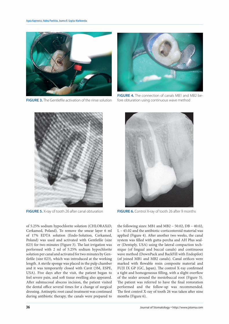
A patient, aged 29 years, was referred by a general dental practitioner in April 2017 to have the specialist endodontic treatment of tooth 26 performed. The med-ical history showed no clinical significance. The root ca-nal treatment had been started about six months earlier. The patient admitted that for financial reasons and due to his negligence, he did not continue the treatment. On the day of the appointment, the tooth was asymptomat-ic. The X-ray revealed extensive destruction of the tooth crown and inflammatory lesions in the periapical tissues (Figure 2). The examination confirmed the radiographic result, and palpation showed a slight and tender swelling
in the vestibule region. However, no pathological mo-bility of tooth 26 was observed. The shared patient-doc-tor decision was made to perform root canal treatment. After the preparation of the carious lesion, the wall on the disto-occlusal surface was restored with Evetric A2 (Ivoclar Vivadent, Liechtenstein), followed by rubber dam placement. Using an OPMI PROergo microscope (Zeiss, Germany), the additional canal (MB2) was found and the localisation of the other root canals, i.e. me-siobuccal, distobuccal, and lingual (MB1, DB, and L), was confirmed; their working length was measured using an Endopilot (Schlumbohm GmbH & Co., KG, Germany). Canal preparation was performed with C-pilot hand files (VDW, Germany): 8.02, 10.02, 15.02 and Reciproc R25 file (VDW, Germany). During the me-chanical preparation each canal was rinsed with 6 ml
FIGURE 1. The Gentlefile system: files and micro-motor
FIGURE 2. X-ray of tooth 26 before root canal treatment (April 2017)
Journal of Stomatology * http://www.jstoma.com36
Agata Koprowicz, Halina Pawlicka, Joanna B. Grącka-Mańkowska
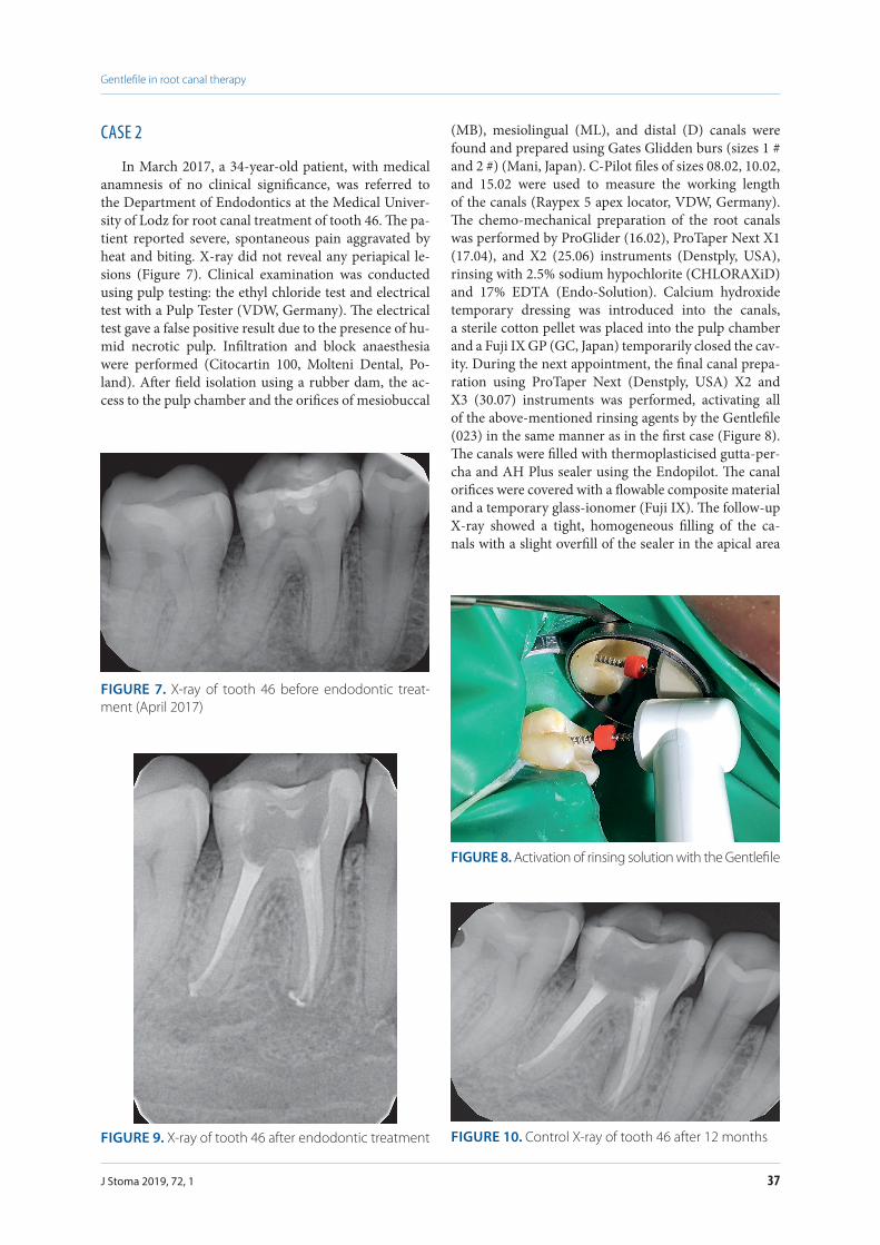
of 5.25% sodium hypochlorite solution (CHLORAXiD, Cerkamed, Poland). To remove the smear layer 6 ml of 17% EDTA solution (Endo-Solution, Cerkamed, Poland) was used and activated with Gentlefile (size 023) for two minutes (Figure 3). The last irrigation was performed with 2 ml of 5.25% sodium hypochlorite solution per canal and activated for two minutes by Gen-tlefile (size 023), which was introduced at the working length. A sterile sponge was placed in the pulp chamber and it was temporarily closed with Cavit (3M, ESPE, USA). Five days after the visit, the patient began to feel severe pain, and soft tissue swelling also appeared. After submucosal abscess incision, the patient visited the dental office several times for a change of surgical dressing. Antiseptic root canal treatment was continued during antibiotic therapy, the canals were prepared to
the following sizes: MB1 and MB2 – 50.02, DB – 40.02, L – 45.02 and the antibiotic-corticosteroid material was applied (Figure 4). After another two weeks, the canal system was filled with gutta-percha and AH Plus seal-er (Dentsply, USA) using the lateral compaction tech-nique (of lingual and buccal canals) and continuous wave method (DownPack and BackFill with Endopilot) (of joined MB1 and MB2 canals). Canal orifices were marked with flowable resin composite material and FUJI IX GP (GC, Japan). The control X-ray confirmed a tight and homogeneous filling, with a slight overflow of the sealer around the mesiobuccal root (Figure 5). The patient was referred to have the final restoration performed and the follow-up was recommended. The first control X-ray of tooth 26 was taken after nine months (Figure 6).
FIGURE 3. The Gentlefile activation of the rinse solutionFIGURE 4. The connection of canals MB1 and MB2 be-fore obturation using continuous wave method
FIGURE 5. X-ray of tooth 26 after canal obturation FIGURE 6. Control X-ray of tooth 26 after 9 months
37
Gentlefile in root canal therapy
J Stoma 2019, 72, 1
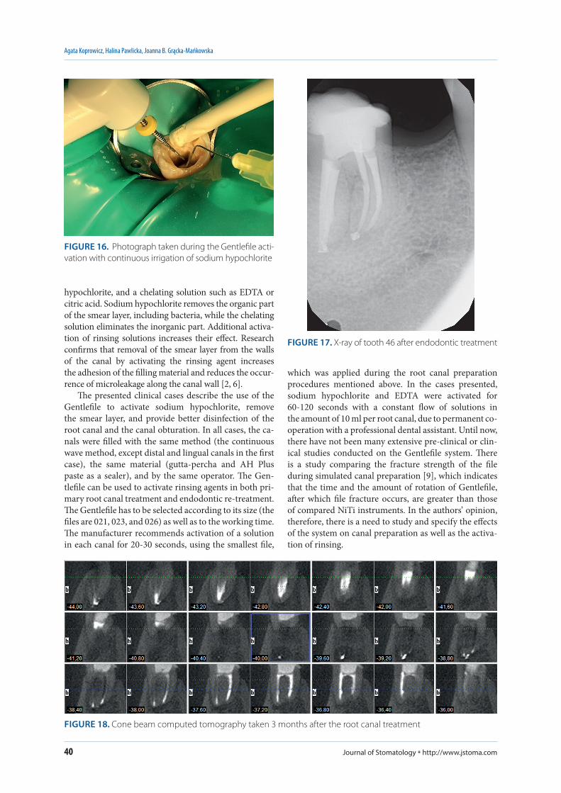
(MB), mesiolingual (ML), and distal (D) canals were found and prepared using Gates Glidden burs (sizes 1 # and 2 #) (Mani, Japan). C-Pilot files of sizes 08.02, 10.02, and 15.02 were used to measure the working length of the canals (Raypex 5 apex locator, VDW, Germany). The chemo-mechanical preparation of the root canals was performed by ProGlider (16.02), ProTaper Next X1 (17.04), and X2 (25.06) instruments (Denstply, USA), rinsing with 2.5% sodium hypochlorite (CHLORAXiD) and 17% EDTA (Endo-Solution). Calcium hydroxide temporary dressing was introduced into the canals, a sterile cotton pellet was placed into the pulp chamber and a Fuji IX GP (GC, Japan) temporarily closed the cav-ity. During the next appointment, the final canal prepa-ration using ProTaper Next (Denstply, USA) X2 and X3 (30.07) instruments was performed, activating all of the above-mentioned rinsing agents by the Gentlefile (023) in the same manner as in the first case (Figure 8). The canals were filled with thermoplasticised gutta-per-cha and AH Plus sealer using the Endopilot. The canal orifices were covered with a flowable composite material and a temporary glass-ionomer (Fuji IX). The follow-up X-ray showed a tight, homogeneous filling of the ca-nals with a slight overfill of the sealer in the apical area
CASE 2
In March 2017, a 34-year-old patient, with medical anamnesis of no clinical significance, was referred to the Department of Endodontics at the Medical Univer-sity of Lodz for root canal treatment of tooth 46. The pa-tient reported severe, spontaneous pain aggravated by heat and biting. X-ray did not reveal any periapical le-sions (Figure 7). Clinical examination was conducted using pulp testing: the ethyl chloride test and electrical test with a Pulp Tester (VDW, Germany). The electrical test gave a false positive result due to the presence of hu-mid necrotic pulp. Infiltration and block anaesthesia were performed (Citocartin 100, Molteni Dental, Po-land). After field isolation using a rubber dam, the ac-cess to the pulp chamber and the orifices of mesiobuccal
FIGURE 7. X-ray of tooth 46 before endodontic treat-ment (April 2017)
FIGURE 8. Activation of rinsing solution with the Gentlefile
FIGURE 9. X-ray of tooth 46 after endodontic treatment FIGURE 10. Control X-ray of tooth 46 after 12 months

Journal of Stomatology * http://www.jstoma.com38
Agata Koprowicz, Halina Pawlicka, Joanna B. Grącka-Mańkowska
(Figure 9). The control X-ray of tooth 46 was taken after 12 months (Figure 10).
CASE 3
A patient, aged 45 years, presented to the dental of-fice for comprehensive dental treatment including pros-thetic and implant reconstruction. The medical history was collected, and a clinical examination was completed with intraoral photographs and X-rays (Figures 11 and 12). The patient did not report any pain. Caries reaching the pulp horn was observed in tooth 26. During the re-moval of caries, the pulp tissue was exposed. After caries tissue removal, the missing wall was rebuilt with Evetric A3 (Ivoclar, Vivadent, Liechtenstein) and the pulp was covered with antibiotic-steroid cream. During the next visit, local anaesthesia was performed with Ubistesin
forte (1 amp, 3M ESPE, USA), and then the tooth was isolated with a rubber dam, and straight-line access to four canals (MB1, MB2, DB, L) was achieved using magnifying loupes (2.5x, Heine, Germany). Single-ap-pointment root canal treatment was performed. Che-mo-mechanical canal preparations were used with Re-ciproc R25 (25.08) and R50 (50.05) (VDW, Germany) and rinsing agents in following sequence as 2.5% sodi-um hypochlorite (CHLORAXiD) and 17% EDTA (En-do-Solution) and 2.5% sodium hypochlorite (CHLO-RAXiD) (10 ml NaOCl and 2 ml EDTA per canal). Both irrigating agents were activated with the Gentlefile (026) for two minutes at the working lengths. Canal obtura-tion was performed using 4% taper gutta-percha cones previously coated with AH Plus sealer, and then the ca-nals were filled with gutta-percha heated to 150°C with the use of an Endopilot. The canal orifices were covered with a flowable composite material and the pulp cham-ber with a RIVA LC material (SDI, Australia). The X-ray image confirmed the correct filling with a small amount of the gutta-percha material extruded beyond the apex of the MB root canal (Figure 13). Due to the further planned dental treatment CBCT was performed after seven months (Figure 14).
CASE 4
A 40-year-old patient came to the dental office in April 2017 to continue treatment of tooth 46. Canal treatment had been started in 2015; however, the patient did not continue the treatment. After a detailed conver-sation with the patient, a shared patient-doctor decision was made to perform re-treatment. The current X-ray revealed the presence of secondary caries and periapi-cal lesions (Figure 15). The patient did not report any pain and no oedema was found on an intra-oral ex-amination. Endodontic re-treatment was performed in a rubber dam under the operating microscope (Leica, Germany). The mechanical canal preparation of four root canals (MB, ML, DB, and DL) was performed us-ing Reciproc R50 files (50.05) (Endopilot) and 5.25% sodium hypochlorite solution (CHLORAXiD) as well as 15% EDTA (Endo-Solution) (10 ml NaOCl and 2 ml EDTA per canal). The Gentlefile (021) was used for both solution activations at the working lengths for two min-utes (Figure 16). The canals were filled with gutta-percha and AH Plus root canal sealer using continuous wave compaction (Endopilot), and the orifices were covered with Flow Berry composite material (Arkona, Poland). The cavity was filled with Ketac Molar glass-ionomer (3M ESPE, USA). The control X-ray showed a tight, homogeneous filling of the canals with a slight over-flow of the material into the periapical region (Figure 17). According to the implantologist’s and prosthodon-tist’s reconstructive dental plan, CBCT was taken three months after the root canal treatment (Figure 18).
FIGURE 11. X-ray of tooth 26 before root canal treatment (July 2016)
FIGURE 12. Photograph of teeth 25 and 26 before endo-dontic therapy of tooth 26

39
Gentlefile in root canal therapy
J Stoma 2019, 72, 1
DISCUSSION
The use of nickel-titanium rotary files for endodontic therapy resulted in a significant reduction of treatment time dedicated to root canal shaping [14]. The research proves that during the mechanical preparation of root canals, the contact surface of the rotary files with the ca-nal wall is about 40-45% [12]. To achieve the best possi-ble disinfection of the root canal system, the saved time should be used for so-called active irrigation. Recently, the importance of using special syringe infusion pumps (B. Braun, Germany), which continuously provide rins-ing agents, or the activation of solutions by using various devices have been discussed.
After mechanical preparation, canal walls are cov-ered with the smear layer that contains dentinal or pulpal residues and microorganisms [17]. The presence of this layer, particularly in the apical region of the canal, is disadvantageous from the clinical point of view because the bacteria colonising the smear layer may contrib-ute to the failure of the root canal therapy. In addition, this layer obliterates the dentinal tubules and reduces the effect of rinsing agents, having an impact on both the quality of filling and the effect of endodontic treat-ment [16]. Physical and chemical properties of the rins-ing solutions allow the smear layer to be dissolved and partially removed [13]. It is necessary to use a combina-tion of two rinsing agents, a disinfectant such as sodium
FIGURE 13. X-ray of tooth 26 after canal obturation (March 2017)
FIGURE 14. Cone beam computed tomography after 7 months
FIGURE 15. X-ray of tooth 46 before treatment
Journal of Stomatology * http://www.jstoma.com40
Agata Koprowicz, Halina Pawlicka, Joanna B. Grącka-Mańkowska
hypochlorite, and a chelating solution such as EDTA or citric acid. Sodium hypochlorite removes the organic part of the smear layer, including bacteria, while the chelating solution eliminates the inorganic part. Additional activa-tion of rinsing solutions increases their effect. Research confirms that removal of the smear layer from the walls of the canal by activating the rinsing agent increases the adhesion of the filling material and reduces the occur-rence of microleakage along the canal wall [2, 6].
The presented clinical cases describe the use of the Gentlefile to activate sodium hypochlorite, remove the smear layer, and provide better disinfection of the root canal and the canal obturation. In all cases, the ca-nals were filled with the same method (the continuous wave method, except distal and lingual canals in the first case), the same material (gutta-percha and AH Plus paste as a sealer), and by the same operator. The Gen-tlefile can be used to activate rinsing agents in both pri-mary root canal treatment and endodontic re-treatment. The Gentlefile has to be selected according to its size (the files are 021, 023, and 026) as well as to the working time. The manufacturer recommends activation of a solution in each canal for 20-30 seconds, using the smallest file,
FIGURE 18. Cone beam computed tomography taken 3 months after the root canal treatment
FIGURE 16. Photograph taken during the Gentlefile acti-vation with continuous irrigation of sodium hypochlorite
FIGURE 17. X-ray of tooth 46 after endodontic treatment
which was applied during the root canal preparation procedures mentioned above. In the cases presented, sodium hypochlorite and EDTA were activated for 60-120 seconds with a constant flow of solutions in the amount of 10 ml per root canal, due to permanent co-operation with a professional dental assistant. Until now, there have not been many extensive pre-clinical or clin-ical studies conducted on the Gentlefile system. There is a study comparing the fracture strength of the file during simulated canal preparation [9], which indicates that the time and the amount of rotation of Gentlefile, after which file fracture occurs, are greater than those of compared NiTi instruments. In the authors’ opinion, therefore, there is a need to study and specify the effects of the system on canal preparation as well as the activa-tion of rinsing.

41
Gentlefile in root canal therapy
J Stoma 2019, 72, 1
In literature, traditional rinsing activation methods, such as manual irrigation with a gutta-percha cone or needle irrigation, are compared extensively with passive ultrasound irrigation or laser irradiation [7, 15]. Research shows that none of the devices used removes the bio-film completely; however, the best results are achieved with Passive Ultrasonic Irrigation (56.6% of the non-smear layer surface compared to Nd: Yag laser 30%, CanalBrush, Coltene Whaledent, Germany – 23.4%, and ProTaper Universal, Denstply Maillefer, Switzerland – 13.4%) [15]. Another study comparing Aseptim Plus, Laser Diode, and ultrasonic activation confirms these results [4]. The comparison of the efficacy of removal of calcium hydroxide from the root canals using different methods of rinsing activation shows that the cleanliness was obtained in 80.9 ± 25.9% in the PUI group, while in the CanalBrush and EndoActivator groups (Dentsply Maillefer, Switzerland) it was from 45% to 49.1% [18].
Various root canal irrigation devices are currently present on the dental market. Among others, the pro-posed file is XP-endo finisher (FKG, Switzerland), which, according to studies, enables effective removal of the bio-film from hard-to-reach areas [19], and provides better results than passive ultrasonic irrigation [10]. The Self- Adjusting File (Re-Dent-Nova, Israel) may be a competi-tive product because there are studies proving its superi-ority over other methods (EndoVac, Discus Dental, Cul-ver City, CA or passive ultrasonic irrigation) [3].
The Gentlefile, which is a rotary endodontic sys-tem, serves for the mechanical canal preparation. According to the manufacturer’s instructions, it may agitate canal irrigation solutions as well. Therefore, the authors are introducing this system to clinics gradually. The file’s properties ensure good results in efficacy of rinsing activation; however, it is a new system that still requires clinical and laboratory anal-yses. Compared to NiTi files, the production of stain-less-steel files is more cost-effective, and therefore the price of the system is reduced. Thanks to its inno-vative design and high elasticity, the instrument can reach deeper areas in the canal system, improving its cleanliness. Due to the small number of studies pub-lished on the use of the system, further observations and comparative analyses are required.
CONCLUSIONS
Activation of rinsing agents using the Gentlefile system is one of the new methods available in the den-tal market. In addition to the mechanical preparation of root canals, the system is said by the manufacturer to enhance the rinsing effect during root canal treat-ment. Because of its ease of use, low cost, and positive therapeutic effects, it can become an alternative tool for dentists. However, further research is required because the Gentlefile is a novel product.
CONFLICT OF INTEREST
The authors declare no potential conflict of interests with respect to the authorship and/or publication of this article.
References1. Ahmetoǧlu F, ŞImşek N, Keleş A, Ocak MS, Er K. Efficacy
of self-adjusting file and passive ultrasonic irrigation on remov-ing calcium hydroxide from root canals. Dent Mater J 2013; 32: 1005-1010.
2. Baldissera R, Rosa RA, Wagner MH, et al. Adhesion of real seal to human root dentin treated with different solutions. Braz Dent J 2012; 23: 521-526.
3. Bao P, Shen Y, Lin J, Haapasalo M. In vitro efficacy of XP-endo Finisher with 2 different protocols on biofilm removal from apical root canals J Endod 2017; 43: 321-325.
4. Da Costa Lima GA, Aguiar CM, Câmara AC, Alves LC, Dos Santos FAB, Do Nascimento AE. Comparison of smear layer removal using the Nd:YAG Laser, ultrasound, ProTaper Universal System, and Can-alBrush methods: an in vitro study. J Endod 2015; 41: 400-404.
5. Fleming CH, Litaker MS, Alley LW, Eleazer PD. Comparison of classic endodontic techniques versus contemporary techniques on endodontic treatment success. J Endod 2010; 36: 414-418.
6. Fróes JA, Horta HG, da Silveira AB. Smear layer influence on the apical seal of four different obturation techniques. J Endod 2000; 26: 351-354.
7. Guerisoli DM, Marchesan MA, Walmsley AD, Lumley PJ, Pecora JD. Evaluation of smear layer removal by EDTAC and sodium hy-pochlorite with ultrasonic agitation. Int Endod J 2002; 35: 418-421.
8. Kamel WH, Kataia EM. Comparison of the efficacy of smear clear with and without a canal brush in smear layer and debris removal from instrumented root canal using WaveOne versus ProTaper: a scanning electron microscopic study. J Endod 2014; 40: 446-450.
9. Moreinos D, Dakar A, Moshonov J, Stone NJ. Evaluation of time to fracture and vertical forces applied by a novel gentlefile system for root canal preparation in simulated root canals J Endod 2016; 42: 505-508
10. Muhammad Omid H, Chevalier M, Rocca JP, Brulat-Bouchard N, Medioni E. Photodynamic therapy versus ultrasonic irrigation: Interaction with endodontic microbial biofilm, an ex vivo study. Photodiagnosis Photodyn Ther 2014; 11: 171-185.
11. Neuhaus KW, Liebi M, Stauffacher S, Sigrun E, Lussi A. Antibac-terial efficacy of a new sonic irrigation device for root canal disin-fection. J Endod 2016; 42: 1799-1803.
12. Poggio C, Dagna A, Chiesa M, Scribante A, Beltrami R, Colombo M. Effects of NiTi rotary and reciprocating instruments on debris and smear layer scores: an SEM evaluation. J Appl Biomater Funct Ma-ter 2014; 12: 256-262.
13. Ruddle CJ. Cleaning and shaping root canal system. Cohen S, Burns RC. Pathways of the pulp. 8th ed. St. Louis: Mosby; 2002: 231-291.
14. Sharma G, Kakkar P, Vats A. A comparative SEM investigation of smear layer remaining on dentinal walls by three rotary NiTi files with different cross sectional designs in moderately curved canals. J Clin Diagn Res 2015; 9: 43-47.
15. Takeda FH, Harashima T, Kimura Y, Matsumoto K. Efficacy of Er:YAG laser irradiation in removing debris and smear layer on root canal walls. J Endod 1998; 24: 548-551.
16. Torabinejad M, Handysides R, Khademi AA, Bakland LK. Clinical implications of the smear layer in endodontics: a review. Oral Surg Oral Med Oral Pathol Oral Radiol Endod 2002; 94: 658-666
17. Violich DR, Chandler NP. The smear layer in endodontics – a re-view. Int Endod J 2010; 43: 2-15.
18. Wujec P, Pawlicka H. Skuteczność usuwania wodorotlenku wap-nia z kanałów korzeni zębów. E-Dentico 2012; 40: 66-77.
19. Živković S, Nešković J, Jovanović-Medojević M, Popović-Bajić M, Živković-Sandić M. XP-endo Finisher: a new solution for smear layer removal. Serbian Dent J/Stom Glas Srb 2015; 62: 122-129.