accredited by dikti no : 58/dikti/kep/2013eprints.unsri.ac.id/5871/1/jurnal_ijc_2014_(14).pdf ·...
TRANSCRIPT


Accredited by DIKTI No : 58/DIKTI/Kep/2013Date : 22 August 2013
ISSN 1411-9420INDONESIAN JOURNAL OF CHEMISTRYVol. 14, No. 3, November 2014
Editor in Chief
Prof. Dr.rer.nat. Nuryono, MS Email: [email protected] of Chemistry, Universitas Gadjah MadaSekip Utara, Yogyakarta Indonesia 55281, Tel/Fax (0062-274)-545188.Website : http://www.ijc.chemistry.ugm.ac.idEmail : [email protected] or [email protected]
Vice Editor in Chief
Prof. Dr. Harno Dwi Pranowo, M.Si Email: [email protected] or [email protected]. Dr. Mudasir, M.Eng Email: [email protected] or [email protected]
Editorial Board
Prof. Dr. Karna Wijaya, M.Eng. (Physical/Material Chemistry)Drs. Iqmal Tahir, M.Si. (Computational/Physical Chemistry)Dr. Ria Armunanto, M.Si. (Computational/Physical Chemistry)Dr. Tri Joko Raharjo, M.Si. (Biochemistry/Bioanalysis)Dr. Nurul Hidayat Aprilita (Analytical/Environmental Chemistry)
Advisory Editorial Board
Dr. Brian Williams (Adelaide University Australia) Prof. Dr. Muhammad Idiris Saleh (University SainsProf. Dr. Dr. Hc. Bernd M Rode (University of Malaysia)Innsbruck, Austria) Dr. Hery Haerudin (Pertamina, Indonesia)Dr. Dirk Bax (Utrecht University, Netherlands) Prof. Dr. Iip Izul Falah (Universitas Gadjah Mada,Prof. Dr. M. Gross (Louis Pasteur University, France) Indonesia)Prof. Dr. Hardjono Sastrohamidjojo (Universitas Prof. Sri Juari Santosa, M.Eng, Ph.D. (UniversitasGadjah Mada, Indonesia) Gadjah Mada, Indonesia)Prof. Dr. David. St. C. Black (University of New South Prof. Dr. Endang Tri Wahyuni, MS (Universitas GadjahWales, Australia) Mada, Indonesia)Prof. Dr. Max Lu (University of Queensland, Australia) Prof. Dr. Bambang Rusdiarso, DEA (Universitas GadjahProf. Dr. Buchori (Bandung Institute of Technology, Mada, Indonesia)Indonesia) Prof. Dr. Wega Trisunaryanti, M.S., Ph.D. EngProf. Dr. Abdul Ra’uf Pathong (Hasanudin University, (Universitas Gadjah Mada, Indonesia)Indonesia) Prof. Dr. Sabirin Matsjeh (Universitas GadjahProf. Dr. Naoki Yoshioka (Keio University, Japan) Mada, Indonesia)Assoc. Prof. Dr. Wan Ahmad Kamil Mahmood Dr. Winarto Haryadi, M.Si. (Universitas Gadjah Mada,(University Sains Malaysia) Indonesia)
Administrator
Dr. Akhmad Syoufian Robby Noor Cahyono, S.Si., M.ScWarakustarti Listyariwangi, A.Md Nurzanah Hidayanti, A.MdDjoko Prihandono
Aims and Scope
Indonesian Journal of Chemistry is an international journal covering all aspects of Chemistry, includingChemical Education and Chemical Engineering. The journal publishes original research papers, shortcommunications, and review articles, and has been indexed by SCOPUS since 2012. The paper published in thisjournal implies that the work described has not been, and will not be published elsewhere, except in abstract, aspart of a lecture, review or academic thesis.
Indonesian Journal of Chemistry (ISSN 1411-9420) is published by the Department of Chemistry, Faculty ofMathematics and Natural Sciences, Universitas Gadjah Mada, Sekip Utara, Yogyakarta, Indonesia. All ordersaccompanied by payment should be sent directly to The Department of Chemistry, Universitas Gadjah Mada.Annual subscription rate is IDR 200,000.00 (Java-Bali), IDR 250,000.00 (outside Java-Bali) (shipping included).Reprint order price is IDR 50,000.00 per article (shipping not included). Customers may make payments by transferon Mandiri Universitas Gadjah Mada (Dr. Nuryono, Account Nr. 137-00-1061199-0).

Accredited by DIKTI No : 58/DIKTI/Kep/2013Date : 22 August 2013
ISSN 1411-9420INDONESIAN JOURNAL OF CHEMISTRYVol. 14, No. 3, November 2014
CONTENTS
Photocatalytic Decolorization Study of Methyl Orange by TiO2-Chitosan Nanocomposites byImelda Fajriati, Mudasir, and Endang Tri Wahyuni
209-218
Simulation of Pollutant Gas Dispersion on Case Study of Lontar 3 Coal Fired Power Plant inAddition into 1 X 660 MW Capacity in Kemiri, Tangerang District, Banten Province by EkoSugiharto, Taufik Abdillah Natsir, and Abdul Rozaq
219-225
Adsorption Isotherm Studies on Acid Orange-10 Dye Removal Using Cerium DioxideNanoparticles by Harry Budiman and Oman Zuas
226-232
Production of Reducing Sugar from Cassava Solid Waste by Simultaneous Ultrasonication andAcid Hydrolysis by Wasinton Simanjuntak, Heri Satria, and Nurul Utami
233-238
Theoretical Analysis of Interaction Energy in Alginate-Capped Gold Nanoparticles ColloidalSystem by Foliatini, Yoki Yulizar, and Mas Ayu Elita Hafizah
239-245
Fast Swelling Superabsorbent Hydrogels Starch Based Prepared by Gamma RadiationTechniques by Erizal, Dian Pribadi Perkasa, Basril Abbas, Sudirman, and Sulistioso G.S.
246-252
The Effect of Caramelization and Carbonization Temperatures toward Structural Properties ofMesoporous Carbon from Fructose with Zinc Borosilicate Activator by Tutik Setianingsih,Indriana Kartini, and Yateman Arryanto
253-261
Hydrogel Based on Crosslinked Methylcellulose Prepared by Electron Beam Irradiation forWound Dressing Application by Ambyah Suliwarno
262-268
Oxidation and Acetylation of Ursolic and Oleanolic Acids Isolated from Fragraea fragransFruits: Antiproliferation of P388 Leukemia Cells by Dasril Basir, Julinar, Eva Agustriana, and BudiUntari
269-276
Cytotoxic Isobractatin (Prenylated Xanthone) Epimer Mixture of Garcinia eugenifolia by SriHartati, I Ketut Triyono, and Sri Handayani
277-282
Synthesis and Thermal-Stability Study of Polybutylene Itaconate Modified with Divinyl Benzeneand Glycerol by Atmanto Heru Wibowo, Ninis Makhnunah, Deny Irawati, Candra Purnawan, NanikDwi Nurhayati, and Henning Storz
283-289
Di-(2-Ethylhexyl)Phthalate and Pyranon Derivated from Endophytic Fungi Penicillium sp theLeave of Kunyit Putih (Curcuma zedoaria) by Muharni, Fitrya, Milanti Okta Ruliza, Dwi AnjarSusanti, and Elfita
290-296
Major Anthocyanin Pigments in the Ficus padana Fruits: HPLC-DAD-ESI-MS Identification andAntioxidant Activity by Daimon Syukri, Djaswir Darwis, and Adlis Santoni
297-303
Isolation of Bioactive Compounds from Aspergillus terreus LS07 by Rizna Triana Dewi, SanroTachibana, Puspa Dewi, L.B.S. Kardono, and Muhammad Ilyas
304-310
Short Communication: Synthesis and Characterization of [Fe(Picolinate)3][MnNi(Oxalate)3].CH3
OH Polymeric Complex by Fahimah Martak, Djulia Onggo, Ismunandar, and Agung Nugroho311-314

Accredited by DIKTI No : 58/DIKTI/Kep/2013Date : 22 August 2013
ISSN 1411-9420INDONESIAN JOURNAL OF CHEMISTRYVol. 14, No. 3, November 2014
CONTENTS (Continued)
Cover picture :
See Imelda Fajriati et al., page 212 & 215
The hypothetic interaction between TiO2 andchitosan (above) & SEM images of chitosan bulkand TiO2-chitosan nanocomposite (NK 0.13)(below)
Accredited by DIKTI No : 58/DIKTI/Kep/2013Date : 22 August 2013
ISSN 1411-9420INDONESIAN JOURNAL OF CHEMISTRYVol. 14, No. 3, November 2014
Organization of Manuscripts
The submitted manuscripts are classified into three categories: original paper which presents original works indetail, notes and/or short communications which present novel and/or valuable information and reviews whichpresent a general survey of specialized subject in chemistry. All manuscripts should be written in concise and clearEnglish and suggested to be typed with full justification, singled spaced for abstract, references, figure captions andtables (tables and figures should be typed on separate sheets at the end of the manuscript): double spaced for text,in Arial 11, using no more than 20 pages for original papers, 10 pages for notes and/or short communication and 30pages for reviews. Left and right margins should be 3.0 cm length. The title should be typed in Arial 12 bold. Thenames of the authors and addresses at which the research was done, including postal code, should appear underthe title. Use Arabic number typed as superscript to link authors to their addresses and asterisk to indicate theauthor(s) to whom correspondence should be addressed. Main headings (Abstract, Introduction, Experimental,Results and Discussion, Conclusions) are typed in bold and capital italics. Type all headings aligned left and lowercase except the first letter of the first word or any proper name. The manuscripts should be written in English or inIndonesian, but the abstract must be written in English and contains no more than 200 words followed by 3-5keywords. All references should be prepared according to the following style: Article in Journal: Barrer, R.M. andCraven, R.J.B., 2000, Phys. Chem., 2, 545–550. Chapter in a Book: Rao, C.N.R, and Rao, K.J., “Ferroics” in SolidState Chemistry Compounds. Eds. Cheetam, A.K., and Day, P, P., Clarendon Press, Oxford, 1992, 281-96. WholeBook: Barrer, R.M. and Craven, R.J.B., 1986, New Developments in Zeolite Science and Technology, ed.Murakame, Y, Iijima, A. and Ward, J.W., Kodansha, Tokyo, p.521. Text references to the literature must benumbers in square brackets. Journals titles should be abbreviated according to the Chemical Abstract ServiceSource Index (CASSI).
Template file of the article could be downloaded in the website:http://www.ijc.chemistry.ugm.ac.id/author/ijctemplate.doc
Acknowledgment
All other contributing individuals should be typed and acknowledged at the end of the manuscript.
Submission of Manuscripts
Please submit your article by online submission at http://pdm-mipa.ugm.ac.id/ojs/index.php/ijc/ orvia email: [email protected] or [email protected]
An IDR 800,000.00 (Java-Bali) and IDR 1,000,000.00 (outside Java-Bali) fee per article may be paid for paperspublished in this journal. This fee includes automatic subscription of the journal for one volume (3 issues).Subscription fee charged to overseas is USD 100 for 1 (one) copy of the journal and the article in pdf format.Articles of more than 7 pages (after layout) are subject to additional cost of IDR 75,000.00 per extra page. Colorprinting fee is IDR 100,000.00 per page.
Author may reproduce/republish portions of their published contribution without seeking permission from the Dept.of Chemistry, Universitas Gadjah Mada (UGM), provided that any such republication is accompanied by anacknowledgement in the form: (Original Citation-Reproduced by Permission of The Dept. of Chemistry, UniversitasGadjah Mada).
Advertising
Inquires concerning advertising should be addressed to Dr. Tri Joko Raharjo, M.Si, Department of Chemistry,Universitas Gadjah Mada, Sekip Utara, Yogyakarta, Indonesia. Email: [email protected] [email protected]

Indo. J. Chem., 2014, 14 (3), 290 - 296
Muharni et al.
290
* Corresponding author.Email address : [email protected]
DI-(2-ETHYLHEXYL)PHTHALATE AND PYRANON DERIVATED FROM ENDOPHYTIC FUNGIPenicillium sp THE LEAVE OF KUNYIT PUTIH (Curcuma zedoaria)
Muharni1,*, Fitrya2, Milanti Okta Ruliza1, Dwi Anjar Susanti1, and Elfita1
1Department of Chemistry, Faculty of Mathematics and Natural Sciences , Sriwijaya University,
Jl. Raya Palembang Prabumulih Km 32, Indralaya, Ogan Ilir, South Sumatera 30662, Indonesia
2Department of Pharmacy, Faculty of Mathematics and Natural Sciences , Sriwijaya University,
Jl. Raya Palembang Prabumulih Km 32, Indralaya, Ogan Ilir, South Sumatera 30662, Indonesia
Received October 24, 2013; Accepted July 18, 2014
ABSTRACT
Two compounds from cultivation of the endophytic fungi Penicillium sp of leaves of kunyit putih (Curcumazedoaria have been isolated. The endophytic fungus was cultivated on 5 L of Potatos Dextrose Broth (PDB) mediumat room temperature (no shaking) for 3 weeks. The cultures were extracted with ethyl acetate to afford 3.0 g ofresidue after removal of the solvent under reduced pressure. The extract was separated and purified by silica gelcolumn chromatography (CC) and afforded two pure compounds as colorless oily liquid (compound 1) and yellowcrystal (compound 2). The structure of these compounds were characterized by detailed UV, IR, and NMRspectroscopic analysis and compound 1 as well as comparison with the reported data. Base on spectra analysis thecompound 1 was determined as Di-(2-ethylhexyl)phthalate and compound 2 as 5-(4’-ethoxy-2’-hydroxy-5’-methyl-2’,3’-dihydrofuran-3’-il (hydroxy) methyl-4-isopropyl-3-methyl-2-pyran-2-on). Compound 1 is not new compound, butit is new for endophytic fungus from C. zeodoria and compound 2 is new compound.
Keywords: endophytic fungi; Penicillium sp; Curcuma zedoaria
ABSTRAK
Telah dilakukan isolasi dua senyawa dari kultifat jamur endofitik Penicillium sp dari daun kunyit putih (Curcumazedoaria. Jamur endofit dikultur dalam 5 L medium Potatos Dextrose Broth (PDB) pada suhu kamar (keadaan statis)selama 3 minggu. Kultur kemudian diekstraksi dengan etil asetat dan dipekatkan dengan rotary evaporator sehinggadidapatkan ekstrak pekat etil asetat 3,0 g. Ekstrak dipisahkan dan dimurnikan dengan kromatografi kolommenggunakan fasa diam silika gel dan didapatkan dua senyawa murni berupa cairan minyak bening (senyawa 1)dan kristal kuning (senyawa 2). Struktur senyawa hasil isolasi ditentukan berdasarkan analisis data spektroskopi UV,IR, dan NMR, dan senyawa 1 juga dikonfirmasi dengan membandingkan data yang telah dilaporkan. Berdasarkananalisis data spektroskopi disimpulkan senyawa 1 adalah Di-(2-ethylhexyl)phthalate dan senyawa 2 adalah 5-(4’-etoksi-2’-hidroksi-5’-metil-2’,3’-dihidrofuran-3’-il (hidroksi) metil-4-isopropil-3-metil-2-piran-2-on). Senyawa 1 bukanmerupakan senyawa baru, tetapi untuk pertama kalinya ditemukan pada jamur endofitik pada C. zeodoria dansenyawa 2 merupakan senyawa baru.
Kata Kunci: jamur endofitik; Penicillium sp; Curcuma zedoaria
INTRODUCTION
Endophytic microorganisms that redise in thetissues of living plants and may produce secondarymetabolites of biologically active [1]. Novel antibiotics,antimycotics, immunosuppressants, anticancercompound are only a few examples of what has beenfound after the isolation, culture and purification andcharacterization of some choice endophytes in therecent past. Isolation of their bioactive secondarymetabolites of endophytic fungus from plant could be
selected mainly something on ethonobotanical history[2].
Curcuma zedoaria, a medicinal tuber plantbelonging to the family Zingiberaceae has been used inthe traditional system of medicine [3]. These plantswere used for curing stomach diseases, toothache,blood stagnation, leucoderma, tuberculosis,enlargement of spleen, and for promoting menstruationin traditional medicine in Asia [4]. Antiinflammatoryactivity [5], antiulcer activity [6], and antimicrobial effect[7], of this plant rhizome have been reported.

Indo. J. Chem., 2014, 14 (3), 290 - 296
Muharni et al.
291
In our research of endophytic fungus, manybioactive compounds and new compounds were isolated[8-9]. In this paper we reported the isolation andstructural identification one known compound namely Di-(2-ethylhexyl)phthalate (1) and one new compound as 5-(4’-ethoxy-2’-hydroxy-5’-methyl-2’,3’-dihydrofuran-3’-il(hydroxy) methyl-4-isopropyl-3-methyl-2-pyran-2-on) (2)of Penicillium sp from the leaves of C. zedoaria.Penicillium species isolated as endophytic usually befound in plants zingiberaceae [10] and meliaceae family,although in marine organisms, three meroterpenespreaustinoids, A, B, A1, A2, and B1 have been reportedto be isolated from Penicillium sp associated with theMelia azedarach [11]. Penicillium commune from thesemi-mangrove plant Hibiscus tiliaceus, have beenisolated one new compound 1-O-(2,4-dihydroxy-6-methylbenzoyl)-glycerol along with thirteen knownproducts including 1-O-acetylglycerol, N-acetyltryptophan, 3-indolylacetic acid methyl ester, 1-(2,4-dihydroxy-3,5-dimethylphenyl)ethanone, 2-(2,5-dihydroxyphenyl)acetid acid, (4R,5S)-5-hydroxyhexan-4-olide,thymidine, uracil, thymine, ergosterol, β-sitosterol, β-daucosterol, and ergosta-7,22-dien-3β,5α,6β-triol [12].
EXPERIMENTAL SECTION
Materials
The leaves of kunyit putih were collected on May2013 from the Indralaya, Ogan Ilir, South Sumatra.Material for isolation endophytic fungi: ethanol 70%,NaOCl, chloramphenicol, potato dextrose broth (PDB),potato dextrose agar (PDA), silica gel 60 (70-230 mesh),thin layer chromatography (TLC) from Merck (Art.5554)silica gel 60 F254, n-hexane, ethyl acetate, and methanol.The organic solvents were used from distilled technicalgrade.
Instrumentation
The apparatus in the research were countercolony, autoclave, incubator, water bath, microscope,magnetic hotplate, UV lamp, column chromatographyand generally apparatus in organic and microbiologylaboratory, melting point was determined using FisherJohn Apparatus. UV spectra were determined withVarian Conc 100 instrument. IR spectra weredetermined on FTIR-Perkin Elmer-Spectrum One andNMR spectra were recorded at 500 MHz (
1H) and
125 MHz (13
C) on JEOL JNM ECA-500 spectrometer,UV light at λ 254 nm and 365 nm.
Procedure
Isolation of endophytic fungusThe leaves sample was washed before it was
processed and surface sterilized in 70% ethanol for3 min and 0.5% NaOCl for 1 min and rinsed thoroughlywith sterile distilled water. The segment sample placedon petri-plates containing potato dextrose agar medium(PDA) (200 g potato, 20 g dextrose, and 15 g agar in1 L of H2O, supplemented with 100 mg/L ofchloramphenicol to suppress bacterial growth). Theplates were incubated at 25 ± 2 °C until fungus growthappeared. The plant segments were observed once aday for the growth of endophytic fungus. Colony fungusshowed difference characteristic furthermore to purewith the plated segments were immediately transferredinto new PDA plates and then subcultured until purecultures were obtained [13].
Identification of the endophyteThe endophytic fungal strain was identified by the
morphological method. The morphological examinationwas performed by scrutinizing the fungal culture, themechanism of spore production, and the characteristicsof the spores. All experiments and observations wererepeated at twice [14].
Cultivation of pure fungal strainThe purified fungus (a small park) was transferred
under sterile conditions to the PDB medium. Forchemical investigations, the fungal strains were staticcultivated into 15 flasks (1 L each) containing 400 mLof PDB medium for 3 weeks at room temperature[12-14].
Extraction, isolation, purification, and structureelucidation
Fungus in the 3 weeks cultures were vacuum-filtered and the filtrate fractionated thrice by liquid-liquidpartition with ethyl acetate (1:1). Then the solventphase was evaporated under reduced pressure usingrotary vacuum evaporator at 40 °C to produce the ethylacetate fraction of liquid cultures. The EtOAc fraction(3.0 g) was preabsorbed on silica gel and thenpurification by column chromatography (silica gel,eluted n-hexane : EtOAc = 5:5 – 1:9), EtOAc 100%,EtOAc : MeOH = 9:1 – 1:9 and MeOH 100%). Basedon detection by TLC using the eluent system, to givefive fractions F1–F5. The 1
stfraction to yield compound
1 (20 mg). Furthermore, fraction 2nd
(0.2 g) wasrechromatographed using the same method (silica gel,eluted with EtOAc : MeOH (8:2 – 1:9) and MeOH(100%) to yield three fractions F2.1 – F2.3. FractionF2.1 to yield pure compound 2 (10 mg). The molecularstructure of compounds were established on the basis
Indo. J. Chem., 2014, 14 (3), 290 - 296
Muharni et al.
292
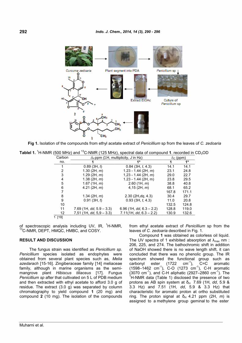
Fig 1. Isolation of the compounds from ethyl acetate extract of Penicillium sp from the leaves of C. zedoaria
Tablel 1.1H-NMR (500 MHz) and
13C-NMR (125 MHz), spectral data of compound 1. recorded in CD3OD
H ppm (H, multiplicity, J in Hz) C (ppm)Carbonno. 1 1* 1 1*1 0.89 (3H, t) 0.84 (3H, t, 4.3) 14.1 14.12 1.30 (2H, m) 1.23 - 1.44 (2H, m) 23.1 24.83 1.29 (2H, m) 1,23 - 1.44 (2H, m) 29.0 22.74 1.38 (2H, m) 1.23 - 1.44 (2H, m) 23.8 29.55 1.67 (1H, m) 2.60 (1H, m) 38.8 40.86 4.21 (2H, m) 4,15 (2H, m) 68.1 65.27 167.8 171.18 1.34 (2H, m) 2.30 (2H,dq, 4.3) 30.4 29.79 0.91 (3H, t) 0.93 (3H, t, 4.3) 11.0 20.8
10 132.5 124.811 7.69 (1H, dd, 5.9 – 3.3) 6.96 (1H, dd, 6.3 – 2.2) 128.8 119.012 7,51 (1H, dd, 5,9 – 3.3) 7.11(1H, dd, 6.3 – 2.2) 130.9 132.6
1* [19]
of spectroscopic analysis including UV, IR,1H-NMR,
13C-NMR, DEPT, HMQC, HMBC, and COSY.
RESULT AND DISCUSSION
The fungus strain was identified as Penicillium sp.Penicillium species isolated as endophytes wereobtained from several plant species such as, Meliaazedarach [15-16]. Zingiberaceae family [14] meliaceaefamily, although in marine organisms as the semi-mangrove plant Hibiscus tiliaceus [17]. FungusPenicillium sp after that cultivated on 5 L of PDB mediumand then extracted with ethyl acetate to afford 3.0 g ofresidue. The extract (3.0 g) was separated by columnchromatography to yield compound 1 (20 mg) andcompound 2 (10 mg). The isolation of the compounds
from ethyl acetate extract of Penicillium sp from theleaves of C. zedoaria described in Fig. 1.
Compound 1 was obtained as colorless oil liquid.The UV spectra of 1 exhibited absorption at λmax nm :206, 225, and 274. The bathochromic shift in additionof NaOH showed there is no wave length shift, it canconcluded that there was no phenolic group. The IRspectrum showed the functional group such ascarbonyl ester (1722 cm
-1), C=C aromatic
(1598–1462 cm-1
), C-O (1273 cm-1
), C-H aromatic(3070 cm
-1), and C-H aliphatic (2927–2860 cm
-1). The
1H-NMR data (Table 1) disclosed the presence of two
protons as AB spin system at δH 7.69 (1H, dd, 5.9 &3.3 Hz) and 7.51 (1H, dd, 5.9 & 3.3 Hz) thatcharacteristic for aromatic proton at ortho substitutedring. The proton signal at δH 4.21 ppm (2H, m) isassigned to a methylene group geminal to the ester

Indo. J. Chem., 2014, 14 (3), 290 - 296
Muharni et al.
293
Table 2.1H-NMR (500 MHz) and
13C-NMR (125 MHz), spectral data of compound 2 recorded in CDCl3
Carbon no. C ppm DEPT H ppm (H, multiplicity, J in Hz) HMBC COSY2 183.4 C3 123.1 C4 139.1 C5 107.4 C6 162.8 CH 8.23 (1H, s) 81.7, 107.4, 139.17 34.6 CH 2.98 (1H, q, J = 7.15 Hz) 107.4, 123.1, 139.1 1.228 18.5 CH3 1.22 (3H, d, J = 7.15 Hz) 139.1, 34.6, 81.79 18.2 CH3 1.34 (3H, d, J = 7.15 Hz) 34.6
10 9.5 CH3 2.01 (3H, s) 123.1, 139.1, 183.82’ 174.5 C3’ 100.3 C4’ 171.2 C5’ 177.2 C6’ 60.4 CH2 4.11 (2H, q, J = 7.15 Hz) 171.2 1.257’ 14.2 CH3 1.25 (3H, t) 60.48’ 21.1 CH3 2.04 (3H, s) 171.29’ 81.7 CH 4.77 (1H, q, J = 7.15 Hz) 139.1, 162.8 1.34
Fig 2. The1H-NMR spectrum of compound 2
alcohol group. Furthermore, the presence proton signalat δH 1.67 ppm (1H, m) for proton methine, signal at1.2–1.4 ppm for four methilene group, and signal at 0.89and 0.91 as pair of multiplate (3H, m) for two methylgroups.
The13
C-NMR spectrum of compound 1 (Table 1),confirming the symmetry of the molecule, exhibited theexpected 12 carbon resonance. DEPT spectrum showedto two quaterner, three methane, five methylenecarbons, and two methyl groups. These spectroscopicdata, by comparison of
1H and
13C-NMR data to those
published in literature [19] and showed similarity, inconclusion compound 1 was identified asDi-(2-ethylhexyl)phthalate (DEHP). DEHP (compound1) is a well known synthetic plasticizes, so alreadyreported to be present in Calotropis gigantean [15],Alchornea cordifolia [16], and Aloe vera [17]. Theeffective presence of compound 1 in endophytic fungusPenicillium sp of leave C. zedoaria, not as acontaminant from solvents and endophytic fungusPenicillium sp not cultivated in plastic bags, so thesecould be discounted as a source of DEHP.

Indo. J. Chem., 2014, 14 (3), 290 - 296
Muharni et al.
294
Fig 3. Spectrum13
C-NMR compound 2
Fig 4. HMQC correlation of proton at δH 1.23–8.23 ppm (A) and HMBC correlation of proton at δH 1.23–2.98 ppm (B)compound 2
Compound 2 was obtained as a yellow crystal, mp.171-172 °C. The Spectra UV (MeOH) of 2 exhibitedabsorption at λmax nm: 213, 253, and 321. Thebathochromic shift in addition of NaOH exhibitedabsorption at λmax nm: 213, 253, and 321. Base onSpectroscopic data UV indicate this compound was no
phenolic group. The IR spectra (KBr) showed νmax cm-1
:3466.08 (OH), 2980.02 and 2935.66 (C-H aliphatic),1625.99 (conjugated C=O), 1579.70; 1521.12; 1438.90(C=C conjugation), and 1180.44 (C-O ether).
1H-NMR
(DMSO, 500 MHz) δH ppm and13
C-NMR (DMSO,125 MHz) δC ppm (see Table 2).

Indo. J. Chem., 2014, 14 (3), 290 - 296
Muharni et al.
295
Fig 5. HMBC correlation of proton at 4.01–8.23 ppm and COSY correlation compound 2
Fig 6. HMBC (A), and COSY (B) correlations and δ-assignment of compound 2
The1H-NMR spectrum, (Fig. 2) showed signal two
methyl doublet at δH 1.22 and 1.34 ppm (3H, d, 7.15 Hz)and signal methine quartet at δH 2.98 ppm (1H, q,7.15 Hz) and one methine singlet at δH 8.23 (1H, s). Atspectrum also showed signal for methyl triplet atδH 1.25 ppm (3H, t, 7.15 Hz), and two methyl singlet atδH 2.01 and 2.04 ppm, (3H, s) and one signal methylenequartet at δH 4.11 ppm (2H, q, 7.15 Hz).
The13
C-NMR (Fig. 3), DEPT 135 spectrum, andHMQC spectrum (Fig. 4) showed 17 signal consist thatnine signal as C sp
2and 8 signal as C sp
3. Analysis
spectrum DEPT 135 showed 8 signal C quarternary atδC 100.3; 107.4; 123.1; 139.1; 171.2; 174.5; 177.2 and183.8 ppm, 5 signal methyls carbon at δC 9.5; 14.2;18.2; 18.5 and 21.1 ppm, 3 signal methines carbon atδC 34.6; 81.7 and 162.8 ppm, and one signal
Indo. J. Chem., 2014, 14 (3), 290 - 296
Muharni et al.
296
methylene carbon at δC 60.4 ppm. Signal carbon atδC 183.4 ppm indicated these compound have C=Ocarbonyl.
NMR 2D analysis for HMQC spectrum (Fig. 4)showed the proton at δH 1.34 ppm correlation to carbonat δH 18.2 ppm and proton at δH 1.22 ppm correlation tocarbon at δC 18.5. HMBC spectrum showed correlationfrom proton at δH 1.22 and 1.34 ppm to carbon atδC 34.6 and 139.1 ppm. Proton at δH 1.22 alsocorrelation to carbon at δC 18.2 ppm and proton atδH 1.34 ppm showed correlation with carbon atδC 18.5. This data to indicated that proton δH 1.22 and1.34 ppm bound to carbon fasten carbon δC 34.6 ppm.Proton at 1.25 ppm correlation to carbon at δC 60.4 ppm.Further HMBC spectrum showed correlation proton at δH
2.01 (3H, s) to carbon at δC 123.1; 139.1 and 183.8 ppm,and correlation proton at δH 2.04 ppm to carbon atδC 171.2 ppm. Proton at δH 2.01 and 2.04 (3H, s) atHMQC spectrum showed fastened with carbon atδC 9.5 and 21.1 ppm.
Proton at δH 4.11 correlation to carbon at δC 171.2,proton at δH 4.77 ppm showed correlation to carbon atδC 139.1; 162.8 ppm, while proton at δH 8.23 ppm tocorrelation to carbon at δC 81.7; 107.4 and 139.1 ppm.Analysis of
1H–
1H COSY spectrum (Fig. 5) also to
indication of two proton spin system corresponding at δH
1.22 with proton at δH 2.98 ppm. And proton at δH 1.25 tocorrelation to proton at 4.11 ppm. HMBC and COSYcorrelation and δ-assignment of compound showed Fig. 6. These spectroscopic data, therefore suggested thatcompound 2 is 5-(4’-ethoxy-2’-hydroxy-5’-methyl-2’,3’-dihydrofuran-3’-il (hydroxy) methyl-4-isopropyl-3-methyl-2-pyran-2-on).
Compound 1 is not new compound, but it is new forendophytic fungus from C. zedoaria and base onDictionary Natural Products data base, 5-(4’-ethoxy-2’-hydroxy-5’-methyl-2’,3’-dihydrofuran-3’-il (hydroxy)methyl-4-isopropyl-3-methyl-2-pyran-2-on) (2) is newcompound. Exploration of secondary metabolitesresearch needs to be done in order to get the profile oforganic compounds produced by endophytic fungus ofC. zedoaria.
CONCLUSION
Two compounds have been isolated from theendophytic fungus Penicillium sp from the leaves ofkunyit putih (C. zedoaria). Based on spectroscopicanalysis and comparison data to those published inliterature compound 1 was identified asDi-(2-ethylhexyl)phthalate and compound 2 as
5-(4’-ethoxy-2’-hydroxy-5’-methyl-2’,3’-dihydrofuran-3’-il(hydroxy) methyl-4-isopropyl-3-methyl-2-pyran-2-on).
ACKNOWLEDGEMENT
The authors are statement grateful to theDirectorate General of Higher Education whichresearch grant Fundamental 2013 was supported thisresearch.
REFERENCES
1. Strobel, G., Daisy, B., and Castillo, U., 2005, PlantPathol. J., 4 (2), 161–176.
2. Premjanu, N., and Jayanthy, C., 2012, Int. J. Inst.Pharm. Life Sci., 2 (1), 135–162.
3. Lakshmi, S., Padmaja, G., and Remani, P., 2011,Int. J. Med. Chem., 2011, 1–13.
4. Saikia, N., and Nath, S.C., 2003, J. Econ. Taxon.Bot., 27, 430–433.
5. Jang, M.K., Sohn, D.H., and Ryu, J-H., 2001,Planta Med., 67 (6), 550–552.
6. Wilson, B., Abraham, G., Manju, V.S., Mathew, M.,Vimala, B., Sundaresan, S., and Nambisan, B.,2005, J. Ethnopharmacol., 99 (1), 147–151.
7. Bugno, A., Nicoletti, M.A., Almodóvar, A.A.B.,Pereira, T.C., and Auricchio, M.T., 2007, Braz. J.Microbiol., 28, 440–445.
8. Elfita, Muharni, Munawar, Legasari, L., andDarwati, 2011, Indo. J. Chem., 11 (1), 53–58.
9. Elfita, Muharni, Munawar, and Aryani, S., 2012,Indo. J. Chem., 12 (2), 195–200.
10. Xu, L., Zhou, J., Zhao, J., Li, X., and Wang, J.,2008, Lett. Appl. Microbiol., 46 (1), 68–72.
11. dos Santos, R.M.G., and Rodrigues-Fo, E., 2003,Z. Naturforsch., 58c, 663–669.
12. Yan, H-J., Gao, S-S., Li, C-S., Li, X-M., and Wang,B-G., 2010, Molecules, 15 (5), 3270–3275.
13. Barik, B.P., Tayung, K., Jagadev, P.N., and Dutta,S.K., 2010, Eur. J. Biol. Sci., 2 (1), 8–16.
14. Guo, L., Wu, J-Z., Han, T., Cao, T., Rahman, K.,and Qin, L-P., 2008, Molecules, 13 (9), 2114–2125.
15. Habib, M.R., and Karim, M.R., 2009, Micobiology,37 (1), 31–36.
16. Mavar-Manga, H., Haddad, M., Pieters, L., Baccelli,C., Penge, A., and Quetin-Leclercq, J., 2008, J.Ethnopharmacol., 115 (1), 25–29.
17. Lee, K.H., Kim, J.H., Lim, D.S., and Kim, C.H.,2000, J. Pharm. Pharmacol., 52 (5), 593–598.