accreditation in adult transthoracic echocardiographysotirmarchev.tripod.com/instr.pdf · letter to...
TRANSCRIPT

Accreditation Info for Candidates Version 4 Sept 2003
Accreditation in Adult Transthoracic Echocardiography
The European Association of Echocardiography A Registered Branch of the ESC
(formerly the Working Group on Echocardiography
of the European Society of Cardiology)
Information for Candidates Seeking Accreditation Contents:
1. Introduction and aims
2. Summary of process and requirements
3. Details of the exam and practical assessment
4. Example questions
5. Suggested format for a report
6. Suggested reading list and syllabus
7. Enrolment form
8. Supervisor’s form to accompany enrolment
9. Log book summary sheet
10. Supervisor’s mark sheet
11. Letter to accompany log book submission
12. Check list for log book submission

Accreditation Info for Candidates Version 3.1 June 2003 1
Introduction and Aims 1. Accreditation is run as a service by the European Association of
Echocardiography and is not necessarily a compulsory or regulatory certificate of competence or excellence.
2. Applications for accreditation are welcomed from Sonographers (Technicians) and Doctors
3. The goals of accreditation are to protect patients from undergoing echocardiographic examinations performed by unqualified persons and to set a European standard for competency and excellence in echocardiography.
4. Accredited echocardiographers are expected to be able to perform and report routine echocardiographic studies unsupervised.
5. While European Accreditation is designed to test the competency of an individual to be able to perform, interpret and report routine echocardiographic studies unsupervised, the right to report and sign clinical studies in individual countries will be defined by national laws and regulations.
6. Accreditation in echocardiography should bring credibility and professional legitimacy to an individual by demonstrating competency by the successful passage of examinations.
7. The accreditation process will includes a written and a practical component. 8. The accreditation process will identify qualified practitioners of
echocardiography and should enhance the professional image of echocardiographers. It will also provide statistics and records about echocardiography that can be easily accessed.
9. As echo skills can only be maintained by continued education and practical involvement, a re-accreditation process will be required. Accreditation will be granted for a period of five years.

Accreditation Info for Candidates Version 3.1 June 2003 2
Summary of process and requirements 1. Enrolment for the accreditation process will be through the European
Association of Echocardiography administrator at the European Heart House.
2. The exam can be taken only after acceptance for enrolment. 3. The accreditation process should be completed within two years from the
date of enrolment. During this period both the written and the practical assessment should be completed.
4. The practical assessment must be completed in a 12-month period, within 12 months before or after completing the exam assessment. The following must be submitted:
• Copies of reports on 250 clinical cases performed and reported (in the national language) by the candidate (anonymised)
• Summary Sheet (enclosed) • A letter from a supervisor testifying that the studies were performed and
reported by the candidate (example enclosed).
• A letter from the supervisor documenting training and the review of studies undertaken by the candidate
5. A logbook should be submitted within 12 months after the date of completing the exam. Failure to do so will necessitate repeating the entire process from the beginning.
6. The fee for the complete Accreditation process is €150. This fee is payable, in advance, upon enrolment, and will cover the exam and logbook submission. Candidates who are unsuccessful in the exam will be charged a reduced fee to re-sit this section.
To obtain accreditation you must pass both the written and practical parts of the assessment.

Accreditation Info for Candidates Version 3.1 June 2003 3
Details of the Exam and Practical Assessment for Accreditation in Adult Transthoracic Echocardiography
1 The Exam
1.1 The exam will be held twice each year. Full details of dates and venues, and registration forms, can be obtained through the European Association of Echocardiography Administrator at the European Heart House.
1.2 The exam will be sat under formal examination conditions. It will be comprised of two parts: a section testing theoretical knowledge and a section of questions on clinical cases using digital clips and stills
1.3 Candidates need a simple calculator for the exam (NOT a laptop / palm computer or mobile phone calculator)
2 Theory section
2.1 This will consist of a series of 100 multiple choice questions which must be answered within 90 minutes. The questions will test the candidate’s knowledge of the principles and practice of echocardiography. The first 20 questions will relate to ultrasound physics and the remaining 80 to general echocardiography. Each question will have 4 possible responses, and candidates will be asked to select the best answer.
2.2 The examination will be written in a straightforward way to test knowledge. Clinical cardiology unrelated to echocardiography will not be tested. Some example questions are included with this document
3 Echo Reporting Section
3.1 This will consist of 50 questions, typically 5 questions on each of 10 case studies. Each question will have 4 possible responses and candidates will be asked to select the best answer.
3.2 Questions will be based on imaging material reflecting the range of clinical conditions seen in current echocardiographic practice. Normal or near-normal studies may also be presented. Some example questions are included with this document

Accreditation Info for Candidates Version 3.1 June 2003 4
4 Passing the exam
4.1 Both parts of the exam will be graded by an appropriate independent body.
4.2 It is necessary to pass both the theory section and the echo reporting section. An appropriate pass mark for the multiple choice and for the echo reporting section questions will be set by the examining board to ensure consistency and validity of the accreditation standard. The provisional pass mark for the theory section will be approximately 65 / 100 and for the case evaluation section 30 / 50 but this may be adjusted by a meeting of the accreditation assessment committee.
4.3 There is no bar to re-sitting the written assessment.
4.4 Accreditation will only be awarded once a candidate has also completed the practical assessment. A satisfactory performance at the exam alone does not allow ‘partial accreditation’.
4.5 Feedback from the examining board and from the candidates will be collected in the form of a questionnaire, to be used for improving the quality of the process.
5 Practical assessment - General
5.1 The log book must be submitted within 12 months of passing the exam.
5.2 The submission should include a letter from the candidates supervisor, the summary sheet and check list (enclosed).
6 Log-book
6.1 The log-book (portfolio) should comprise details of 250 transthoracic cases personally performed and reported by the candidate during a period of 12 months either prior or after the date of enrolment.
6.2 The preferred format for the log book is a set of anonymised copies of actual clinical reports in the national language (numbered 1 – 250), sent electronically or enclosed in a folder or binder. The reports should be anonymised. The reports should include cavity and Doppler measurements, objective observations and a comment (see suggested format).
6.3 All reports submitted must carry the signature of the candidate and they should include reports primarily by the candidate alone although they may be checked by another operator.

Accreditation Info for Candidates Version 3.1 June 2003 5
6.4 The studies should reflect the normal case-load of a general echocardiography department and should include cases of:
• Assessment of left ventricular function (including regional wall motion abnormalities)
• Valvular heart disease • Prosthetic valves • Pericardial disease (including constriction and tamponade) • Diseases of the aorta • Examples of congenital disease (e.g. ASD) • Suspected endocarditis • Cardiomyopathies (including HCM) • Not more than 1/3 of the studies should be completely normal.
6.5 A count of the primary diagnosis assigned to each case must be entered on the appropriate enclosed summary sheet.
6.6 If possible there should be one or more examples of unusual diagnoses. More than one candidate from the same institution is permitted to study the same patient if the diagnosis is unusual.
6.7 If the candidate has problems finding enough specific cases, he/she should discuss this with his/her supervisor who may consider arranging for the candidate to attend a larger centre.
6.8 A letter from the supervisor must be submitted with the completed log-book certifying that the candidate has recorded the studies her(him)self.
7 Assessment of Studies Performed by the Candidate. 7.1 The supervisor will review 10 studies performed by the candidate to
confirm they have been appropriately performed and reported. It is also strongly recommended that the supervisor observes the candidate performing echocardiographic studies.
7.2 A mark sheet will be provided for the supervisor to use and submit with the candidate’s log book.
Accreditation Info for Candidates Version 3.1 June 2003 6
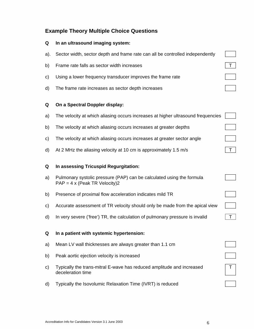
Example Theory Multiple Choice Questions Q In an ultrasound imaging system: a). Sector width, sector depth and frame rate can all be controlled independently b) Frame rate falls as sector width increases T c) Using a lower frequency transducer improves the frame rate d) The frame rate increases as sector depth increases Q On a Spectral Doppler display: a) The velocity at which aliasing occurs increases at higher ultrasound frequencies b) The velocity at which aliasing occurs increases at greater depths c) The velocity at which aliasing occurs increases at greater sector angle d) At 2 MHz the aliasing velocity at 10 cm is approximately 1.5 m/s T Q In assessing Tricuspid Regurgitation: a) Pulmonary systolic pressure (PAP) can be calculated using the formula PAP = 4 x (Peak TR Velocity)2 b) Presence of proximal flow acceleration indicates mild TR c) Accurate assessment of TR velocity should only be made from the apical view d) In very severe (’free’) TR, the calculation of pulmonary pressure is invalid T Q In a patient with systemic hypertension: a) Mean LV wall thicknesses are always greater than 1.1 cm b) Peak aortic ejection velocity is increased c) Typically the trans-mitral E-wave has reduced amplitude and increased
deceleration time T
d) Typically the Isovolumic Relaxation Time (IVRT) is reduced

Accreditation Info for Candidates Version 3.1 June 2003 7
Example Echo Reporting Section MCQs Question 1 The clips and stills show a case of severe AR due to a dilated aortic root in a hypertensive patient with poor LV function 65 year old male Request: Hypertensive patient - breathlessness and a murmur Data: Ao root at sinotubular junction = 4.9cm, P1/2 time 251ms 1. The left ventricular function is
a. Normal
b. Mildly impaired
c. Moderately impaired
d. Severely impaired X
2. Chose the phrase which best describes the left ventricle
a. Not dilated and not hypertrophied
b. Dilated and hypertrophied
c. Dilated and not hypertrophied X
d. Thin walled
3. The aortic regurgitation is:
a. Uninterpretable
b. Mild
c. Moderate
d. Severe X
4. The mechanism of the AR is
a. Secondary to aortic root dilatation X
b. Due to a bicuspid aortic valve
c. Due to dilatation of the LV
d. Due to rheumatic disease
5 The patient subsequently presents with an embolic stroke
a. Although no cardiac source is seen, a cardiac source is likely b. No cardiac source is seen and is unlikely based of this study X c. The aortic valve is a likely source of emboli
d. The left ventricle is a likely source of emboli
Accreditation Info for Candidates Version 3.1 June 2003 8
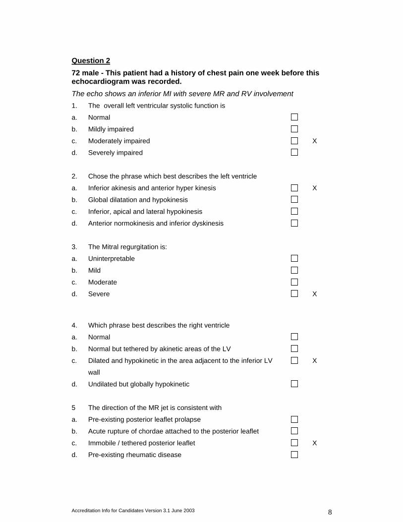
Question 2 72 male - This patient had a history of chest pain one week before this echocardiogram was recorded. The echo shows an inferior MI with severe MR and RV involvement 1. The overall left ventricular systolic function is
a. Normal
b. Mildly impaired
c. Moderately impaired X
d. Severely impaired
2. Chose the phrase which best describes the left ventricle
a. Inferior akinesis and anterior hyper kinesis X
b. Global dilatation and hypokinesis
c. Inferior, apical and lateral hypokinesis
d. Anterior normokinesis and inferior dyskinesis
3. The Mitral regurgitation is:
a. Uninterpretable
b. Mild
c. Moderate
d. Severe X
4. Which phrase best describes the right ventricle
a. Normal
b. Normal but tethered by akinetic areas of the LV
c. Dilated and hypokinetic in the area adjacent to the inferior LV
wall
X
d. Undilated but globally hypokinetic
5 The direction of the MR jet is consistent with
a. Pre-existing posterior leaflet prolapse
b. Acute rupture of chordae attached to the posterior leaflet
c. Immobile / tethered posterior leaflet X
d. Pre-existing rheumatic disease
Accreditation Info for Candidates Version 3.1 June 2003 9
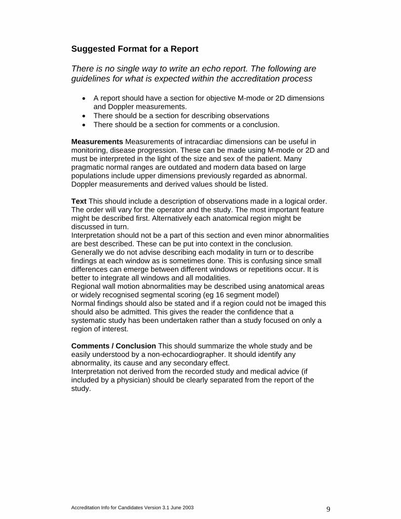
Suggested Format for a Report There is no single way to write an echo report. The following are guidelines for what is expected within the accreditation process
• A report should have a section for objective M-mode or 2D dimensions and Doppler measurements.
• There should be a section for describing observations • There should be a section for comments or a conclusion.
Measurements Measurements of intracardiac dimensions can be useful in monitoring, disease progression. These can be made using M-mode or 2D and must be interpreted in the light of the size and sex of the patient. Many pragmatic normal ranges are outdated and modern data based on large populations include upper dimensions previously regarded as abnormal. Doppler measurements and derived values should be listed. Text This should include a description of observations made in a logical order. The order will vary for the operator and the study. The most important feature might be described first. Alternatively each anatomical region might be discussed in turn. Interpretation should not be a part of this section and even minor abnormalities are best described. These can be put into context in the conclusion. Generally we do not advise describing each modality in turn or to describe findings at each window as is sometimes done. This is confusing since small differences can emerge between different windows or repetitions occur. It is better to integrate all windows and all modalities. Regional wall motion abnormalities may be described using anatomical areas or widely recognised segmental scoring (eg 16 segment model) Normal findings should also be stated and if a region could not be imaged this should also be admitted. This gives the reader the confidence that a systematic study has been undertaken rather than a study focused on only a region of interest. Comments / Conclusion This should summarize the whole study and be easily understood by a non-echocardiographer. It should identify any abnormality, its cause and any secondary effect. Interpretation not derived from the recorded study and medical advice (if included by a physician) should be clearly separated from the report of the study.

Accreditation Info for Candidates Version 3.1 June 2003 10
Suggested Reading List and Syllabus • The European Association of Echocardiography Education Committee will
run a teaching course in October which will provide preparation for the exam.
• The syllabus is set by the Accreditation Committee of the European Association of Echocardiography and is presented as a guide to candidates
• The reading list is provided by the Education Subcommittee of the European Association of Echocardiography
There are many excellent books on echo and just some examples are listed below. In addition to those listed there are many small basic texts which are a useful introduction to the subject.
Authoritative textbooks: A.E.Weyman, Principles and Practice of Echocardiography, 2nd ed. 1994 Lea & Febiger H.Feigenbaum, Echocardiography, 5th ed.1994 Lea & Febiger JRTC Roelandt, NG Pandian. Multiplane transesophageal echocardiography. Churchill LIvingstone 1996 Otto C. The Practice of Clinical Echocardiography. 2nd ed. Philadelphia: W. B. Saunders 2002. Marwick TH. Stress Echocardiography: Its Role in the Diagnosis and Evaluation of Coronary Artery Disease (Book with CD-ROM) Kluwer 2003 Useful review articles: Wranne B, Baumgartner H, Flachskampf FA, Hasenkam M, Pinto F. Stenotic lesions (editorial). Heart 1996; 75 (Suppl.2);36-42. Downloadable from http://heart.bmjjournals.com/supplements.shtml M Enriquez-Sarano,C Tribouilloy.Quantitation of mitral regurgitation: rationale, approach, and interpretation in clinical practice.Heart 2002; 88 (Suppl 4): iv1-iv3. Downloadable from http://heart.bmjjournals.com/supplements.shtml S Y Ho Anatomy of the mitral valve. Heart 2002; 88 (Suppl 4): iv5-iv10. Downloadable from http://heart.bmjjournals.com/supplements.shtml T Irvine, X K Li, D J Sahn, A Kenny. Assessment of mitral regurgitation. Heart 2002; 88 (Suppl 4): iv11-iv19. Downloadable from http://heart.bmjjournals.com/supplements.shtml

Accreditation Info for Candidates Version 3.1 June 2003 11
D Pellerin, S Brecker, and C Veyrat. Degenerative mitral valve disease with emphasis on mitral valve prolapse. Heart 2002; 88 (Suppl 4): iv20-iv28. Downloadable from http://heart.bmjjournals.com/supplements.shtml Flachskampf FA, Decoodt P, Fraser AG, Daniel WG, Roelandt JRTC. Recommendations for performing transesophageal echocardiography. Eur J Echocardiography 2001;2;8-21. Downloadable from www.escardio.org Gardin JM, Adams DB, Douglas PS, Feigenbaum H, Forst DH, Fraser AG, Grayburn PA, Katz AS, Keller AM, Kerber RE, Khandheria BK, Klein AL, Lang RM, Pierard LA, Quinones MA, Schnittger I; American Society of Echocardiography. Recommendations for a standardized report for adult transthoracic echocardiography: a report from the American Society of Echocardiography's Nomenclature and Standards Committee and Task Force for a Standardized Echocardiography Report. J Am Soc Echocardiogr. 2002 Mar;15(3):275-90. Downloadable from www.asecho.org Zoghbi WA, Enriquez-Sarano M, Foster E, Grayburn PA, Kraft CD, Levine RA, Nihoyannopoulos P, Otto CM, Quinones MA, Rakowski H, Stewart WJ, Waggoner A, Weissman NJ. Recommendations for Evaluation of the Severity of Native Valvular Regurgitation with Two-dimensional and Doppler Echocardiography. J Am Soc Echocardiogr 2003;16:777-802. Downloadable from http://www.asecho.org/freepdf/valvularregurg.pdf Cheitlin MD, Armstrong WF, Aurigemma GP, Beller GA, Bierman FZ, Davis JL, Douglas PS, Faxon DP, Gillam LD, Kimball TR, Kussmaul WG, Pearlman AS, Philbrick JT, Rakowski H, Thys DM. ACC/AHA/ASE 2003 guideline update for the clinical application of echocardiography—summary article: a report of the American College of Cardiology/American Heart Association Task Force on Practice Guidelines (ACC/AHA/ASE Committee to Update the 1997 Guidelines on the Clinical Application of Echocardiography). Circulation.2003 http://www.acc.org/clinical/guidelines/echo/summary_article.pdf

Accreditation Info for Candidates Version 3.1 June 2003 12
Syllabus
GENERAL
General Concepts
The place of echocardiography Clinical role of echocardiography and Doppler
• Information that echocardiography can, and cannot provide • ‘Ruling out’ pathology (sensitivity, specificity & Baye’s theorem) • Likelihood of findings influencing patient management • Undesirable outcomes: inaction while waiting for results, clinical ‘red
herrings’ Indications for echocardiography Competing and complementary technology • Cardiac catheterisation • X-ray ventriculography and coronary angiography • contrast C-T • Magnetic resonance imaging • Nuclear Cardiology
Service Provision Advantages/disadvantages of technician-led versus physician-led service Costs: fixed and variable Provision and indication for specialised techniques, e.g. TOE. Stress echo, Contrast echo Availability and access Controlling workload Training & motivation of staff Audit, Quality Control, Clinical Governance
Relationship with patients Explaining the procedure in terms relevant to the particular patient Respect for patients’ dignity and cultural backgrounds Relationships with colleagues. Handling requests for information about the study findings
Reporting and Documentation Standard methods & terminology Distinction between Technical and Clinical reports Responsibility for reporting Medico-legal considerations (Data Protection Act)
Imaging Physics & Instrumentation
Concepts and Terminology Concept of compression waves Definitions: frequency, wavelength, propagation velocity Units of measurement: Hz and MHz, Decibel Comparison of Ultrasound with audible sound.
Propagation of ultrasound through tissues Speed of sound in different body tissues. Frequency range used for diagnostic imaging Distinction between specular reflection and backscatter Principles of attenuation and scattering
Accreditation Info for Candidates Version 3.1 June 2003 13
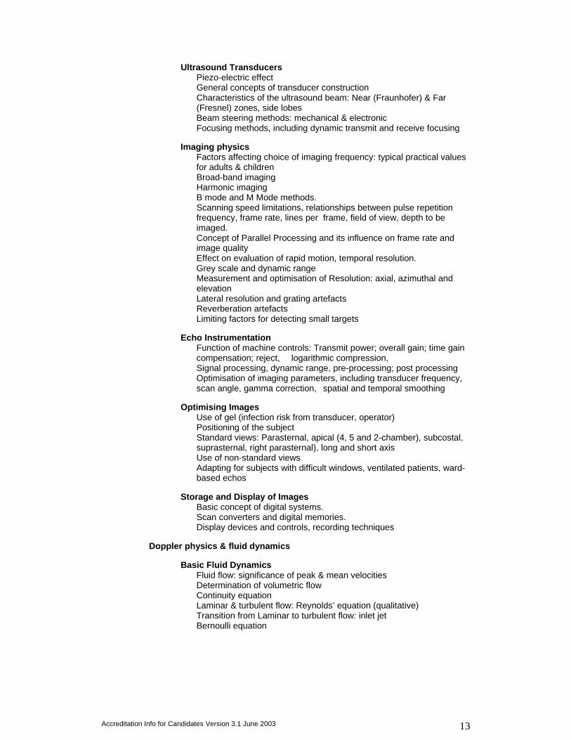
Ultrasound Transducers Piezo-electric effect General concepts of transducer construction Characteristics of the ultrasound beam: Near (Fraunhofer) & Far (Fresnel) zones, side lobes Beam steering methods: mechanical & electronic Focusing methods, including dynamic transmit and receive focusing
Imaging physics Factors affecting choice of imaging frequency: typical practical values for adults & children Broad-band imaging Harmonic imaging B mode and M Mode methods. Scanning speed limitations, relationships between pulse repetition frequency, frame rate, lines per frame, field of view, depth to be imaged. Concept of Parallel Processing and its influence on frame rate and image quality Effect on evaluation of rapid motion, temporal resolution. Grey scale and dynamic range Measurement and optimisation of Resolution: axial, azimuthal and elevation Lateral resolution and grating artefacts Reverberation artefacts Limiting factors for detecting small targets
Echo Instrumentation Function of machine controls: Transmit power; overall gain; time gain compensation; reject, logarithmic compression, Signal processing, dynamic range, pre-processing; post processing Optimisation of imaging parameters, including transducer frequency, scan angle, gamma correction, spatial and temporal smoothing
Optimising Images Use of gel (infection risk from transducer, operator) Positioning of the subject Standard views: Parasternal, apical (4, 5 and 2-chamber), subcostal, suprasternal, right parasternal), long and short axis Use of non-standard views Adapting for subjects with difficult windows, ventilated patients, ward-based echos
Storage and Display of Images Basic concept of digital systems. Scan converters and digital memories. Display devices and controls, recording techniques
Doppler physics & fluid dynamics
Basic Fluid Dynamics Fluid flow: significance of peak & mean velocities Determination of volumetric flow Continuity equation Laminar & turbulent flow: Reynolds’ equation (qualitative) Transition from Laminar to turbulent flow: inlet jet Bernoulli equation
Accreditation Info for Candidates Version 3.1 June 2003 14
Basic Principles of Doppler Interaction of ultrasound waves with moving blood: the Doppler effect The Doppler equation: factors influencing magnitude of Doppler shift Spectral analysis: fast Fourier transform (qualitative) The spectral Doppler display: determination of mean, modal and peak velocities Limitation of CW Doppler caused by lack of depth discrimination Audible range of Doppler shift frequencies The effect of beam angle errors on Doppler velocities Aliasing: how it is caused and how it manifests in practice: the Nyquist limit Influence on aliasing of: transducer frequency; sample depth (range x velocity product); and beam angle High pulse repetition frequency (extended range) PW Doppler Relative advantages and disadvantages of CW, PW and HPRF modes Concept of colour flow imaging as multi-sampled PW Velocity estimation, by moving target indication and autocorrelation (qualitative) Limitations of mean velocity: use of velocity variance to show high velocities/turbulence Aliasing in colour Doppler Packet size, colour mode and sector size and their effect on frame rate and aliasing
Doppler instrumentation
Spectral Doppler Instrumentation ‘Simultaneous’ Doppler using imaging transducers The ‘Stand-alone’ Doppler probe Features of the spectral display: positive & negative velocities; scale & baseline controls. Effect of high- and low-pass filter and intensity threshold (‘reject’) settings Pulsed Doppler sample volume: influence of gate length and distance (beam width) Representation of signal strength by image intensity How aliasing manifests on the spectral display
Colour Flow Instrumentation The colour display: BART convention Colour maps to show velocity scales Image domination and additive colour modes Basic principles of Tissue Doppler Imaging, including optimisation of filters for detecting tissue versus blood velocities Difference between velocity and power (signal amplitude) displays
TOE Instrumentation Transducer types: single plane, biplane, multiplane Optimising machine settings for TOE Patient monitoring for TOE and general safety considerations Control of infection
Safety of ultrasound Potential hazardous biological effects: heating, resonance and cavitation effects Measurement of beam intensity (SPTM) Practical precautions: power levels, use of colour and CW Doppler

Accreditation Info for Candidates Version 3.1 June 2003 15
Recording methods Advantages/disadvantages of recording on: videotape, photographic or dye-transfer prints, thermal strip chart Basic understanding of digital image processing and recording methods: pixel density, volume of data, concept of data compression, storage in RAM or magneto-optical disc format
Cardiac Anatomy and Physiology
Anatomy of the thorax Thorax contained by rib cage & diaphragm Lungs & pleura; heart & pericardium; mediastinum Blood vessels within the thorax
Gross anatomy of the heart Basic cardiac embryology Nomenclature of chambers and valves Major relationships of chambers, valves and blood vessels Distinguishing features of valves and chambers as related to echocardiography The pericardial sac
Cardiac anatomy and physiology as demonstrated by echocardiography Detailed structural anatomy of the heart, great vessels and pericardium Visualisation of normal cardiac anatomy and normal variants in standard echocardiographic planes Normal valve function, normal Doppler parameters and normal variants
The Cardiac Cycle Temporal relationships of the ECG, chamber pressures and valve movements Typical values for intracardiac pressures Relationship of valve movements to heart sounds
Cardiac functional parameters
Measurements and calculations On-screen measurement of length, slope, area, volume and time interval, and their significance for 2-D images, M-mode and spectral Doppler displays Standard M-mode measurements and calculations, both using machine software and manual methods Derivation of Stroke Volume, Ejection Fraction and LV Mass Methods of measuring LV volume, including biplane area, area-length and Simpson’s rule methods Limitations of measurement and/or calculation validity in presence of poor quality and/or off-axis images
Doppler determination of cardiac output, ejection time and velocity acceleration Methods of measuring diastolic dysfunction: E/A ratio, deceleration time, pulmonary venous flow patterns Peak and Mean pressure gradient measurements by Doppler and their relationship to catheterisation data Measurement of pulmonary pressures from tricuspid and pulmonary regurgitant flow velocities and assessment of inferior vena cava contraction

Accreditation Info for Candidates Version 3.1 June 2003 16
Contrast Studies Significance of spontaneous echo contrast Optimisation of machine control settings for detecting contrast Indications for a bubble contrast study Technique for performing a hand-agitated contrast study Clinical precautions
Awareness of encapsulated contrast agents and techniques Interaction of ultrasound with encapsulated agents Generation of harmonic energy by bubble distortion and fracture Doppler signals generated by bubbles (Power Mode) Clinical application for LV opacification and Doppler enhancement
PATHOLOGY
Mitral Valve Disease
2D, M-mode and Doppler features of the normal mitral valve
Mitral Stenosis Qualitative description of valve and sub-valve calcification and fibrosis Measurement of orifice area by planimetry Factors favouring successful balloon valvuloplasty Doppler assessment of mean and end-diastolic gradient Doppler assessment of area by ‘pressure half-time’: technique and limitations
Rheumatic mitral stenosis Assessment of severity (see 2.1.2)
Mitral regurgitation Assessment of severity by:
• Chamber sizes and volume overload • CW Doppler • PISA • Pulmonary vein flow patterns • Indirect effects
Aetiologies and typical echocardiographic features of:
• rheumatic • mitral annular calcification • ‘Floppy MV’ • ischaemic • functional • infective endocarditis
Aortic Valve Disease
2D, M-mode and Doppler features of the normal aortic valve
Aortic Stenosis Assessment by CW Doppler
• Peak and Mean gradients • Apical, right parasternal and suprasternal positions • Continuity equation • Assessment of left ventricular hypertrophy and function
Aetiologies and echocardiographic features: • Rheumatic

Accreditation Info for Candidates Version 3.1 June 2003 17
• Bicuspid • Senile degenerative • Sub- and supra-valve obstruction
Aortic regurgitation Aetiologies and typical echocardiographic features of:
• rheumatic • bicuspid valve • aortic root disease • infective endocarditis (including root abscesses)
Assessment of severity by:
• Chamber sizes/volume overload • CW Doppler • Colour Doppler • Indirect effects Role of TOE in assessing aetiology and severity
Tricuspid Valve Disease
2D, M-mode and Doppler features of the normal tricuspid valve
Rheumatic tricuspid valve stenosis Echocardiographic features Assessment of severity by imaging and Doppler
Tricuspid Regurgitation Assessment of severity by:
• 2D imaging and M-mode • CW Doppler • Colour Doppler • Indirect effects
Aetiologies and echocardiographic features of: • rheumatic • prolapse • congenital • endocarditis • carcinoid • functional
Pulmonary Valve Disease
2D, M-mode and Doppler features of the normal pulmonary valve
Pulmonary Valve Stenosis Echocardiographic features Assessment of severity by spectral Doppler Detection of infundibular obstruction by spectral Doppler
Pulmonary Regurgitation Aetiologies and echocardiographic features Assessment of severity by
• CW Doppler • Colour Doppler • Indirect effects

Accreditation Info for Candidates Version 3.1 June 2003 18
Infective Endocarditis Typical echocardiographic appearance of vegetations in bacterial and fungal endocarditis Preferred locations for vegetations ’Jet’ lesions Endocarditis associated with congenital disease and HCM Complications: abscess, fistula, perforation Role of TOE in suspected endocarditis
Prosthetic Valves
2D, M-Mode and Doppler features of the main types of replacement valves
• Ball & cage • Tilting Disc • Bi-leaflet • Stented Bioprostheses
Age-related deterioration of bioprostheses Role of TOE in examining normal and malfunctioning prosthetic valves
Prosthetic valve stenosis Assessment by 2D, M-mode and Doppler Normal ranges Use of Continuity Equation for aortic prostheses
Prosthetic valve regurgitation Trans- versus para-valvar regurgitation Normal versus abnormal regurgitation Assessment by CW, PW and Colour Doppler Colour artefacts from mechanical prostheses
Cardiomyopathies
Dilated Cardiomyopathy 2D, M-mode and Doppler features of dilated cardiomyopathy Detection and assessment of associated lesions:Functional valve regurgitation Thrombus in cardiac chambers Pericardial effusions Role of echocardiography in assessment and follow-up
Hypertrophic Cardiomyopathy 2D, M-mode and Doppler features of Hypertrophic Cardiomyopathy Differentiation from other causes of hypertrophy, e.g. ‘athletic heart’ Techniques for measurement of left ventricular wall thickness, detection of intracavity flow acceleration Assessment of right ventricular involvement Associated abnormalities, e.g. mitral regurgitation
Intracardiac Masses Typical locations for formation of intracardiac thrombus Echocardiographic features of typical LA Myxoma Differentiation of myxoma from other cardiac tumours Features suggestive of malignancy Role of TOE in assessment of intracardiac masses
Accreditation Info for Candidates Version 3.1 June 2003 19
Pericardial Disease
Anatomy of the normal pericardium Relationships of serous pericardium to heart and great vessels Transverse and oblique sinuses of the pericardium
Echocardiographic features of pericardial fluid Location of fluid in relation to patient position and fluid volume Differentiation from pleural effusion Assessment of volume of pericardial fluid Role of echocardiography in pericardiocentesis
Features of tamponade Collapse of RA and/or RV walls Effect on IVC Effect on A-V valve flow velocities Features of pericardial constriction Effect on A-V valve flow velocities Effect of respiration SVC/hepatic vein flow Differentiation from restrictive cardiomyopathy
Coronary Artery Disease and Systolic LV function Anatomy & nomenclature of the major branches of the coronary arteries Relationship of coronary anatomy to standard echocardiographic imaging planes Nomenclature for describing myocardial segments (ASE convention) Analysis of segmental systolic myocardial function Diastolic dysfunction in coronary artery disease Global measures of LV function:
• Ejection Fraction • Stroke Distance • Stroke Volume
Myocardial Infarction and its sequelae 2D, M-mode and Doppler features of:
• post-infarction VSD • mitral papillary muscle rupture • tamponade • mural thrombus • myocardial scarring • Dressler’s syndrome • left ventricular aneurysm – true aneurysm vs pseudoaneurysm
Pulmonary Hypertension 2-D, M-mode and Doppler features of pulmonary hypertension Aetiologies: primary; post pulmonary embolism; secondary to left-sided lesions; lung disease
Diseases of the Aorta Technique for examining the ascending and descending thoracic aorta Echocardiographic features of the normal aortic root, sinuses of Valsalva, ascending aorta and aortic arch 2-D, M-mode and Doppler features of:
• Marfan’s syndrome • sinus of Valsalva aneurysm • thoracic aortic aneurysm • aortic dissection • Additional features related to aortic dissection:

Accreditation Info for Candidates Version 3.1 June 2003 20
• aortic cusp prolapse • aortic regurgitation • fluid in pericardium
Role of transoesophageal echocardiography in the diagnosis of aortic dissection
Adult Congenital Heart Disease
Anatomy, pathophysiology and natural history of common congenital lesions present in adults: 2-D, M-mode and Doppler features of the following, pre-operatively and post-operatively, as seen in the older child or adult
• Ostium Secundum Atrial septal defects • Perimembranous and muscular ventricular septal defects • Partial and complete atrio-ventricular septal defects • Persistent ductus arteriosus • Bicuspid aortic valve • Sub- and supra-valve aortic stenosis • Aortic coarctation • Pulmonary stenosis • Ebstein’s anomaly • Fallot’s tetralogy
Role of contrast echocardiography in evaluating shunts in adults
Calculation of shunts
Role of TOE in adult congenital disease
Likely echocardiographic findings for common clinical presentations: Heart failure or breathlessness Arrhythmia Ejection systolic murmur Hypertension Collagen abnormalities Renal failure Stroke

Accreditation Info for Candidates Version 3.1 June 2003 21
Enrolment Form for European Accreditation In Adult Transthoracic Echocardiography
Name Correspondence Address
(City) (Country)
(Telephone) (email)
How long have you been performing echocardiograms? yrs months Approximately how many studies have you performed as first or second operator? About your department: Institution Name City / Country • How many Cardiologists or Physicians with an Interest in Cardiology are there • How many transthoracic echocardiograms are performed each year? • How many trained operators perform echocardiograms? • How many of the echocardiographers are: Training:Fellows/ Junior Doctors…….. Sonographers…….. Others • Do you have regular departmental reporting sessions? Yes / No • Are there formal teaching sessions? ……………………………Yes / No List the main machines in your department:
Manufacturer Model Approx. Age Does your unit have the ability to perform: Transesophageal..Yes/ No Stress echo Yes/ No Contrast echo Yes/ No

Accreditation Info for Candidates Version 3.1 June 2003 22
Statement of supervisor: (see also enrolment form for supervisor) • Has the candidate enough basic knowledge and skill to start the
accreditation process? • Is the candidate actively involved in scanning and reporting? I undertake to supervise and train the above candidate in echocardiography. I understand that 250 cases must be performed and reported by the candidate (the report may be checked by a second operator)' Name of supervisor Position held: Signature of supervisor Date Signature of candidate: Signature Date
Please send the fee of €150 by bank transfer (details below), specifying the purpose of your transfer, i.e. “Accreditation 12/03”:
Account owner: SEC WG7 ECHOCARDIO
Bank name: Banque Populaire de la Côte d’Azur, Nice Account number: 37019042359
IBAN FR76 1560 7000 6537 0190 4235 927 SWIFT CCBPFRPPNCE
and send the enrolment form by 5 November 2003 to
European Association of Echocardiography Administration
Attn.: Barbara Lefèvre The European Heart House
2035, Route des Colles – Les Templiers – BP 179 06903 Sophia Antipolis Cedex – France
Email: [email protected]

Accreditation Info for Candidates Version 3.1 June 2003 23
European Association of Echocardiography A Registered Branch of the ESC Accreditation in Transthoracic Echocardiography: Supervisor’s Enrolment form The role of the local supervisor is of great importance Supervisors would normally be expected to have held accreditation for at least 12 months While at present few individuals hold accreditation candidates may nominate supervisors. Supervisors would normally be locally or nationally recognised practicing echocardiographers In cases of doubt about the suitability of a supervisor the Accreditation Assessment committee may consult with the National Society and Working Group. The final decision regarding the suitability of the supervisor rests with the Accreditation Assessment Committee Application to act as Supervisor (to be submitted with candidates enrolment form) Name: Qualifications: Position: European Association of Echocardiography Accreditation: Yes No If Yes: Year first accredited: Last year of re-accreditation: If ‘No’:Please give a brief summary of your experience in echocardiography (years of practice, echo lab director, teaching and training responsibilities, membership of Working Groups or Societies relating to echo) I declare the information given to be true and accurate and I apply to act as a supervisor for accreditation in transthoracic echocardiography Signature: Date:
Accreditation Info for Candidates Version 3.1 June 2003 24
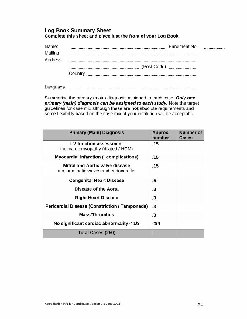
Log Book Summary Sheet Complete this sheet and place it at the front of your Log Book Name: Enrolment No. Mailing Address (Post Code) Country Language Summarise the primary (main) diagnosis assigned to each case. Only one primary (main) diagnosis can be assigned to each study. Note the target guidelines for case mix although these are not absolute requirements and some flexibility based on the case mix of your institution will be acceptable
Primary (Main) Diagnosis Approx. number
Number of Cases
LV function assessment inc. cardiomyopathy (dilated / HCM)
/15
Myocardial Infarction (+complications) /15
Mitral and Aortic valve disease inc. prosthetic valves and endocarditis
/15
Congenital Heart Disease /5
Disease of the Aorta /3
Right Heart Disease /3
Pericardial Disease (Constriction / Tamponade) /3
Mass/Thrombus /3
No significant cardiac abnormality < 1/3 <84
Total Cases (250)

Accreditation Info for Candidates Version 3.1 June 2003 25
European Association of Echocardiography A Registered Branch of the ESC Accreditation in Transthoracic Echocardiography Supervisors Marking Sheet for Cases Performed by Candidate Candidate No: ………………………………… Summary Sheet:
Case No. Indication Conclusion Acceptable Initials
I
II
III
IV
V
VI
VII
VIII
IX
X
Supervisor (signed) …………………………………… Date …… / ……./ ……..
Accreditation Info for Candidates Version 3.1 June 2003 26
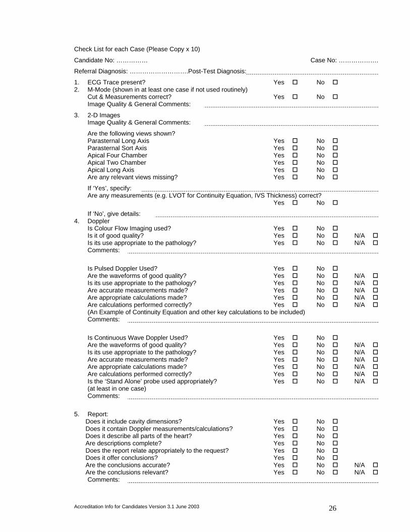
Check List for each Case (Please Copy x 10)
Candidate No: …………… Case No: ……………….
Referral Diagnosis: ……………………….Post-Test Diagnosis:
1. ECG Trace present? Yes ! No ! 2. M-Mode (shown in at least one case if not used routinely)
Cut & Measurements correct? Yes ! No ! Image Quality & General Comments:
3. 2-D Images Image Quality & General Comments:
Are the following views shown? Parasternal Long Axis Yes ! No ! Parasternal Sort Axis Yes ! No ! Apical Four Chamber Yes ! No ! Apical Two Chamber Yes ! No ! Apical Long Axis Yes ! No ! Are any relevant views missing? Yes ! No !
If ‘Yes’, specify: Are any measurements (e.g. LVOT for Continuity Equation, IVS Thickness) correct? Yes ! No !
If ‘No’, give details: 4. Doppler
Is Colour Flow Imaging used? Yes ! No ! Is it of good quality? Yes ! No ! N/A ! Is its use appropriate to the pathology? Yes ! No ! N/A ! Comments:
Is Pulsed Doppler Used? Yes ! No ! Are the waveforms of good quality? Yes ! No ! N/A ! Is its use appropriate to the pathology? Yes ! No ! N/A ! Are accurate measurements made? Yes ! No ! N/A ! Are appropriate calculations made? Yes ! No ! N/A ! Are calculations performed correctly? Yes ! No ! N/A ! (An Example of Continuity Equation and other key calculations to be included) Comments:
Is Continuous Wave Doppler Used? Yes ! No ! Are the waveforms of good quality? Yes ! No ! N/A ! Is its use appropriate to the pathology? Yes ! No ! N/A ! Are accurate measurements made? Yes ! No ! N/A ! Are appropriate calculations made? Yes ! No ! N/A ! Are calculations performed correctly? Yes ! No ! N/A ! Is the ‘Stand Alone’ probe used appropriately? Yes ! No ! N/A ! (at least in one case) Comments:
5. Report:
Does it include cavity dimensions? Yes ! No ! Does it contain Doppler measurements/calculations? Yes ! No ! Does it describe all parts of the heart? Yes ! No ! Are descriptions complete? Yes ! No ! Does the report relate appropriately to the request? Yes ! No ! Does it offer conclusions? Yes ! No ! Are the conclusions accurate? Yes ! No ! N/A ! Are the conclusions relevant? Yes ! No ! N/A ! Comments:

Accreditation Info for Candidates Version 4 Sept 2003
Suggested format for the letter to accompany submission of your Practical Assessment
Re: (Candidate name)__________________________________________ Initial We certify that the candidate has undergone a programme of training in echocardiography
We certify that the candidate has reached a standard of training to be able to independently perform and report a trans thoracic echocardiographic study
We certify that the candidate above has performed and reported the 250 cases included in the accompanying log book within a 12 month period.
We enclose a mark sheet assessing 10 studies performed and reported by the candidate (including studies performed with the supervisor present)
Supervisor
Signature: Date:

Accreditation Info for Candidates Version 3.1 June 2003 1
Practical Assessment – Final Check List Before submitting your Practical Assessment, check the following requirements:
I have passed the written examination within the last 12 months !
The letter and mark sheet from my Supervisor is enclosed !
My Name and address are clearly marked on the Log Book !
The completed Log Book Summary Sheet is at the front of the book !
All materials are securely packed addressed to: European Association of Echocardiography Administration, European Heart House 2035, Route des Colles – Les Templiers – BP 179 06903 Sophia Antipolis Cedex - France and my return address is also on the outside of the package. !
Accreditation Info for Candidates Version 4 Sept 2003
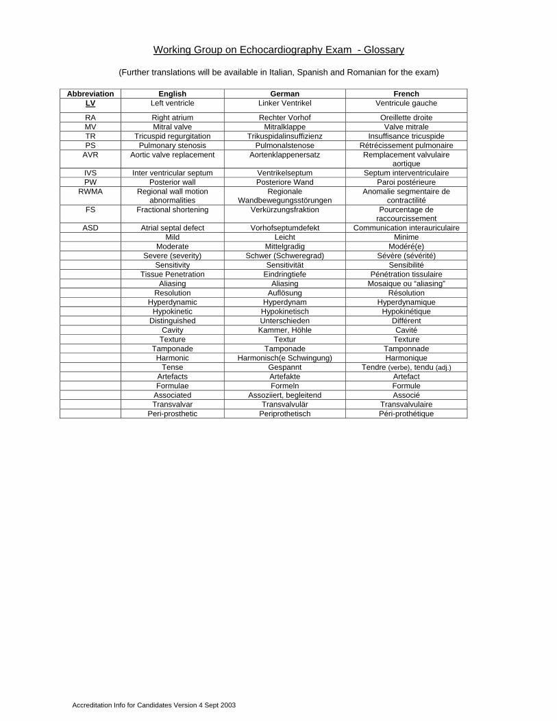
Working Group on Echocardiography Exam - Glossary
(Further translations will be available in Italian, Spanish and Romanian for the exam)
Abbreviation English German French LV Left ventricle Linker Ventrikel Ventricule gauche
RA Right atrium Rechter Vorhof Oreillette droite MV Mitral valve Mitralklappe Valve mitrale TR Tricuspid regurgitation Trikuspidalinsuffizienz Insuffisance tricuspide PS Pulmonary stenosis Pulmonalstenose Rétrécissement pulmonaire
AVR Aortic valve replacement Aortenklappenersatz Remplacement valvulaire aortique
IVS Inter ventricular septum Ventrikelseptum Septum interventriculaire PW Posterior wall Posteriore Wand Paroi postérieure
RWMA Regional wall motion abnormalities
Regionale Wandbewegungsstörungen
Anomalie segmentaire de contractilité
FS Fractional shortening Verkürzungsfraktion Pourcentage de raccourcissement
ASD Atrial septal defect Vorhofseptumdefekt Communication interauriculaire Mild Leicht Minime Moderate Mittelgradig Modéré(e) Severe (severity) Schwer (Schweregrad) Sévère (sévérité) Sensitivity Sensitivität Sensibilité Tissue Penetration Eindringtiefe Pénétration tissulaire Aliasing Aliasing Mosaique ou “aliasing” Resolution Auflösung Résolution Hyperdynamic Hyperdynam Hyperdynamique Hypokinetic Hypokinetisch Hypokinétique Distinguished Unterschieden Différent Cavity Kammer, Höhle Cavité Texture Textur Texture Tamponade Tamponade Tamponnade Harmonic Harmonisch(e Schwingung) Harmonique Tense Gespannt Tendre (verbe), tendu (adj.) Artefacts Artefakte Artefact Formulae Formeln Formule Associated Assoziiert, begleitend Associé Transvalvar Transvalvulär Transvalvulaire Peri-prosthetic Periprothetisch Péri-prothétique