accepted author version posted online: 3 august 2018 the ... · introduction honey bees use...
TRANSCRIPT

Accepted author version posted online: 3 August 2018
The Phosphofructokinase and Pyruvate Kinase Genes In Apis andreniformis and Apis cerana
indica: Exon Intron Organisation and Evolution
Nurul I. Shullia, Rika Raffiudin*, Berry Juliandi
Department of Biology, Faculty of Mathematics and Natural Sciences, Bogor Agricultural University,
Dramaga Campus, Bogor 16680, Indonesia
*Corresponding author: [email protected], [email protected]
Running title: PFK and PK Genes Organisation in Honey bee
Abstract: Genes related to carbohydrate metabolism have evolved rapidly in eusocial bees, including
honey bees. However, the characterisation of carbohydrate metabolism genes has not been reported
in Apis andreniformis or Apis cerana indica. This research aimed to characterise phosphofructokinase
(PFK) and pyruvate early view – July kinase (PK) genes in these honey bee species and to analyse
the evolution of the genus Apis using these genes. This study found the first data regarding A.
andreniformis PFK and PK-like nucleotide sequences. A BLAST-n algorithm-based search showed
that A. andreniformis and A. c. indica PFK and PK genes were homologous with those of Apis florea
and Apis cerana cerana from Korea, respectively. Multiple alignments of PFKs from five Apis species
showed many exon gains and losses, but only one among the PKs. Thus, the exon–intron
organisation of the PK genes may be more conserved compare with that of the PFKs. Another
evolutionary pattern indicated that more nucleotide substitutions occurred in Apis’ PK than PFK genes.
Deduced PFK amino acid sequences revealed a PFK consensus pattern of 19 amino acids, while the
deduced PK amino acid sequences were predicted to have barrel and alpha/beta domains. Based on
these two metabolism-related genes, The Neighbour-joining and Maximum likelihood phylogenetic
trees are congruent and revealed that the A. andreniformis and A. florea group were in the basal
position. Apis mellifera, A. cerana, and Apis dorsata formed a monophyletic clade, although the
positions of A. mellifera and A. dorsata were different in the nucleotide- and amino acid-based
phylogenetic trees.
Keywords: Apis cerana indica, Apis andreniformis, phosphofructokinase gene, pyruvate kinase gene,
exon gain and loss

INTRODUCTION
Honey bees use carbohydrates as the main fuel for flight and produce modified stored sugar (honey)
to maintain the optimal hive temperature (Fischman et al. 2011). Molecular data has indicated that
carbohydrate metabolism-related genes are among the most rapidly evolving genes in eusocial bees,
including honey bees (Woodard et al. 2011). Phosphofructokinase (PFK; EC 2.7.1.11) plays a key
regulatory role in the glycolytic pathway. It catalyses the reaction of fructose 6-phosphate using ATP to
generate 1,6-diphosphate and ADP (Voet & Voet 1995). Pyruvate kinase (PK; EC 2.7.1.40) is involved
in glycolytic flux and catalyses the reaction of pyruvate phosphoenol to generate ATP and pyruvate by
transferring the phosphate group to ADP (Voet & Voet 1995).
The first eukaryotic PFK sequence was characterised in cloned rabbit muscle and its 17-kb
length was split into 22 exons, encoding 780 amino acids (Lee et al. 1987). The exon–intron
organisation was the same among human liver (Elson et al. 1990), human muscle (Vaisanen et al.
1992) and mouse liver (Rongnoparut et al. 1991) PFKs. In insects, the PFK gene has been
characterised in Drosophila melanogaster and spans 6.5 kb, which is split into 8 exons and encodes
787 amino acids. The amino acid sequence of D. melanogaster PFK showed a 50.9% identity with the
human muscle PFK (Currie & Sullivan 1994).
PK genes have also been characterised as 20 kb in rat muscle (Takenaka et al. 1989), and 32
kb in human muscle, consisting of 12 exons and 11 introns (Takenaka et al. 1991), whereas chicken
PK has at least 10 introns (Lonberg & Gilbert 1985). Complementary DNA cloning of the PK gene in
D. melanogaster revealed a 1,602-bp coding region split into four exons encoding a predicted 533
amino acids (Chien et al. 1999).
Database entries from GenBank showed that the PFK gene in the genus Apis has different
exon numbers in different species. The whole genome of Apis mellifera from NCBI showed that ATP-
dependent 6-phosphofructokinase has 13 exons (GenBank NC_007079). However, this gene in the
Apis cerana cerana strain from Korea (GenBank NW_016019786) has 7 isoforms and 24 exons. The
giant honey bee Apis dorsata (GenBank NW_006263741) has 7 isoforms and 22 exons, and the dwarf
honey bee Apis florea (GenBank NW_003790158) has 14 exons. However, almost all of the Apis PK
genes have similar exon–intron organisations. GenBank database entries showed that A. dorsata
(GenBank NW_006263478), A. florea (GenBank NW_003790664), and A. c. cerana (GenBank
NW_016019308) predicted PK (PK-like) genes have 8 exons, while Apis mellifera (GenBank
NC_007073) has 2 isoforms and 10 exons.
Sequences that have genetic variants are invaluable in documenting evolutionary history.
Honey bee phylogenetic studies have been performed based on molecular data from mitochondrial
genes, such as cytochrome c oxidase subunit I (COI) (Tanaka et al. 2001), cytochrome c oxidase
subunit II (COII), rRNA gene for the large ribosomal subunit rrnL, and NADH dehydrogenase subunit
2 (nad2), or from nuclear genes, such as inositol 1,4,5-triphosphate receptor (itpr) (Raffiudin & Crozier
2007) and the elongation factor 1-alpha (EF1-α) intron (Arias & Sheppard 2005). The position of dwarf
bees (the A. andreniformis and A. florea group) is almost at the tree’s base, and the giant (the A.
dorsata and Apis laboriosa group) and medium-sized bees (the A. mellifera and Apis cerana group)
form a monophyletic clade. A phylogenetic study based on several genes, including carbohydrate
metabolism-related genes, has been reported for eusocial bees (Woodard et al. 2011), but the
evolution of PFK and PK genes has not been explored in honey bees at the species level.
Indonesia has the most diverse honey bee population in the world, with five, A. dorsata, A.
cerana (Ruttner 1988), A. andreniformis (Wu & Kuang 1987), Apis koschevnikovi (Tingek et al. 1988),
and Apis nigrocincta (Hadisoesilo et al. 1995), of nine species of honey bee being native to Indonesia.
A. cerana is distributed in the most of the Indonesian islands. Four subspecies of A. cerana are
distributed in the old world, and A. c. indica is established in Indonesia (Ruttner 1988). The PFK and
PK genes of the dwarf honey bee A. florea (Lowe & Eddy 1997) and the other subspecies A. c. cerana
(Park et al. 2015) have been submitted as GenBank database entries, but those of A. andreniformis
and A. c. indica from Indonesia have not been reported. This study aimed to characterise PFK and PK
genes in A. andreniformis and A. c. indica and also to analyse the evolution of honey bees based on
these genes.
MATERIAL AND METHODS
Samples and DNA Extraction, Amplification and Sequencing
Apis andreniformis was collected from Padang Pariaman, West Sumatra and A. c. indica was
collected from Bogor, West Java. Total DNA was extracted from the thoraxes using a standard
phenol–chloroform extraction method and ethanol precipitation (Sambrook et al. 1989), with minor
modifications (Raffiudin & Crozier 2007).
The partial regions of PFK and PK-like gene primers were designed manually from A. mellifera
(GenBank NC_007079, NC_007073), A. dorsata (GenBank NW_006263741, NW_006263478), and A.
florea (GenBank NW_003790158, NW_003790664) genomic sequences. Due to an obstacle in primer
design involving the 1,099 bp of Intron 3 in the A. mellifera PFK gene, the targeted gene was divided
into two regions, Part A (exons 1–3) and Part B (exon 4–7) (Table 1). The PCR conditions were as
follows: initial denaturing at 95°C for 3 min, 30 cycles of 95°C for 1 min, 48–53°C for 30 s, and 72°C
for 1 min, followed by a final extension at 72°C for 2 min. PCR products were electrophoreses in 1.5%
agarose gel and stained using Diamond Nucleic Acid Dye (Promega, Madison, WI, USA). The PCR
products were sequenced by a company sequencing service (First BASE, Selangor, Malaysia).
Gene Structure, Motif, and Phylogenetic Analyses
The sequences of the PFK and PK-like genes from A. andreniformis and A. c. indica were aligned with
homologues from Apidae database entries in GenBank identified using a BLAST-n algorithm-based
search of the nucleotide collection (nt/nt) (http://blast.ncbi.nlm.nih.gov/Blast.cgi). Based on homology
analyses of the DNA coding regions and genomics, the closest related species to A. andreniformis
and A. c. indica were aligned using ClustalX 2 (Larkin et al. 2007) and were used to determine the
exon–intron organisation. Protein motifs and families of the putative amino acid sequences were
explored using PROSITE (http://prosite.expasy.org/) and Pfam (http://pfam.sanger.ac.uk/),
respectively. The number of substitutions and pairwise distances of Apis PFK and PK-like nucleotide
sequences were analysed using MEGA6 (Tamura et al. 2013). The obtained sequences, combined
with other Apidae sequences in GenBank, were chosen for phylogenetic analysis (Table 2). The
nucleotide-based phylogenetic trees were constructed using the Neighbour-joining (NJ) with Tamura-
Nei Model and Maximum likelihood (ML) method with suggested Best Model Tamura-Nei implemented
in MEGA6 with 1,000 bootstrap replicates (Tamura et al. 2013). The amino acid-based phylogenetic
trees were also constructed using NJ method with Poisson model and ML method with suggested
Best model cpREV+G implemented in MEGA6 with 1,000 bootstrap replicates (Tamura et al. 2013).
RESULTS
Characterisation of Partial PFK and PK-Like Genes
This study successfully amplified the targeted PFK and PK-like genes in A. andreniformis from
Padang Pariaman, West Sumatra and A. c. indica from Bogor (DDBJ LC318660, LC318759-63).
BLAST-n algorithm-based searches using the nucleotide collection (nr/nt) database showed that A.
andreniformis is closely related to A. florea, with 100% (GenBank XM_012485123.1) and 95%
(GenBank XM_012487945.1) identities for PFK and PK-like genes. Based on this homology and
previous morphology (Alexander 1991), a combined behavioural and molecular phylogenetic study
(Raffiudin and Crozier, 2007) revealed that A. andreniformis and A. florea are sister species. Thus, A.
florea PFK and PK coding regions and genomes were used to determine A. andreniformis’ exon–
intron organisation. There were six exons (Exons 2, 3, 4, 6, 7, and 8) and four introns (Introns 2, 3, 6,
and 7) of A. andreniformis in the A and B PFK sequences (Fig. 1), while the A. andreniformis partial
PK-like sequence had seven exons (Exons 2–8) and six introns (Introns 2–7) (Fig. 2). The putative
exon regions of A. andreniformis’ PFK parts A and B, and PK-like sequences revealed 543, 435 and
1,446 bp, respectively. Translations of A. andreniformis part A, PFK part B, and PK-like exon regions
revealed 181, 151, and 482 putative amino acids, respectively.
There was also a high similarity between A. c. indica from Bogor, Indonesia and A. cerana from
Korea, with 100% (GenBank XM_017065124.1) and 99% (GenBank XM_017058664.1) identities for
the PFK and PK-like genes, respectively. Using the sequence of the A. cerana strain from Korea
revealed that A. c. indica PFK parts A and B contained seven exons (Exons 6–12) and five introns
(Intron 6, 7, 9, 10, and 11). The exon region of A. c. indica PFK parts A and B cover 537 and 510 bp,
respectively. The partial PK-like sequence of A. c. indica s consists of seven exons (Exons 2–8) and
six introns (Exons 2–7), which corresponds to 1,380 bp. The complete translations of A. c. indica’ PFK
parts A and B, and the PK-like sequence encompassed 179, 170, and 490 putative amino acids,
respectively.
Schematic representations of the PFK and PK-like exon–intron organisation in the genus Apis
(Fig. 3) showed that the former had more variation than the latter in the genus Apis, even though their
exon lengths are the same. Exons 6–12 in the A. c. indica PFK gene had similar sequences to Exons
1–7 of A. mellifera (GenBank NC_007079). The sequence of Exon 9 in A. cerana (GenBank
NW_016019786) or Exon 4 in A. mellifera PFK gene was part of Intron 5 in the dwarf honey bee
(GenBank NW_003790158). Thus, there is only one exon gain and one PK-like gene loss among
these five species in the genus Apis.
All exon–intron boundaries in the PFK and PK-like genes were confirmed using GT–AG rules
(Tables 3 and 4). Although the ranges of PFK and PK-like intron lengths were different in A.
andreniformis and A. c. indica, the homologous introns had the same intron phase. Intron 5 of the A.
andreniformis PFK gene was incomplete because the region was unamplified. The differences in
intron lengths between A. andreniformis and A. c. indica were caused by base insertions and
deletions.
Motifs of Partial PFK and PK-like Genes
Motif searches using PROSITE (http://prosite.expasy.org/) showed a consensus pattern, [RK]-x(4)-
[GAS]-H-x-[QL]-[QR]-[GS]-[GF]-x(5)-[DE]-[RL] PFK (PS00433), in both A. andreniformis and A. c.
indica partial PFK sequences. This study also found a conserved PFK-related consensus pattern in
the genus Apis (Fig. 4). Analysis of the protein family using Pfam indicated that A. andreniformis and
A. c. indica partial PK-like amino acid sequences formed a pattern of a pyruvate kinase barrel domain
at amino acids 2–323 and a pyruvate kinase alpha/beta domain format amino acids 345–463.
Phylogenies of The Genus Apis’ PFK and PK-Like Genes
The comparisons between the number of substitutions and the Tamura–Nei corrected p-distances
showed that transitions occurred more often than transversions in the PFK and PK-like genes of these
five Apis species. The p-distances corrected by Tamura-Nei were greater in the PK-like gene than in
the PFK gene (Fig. 5). Analyses of pairwise comparisons revealed that the 3rd codon substitution
number (transition and transversion) was the highest in both PFK and PK-like gene sequences (Figs.
6 and 7). The range of the number substitutions in the exon regions in Apis PK-like gene sequences
was wider than in the PFK gene.
Using a combination of PFK and PK-like nucleotide (Fig. 8) and amino acid (Fig. 9) sequences
in the genus Apis and out group, this study found two topologies based on nucleotide sequence and
amino acid phylogenetic tree. The topology of both phylogenetic trees based on NJ and ML for
nucleotide and amino acid sequences are the same. All of the trees supported the dwarf honey bee’s
(A. florea and A. andreniformis) basal position. The nucleotide-based topology showed that the giant
honey bee A. dorsata is the sister clade of the medium honey bee (A. c. cerana, A c. indica, and A.
mellifera) (Fig. 8 A-B), but the amino acid-based topology placed A. mellifera and A. dorsata in a
separate clade (Fig. 9 A-B).
DISCUSSION
Motifs in PFK and PK Genes in Apis
This study aimed to characterise PFK and PK-like genes, which are key regulatory enzymes in
glycolysis and control the flux through this pathway (Voet & Voet 1995). We studied these two genes
in the native Indonesian honey bee A. andreniformis and the widely distributed A. c. indica. This is the
first data regarding A. andreniformis PFK and PK-like nucleotide sequences. Analyses of deduced A.
andreniformis and A. c. indica PK-like amino acids determined that these sequences have barrel and
alpha/beta domains. Muirhead (1990) found that the cat PK gene in muscle consists of four domains:
N-terminal, A (A1 and A2), B, and C. The complementary DNA of the Drosophila PK gene also has
four domains and a conserved amino acid in the active site (Chien et al. 1999).
A PROSITE analysis determined that the PFK sequences contain the [RK]-x(4)-[GAS]-H-x-[QL]-
[QR]-[GS]-[GF]-x(5)-[DE]-[RL] PFK (PS00433) consensus pattern. This corroborates our investigation
of the Apis PFK gene in which a multiple alignment revealed the consensus pattern of
RITVLGHVQRGGNPSAFDR. The R or K amino acid, and the H and Q or R amino acids are important
because of their involvement in fructose-6-phosphate binding (http://prosite.expasy.org/). The R and H
amino acids were also found in the N- and C-halves of two adjacent subunits in the rabbit muscle PFK
and defined the binding-site of fructose-6-phosphate (Poorman et al. 1984).
Exon Gain and Loss in The PFK and PK Genes of Apis
The NCBI database entries for PFK genes in the genus Apis showed variations in number of exons,
with 13 exons in A. mellifera (GenBank NC_007079) and up to 24 exons in A. c. cerana (Genbank
NW_016019786). This variation indicated a phenomenon of exon gain and loss in the PFK gene. This
lead to the sequence of Exon 9 from A. c. indica and A. c. cerana or Exon 4 from the A. mellifera PFK
gene being part of Intron 5 in A. florea and A. andreniformis.
Like the PFK exon number among the genus Apis, human (Vaisanen et al. 1992) and rabbit
PFK genes in muscle have up to 22 exons (Lee et al. 1987). However, the PFK gene of D.
melanogaster that contains 6.5 kb, only has half the Apis PFK exon number (eight exons and seven
introns) (Currie & Sullivan 1994). This suggests that the PFK gene in the genus Apis was more
evolved than that of Drosophila. The losses of exons might be caused by frame shift mutations or
splice junctions that resulted in intron sliding (Currie & Sullivan 1994).
A. andreniformis and A. c. indica have eight exons in their PK-like genes and show similar exon-
intron organisations. However, the Drosophila PK gene has only half the exon number compared with
Apis (Chien et al. 1999). Although the Apis PK-like genes have more similar exon-intron organisations
than the PFK genes, another study revealed that A. mellifera and Drosophila PFK genes had a 1:1
orthology, while the PK gene had a 2:6 orthology (Kunieda et al. 2006). The greater diversity level of
the PK-like gene may be a result of its position at the end of glycolysis pathway, before pyruvate
enters the citrate cycle or other pathways (Kunieda et al. 2006). The PFK gene evolved by gene
duplications and the amino acid sequence is highly homologous between prokaryotes and mammals
(Poorman et al. 1984). The presence of orthologous PK-like genes in the genus Apis might be caused
by the high nucleotide substitution rate in the PK-like gene compared with that of the PFK gene.
The Evolution of Apis PFK and PK Genes
Here, the PK-like gene had more substitutions than the PFK gene. Thus, we analysed the evolution of
the genus Apis based on the combined data regarding PFK and PK-like genes. The resulting
Neighbour-joining phylogenetic tree of the honey bee that confirmed by Maximum likelihood
phylogenetic showed that the dwarf honey bee (the A. andreniformis and A. florea group) was always
in basal position. The tree also grouped the medium-sized honey bee (the A. cerana and A. mellifera
group) and giant honey bee (A. dorsata) into a monophyletic clade, but A. mellifera and A. dorsata
formed two topologies. The first topology built from combined PFK and PK-like nucleotide sequences
was congruent with phylogenetic tree based on the molecular sequences of five genes and the
behavioural states (Raffiudin & Crozier 2007). Almost all of the phylogenetic trees based on the
molecular data grouped honey bees into three major clusters based on body size: giant bees, dwarf
bees, and medium bees. Molecular-based honey bee phylogenetic trees were also congruent with the
morphology-based phylogenetic tree (Alexander 1991). This indicated that nucleotide variations in
intron regions also had roles in building the phylogenetic tree. The substitution rates in PFK and PK-
like genes were greater in the third and first codons, respectively, than in the second codon. This
result supported the finding that transitions in 16S rRNA, COI, and COII genes were more common
than transversions in the genus Apis (Tanaka et al. 2001). In a future study, an analysis of the cDNAs
of these genes in the honey bee is needed to fully analyse the phenomenon of exon gain and loss in
Apis evolution.
CONCLUSIONS
Characterisations of A. andreniformis and A. c. indica PFK and PK-like genes revealed that they have
same exon–intron organisation as A. florea and A. c. cerana from Korea, respectively. Moreover,
multiple alignments of these genes among five Apis species revealed that exon gain and loss occurred
more often in PFK than in PK-like genes, even though the nucleotide substitution rate in the former
was higher than in the latter. The nucleotide-based phylogenetic tree generated from the combination
of data on the two carbohydrate metabolism-related genes was congruent with molecular and
morphological phylogenetic trees, and clustered A. mellifera and A. cerana groups with A. dorsata to
form a monophyletic clade, while the A. florea and A. andreniformis group was basal.
ACKNOWLEDGMENTS
We thank the Indonesia Ministry of Research Technology and Higher Education for the research grant
(Grant number 079/SP2H/LT/DRPM/II/2016) for Rika Raffiudin.
REFERENCES
Alexander B A. (1991). Phylogenetic analysis of the genus Apis (Hymenoptera : Apidae).
Entomological Society of America 84: 137–149. https://doi.org/10.1093/aesa/84.2.137
Arias M C and Sheppard W S. (2005). Phylogenetic relationships of honey bees (Hymenoptera :
Apinae : Apini) inferred from nuclear and mitochondrial DNA sequence data. Molecular
Phylogenetic Evolution 37: 25–35. https://doi.org/10.1016/j.ympev.2005.02.017
Chien Y, Zhu Y and Chuen C. (1999). Complementary DNA cloning and analysis of gene structure of
Pyruvate kinase from Drosophila melanogaster. Zoological Studies 38(3): 322-332.
Currie P D and Sullivan D T. (1994). Structure and expression of the gene encoding PFK in
Drosophila melanogaster. The Journal of Biological Chemistry 269(40): 24679-24687.
Elson A, Levanon D, Brandies M, Dafni N, Bernstein Y, Danciger E and Groner Y. (1990). The
Structure of the human liver-type Phosphofructokinase gene. Genomics 7: 47-56.
https://doi.org/10.1016/0888-7543(90)90517-x
Fischman B J, Woodard S H and Robinson G E. (2011). Molecular evolutionary analyses of insect
societies. Proceedings of the National Academy Sciences 108: 10847–10854.
https://doi.org/10.1073/pnas.1100301108
Hadisoesilo S, Otis G W and Meixner M. (1995). Two distinct populations of cavity-nesting honey bees
(Hymenoptera, Apidae) in South Sulawesi, Indonesia. Journal of the Kansas Entomological
Society 68: 399-407.
Kunieda T, Fujiyuki T, Kucharski R, Foret S, Ament S A, Toth A L, Ohashi K et al. (2006)
Carbohydrate metabolism genes and pathways in insects: insights from the honey bee
genome. Insect Molecular Biology 15: 563–576. https://doi.org/10.1111/j.1365-
2583.2006.00677.x
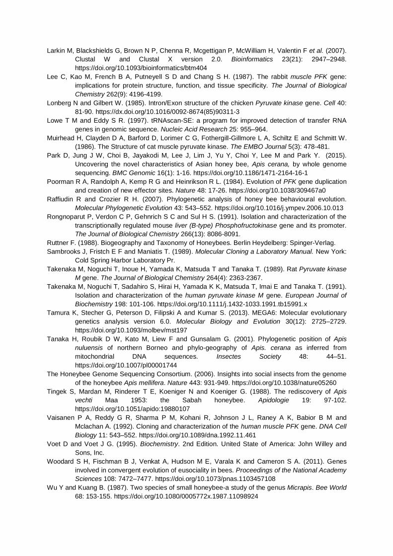
Larkin M, Blackshields G, Brown N P, Chenna R, Mcgettigan P, McWilliam H, Valentin F et al. (2007).
Clustal W and Clustal X version 2.0. Bioinformatics 23(21): 2947–2948.
https://doi.org/10.1093/bioinformatics/btm404
Lee C, Kao M, French B A, Putneyell S D and Chang S H. (1987). The rabbit muscle PFK gene:
implications for protein structure, function, and tissue specificity. The Journal of Biological
Chemistry 262(9): 4196-4199.
Lonberg N and Gilbert W. (1985). Intron/Exon structure of the chicken Pyruvate kinase gene. Cell 40:
81-90. https://dx.doi.org/10.1016/0092-8674(85)90311-3
Lowe T M and Eddy S R. (1997). tRNAscan-SE: a program for improved detection of transfer RNA
genes in genomic sequence. Nucleic Acid Research 25: 955–964.
Muirhead H, Clayden D A, Barford D, Lorimer C G, Fothergill-Gillmore L A, Schiltz E and Schmitt W.
(1986). The Structure of cat muscle pyruvate kinase. The EMBO Journal 5(3): 478-481.
Park D, Jung J W, Choi B, Jayakodi M, Lee J, Lim J, Yu Y, Choi Y, Lee M and Park Y. (2015).
Uncovering the novel characteristics of Asian honey bee, Apis cerana, by whole genome
sequencing. BMC Genomic 16(1): 1-16. https://doi.org/10.1186/1471-2164-16-1
Poorman R A, Randolph A, Kemp R G and Heinrikson R L. (1984). Evolution of PFK gene duplication
and creation of new effector sites. Nature 48: 17-26. https://doi.org/10.1038/309467a0
Raffiudin R and Crozier R H. (2007). Phylogenetic analysis of honey bee behavioural evolution.
Molecular Phylogenetic Evolution 43: 543–552. https://doi.org/10.1016/j.ympev.2006.10.013
Rongnoparut P, Verdon C P, Gehnrich S C and Sul H S. (1991). Isolation and characterization of the
transcriptionally regulated mouse liver (B-type) Phosphofructokinase gene and its promoter.
The Journal of Biological Chemistry 266(13): 8086-8091.
Ruttner F. (1988). Biogeography and Taxonomy of Honeybees. Berlin Heydelberg: Spinger-Verlag.
Sambrooks J, Fristch E F and Maniatis T. (1989). Molecular Cloning a Laboratory Manual. New York:
Cold Spring Harbor Laboratory Pr.
Takenaka M, Noguchi T, Inoue H, Yamada K, Matsuda T and Tanaka T. (1989). Rat Pyruvate kinase
M gene. The Journal of Biological Chemistry 264(4): 2363-2367.
Takenaka M, Noguchi T, Sadahiro S, Hirai H, Yamada K K, Matsuda T, Imai E and Tanaka T. (1991).
Isolation and characterization of the human pyruvate kinase M gene. European Journal of
Biochemistry 198: 101-106. https://doi.org/10.1111/j.1432-1033.1991.tb15991.x
Tamura K, Stecher G, Peterson D, Filipski A and Kumar S. (2013). MEGA6: Molecular evolutionary
genetics analysis version 6.0. Molecular Biology and Evolution 30(12): 2725–2729.
https://doi.org/10.1093/molbev/mst197
Tanaka H, Roubik D W, Kato M, Liew F and Gunsalam G. (2001). Phylogenetic position of Apis
nuluensis of northern Borneo and phylo-geography of Apis. cerana as inferred from
mitochondrial DNA sequences. Insectes Society 48: 44–51.
https://doi.org/10.1007/pl00001744
The Honeybee Genome Sequencing Consortium. (2006). Insights into social insects from the genome
of the honeybee Apis mellifera. Nature 443: 931-949. https://doi.org/10.1038/nature05260
Tingek S, Mardan M, Rinderer T E, Koeniger N and Koeniger G. (1988). The rediscovery of Apis
vechti Maa 1953: the Sabah honeybee. Apidologie 19: 97-102.
https://doi.org/10.1051/apido:19880107
Vaisanen P A, Reddy G R, Sharma P M, Kohani R, Johnson J L, Raney A K, Babior B M and
Mclachan A. (1992). Cloning and characterization of the human muscle PFK gene. DNA Cell
Biology 11: 543–552. https://doi.org/10.1089/dna.1992.11.461
Voet D and Voet J G. (1995). Biochemistry. 2nd Edition. United State of America: John Willey and
Sons, Inc.
Woodard S H, Fischman B J, Venkat A, Hudson M E, Varala K and Cameron S A. (2011). Genes
involved in convergent evolution of eusociality in bees. Proceedings of the National Academy
Sciences 108: 7472–7477. https://doi.org/10.1073/pnas.1103457108
Wu Y and Kuang B. (1987). Two species of small honeybee-a study of the genus Micrapis. Bee World
68: 153-155. https://doi.org/10.1080/0005772x.1987.11098924

Table 1. PFK and PK-like gene primers designed based on the Apis mellifera whole genome.
No. Gene/ Exon* Primer names Primer Nucleotide (5'–3') A. mellifera Acc number
1 Part A PFK
Am_PFK1_F GGAATGAATGCAGCAGTTCGAG GenBank
NC_007079
(Exons 1–3) Am_PFK1_R CAATGCCGACCCATAACTTCCA
2 Part B PFK Am_PFK3_F GCAGCCATATGTTCTGAAGCTG
(Exons 4–5) Am_PFK3_R ACCACCTCTTTGAACATGACCA
3 Part B PFK Am_PFK4_F GAAAGACTTATGGGACAACGACT
(Exons 5–7) Am_PFK4_R ATAGGTGCTTTCAGACGGGTCA
4 PK-like
Amel_PK1_F TGGCCGGTGCAAATATAGTTCG
GenBank NC_007073
(Exons 1–2) Amel_PK1_R AACTTGTAATAAAACGGCTCCTC
5 PK-like Amel_PK2_F AGGACGTGCAAACTGTTTCTGG
(Exons 2–4) Amel_PK2_R TGTCGAGGAGAATAGCGTCAG
6 PK-like Amel_PK3ex_F GATTACTAGAATTTGCGTTATAGC
(Exons 4–6) Amel_PK3in_F ATCTCGTCTCAACAAGGTTTGGA
Amel_PK3_R GCGAGAATGGGACATCTAGGAC
7 PK-like Amel_PK4_F CACTCGATCATAATTGGTGGTGT
(Exon 7) Amel_PK4in_R GGGACTTGATCCTTTCTGCATC
Amel_PK4ex_R TCACCAGCAAGACTCAAAATCTG
Table 2. Species used for phylogenetic analysis from current research and GenBank.
No. Species Abbreviation Accession Number References
PFK PK-like
In group
DDBJ
1 A. c. indica Ace LC318760-61 LC318660 current research 2 A. andreniformis Aan LC318762-63 LC318759 current research
GenBank
3 A. c. cerana Acc NW_016019786 NW_016019308 (Park et al. 2016) 4 A. mellifera Ame NC_007079 NC_007073 (Honeybee
Genome Sequencing Consortium 2006)
5 A. florea Afl NW_003790158 NW_003790664 (Lowe and Eddy 1997)
6 A. dorsata Ado NW_006263741 NW_006263478 (Lowe and Eddy 1997)
Out group
7 Bombus terrestris Bte NC015771 NC015765 -
Table 3. Intron lengths in partial A. andreniformis PFK and PK-like genes.
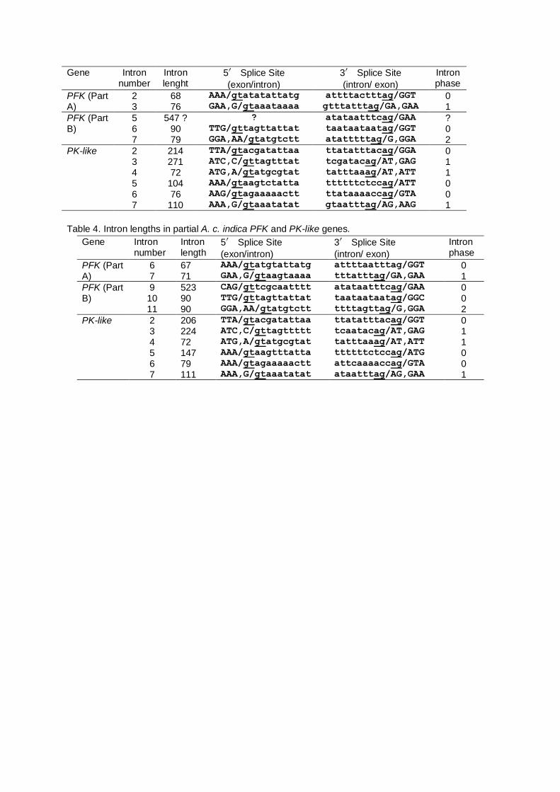
Gene Intron number
Intron lenght
5′ Splice Site
(exon/intron)
3′ Splice Site
(intron/ exon)
Intron phase
PFK (Part A)
2 68 AAA/gtatatattatg attttactttag/GGT 0 3 76 GAA,G/gtaaataaaa gtttatttag/GA,GAA 1
PFK (Part B)
5 547 ? ? atataatttcag/GAA ? 6 90 TTG/gttagttattat taataataatag/GGT 0 7 79 GGA,AA/gtatgtctt atatttttag/G,GGA 2
PK-like 2 214 TTA/gtacgatattaa ttatatttacag/GGA 0 3 271 ATC,C/gttagtttat tcgatacag/AT,GAG 1 4 72 ATG,A/gtatgcgtat tatttaaag/AT,ATT 1 5 104 AAA/gtaagtctatta ttttttctccag/ATT 0 6 76 AAG/gtagaaaaactt ttataaaaccag/GTA 0 7 110 AAA,G/gtaaatatat gtaatttag/AG,AAG 1
Table 4. Intron lengths in partial A. c. indica PFK and PK-like genes.
Gene Intron number
Intron length
5′ Splice Site
(exon/intron)
3′ Splice Site
(intron/ exon)
Intron phase
PFK (Part A)
6 67 AAA/gtatgtattatg attttaatttag/GGT 0 7 71 GAA,G/gtaagtaaaa tttatttag/GA,GAA 1
PFK (Part B)
9 523 CAG/gttcgcaatttt atataatttcag/GAA 0 10 90 TTG/gttagttattat taataataatag/GGC 0 11 90 GGA,AA/gtatgtctt ttttagttag/G,GGA 2
PK-like 2 206 TTA/gtacgatattaa ttatatttacag/GGT 0 3 224 ATC,C/gttagttttt tcaatacag/AT,GAG 1 4 72 ATG,A/gtatgcgtat tatttaaag/AT,ATT 1 5 147 AAA/gtaagtttatta ttttttctccag/ATG 0 6 79 AAA/gtagaaaaactt attcaaaaccag/GTA 0 7 111 AAA,G/gtaaatatat ataatttag/AG,GAA 1
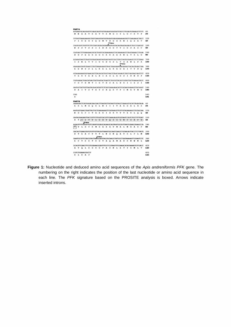
Figure 1: Nucleotide and deduced amino acid sequences of the Apis andreniformis PFK gene. The
numbering on the right indicates the position of the last nucleotide or amino acid sequence in
each line. The PFK signature based on the PROSITE analysis is boxed. Arrows indicate
inserted introns.
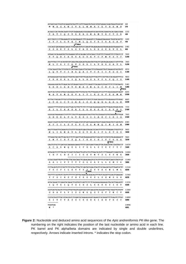
Figure 2: Nucleotide and deduced amino acid sequences of the Apis andreniformis PK-like gene. The
numbering on the right indicates the position of the last nucleotide or amino acid in each line.
PK barrel and PK alpha/beta domains are indicated by single and double underlines,
respectively. Arrows indicate inserted introns. * indicates the stop codon.
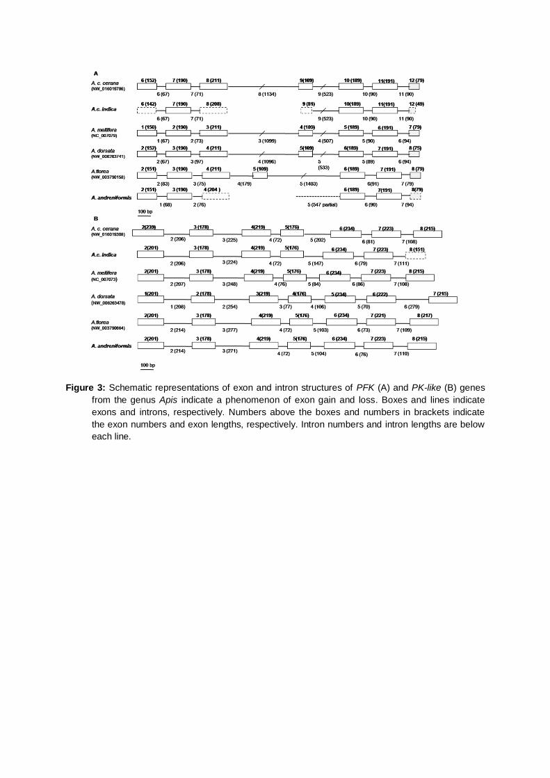
Figure 3: Schematic representations of exon and intron structures of PFK (A) and PK-like (B) genes
from the genus Apis indicate a phenomenon of exon gain and loss. Boxes and lines indicate
exons and introns, respectively. Numbers above the boxes and numbers in brackets indicate
the exon numbers and exon lengths, respectively. Intron numbers and intron lengths are below
each line.

Figure 4: Multiple alignment of Apis PFK amino acid sequences. The number indicates the position of
the last amino acid in the line. The PFK signature based on the PROSITE analysis is boxed. †
indicates GenBank Accession numbers found in Table 2. * indicates conserved amino acid
sequences.
Figure 5: The relative transition and transversion rates of PK-like gene are higher than PFK gene in
Apis.

Figure 6: The difference of PFK exon substitution numbers for each codon position in Apis.
Figure 7: The difference of PK-like exon substitution numbers for each codon position in Apis
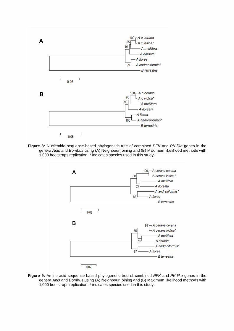
Figure 8: Nucleotide sequence-based phylogenetic tree of combined PFK and PK-like genes in the
genera Apis and Bombus using (A) Neighbour joining and (B) Maximum likelihood methods with 1,000 bootstraps replication. * indicates species used in this study.
Figure 9: Amino acid sequence-based phylogenetic tree of combined PFK and PK-like genes in the
genera Apis and Bombus using (A) Neighbour joining and (B) Maximum likelihood methods with 1,000 bootstraps replication. * indicates species used in this study.