accelerated fat cell aging links oxidative stress and insulin resistance in adipocytes
TRANSCRIPT
Accelerated fat cell aging
links oxidative stress and
insulin resistance in
adipocytesFinny Monickaraj, Sankaramoorthy Aravind, Pichamoorthy
Nandhini,
Paramasivam Prabu, Chandrakumar Sathishkumar, Viswanathan
Mohan and Muthuswamy Balasubramanyam
Natalia Perilla Hernández
Salomé Rave DiosaThird semester of medicine
Molecular Biology

INTRODUCTION
Recent studies have shown the importance of fat tissue in telomere
shortening and its relationship with obessity and type 2 diabetes.
This study was made inducing oxidative stress in 3T3-L1 adipocytes to
test shortened telomeres, senescence and functional impairment. This
adipocytes showed ROS, DNA damage, mRNA and protein expression
of senescence and pro-inflamatory markers, telomere length and
glucose uptake.
The researchers wanted to study adipocytes rather than white blood
cells due to its reflection on target tissues.
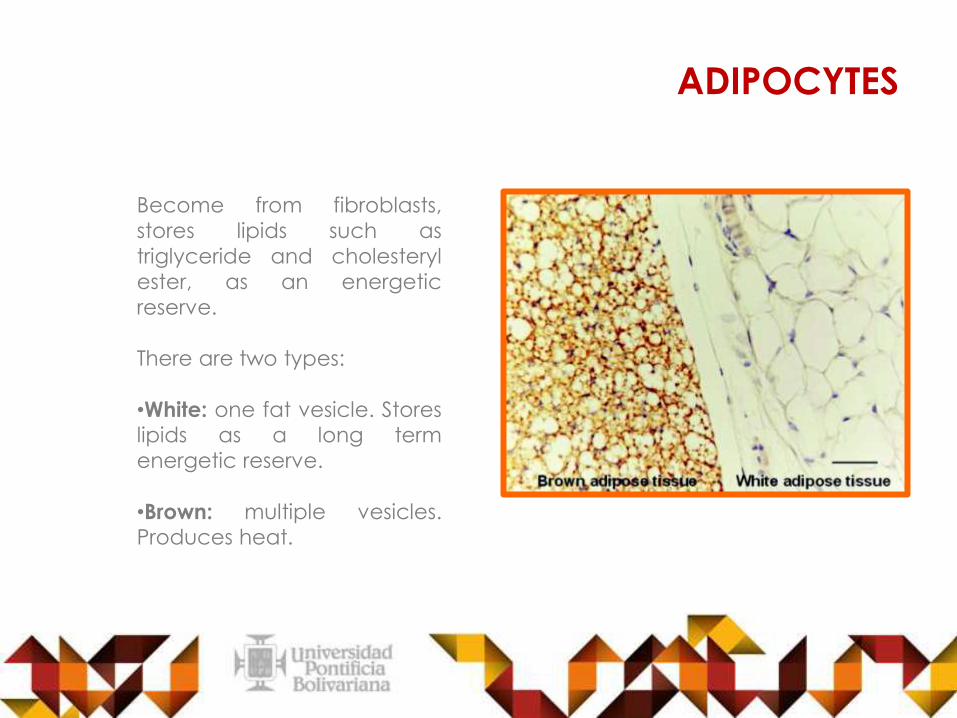
Become from fibroblasts,
stores lipids such as
triglyceride and cholesteryl
ester, as an energetic
reserve.
There are two types:
•White: one fat vesicle. Storeslipids as a long term
energetic reserve.
•Brown: multiple vesicles.Produces heat.
ADIPOCYTES
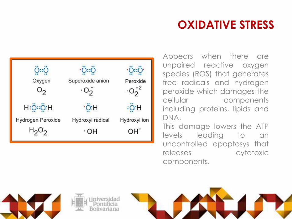
OXIDATIVE STRESS
Appears when there are
unpaired reactive oxygen
species (ROS) that generates
free radicals and hydrogen
peroxide which damages the
cellular components
including proteins, lipids and
DNA.
This damage lowers the ATP
levels leading to an
uncontrolled apoptosys that
releases cytotoxic
components.

INSULIN RESISTANCE
Its a genetic or acquired cell
inability to uptake insulin-
dependent glucose in tissues
such as liver, muscle and fat.
It leads to hyperglycemia
and disminished glucose
uptake if is not treated.
Some risk factors are obesity
and low physical activity.

ADIPOCYTE

OBJECTIVE
•Determine how oxidative stress and accelerated
senescence impacts fat tissue function and how
this, in turn, leads to age-related diseases.
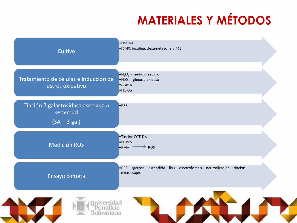
MATERIALES Y MÉTODOS
•DMEM
•IBMX, insulina, dexametasona y FBS Cultivo
•H2O2 - medio sin suero
•H2O2 - glucosa oxidasa
•ADMA
•HG-LG
Tratamiento de células e inducción de estrés oxidativo
•PBSTinción β galactosidasa asociada a senectud
(SA – β-gal)
•Tinción DCF-DA
•HEPES
•PMA ROSMedición ROS
•PBS – agarosa – extendido – lisis – electroforesis – neutralización – tinción –microscopio.
Ensayo cometa
MATERIALES Y MÉTODOS
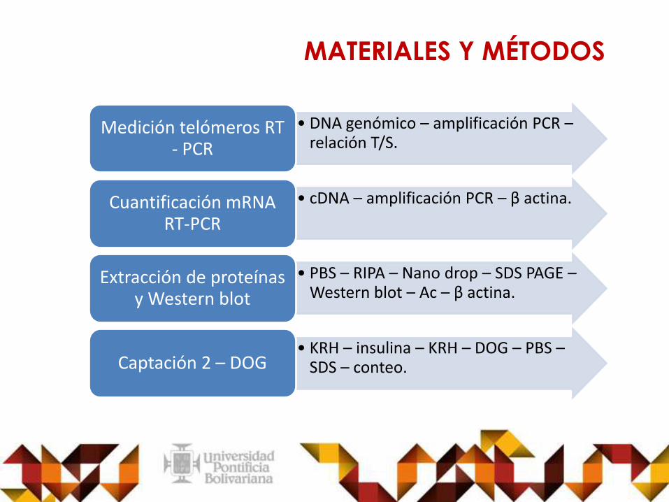
• DNA genómico – amplificación PCR –relación T/S.
Medición telómeros RT - PCR
• cDNA – amplificación PCR – β actina.Cuantificación mRNART-PCR
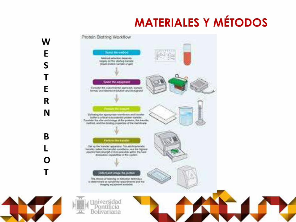
• PBS – RIPA – Nano drop – SDS PAGE –Western blot – Ac – β actina.
Extracción de proteínas y Western blot
• KRH – insulina – KRH – DOG – PBS –SDS – conteo. Captación 2 – DOG
MATERIALES Y MÉTODOS
MATERIALES Y MÉTODOS
ABI 7000
MATERIALES Y MÉTODOS
WESTERN
BLOT
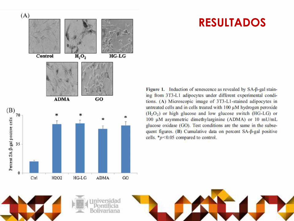
RESULTADOS

RESULTADOS
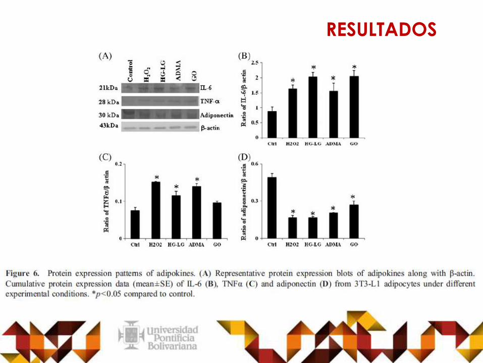
RESULTADOS

Morley.2008
“In fact, diabetes mellitus has recently beenconsidered as a cause of accelerated aging”.
Monickaraj agrees
Tchkonia et al. 2010
“Despite the fact that senescence in adipocytescould have profound clinical consequences becauseof the large size of the fat organ and its centralmetabolic role, there are only very few studies thathave looked at the senescence mechanisms inadipocytes”.
Monickaraj agrees
Beliveau and Yaswen.2007
“In fact, ectopic expression of hTERT was shown toreduce the p53-dependent cellular stressresponses”.
Monickaraj agrees
Campisi.2005
Minamino and Komuro.2007
“Senescent cells are known to secrete moleculesthat can alter the local microenvironment, such aspro – inflammatory cytokines”.
Monickaraj agrees
DISCUSSION

CONCLUSIONS
•Adipocytes subjected to glucose levels oscillation causes a
hyperactivation of p53 which is associated with insulin resistance, pro-
inflamatory effects and premature cell-aging.
•Adipocytes exposed to oxidative stress showed increased cellular
aging due to the following:
•Telomere shortening
•Increased expression of mRNA of p53 and p21
•Decreased adiponectin expression.

CONCLUSIONS
•The oxidative stress cause a increased secretion of IL6,
TNFa and decreased secretion of adiponectin and thatwoul be reflected in insulin resistance.
•Type 2 diabetes and obesity induce oxidative stress
which causes a shortened telomeres, aging and
functional impairment

CONCEPTUAL MAP
NATALIAObesity and
type 2 diabetes
Insulineresistance
• Shortenedtelomeres
• Functionalimpairment
AgingOxidative
stress
Apoptosis
IncreaseTNFa, IL6
ROS
Hyperglycemia
Dcreasedadiponectin
Damageof:
DNAPROTEIN
LIPIDS
Increasep53
Increasep21
Pro-inflamatorycytokines
Induced:H202, HG-LG,
ADMA,GONATALIA

CONCEPTUAL MAP
SALOME

GRACIAS
THANKS