accelerated alcoholic fermentation of intact grapes by
TRANSCRIPT

Accelerated Alcoholic Fermentation of IntactGrapes by Saccharomyces Cerevisiae in Symbiosiswith Microbial Community Inhabiting Grape-skinDaisuke Watanabe
Kyoto University https://orcid.org/0000-0002-8831-5765Wataru Hashimoto ( [email protected] )
Kyoto University
Article
Keywords: Accelerated Alcoholic Fermentation, Intact Grapes, Saccharomyces Cerevisiae, Symbiosis
Posted Date: September 23rd, 2021
DOI: https://doi.org/10.21203/rs.3.rs-916454/v1
License: This work is licensed under a Creative Commons Attribution 4.0 International License. Read Full License

1
ARTICLE submitted to Communications Biology 1
Accelerated alcoholic fermentation of intact grapes by Saccharomyces 2
cerevisiae in symbiosis with microbial community inhabiting grape-skin 3
4
Daisuke Watanabe1 & Wataru Hashimoto1* 5
6
1Laboratory of Basic and Applied Molecular Biotechnology, Division of Food Science 7
and Biotechnology, Graduate School of Agriculture, Kyoto University, Uji, Kyoto, Japan 8
9
*email: [email protected] 10

2
Abstract 1
Saccharomyces cerevisiae, an essential player in alcoholic fermentation during 2
winemaking, is rarely found in intact grapes. Here, we addressed symbiotic interactions 3
between S. cerevisiae and grape-skin residents upon spontaneous wine fermentation. 4
When glucose was used as a carbon source, the yeast-like fungus Aureobasidium 5
pullulans, a major grape-skin resident, had no effect on alcoholic fermentation by S. 6
cerevisiae. In contrast, when intact grape berries as a sole carbon source, coculture of S. 7
cerevisiae and A. pullulans accelerated alcoholic fermentation. Thus, grape-inhabiting 8
microorganisms may increase carbon availability by degrading and/or incorporating 9
grape-skin materials, such as cell wall and cuticles. A. pullulans exhibited broad spectrum 10
assimilation of plant-derived carbon sources, including ω-hydroxy fatty acids, arising 11
from degradation of cutin. In fact, yeast-type cutinase was produced from A. pullulans 12
EXF-150 strain. The degradation and utilization of grape-skin materials by fungal 13
microbiota may account for their colonization on grape-skin and symbiotic interactions 14
with S. cerevisiae. 15

3
Introduction 1
The origin of wine yeasts, taxonomically categorized into Saccharomyces cerevisiae or 2
its closely related species, has been an issue of great controversy. Historically, wine yeasts 3
were first found on grape surfaces1, whereas Saccharomyces species are absent or rare in 4
fresh grape berries2–4. Resident microbiota in intact grapes predominantly consists of the 5
yeast-like ascomycetous fungi Aureobasidium pullulans and basidiomycetous yeasts, 6
such as Cryptococcus, Rhodosporidium, Rhodotorula, and Sporobolomyces (a.k.a. 7
Sporidiobolus as teleomorphic forms) species, which are irrelevant to winemaking due to 8
their lack of alcoholic fermentation ability3–5. Assuming that Saccharomyces yeast species 9
are vectored by insects or migratory birds6,7, the first colonist wine yeasts must endure 10
and adapt in vineyard environments, nutritionally undesirable for survival and 11
proliferation. 12
The grape-skin, the natural habitat for non-Saccharomyces oligotrophic 13
microorganisms, covers and protects the pulp for nutrient storage8,9. The grape-skin 14
occupies approximately 10% of the dry weight of grape berries and acts as a barrier 15
against dehydration, physical damage, and microbial penetration. The plant primary cell 16
wall components, cellulose, hemicellulose, and pectin, contribute to the structural 17
integrity. The grape-skin is particularly abundant in pectinic acid, the methyl-esterified 18
form of polygalacturonic acid10,11. As previously reported, many grape-inhabiting 19
microorganisms secrete cellulase, pectinase, and relevant degrading enzymes to use the 20
cell wall decomposition products as nutrients12–15. The microbial targeting ability to 21
degrade and assimilate the plant cell wall compounds is likely a prerequisite for 22
colonization and adaptation to vineyard environments. Wine yeasts of the Saccharomyces 23
genus may need the aid of grape-skin residents to survive on the surface of grapes due to 24

4
the absence of degrading or metabolizing enzymes for plant cell wall and decomposition 1
products16,17. 2
The outermost layer of grape-skin is the cuticle, mainly composed of the lipid 3
polyester cutin (i.e., ester-linked ω-hydroxy C16 and C18 fatty acids) and the chemically 4
highly resistant biopolymer cutan18,19. On all aerial surfaces of land plants, thick and 5
hydrophobic cuticle layers prevent desiccation, UV damage, and pathogen infection. At 6
the frontline, the plant cuticle also serves multifunctional roles in triggering immunity 7
during plant-pathogen interactions20. Thus, cuticle-degrading virulent pathogens can 8
cause severe damage to terrestrial plants. The cutin-hydrolyzing enzyme cutinase has 9
been extensively studied in typical plant pathogens of Fusarium species and in a limited 10
number of molds, yeasts, and bacteria21–23. However, the degradation and assimilation of 11
the plant cuticle compounds by non-pathogenic, grape-skin microorganisms are yet to be 12
described. 13
This study proposed a novel tripartite relationship between grape berries, grape-14
skin microbiota, and S. cerevisiae during alcoholic fermentation. Similar plant-microbial 15
interactions may typically occur in traditional production processes of fermented foods 16
and beverages, where humans have optimized the growth of characteristic microbial 17
communities over thousands of years24. Thus, this study provides an important clue to 18
understand the dynamics and mechanism of plant-microbial ecosystems by analyzing the 19
origin of wine fermentation as an experimentally tractable model. 20
21
Results 22
Isolation of yeasts and related microorganisms inhabiting grape-skin. Among 23
chloramphenicol-tolerant, yeast-like colonies isolated from grape juice, surface-washed 24

5
suspensions, or enrichment cultures, 76 clones were identified to species level by 1
sequencing the internal transcribed spacer (ITS) region of the nuclear rRNA gene 2
(Supplementary Table S1). Isolated microorganisms were classified as yeast-like fungi, 3
basidiomycetous yeasts, or ascomycetous yeasts. The yeast-like fungi included a single 4
ascomycetous species A. pullulans25–27, also known as “black yeast”, frequently isolated 5
from grape and wine environments in previous reports3–5. On nutrient-rich yeast extract-6
peptone-dextrose (YPD) medium, A. pullulans earlier produced smooth pale-pink 7
colonies, which developed protrusions after long-term cultivation (Fig. 1a). Due to 8
melanin-like pigment accumulation, A. pullulans cells were easily observed on the skin 9
of spoiled grape berries (Fig. 1b). The isolated basidiomycetous yeasts mainly consisted 10
of Sporobolomyces and Papiliotrema (formerly categorized as Cryptococcus) species3–5. 11
Additionally, Rhodotorula mucilaginosa is a common ecological basidiomycete found in 12
soil, air, water, and foods26,28. Hanseniaspora uvarum26,29 was most frequently isolated 13
from the wine grape variety, Pinot noir, among the ascomycetous yeast species. Several 14
other ascomycetes were assigned to Candida (as anamorphic status) or Pichia genus. In 15
most cases, every grape sample tested in this study contained a few yeasts or associated 16
fungal species, excluding Saccharomyces cerevisiae. Thus, grape environments may be 17
greatly suitable for the colonization and development of non-Saccharomyces oligotrophic 18
fungal microbiota. Yeast-like fungus A. pullulans, three basidiomycetous yeast species 19
(Papiliotrema laurentii, Sporidiobolus pararoseus, and R. mucilaginosa), and five 20
ascomycetous yeast species (H. uvarum, Torulaspora delbrueckii, Meyerozyma caribbica, 21
Debaryomyces hansenii, and Pichia terricola) were used as representative grape-skin 22
microorganisms for further comparative analysis (Fig. 2). 23
24

6
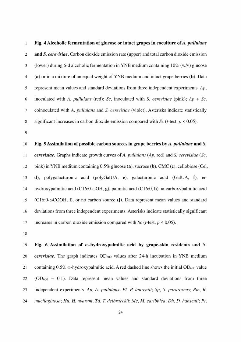
Alcoholic fermentation of glucose by grape-skin fungi and S. cerevisiae. To assess 1
alcoholic fermentation performance of the grape-skin fungi and S. cerevisiae, each 2
microorganism was statically incubated in a 5-mL liquid yeast nitrogen base (YNB) 3
medium, synthetic minimum medium for laboratory yeast strains, supplemented with 4
10% (w/v) glucose as a sole carbon source. As all species grew well in this medium 5
(Supplementary Fig. S1; optical density of 600 nm (OD600) > 1), carbon dioxide emission 6
rate, glucose consumption, and ethanol production were quantified during the 6-d 7
fermentation test (Fig. 3). S. cerevisiae exhibited a robust peak of fermentation rate and 8
full glucose consumption to yield approximately 5% (v/v) ethanol. In contrast, yeast-like 9
fungus A. pullulans, basidiomycetous yeasts, P. laurentii, S. pararoseus, R. mucilaginosa, 10
and ascomycetous yeast D. hansenii showed a constant basal level of carbon dioxide 11
emission, no detectable ethanol production, and little glucose consumption, and was 12
categorized as non-fermenting species. The other ascomycetous yeasts, H. uvarum, T. 13
delbrueckii, M. caribbica, and P. terricola, with intermediate phenotypes, were 14
categorized as weak-fermenting species. 15
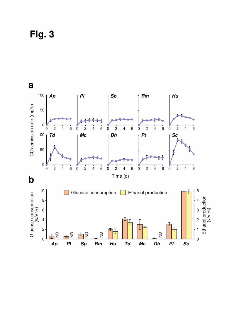
In the coculture experiment in 50-mL YNB medium containing 10% (w/v) 16
glucose as a sole carbon source, the non-fermenting species A. pullulans displayed no 17
significant interaction with S. cerevisiae in alcoholic fermentation (Fig. 4a). Notably, the 18
coculture of A. pullulans and S. cerevisiae had no synergistic effect on the growth of both 19
species (Supplementary Fig. S2). 20
21
Alcoholic fermentation of intact grapes by nonfermentative, grape-skin fungi and S. 22
cerevisiae. In 50-mL YNB minimum medium plus 50-g intact grape berries as a sole 23
carbon source (Fig. 4b), A. pullulans generated almost no carbon dioxide, whereas S. 24

7
cerevisiae slowly progressed alcoholic fermentation, reaching the maximum carbon 1
emission rate 4 d after inoculation. Since robust peaks of fermentation rates were typically 2
observed within 1−2 d after inoculation in the presence of glucose (Figs. 3a and 4a), 3
grape-skin may function as a physical barrier against S. cerevisiae cells to protect 4
fermentable sugars, such as glucose, fructose, and sucrose, inside grape berries. 5
When A. pullulans and S. cerevisiae were co-cultured, the initial carbon emission 6
rate increased, and a maximum fermentation rate was observed at 3 d (Fig. 4b). 7
Considering the minor contribution of A. pullulans to alcoholic fermentation and S. 8
cerevisiae growth, A. pullulans may specifically contribute to an increase in sugar 9
availability for S. cerevisiae, leading to accelerated alcoholic fermentation. Coculture of 10
S. cerevisiae with the nonfermentative basidiomycetous yeast, P. laurentii, S. pararoseus, 11
or R. mucilaginosa, gave similar results (Supplementary Fig. S3). These data suggest that 12
S. cerevisiae cells make use of the ability of grape-skin microbiota to access fermentable 13
sugars in grape berries upon triggering spontaneous wine fermentation. 14
15
Carbon assimilation profiles of grape-skin fungi and S. cerevisiae. To characterize 16
carbon assimilation profile, A. pullulans and S. cerevisiae cells were cultivated in a YNB 17
minimum medium with various possible carbon sources in grape berries (Fig. 5). Both A. 18
pullulans and S. cerevisiae vigorously grew in the presence of fermentable sugars, such 19
as glucose and sucrose (Fig. 5a, b). A major part of grape-skin consists of plant cell wall 20
polymers, cellulose and pectin10. Although carboxymethyl cellulose (CMC), a water-21
soluble cellulose derivative, was used by neither A. pullulans nor S. cerevisiae, the main 22
cellulose degradation product cellobiose was assimilated by A. pullulans (Fig. 5c, d). As 23
previously reported30, the activity of carboxymethyl cellulase (i.e., CMCase) is deficient 24

8
among some A. pullulans strains. The A. pullulans cells identified in this study may be 1
unable to degrade cellulose by themselves but can grow by using cellobiose produced 2
through cellulose degradation by other grape-skin fungi. The major pectic polysaccharide 3
polygalacturonic acid and its building block galacturonic acid were assimilated by A. 4
pullulans, but not by S. cerevisiae (Fig. 5e, f). Thus, A. pullulans likely degrades and 5
assimilates a broad spectrum of plant cell wall-relevant materials, unmetabolized by S. 6
cerevisiae, consistent with previous reports12–15,26,27. 7
The plant cuticle, the outermost hydrophobic layer, is another major component 8
of grape-skin10. Cutin in the plant cuticle is a polyester of ω-hydroxy C16 and C18 fatty 9
acids and their derivatives. We discovered that A. pullulans exhibited weak but 10
reproducible growth using ω-hydroxypalmitic acid as a sole carbon source (Fig. 5g). 11
Since neither palmitic acid or ω-carboxypalmitic acid was assimilated (Fig. 5h, i), A. 12
pullulans may possess a utilization system specific for ω-hydroxy fatty acids. Besides A. 13
pullulans, three basidiomycetous yeasts, P. laurentii, S. pararoseus, and R. mucilaginosa, 14
and two ascomycetous yeasts, M. caribbica and D. hansenii, showed significant growth 15
in the presence of ω-hydroxy palmitic acid (Fig. 6). Notably, most species described 16
above were nonfermentative microorganisms. 17
18
Cutinase-like esterase activity in A. pullulans. The secretion of cutin-degrading 19
enzymes in A. pullulans, basidiomycetous yeasts, and S. cerevisiae was tested using a 20
model polyester polycaprolactone (PCL)-plate (Fig. 7a). Known cutinases from the other 21
species represent PCL degradation activity in previous studies31–33. The supernatant of the 22
fully grown A. pullulans culture in YNB medium plus 2% glucose formed a clear halo, 23
although no halo was observed using the other supernatant samples. Additionally, the 24

9
supernatant of the A. pullulans culture exhibited higher esterase activity toward p-1
nitrophenyl butyrate (pNPB) and p-nitrophenyl palmitate (pNPP) used as substrates than 2
the supernatant of the S. cerevisiae culture (Fig. 7b). These data suggest that the isolated 3
A. pullulans strain from grapes secretes cutinase to assist penetration into plant cuticles. 4
Previous whole-genome analysis of the A. pullulans EXF-150 strain34 revealed 5
nine candidate genes encoding cutinase-like enzymes, designated as ApCut1 to ApCut9 6
(Supplementary Fig. S4). These gene products contained a classical α/β-hydrolase 7
catalytic triad Ser-His-Asp and a Gly-Tyr-Ser-Gln-Gly (GYSQG) motif conserved among 8
cutinase catalytic sites22,27. Additionally, two pairs of cysteine residues forming disulfide 9
bonds, important for spatial conformation22,35, were also conserved in all cutinase 10
candidates except for ApCut4, in which the amino terminus is truncated. Phylogenetic 11
analysis indicated that ApCut1 to ApCut3 form a subgroup with yeast cutinases from A. 12
adeninivorans and Cryptococcus sp. S-231,33, while ApCut5 to ApCut9 form a subgroup 13
with mold cutinases from F. solani, A. oryzae, and B. cinerea (Fig. 8a)36–38. Whole-cell 14
lysate of E. coli expressing recombinant ApCut1 indicated cutinase-like activity, based 15
on the PCL-plate clearing assay and the pNPB hydrolysis assay (Fig. 8b−d). Also, the 16
extracts of ApCut2- or ApCut3-expressing E. coli cells weakly degraded PCL 17
(Supplementary Fig. S5). Since the expression of ApCut2 and ApCut3 was almost 18
undetectable in the coomassie brilliant blue (CBB)-stained gel, more attention should be 19
paid to the protease sensitivity, expression conditions, and synonymous codon usage bias. 20
These results revealed the activity of the yeast-type cutinase isoenzymes in A. pullulans. 21
22
23
24

10
Discussion 1
Based on the experimental data, we propose that the grape-skin-resident microorganisms, 2
including nonfermentative, yeast-like fungus A. pullulans, increase the accessibility to 3
fermentable sugars in intact grape berries by degrading and assimilating the plant cell 4
wall, cuticle compounds, or both. Grape-skin microbiota’s high and versatile abilities to 5
degrade and assimilate plant cell wall and/or cuticle is likely to be essential for their 6
adaptation and proliferation on grape-skin. In contrast, S. cerevisiae cells without such 7
abilities need the aid of grape-skin microbiota to survive in grape environments. Studies 8
of wild populations of S. cerevisiae and its closest relative Saccharomyces paradoxus 9
suggest that woodlands or primeval forests are natural habitats for these yeast species39–10
41. In this study, no S. cerevisiae clone was isolated even from fermented juice or enriched 11
cultures in 5% sucrose. Moreover, S. cerevisiae was unable to assimilate plant cell wall, 12
cuticle, and their components. Altogether, nutrient-poor, intact grape surfaces may be 13
inappropriate for wine yeasts as their stable and permanent habitats. Although 14
Saccharomyces species might have been accidentally brought to the vineyard by yeast-15
carrier animals6,7, the primary cause of wine yeasts still needs to be experimentally 16
explored. Our results provide an important clue to address how S. cerevisiae cells met and 17
conquered grapes upon the origin of spontaneous wine fermentation. 18
What are the key grape-skin compounds that protect fermentable sugars in intact 19
grape berries? Based on the assimilation tests of CMC and cellobiose (Fig. 5c, d), our 20
isolated A. pullulans strain lacks cellulase. This is consistent with a previous report about 21
intraspecific variations of cell wall-degrading enzymes in A. pullulans30. Thus, cellulose 22
degradation may be nonessential for accelerated alcoholic fermentation of intact grapes. 23
Among pectin-degrading enzymes, polygalacturonase is genetically encoded and 24

11
expressed by grape-skin resident species and by S. cerevisiae42,43. Recently, we revealed 1
the importance of pectin as an initial target for the saprophytic bacterium Bacillus subtilis 2
to recognize the surface of dead soybeans44, whereas it is unlikely that pectin degradation 3
by non-Saccharomyces microorganisms specifically accelerated alcoholic fermentation 4
of intact grapes. As shown in Fig. 7a, the PCL-degrading activity was detected in A. 5
pullulans, although not in the examined basidiomycetous yeast strains. Based on these 6
data, the responsible enzymatic activity specific and common to all grape-skin residents 7
is still unidentified. The degradation of the other plant cell wall or cuticle components 8
should be focused on in future research. Alternatively, cooperative or synergistic 9
degradation of plant epidermis by whole grape-skin microbiota needs to be investigated. 10
Such complicated, highly ordered microbial interactions at the chemical, metabolic, 11
genetic, and genomic levels will be the central issue in applied microbiology and 12
microbial ecology. 13
This is the first report on ω-hydroxypalmitic acid assimilation as a carbon source 14
by A. pullulans and several yeast species. Although ω-hydroxylation of fatty acids also 15
occurs in mammals and insects, ω-hydroxy fatty acids play a broad and vital biological 16
role in higher plants as major components of cutin and suberin18–20,45–47. Thus, the 17
microbial ability to assimilate ω-hydroxy fatty acids may mainly contribute to symbiotic 18
interactions with terrestrial plants. In the ω-oxidation process of animals and plants, 19
known as a minor, fatty acid catabolic pathway, a hydroxy group is introduced onto the ω 20
carbon of the medium to long-chain fatty acids46,47. The resultant ω-hydroxy fatty acids 21
are oxidized to ω-carboxy fatty acids (i.e., dicarboxylic fatty acids), further degraded 22
through the β-oxidation pathway. Grape-skin residents may probably metabolize ω-23
hydroxy fatty acids similarly. Based on our data, A. pullulans cells are suggested to have 24

12
an ω-hydroxy fatty acid-specific transporter because they can grow using ω-1
hydroxypalmitic acid as a sole carbon source not palmitic acid or ω-carboxypalmitic acid. 2
Furthermore, this study revealed that A. pullulans may secrete cutinase to hydrolyze cutin 3
into ω-hydroxy fatty acids and other minor components, such as glycerol. The 4
combination of cutin degradation and ω-hydroxy fatty acid assimilation may characterize 5
A. pullulans as the most abundant and persistent resident among grape-skin microbiota. 6
Notably, the A. pullulans EXF-150 strain was first identified as a microorganism that 7
acquired both yeast- and mold-type cutinase genes in the genome, which may be 8
associated with the yeast-to-hyphal dimorphic transition of this species25. Enzymatic 9
analysis of yeast- and mold-type cutinases in A. pullulans will reveal the significance of 10
differences between both cutinase types. 11
Conclusively, this study focused on the symbiotic relationship between grape-12
skin microbiota and S. cerevisiae from the perspective of winemaking origin. 13
Oligotrophic microorganisms, such as A. pullulans, have developed versatile abilities to 14
use plant cell wall polysaccharides and plant cuticular lipids as nutrient sources to 15
establish their ecological niche. Especially, degradation and assimilation of the plant 16
cuticle, the outermost layer interacting with the environment, may be a prerequisite for 17
oligotrophic resident microorganisms to trigger colonization and adaptation. In contrast, 18
eutrophic yeasts, including S. cerevisiae, yield energy through the alcoholic fermentation 19
of sugars in grape berries with the aid of oligotrophic microorganisms. Such tripartite 20
interaction between grape berries, oligotrophic residents, and eutrophic yeasts determines 21
the grape-skin microbiome dynamics and spontaneous wine fermentation. Thus, studying 22
the origins and microbiota ecological succession in fermented foods will help elucidate 23
the key principles governing plant-microbial ecosystems’ emergence and development. 24

13
Materials 1
Materials and strains. To isolate yeasts or yeast-like fungi, (i) juice, (ii) surface-washed 2
suspensions, or (iii) enrichment cultures were obtained from commercially available wine 3
or table grape varieties belonging to Vitis vinifera species or Vitis interspecific hybrids 4
(see Supplementary Table S1). Grape juice was obtained from freshly pressed grape 5
berries using a food-grade juicer (Panasonic, Japan) and was used either immediately or 6
after being incubated at 30°C for 3 d. Surface-washed suspensions were obtained by 7
vigorously shaking the flask containing approximately 50-g grape berries and 25-mL 8
sterilized water at 30°C for 15 min. Enrichment cultures were obtained by statically 9
incubating the flask containing approximately 10-g grape berries and 40-mL sterilized 10
water or 5% (w/v) sucrose at 30°C for 3 d. Each sample was spread on a nutrient-11
rich,YPD (1% yeast extract, 2% peptone, and 2% glucose) medium plate with 0.1% 12
chloramphenicol to inhibit bacterial growth, and was incubated at 30°C. Single colonies 13
representing yeast-like colony morphology were isolated by repeatedly streaking on YPD 14
medium plates with 0.1% chloramphenicol. The isolated clones were identified by DNA 15
sequencing of the rRNA gene ITS region, using the ITS_1F (5’-16
GTAACAAGGTYTCCGT-3’) and ITS_1R (5’-CGTTCTTCATCGATG-3’) primer pair 17
and genomic DNA as PCR templates. The S. cerevisiae X2180 strain, obtained from the 18
American Type Culture Collection (USA), was used for alcoholic fermentation tests or 19
other control experiments. 20
21
Fermentation test. Cells were aerobically precultured at 30°C for 2 d in 0.67% YNB 22
minimum medium containing 2% (w/v) glucose as a carbon source and harvested. For the 23
alcoholic fermentation of glucose, cells were inoculated into a YNB medium containing 24

14
10% (w/v) glucose at a final OD600 of 0.1 and were then further incubated at 30°C without 1
shaking. For the alcoholic fermentation of intact grapes, cells were inoculated into 50-mL 2
YNB medium at a final OD600 of 0.1, mixed with approximately 50-g commercially 3
available intact grape berries of Green Seedless, and were then further incubated at 30°C 4
without shaking. Fermentation was continuously monitored by measuring the weight loss 5
of evolved carbon dioxide for 5-mL test tube-scale tests or using a Fermograph II 6
apparatus (Atto, Japan) for 50-mL-scale coculture tests. Glucose and ethanol 7
concentrations were determined using the LabAssay glucose kit (Fujifilm Wako Pure 8
Chemical, Japan) and the ethanol assay F-kit (Roche, Switzerland), respectively. 9
10
Carbon assimilation test. Cells were aerobically precultured at 30°C for 2 d in a YNB 11
minimum medium containing 2% (w/v) glucose as a carbon source, harvested, washed 12
by sterilized water, and inoculated into a YNB medium containing 0.5% (w/v) of carbon 13
sources as below: glucose, sucrose, CMC, cellobiose, polygalacturonic acid, galacturonic 14
acid, palmitic acid, 16-hydroxyhexadecanoic acid (i.e., ω-hydroxypalmitic acid), or 15
heptadecanedioic acid (i.e., ω-carboxypalmitic acid). Upon inoculation, initial OD600 was 16
adjusted to 0.1. For assimilation tests of palmitic acid, ω-hydroxypalmitic acid, or ω-17
carboxypalmitic acid, 0.05% (w/v) Tween 40 was added to the medium. In advance of 18
measurement of OD600, insoluble fatty acids were removed by washing pellets with 19
hexane three times, and cells were dissolved in sterilized water48. 20
21
Phylogenetic analysis of yeast and mold cutinases. Amino acid sequences of nine 22
putative cutinase gene products in the A. pullulans EXF-150 strain and representative 23
yeast and mold cutinases [Cut2 from Arxula adeninivorans (AaCut2), Cle1 from 24
15
Cryptococcus sp. strain S-2 (CS2Cle1), Cut1 from Fusarium solani (FsCut1), CutL from 1
Aspergillus oryzae (AoCutL), and CutA from Botrytis cinerea (BcCutA)] were obtained 2
from UniPlotKB. Multiple sequence alignment was conducted using Clustal Omega 3
program, and the phylogenetic tree was constructed using Molecular Evolutionary 4
Genetics Analysis v.10.2.2. The putative cutinase genes of A. pullulans were designated 5
as below: M438DRAFT_340638 as ApCUT1, M438DRAFT_351543 as ApCUT2, 6
M438DRAFT_352218 as ApCUT3, M438DRAFT_388226 as ApCUT4, 7
M438DRAFT_264999 as ApCUT5, M438DRAFT_267580 as ApCUT6, 8
M438DRAFT_341517 as ApCUT7, M438DRAFT_347465 as ApCUT8, and 9
M438DRAFT_368700 as ApCUT9. 10
11
Expression of recombinant A. pullulans cutinase. The chemically synthesized ApCUT1 12
gene (Eurofins, Luxembourg) was cloned into the BamHI-EcoRI site of the pET-21b(+) 13
vector (Merck Millipore, USA) to express recombinant ApCut1p tagged with the T7 14
epitope at the amino terminus and the 6 × His epitope at the carboxy terminus. The 15
resultant pET-21b(+)-ApCUT1 plasmid was introduced into Escherichia coli 16
BL21(DE3)pLysS cells (Novagen, Germany). A transformant was inoculated into 100-17
mL Luria-Bertani medium (0.5% yeast extract, 1% tryptone, and 1% sodium chloride) 18
with 100-μg/mL ampicillin and 34-μg/mL chloramphenicol, and was cultured at 37°C to 19
an OD600 of 0.6. Isopropyl thio-β-D-galactoside (IPTG) was added to the culture to a final 20
concentration of 1 mM, and cells were further cultured at 37°C for 4 h. Whole-cell 21
extracts were prepared from cell pellets suspended in the xTractor buffer (TakaraBio, 22
Japan), followed by the addition of DNase I. After protein separation by SDS-PAGE, the 23
proteins were detected by CBB staining or using His-Detect In-Gel Stain (Nacalai Tesque, 24

16
Japan). 1
2
Enzymatic assay. To assay the potential cutinase activity from the culture supernatants 3
of fully grown A. pullulans or yeasts, PCL was used as a model polyester substrate. Turbid 4
agar plates containing 0.05% PCL (Fujifilm Wako Pure Chemical, Japan) were prepared 5
as previously described32,33. The assay for determining esterase activity was conducted 6
according to previous reports33,49, using pNPB and pNPP (Sigma-Aldrich, USA) as 7
substrates. 8
17
References 1
1. Pasteur L. Nouvelles expériences pour démontrer que le germe de la levure qui fait 2
le vin provient de l'extérieur des grains de raisin. C R Acad Sci 1872; 75: 781–793. 3
2. Mortimer R, Polsinelli M. On the origins of wine yeast. Res Microbiol 1999; 150: 4
199–204. 5
3. Barata A, Malfeito-Ferreira M, Loureiro V. The microbial ecology of wine grape 6
berries. Int J Food Microbiol 2012; 153: 243–259. 7
4. Stefanini I, Cavalieri D. Metagenomic approaches to investigate the contribution of 8
the vineyard environment to the quality of wine fermentation: potentials and 9
difficulties. Front Microbiol 2018; 9: 991. 10
5. Loureiro V, Ferreira MM, Monteiro S, Ferreira RB. The microbial community of 11
grape berry. In: Gerós H, Chaves MM, Delrot S (eds). The Biochemistry of the Grape 12
Berry. Bentham Science: 2012, pp 241–268. 13
6. Stefanini I, Dapporto L, Legras J-L, Calabretta A, Di Paola M, De Filippo C et al. 14
Role of social wasps in Saccharomyces cerevisiae ecology and evolution. Proc Natl 15
Acad Sci U S A 2012; 109: 13398–13403. 16
7. Francesca N, Carvalho C, Sannino C, Guerreiro MA, Almeida PM, Settanni L et al. 17
Yeasts vectored by migratory birds collected in the Mediterranean island of Ustica 18
and description of Phaffomyces usticensis f.a. sp. nov., a new species related to the 19
cactus ecoclade. FEMS Yeast Res 2014; 14: 910–921. 20
8. Gao Y, Zietsman AJJ, Vivier MA, Moore JP. Deconstructing wine grape cell walls 21
with enzymes during winemaking: new insights from glycan microarray technology. 22
Molecules 2019; 24: 165. 23
9. Martínez-Lapuente L, Guadalupe Z, Ayestarán B. Properties of wine polysaccharides. 24

18
In: Masuelli M (ed). Pectins - Extraction, Purification, Characterization and 1
Applications. IntechOpen: 2019, DOI: 10.5772/intechopen.85629. 2
10. Lecas M, Brillouet J-M. Cell wall composition of grape berry skins. Phytochemistry 3
1994; 35: 1241–1243. 4
11. González-Centeno MR, Rosselló C, Simala S, Garau MC, López F, Femenia A. 5
Physico-chemical properties of cell wall materials obtained from ten grape varieties 6
and their byproducts: grape pomaces and stems. LWT Food Sci Technol 2010; 43: 7
1580–1586. 8
12. Biely P, Heinrichová K, Kružiková M. Induction and inducers of the pectolytic 9
system in Aureobasidium pullulans. Curr Microbiol 1996; 33: 6–10. 10
13. Strauss ML, Jolly NP, Lambrechts MG, van Rensburg P. Screening for the production 11
of extracellular hydrolytic enzymes by non-Saccharomyces wine yeasts. J Appl 12
Microbiol 2001; 91: 182–190. 13
14. Merín MG, Mendoza LM, Farías ME, Morata de Ambrosini VI. Isolation and 14
selection of yeasts from wine grape ecosystem secreting cold-active pectinolytic 15
activity. Int J Food Microbiol 2011; 147: 144–148. 16
15. Úbeda J, Maldonado Gil M, Chiva R, Guillamón JM, Briones A. Biodiversity of non-17
Saccharomyces yeasts in distilleries of the La Mancha region (Spain). FEMS Yeast 18
Res 2014; 14: 663–673. 19
16. Huisjes EH, Luttik MAH, Almering MJH, Bisschops MMM, Dang DHN, 20
Kleerebezem M et al. Toward pectin fermentation by Saccharomyces cerevisiae: 21
expression of the first two steps of a bacterial pathway for D-galacturonate 22
metabolism. J Biotechnol 2012; 162: 303–331. 23
17. Casa-Villegas M, Polaina J, Marín-Navarro, J. Cellobiose fermentation by 24

19
Saccharomyces cerevisiae: comparative analysis of intra versus extracellular sugar 1
hydrolysis. Process Biochem 2018; 75: 59–67. 2
18. Domínguez E, Heredia-Guerrero JA, Heredia A. The biophysical design of plant 3
cuticles: an overview. New Phytol 2011; 189: 938–949. 4
19. Martin LBB, Rose JKC. There's more than one way to skin a fruit: formation and 5
functions of fruit cuticles. J Exp Bot 2014; 65: 4639–4651. 6
20. Ziv C, Zhao Z, Gao YG, Xia Y. Multifunctional roles of plant cuticle during plant-7
pathogen interactions. Front Plant Sci 2018; 9: 1088. 8
21. Egmond MR, de Vlieg J. Fusarium solani pisi cutinase. Biochimie 2000; 82: 1015–9
1021. 10
22. Chen S, Su L, Chen J, Wu J. Cutinase: characteristics, preparation, and application. 11
Biotechnol Adv 2013; 31: 1754–1767. 12
23. Nyyssölä A. Which properties of cutinases are important for applications? Appl 13
Microbiol Biotechnol 2015; 99: 4931–4942. 14
24. Wolfe BE, Dutton RJ. Fermented foods as experimentally tractable microbial 15
ecosystems. Cell 2015; 161: 49–55. 16
25. Slepecky RA, Starmer WT. Phenotypic plasticity in fungi: a review with observations 17
on Aureobasidium pullulans. Mycologia 2009; 101: 823–832. 18
26. Varela C, Borneman AR. Yeasts found in vineyards and wineries. Yeast 2017; 34: 19
111–128. 20
27. Bozoudi D, Tsaltas D. The multiple and versatile roles of Aureobasidium pullulans 21
in the vitivinicultural sector. Fermentation 2018; 4: 85. 22
28. Wirth F, Goldani LZ. Epidemiology of Rhodotorula: an emerging pathogen. 23
Interdiscip Perspect Infect Dis 2012; 2012: 465717. 24

20
29. Martin V, Valera MJ, Medina K, Boido E, Carrau F. Oenological impact of the 1
Hanseniaspora/Kloeckera yeast genus on wines--a review. Fermentation 2018; 4: 76. 2
30. Sun P-F, Chien I-A, Xiao H-S, Fang W-T, Hsu C-H, Chou J-Y et al. Intraspecific 3
variation in plant growth-promoting traits of Aureobasidium pullulans. Chiang Mai 4
J. Sci. 2019; 46: 15–31. 5
31. Masaki K, Kamini NR, Ikeda H, Iefuji H. Cutinase-like enzyme from the yeast 6
Cryptococcus sp. strain S-2 hydrolyzes polylactic acid and other biodegradable 7
plastics. Appl Environ Microbiol 2005; 71: 7548–7550. 8
32. Murphy CA, Cameron JA, Huang SJ Vinopal RT. Fusarium polycaprolactone 9
depolymerase is cutinase. Appl Environ Microbiol 1996; 62: 456–460. 10
33. Bischoff F, Litwińska K, Cordes A. Three new cutinases from the yeast Arxula 11
adeninivorans that are suitable for biotechnological applications. Appl Environ 12
Microbiol 2015; 81: 5497–5510. 13
34. Gostinčar C, Ohm RA, Kogej T, Sonjak S, Turk M, Zajc J et al. Genome sequencing 14
of four Aureobasidium pullulans varieties: biotechnological potential, stress 15
tolerance, and description of new species. BMC Genomics 2014; 15: 549. 16
35. Kodama Y, Masaki K, Kondo H, Suzuki M, Tsuda S, Nagura T et al. Crystal structure 17
and enhanced activity of a cutinase-like enzyme from Cryptococcus sp. strain S-2. 18
Proteins 2009; 77: 710-717. 19
36. Soliday CL, Dickman MB, Kolattukudy PE. Structure of the cutinase gene and 20
detection of promoter activity in the 5'-flanking region by fungal transformation. J 21
Bacteriol 1989; 171: 1942–1951. 22
37. Ohnishi K, Toida J, Nakazawa H, Sekiguchi J. Genome structure and nucleotide 23
sequence of a lipolytic enzyme gene of Aspergillus oryzae. FEMS Microbiol Lett 24
21
1995; 126: 145–150. 1
38. van der Vlugt-Bergmans CJ, Wagemakers CA, van Kan JA. Cloning and expression 2
of the cutinase A gene of Botrytis cinerea. Mol Plant Microb Interact 1997; 10: 21–3
29. 4
39. Sniegowski PD, Dombrowski PG, Fingerman E. Saccharomyces cerevisiae and 5
Saccharomyces paradoxus coexist in a natural woodland site in North America and 6
display different levels of reproductive isolation from European conspecifics. FEMS 7
Yeast Res 2002; 1: 299–306. 8
40. Fay JC, Benavides JA. Evidence for domesticated and wild populations of 9
Saccharomyces cerevisiae. PLoS Genet 2005; 1: 66–71. 10
41. Duan S-F, Han P-J, Wang Q-M, Liu W-Q, Shi J-Y, Li K et al. The origin and adaptive 11
evolution of domesticated populations of yeast from Far East Asia. Nat Commun 12
2018; 9: 2690. 13
42. Gognies S, Gainvors A, Aigle M, Belarbi A. Cloning, sequence analysis and 14
overexpression of a Saccharomyces cerevisiae endopolygalacturonase-encoding 15
gene (PGL1). Yeast 1999; 15: 11–22. 16
43. Radoi F, Kishida M, Kawasaki H. Endo-polygalacturonase in Saccharomyces wine 17
yeasts: effect of carbon source on enzyme production. FEMS Yeast Res 2005; 5: 663–18
668. 19
44. Sugiura H, Nagase A, Oiki S, Mikami B, Watanabe D, Hashimoto W. Bacterial 20
inducible expression of plant cell wall-binding protein YesO through conflict 21
between Glycine max and saprophytic Bacillus subtilis. Sci Rep 2020; 10: 18691. 22
45. Kolattukudy PE. Biopolyester membranes of plants: cutin and suberin. Science 1980; 23
208: 990–1000. 24

22
46. Miura Y. The biological significance of ω-oxidation of fatty acids. Proc Jpn Acad Ser 1
B Phys Biol Sci 2013; 89: 370–382. 2
47. Wertz PW. Naturally occurring ω-hydroxyacids. Int J Cosmet Sci 2018; 40: 31–33. 3
48. Nakagawa T, Imanaka T, Morita M, Ishiguro K, Yurimoto H, Yamashita A et al. 4
Peroxisomal membrane protein Pmp47 is essential in the metabolism of middle-chain 5
fatty acid in yeast peroxisomes and is associated with peroxisome proliferation. J 6
Biol Chem 2000; 275: 3455–3461. 7
49. Chen S, Tong X, Woodard RW, Du G, Wu J, Chen J. Identification and 8
characterization of bacterial cutinase. J Biol Chem 2008; 283: 25854–25862. 9
10
Acknowledgements 11
This work was partly supported by Grants-in-Aid for Scientific Research from the Japan 12
Society for the Promotion of Science (to D.W.). Addtionally, the authors want to thank 13
Enago (https://www.enago.com) for the English language review. 14
15
Author contributions 16
W.H. designed the study. D.W. performed the experiments. D.W. and W.H. analyzed the 17
data. D.W. and W.H. wrote the manuscript. 18
19
Competing interests 20
The authors declare no competing interests. 21
23
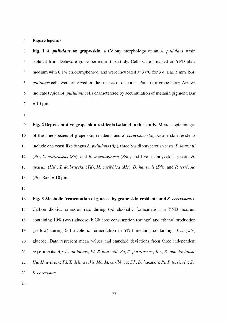
Figure legends 1
Fig. 1 A. pullulans on grape-skin. a Colony morphology of an A. pullulans strain 2
isolated from Delaware grape berries in this study. Cells were streaked on YPD plate 3
medium with 0.1% chloramphenicol and were incubated at 37°C for 3 d. Bar, 5 mm. b A. 4
pullulans cells were observed on the surface of a spoiled Pinot noir grape berry. Arrows 5
indicate typical A. pullulans cells characterized by accumulation of melanin pigment. Bar 6
= 10 μm. 7
8
Fig. 2 Representative grape-skin residents isolated in this study. Microscopic images 9
of the nine species of grape-skin residents and S. cerevisiae (Sc). Grape-skin residents 10
include one yeast-like fungus A. pullulans (Ap), three basidiomycetous yeasts, P. laurentii 11
(Pl), S. pararoseus (Sp), and R. mucilaginosa (Rm), and five ascomycetous yeasts, H. 12
uvarum (Hu), T. delbrueckii (Td), M. caribbica (Mc), D. hansenii (Dh), and P. terricola 13
(Pt). Bars = 10 μm. 14
15
Fig. 3 Alcoholic fermentation of glucose by grape-skin residents and S. cerevisiae. a 16
Carbon dioxide emission rate during 6-d alcoholic fermentation in YNB medium 17
containing 10% (w/v) glucose. b Glucose consumption (orange) and ethanol production 18
(yellow) during 6-d alcoholic fermentation in YNB medium containing 10% (w/v) 19
glucose. Data represent mean values and standard deviations from three independent 20
experiments. Ap, A. pullulans; Pl, P. laurentii; Sp, S. pararoseus; Rm, R. mucilaginosa; 21
Hu, H. uvarum; Td, T. delbrueckii; Mc, M. caribbica; Dh, D. hansenii; Pt, P. terricola; Sc, 22
S. cerevisiae. 23
24
24
Fig. 4 Alcoholic fermentation of glucose or intact grapes in coculture of A. pullulans 1
and S. cerevisiae. Carbon dioxide emission rate (upper) and total carbon dioxide emission 2
(lower) during 6-d alcoholic fermentation in YNB medium containing 10% (w/v) glucose 3
(a) or in a mixture of an equal weight of YNB medium and intact grape berries (b). Data 4
represent mean values and standard deviations from three independent experiments. Ap, 5
inoculated with A. pullulans (red); Sc, inoculated with S. cerevisiae (pink); Ap + Sc, 6
coinoculated with A. pullulans and S. cerevisiae (violet). Asterisks indicate statistically 7
significant increases in carbon dioxide emission compared with Sc (t-test, p < 0.05). 8
9
Fig. 5 Assimilation of possible carbon sources in grape berries by A. pullulans and S. 10
cerevisiae. Graphs indicate growth curves of A. pullulans (Ap, red) and S. cerevisiae (Sc, 11
pink) in YNB medium containing 0.5% glucose (a), sucrose (b), CMC (c), cellobiose (Cel, 12
d), polygalacturonic acid (polyGalUA, e), galacturonic acid (GalUA, f), ω-13
hydroxypalmitic acid (C16:0-ωOH, g), palmitic acid (C16:0, h), ω-carboxypalmitic acid 14
(C16:0-ωCOOH, i), or no carbon source (j). Data represent mean values and standard 15
deviations from three independent experiments. Asterisks indicate statistically significant 16
increases in carbon dioxide emission compared with Sc (t-test, p < 0.05). 17
18
Fig. 6 Assimilation of ω-hydroxypalmitic acid by grape-skin residents and S. 19
cerevisiae. The graph indicates OD600 values after 24-h incubation in YNB medium 20
containing 0.5% ω-hydroxypalmitic acid. A red dashed line shows the initial OD600 value 21
(OD600 = 0.1). Data represent mean values and standard deviations from three 22
independent experiments. Ap, A. pullulans; Pl, P. laurentii; Sp, S. pararoseus; Rm, R. 23
mucilaginosa; Hu, H. uvarum; Td, T. delbrueckii; Mc, M. caribbica; Dh, D. hansenii; Pt, 24

25
P. terricola; Sc, S. cerevisiae. 1
2
Fig. 7 Cutinase-like activity of the culture supernatants of grape-skin residents and 3
S. cerevisiae. a PCL-plate clearing assay. b pNPB and pNPP hydrolysis assay. The 4
supernatants were obtained from fully grown 5-d cultures in a YNB medium containing 5
2% (w/v) glucose. Data represent mean values and standard deviations from three 6
independent experiments. Ap, A. pullulans; Pl, P. laurentii; Sp, S. pararoseus; Rm, R. 7
mucilaginosa; Sc, S. cerevisiae. Asterisks indicate statistically higher specific activity 8
than Sc (t-test, p < 0.05). 9
10
Fig. 8 Cutinase-like activity of recombinant ApCut1. a A phylogenetic tree of A. 11
pullulans cutinase-like gene products. The number corresponds to each cutinase-like gene 12
product in A. pullulans. ApCut1–ApCut3 (magenta) belongs to the yeast cutinase family, 13
while ApCut5 to ApCut9 (cyan) belong to the mold cutinase family. Bar, 0.2 substitutions 14
per nucleotide position. b Expression of recombinant ApCut1 in E. coli. Left and right 15
panels indicate CBB-stained gel and His-detect-stained gel for specific detection of His-16
tagged proteins, respectively. c PCL-plate clearing assay. Bar, 1 cm. d pNPB hydrolysis 17
assay. Data represent mean values and standard deviations from three independent 18
experiments. An asterisk indicates a statistically higher specific activity than the negative 19
control (IPTG (-), t-test, p < 0.05). EV, empty vector. 20

a b
Fig. 1

Ap
Td
Pl
Mc
Sp
Dh
Rm
Pt
Hu
Sc
Yeast-like Basidiomycota
Gra
pe-s
kin
resid
en
ts
Ascomycota
Ascomycota
Fig. 2
Ap TdPl McSp DhRm PtHu Sc
ND
ND
ND
ND
ND
0
2
4
6
8
10
Glu
cose c
onsum
ption
(w/v
%)
Eth
anol pro
duction
(v/v
%)
0
1
2
3
4
5Glucose consumption Ethanol production
Ap
Td
Pl
Mc
Sp
Dh
Rm
Pt
Hu
Sc
0
50
100
0
50
100
CO
2 em
issio
n r
ate
(m
g/d
)
0 2 4 6 0 2 4 6 0 2 4 6 0 2 4 6 0 2 4 6
Time (d)
0 2 4 6 0 2 4 6 0 2 4 6 0 2 4 6 0 2 4 6
a
b
Fig. 3

Fig. 4
Sc
Ap
*
Time (d)
CO
2 em
issio
n r
ate
(mL/0
.5 d
)C
O2 e
mis
sio
n(t
ota
l m
L)
0 1 2 3 4 5 60
250
500
750
1000
0
500
1000
1500
2000
0 1 2 3 4 5 6
0 1 2 3 4 5 6 0 1 2 3 4 5 60
50
100
150
0
100
150
200
250
50
Ap
ApAp
Sc
Sc
Sc
Ap + Sc
Ap + Sc
Ap + Sc
Ap + Sc
*
* * * *
******* * *
baCarbon: Intact grapesCarbon: Glucose

OD
600
0
0.5
1.0
1.5
2.0
2.5
0
0.5
1.0
1.5
2.0
2.5
0
0.5
1.0
1.5
2.0
2.5
0
0.5
1.0
1.5
2.0
2.5
0
5
10
15
20
25
0
5
10
15
20
25
0
5
10
15
20
25
0
5
10
15
20
25
ApSc
Ap
Sc
Ap
Sc
Ap
Sc
Ap
ScAp
Sc
ApSc Ap
Sc
c
g
d
h
e
i
f
j
*
*
*
**
* * *
*
**
0 1 2 3 0 1 2 3 0 1 2 3 0 1 2 3
0 1 2 3 0 1 2 3 0 1 2 3 0 1 2 3
Ap
Sc
*
0
5
10
15
20
25
0
5
10
15
20
25
Ap
Sc
a b
* ** *
0 1 2 3 0 1 2 3
Time (d)
*
(Cel)
(C16:0)
(CMC)
(C16:0-ωOH)
(Glc)
(polyGalUA)
(C16:0-ωCOOH)
(Suc)
(GalUA)
(None)
Fig. 5

Ap TdPl McSp DhRm PtHu Sc0
0.2
0.4
0.6
0.8
OD
600
Fig. 6

a b
ApSc
Sc
Ap
Sc
AppNPP
pNPB
0 5 10 15
Sp. act. (units/mg)
*
*
Pl
Sp
Rm
None
Fig. 7

FsCut1
AoCutL
BcCutA
AaCut2CS2Cle1
12
3
4
5
6
9
8
7
IPTG (-)
IPTG (+)
ApCUT1EV
ApCUT1EV IPTG (-)
IPTG (+)
IPTG (-)
IPTG (+)ApCUT1
EV
0 10 20 30 40
Sp. act. (units/mg)
CBB staining His-detect
IPT
G (
-)
IPT
G (
+)
IPT
G (
-)
IPT
G (
+)
ApCUT1EV
15
20
25
37
50
75100150250kDa IP
TG
(-)
IPT
G (
+)
IPT
G (
-)
IPT
G (
+)
ApCUT1EVa b
c
d
*
Fig. 8

Supplementary Files
This is a list of supplementary �les associated with this preprint. Click to download.
SupplementaryData.pdf