acaadeemmii c sscii eenncesinternational journal of ... institute of biotechnology, amity university...
TRANSCRIPT

Review Article
NEUROPROTEOMICS: ADVANCEMENT AND CHALLENGES FOR BIOMARKER DISCOVERY IN NEURODEGENERATIVE DISEASES
DEEPSHIKHA PANDE KATARE1*, HISAMUDDIN MALIK2 AND MZ ABDIN2
1Amity Institute of Biotechnology, Amity University Uttar Pradesh, Noida 201303, 2Department of Biotechnology, Hamdard University, New Delhi, 110062. Email: [email protected]
Received: 11 Apr 2013, Revised and Accepted: 19 May 2013
ABSTRACT
The common human neurodegenerative disorders such as Amyotrophic lateral sclerosis (ALS), Alzheimer’s (AD) and Parkinson’s diseases (PD) are a heterogeneous group of neurologic disorders that are characterized by the progressive loss of brain function. In ALS, selective and relentless degeneration occurs in both upper and lower motor neurons, resulting in mortality usually within 5 years of symptom onset. However, surviving rates vary among individual patients that can be from a few months to >10 years. Inadequacy in disease detection, treatment as well as lack of diagnostic and prognostic tools have prompted many to turn to proteomics-based biomarker discovery efforts. Proteomics refer to the study of the proteins expressed by a genome at a particular time in a whole cell or a tissue etc. and the proteome can respond to and reflects the status of an organism including health and disease states. Although an emerging field proteomics application promise to uncover biomarkers critical for differentiating patients with neurodegenerative diseases from healthy people and from patients affected by other diseases. These studies will also contribute mechanistic information to facilitate identification of new drug targets for subsequent therapeutic development. In addition to proper experimental conception, standard operating technique for sample procurement, pre-processing, and storage must be developed. Biological samples generally analyzed in proteomic studies of neurologic diseases include both plasma and cerebro spinal fluid. Recent studies have identified individual protein or protein panels from blood plasma and CSF that represent putative biomarkers for neurodegenerative diseases like AD and ALS, although many of these proteins are not unique to this disease. Continued research investigations are required to validate these initial findings and to further pursue the role of these proteins as diagnostic biomarkers or surrogate markers of disease progression. Protein biomarkers specific to amyotrophic lateral sclerosis (ALS) will additionally function to evaluate drug efficacy in clinical trials and to identify novel targets for drug design. It is hoped that proteomic based technologies will soon integrate the basic biology of neurologic disorders with mechanistic disease information to achieve success in the clinical setting.
INTRODUCTION
The term “proteome” was first coined nearly two decades back during the protein mapping studies of Mycoplasma genitalium [1] which was explained as “proteins expressed by a particular genome”. But in the last decade during large scale proteomics investigations, various terms with suffixes “-omic” were introduced, such as metabolomics (studies of metabolites in the cells), ribonomics (proteins binding to mRNA), dependomics (proteome of the dependent organism), peptidomics (peptide pool in the tissue), and lipidomics (lipid pool in the tissue) etc [2-4]. Furthermore proteomics is divided into three main subgroups: (1) Functional proteomics: study of protein-protein interaction and its interaction with other biological molecules. (2) Structural proteomics: 3-D structure determination of a protein and to characterize the functional properties in a cell (3) Clinical proteomics: (Analysis of protein biomarkers of diseases) [5].
The experimental approaches, technology and bioinformatics which facilitate the proteomic research, are evolving rapidly. Proteomics is one of the fastest growing branch of biomedical sciences which allow to fully understanding the key processes in growing, differentiation and regulation occurring at various stages at the cellular and intercellular levels. Proteomic technologies grant powerful tools to study proteinaceous constituents of complex mixture in step by step procedures and to perform qualitative and in some instances quantitative studies or both. Although proteomics is complementary to genomics but there are significant differences between protein and gene expression profiling. Moreover, proteins after translation undergo highly complex and wide range of post-translational modifications. Therefore, genomic data cannot elucidate variations in the level or state of proteins that is mainly influenced from external conditions. For example, a segment of human beta-globin sequence (HbB115-146) exhibits potent bactericidal activity (hemocidins), during menstruation or in case of skin damage release peptides from globins and may prevent bacterial infection [6]. Likewise, some proteins, not their complementary genes, play key role in different types of neurodegenerative and non-neurological disorders [7]. It is possible
now to investigate the entire transcriptome but comparably comprehensive approach to investigate proteins encoded therein is lacking [8-10]. High throughput and ultra-sensitivity make proteomics a promising tool in finding the biomarker for various neurodegenerative and non-neurological diseases.
Human neurodegenerative and non-neurological diseases range from rare to common illnesses. They have huge impact on patient as well as on the society [11]. Pathology associated with protein misfolding are highly prevalent in the population worldwide, including neurodegenerative diseases, cystic fibrosis, certain types of cancer, type II diabetes mellitus [12]. Indeed, some neurodegenerative disorders pose serious public health challenges that will increase in the coming decades. It is estimated that Alzheimer disease affects over 5 million Americans and disaster is spreading, costing 150 billion US$ annually alone in the USA (http://ind.ucsf.edu/ind/). The sole reason of doing research on human diseases is to restore the health and to boost the quality and length of patient’s life. Discovery of novel specific biomarker in neurological disorders will make easy the accomplishment of these purposes by providing sensitive and selective clinical correlation for the evaluation and diagnosis and by providing information about disease mechanisms that can be used to recognize potential therapeutic targets for drug development [13].
Biomarkers are cellular, biochemical and molecular alterations to identify or monitor normal vs abnormal biological process. The NIH officially define the term biomarker as “a characteristic that is objectively measured and evaluated as an indicator of normal biologic processes, pathogenic processes or pharmacologic responses to a therapeutic intervention” [14]. The biomarkers can be based upon any biomolecules (proteins, RNA, DNA, glycan, lipids, metabolites etc). Therefore, various experimental approaches may be applied for biomarkers discovery. Proteins belong to major group of compounds which may be ubiquitously affected in disease and are promising target for biomarker discovery [14]. Biomarker should have one or more of several properties: (1) Selective and specific association with disease in a population; (2) heritability; (3) state independence and presence, whether not the clinical phenotype of
International Journal of Pharmacy and Pharmaceutical Sciences
ISSN- 0975-1491 Vol 5, Issue 3, 2013
AAccaaddeemmiicc SScciieenncceess
Katare et al. Int J Pharm Pharm Sci, Vol 5, Issue 3, 14-22
15
the disease is present; (4) co-segregation with disease within families; (5) presence in relatives of affected individuals at a higher rate than in the general population [13]. Identification of multiple genuine biomarkers will increase diagnostic specificity. Since, there are intrinsic difficulties in characterizing and accessing neurological disorders, biomarkers that satisfy above mentioned criteria have been difficult to identify. This review work will discuss some of the challenges in attempting to identify biomarkers in neurological disorders and how recent advancement in proteomics will help to overcome these challenges and lead to improved diagnostics and therapeutics. Also it will cover the some of the recent developments in biomarker discovery for the neurodegenerative diseases like Parkinson’s (PD), Alzheimer’s (AD), and Amyotrophic lateral sclerosis (ALS).
The challenges of biomarker discovery in neuroproteomics
Biomarker research in neurological diseases are hampered by four basic challenges: (A) the availability of tissue at the site of pathology; (B) poor clinical diagnostics and extent of disease progression at the time of diagnosis; (C) complexity of the brain and tissue heterogeneity; (D) lack of functional endpoints and models for validation [13, 15-17].
a- Tissue availability at the site of pathology
Many of the difficulties in biomarker identification in neurological disorders are related to the acquisition and quality of the necessary tissues, especially from actual site of pathology. The rarity and dangers associate with brain biopsies necessitate the use of post-mortem tissue samples from affected individuals [13, 18]. Due to which the disease is most often at the end stage and the affected tissues have been ravaged by the diseases process, leaving little experimental material of high quality to investigate early etiologies [13, 19]. For the purpose of developing disease diagnostics, peripheral tissues such as blood, urine, and salvia are easily available ante-mortem. However, for discovering etiologically related genes, protein or small molecules, the preferred biological source is often pathologically affected tissues that are more difficult to attain, progress in overcoming the problem of tissue availability and acquisition has been achieved with advances in brain banking. New freezing techniques and shorter post-mortem intervals (PMI) are making higher quality tissue available more rapidly [20-22]. Alternatively, in vitro and animal models of neurological disease have been used to bypass the problem of tissue availability. However, human neurological disorders are highly complex, which are often containing significant behavioural components, these models are unsatisfactory. Thus, where possible well characterized human tissues are preferred substrates for neurological studies, placing significant emphasis on the need for further advance and improvement in brain banking programs [13].
b- Insufficient clinical diagnostics and the extent of disease progression at the time of diagnosis
Clinical diagnostics and classification of patient populations are poorly developed for most neurodegenerative diseases, most notably multiple sclerosis [16, 19, 23, 24], but also to a lesser degree with atypical forms of Parkinson’s disease (PD) and Alzheimer’s disease(AD). Even in the better case scenario of AD, clinic pathological diagnosis has been demonstrated to have a specificity ranging between 76% and 88% and sensitivity between 53% and 65% with a confirmation rate of probable AD as low as 65% [13, 25]. Although clinical diagnosis for neurodegenerative diseases have been reported to be accurate in 70%-80% of cases [13, 26, 27]. Also, the complexity of the neuropathological features that these neurodegenerative disorders have in common hampers in the proper diagnosis. Cases of mixed pathology are common. For example, AD pathology is present in 66% and 70% of LBD and vascular dementia patients, respectively [28-30]. In addition, the deposition of amyloid, a hallmark of AD, has been shown in many cases of PD [31, 32]. Likewise, another hallmark of AD, tau pathology, is seen to be common with frontotemporal dementia (FTD) in Parkinsonism [33, 34], dementia with lewy bodies [35, 36]. Thus, diagnosis has become extremely hard. So, identification of early diagnostic biomarker will be very crucial in proper diagnosis.
c- Complexity of the brain and tissue heterogeneity
The complexity of the brain itself presents a severe bottleneck in the identification of useful biomarkers. In most organs (e.g. liver, muscle), cells are more homogenous in their phenotypes, transcriptomes, proteomes and cellular interactions. However, in the brain above mentioned features vary widely within the neurons and glia [13, 24]. Diverse cellular experiences can be interpreted as differences that manifest on the biochemical and epigenetic level. Also, the complex experiences and interactions of each individual must be considered.
d- Shortage of functional end points and models for validation
The paucity of model systems for functional validation in neurological diseases makes confirmation of candidate biomarkers extremely difficult. The fact that neurological diseases are wholly or partially behavioural in nature makes difficult to ascertain many of the phenotypic characteristics as they occur in vitro or in vivo [37-39]. So in order to improve the above drawbacks, the surrogate endpoints, i.e; biomarkers need to be designated, which can help to identify these characteristic pathologies without necessarily being able to observe the underlying behavioural attributes and these endpoints must be functionally validated. It is important to evaluate whether or not changes in surrogate endpoints like proteins, metabolites, DNA and RNA etc. have any measurable effect on the actual phenotype of a cell or animal model.
Proteome technologies
In the post genomic era, proteomics has evolved as a promising tool for more comprehensive studies of neurodegenerative diseases and for biomarker discovery. In the past decade, proteomic-based applications in neurobiology have increased in accuracy and sensitivity but the development of orthogonal techniques to certify results has fallen behind. Worldwide efforts are involved in the studies of neurodegenerative diseases using proteomic tools. The increasing trend in number of publication found in PubMed/MedLine database, searching for the term ‘‘neurodegenerative, proteomics’’ currently (Jan, 2013) searched 496 articles (Figure 1). Different types of techniques are now available for the analytical separation and identification of proteins from complex mixtures (Figure 2). One dimensional and two dimensional gel electrophoresis (2D), capillary electrophoresis (CE) or HPLC are the most common separation methods, while Mass Spectrometry (MS) is now the gold standard for protein identification. It is generally understood that in proteomics laboratories a single technique will not be sufficient to address all questions about a Proteome. Every technique has limitations as well as advantages.
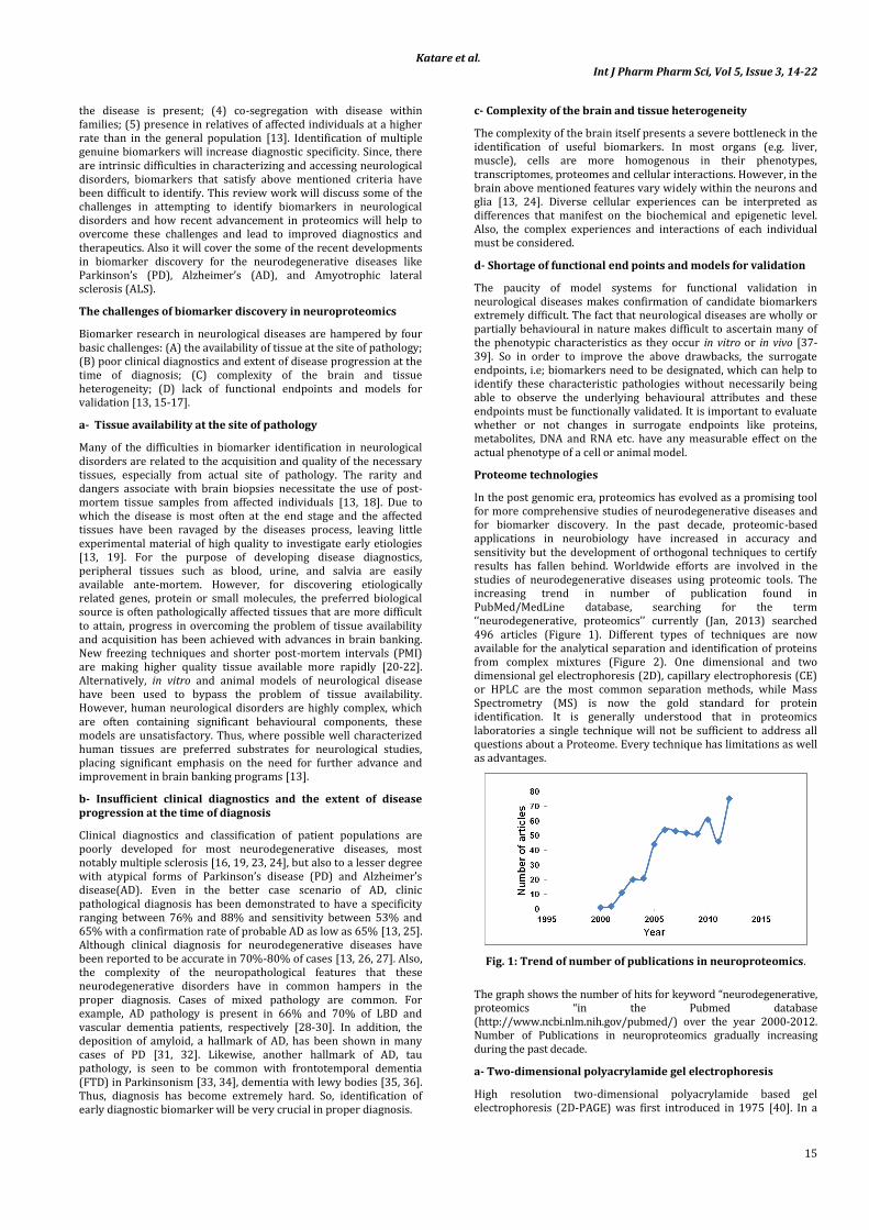
Fig. 1: Trend of number of publications in neuroproteomics.
The graph shows the number of hits for keyword “neurodegenerative, proteomics “in the Pubmed database (http://www.ncbi.nlm.nih.gov/pubmed/) over the year 2000-2012. Number of Publications in neuroproteomics gradually increasing during the past decade.
a- Two-dimensional polyacrylamide gel electrophoresis
High resolution two-dimensional polyacrylamide based gel electrophoresis (2D-PAGE) was first introduced in 1975 [40]. In a

Katare et al. Int J Pharm Pharm Sci, Vol 5, Issue 3, 14-22
16
single experiment, 2D-PAGE allows separation of 102-103 protein based upon proteins isoelectic points in the first dimension (1D) and then separation of proteins according to molecular weight in the second dimension (2D) [41, 42]. Some post-translational modifications of proteins such as oxidation or phosphorylation were identified by western blotting or staining [43-46]. 2D-PAGE combined with mass spectrometer is a powerful technique that represents a remarkable resource for proteomics studies. It offers excellent opportunities to discover potentially new biomarkers for early detection and diagnosis of disease [47]. Both 1D and 2D-PAGE can be combined with liquid chromatography-mass spectrometry based separation of gel extracts which provide an additional dimension of separation and identification for complex mixture. In the past decade several advancements have been made in 2D-PAGE, such as the introduction of fluorescent 2D difference gel electrophoresis (2D-DIGE) and many protein prefractionation techniques. 2D-PAGE is an extremely useful technique, offering significant advantages for fast, sensitive and accurate protein separation and analysis, making it one of the favoured methods for the analysis of protein expression differences in many laboratories.
b- Two-dimensional difference gel electrophoresis
Two-dimensional difference gel electrophoresis (2D-DIGE) is a modified version of 2D-PAGE which utilizes differential labeling of protein samples with upto their fluorogenic tags. 2D-DIGE offers the advancements in the differential labeling of proteins can be applied in 2D-based gel electrophoresis or liquid chromatography. Differential labeling offers the advantage of comparing several samples (control vs experimental) in a single experiment. [48-51]. The test is selected such that it reveals the consequence of a specific condition including the effects of disease, stress, temperature, drugs etc. Differential labeling approach is the most commonly used in 2D-DIGE. Using this technique, thousands of fluorescently labeled proteins are analyzed to determine changes in protein expression and post-translational modifications [43, 51]. Residue (cysteine, tyrosine, lysine, and histidine modifications) specific fluorophores provides great advantage to probe specific properties of a protein. 2D-DIGE is becoming popular to study post-translational modifications such as oxidation, phosphorylation, ubiquitination, and palmitoylation. 2D-DIGE can combine with mass spectrometer or immunoblotting to characterize proteins [52-54].
Fig. 2: Schematic diagram of proteomic approach
The most common approach to separate complex protein mixtures is by 2-dimensional gel electrophoresis (2D), which separates proteins in a pH gradient according to isoelectric point in the first dimension (1D), and in an acrylamide matrix according to molecular weight in the second dimension (2D). Relative levels of expression are compared between gels of different samples using computer algorithms to determine differential protein changes. Proteins of interest are collecting from the gel, trypsin digested, and subjected to mass spectrometry for identification and characterisation. An alternative approach is to pre-label protein mixtures and separate proteins, or more often peptides, by multidimensional liquid chromatography. Differences in peptide levels and protein identification are then performed by mass spectrometry.
c- Mass spectroscopy (MS)
Mass Spectrometer measures the mass to charge ratio of gas-phase ions. Fundamentally, mass spectrometers contains an ion source which converts analyte molecules into gas-phase ions, a mass analyzer which separates ionized analytes based on mass to charge ratio, and a detector that counts the number of ions at each mass to
charge value [55]. Since past several decades mass spectrometry has been popularly used to analyze biological samples and has been evolved into an indispensable tool for proteomics research [56]. This technique is ultra-sensitive, offers tremendous resolution to detect digested peptides or intact proteins in complex mixture [57, 58]. Recent development of a novel mass spectrometer (Orbitrap) and new methods of dissociation (e.g. electron transfer dissociation) has made it applicable in new areas of proteomic application. With the help of stable isotope labelling, mass spectrometry allows to measure dynamic changes in protein expression, modification and interaction. In principle, mass spectrometers can be coupled with 1D, 2D-PAGE, liquid chromatography and capillary electrophoresis and can achieve sensitivity down to the femtomole level [59]. The major obstacles in applying mass spectroscopy to study neuro-proteomics are limitations in availability of samples, which block most neuro-proteomics experiments. Although bottom-up proteomics (analysis of proteolytic peptide mixtures) remains the workhorse for proteomic analysis, middle-down and top-down strategies (analysis of longer peptides and intact proteins, respectively) should allow more complete characterization of protein isoforms and post-translational modifications.
Katare et al. Int J Pharm Pharm Sci, Vol 5, Issue 3, 14-22
17
d- Matrix-assisted ionization-time of flight (maldi-tof-ms)
Matrix-assisted laser desorption/ionization (MALDI) is one of the most common protein and peptide ionization techniques due to the exquisite speed and sensitivity [60]. It allows the identification of protein separated/isolated by 1D, 2D-PAGE or liquid chromatography. The availability of highly sensitive and high-resolution instrumentation and high capacity and fast computers, together with the availability of highly rich protein databases, makes MALDI-TOF-MS technique of choice for protein identification and characterization. In principle, using this technique hundreds to thousands of proteins can be identified in a single run [61]. Proteins are usually digested into smaller fragments (peptides) with a proteinase (e.g. trypsin). Digested proteins are linked with an acidic matrix and applied to a stainless steel plate (MALDI). The mass of peptides are detected by the mass spectrometer in different ways [62]. Time of flight mass spectrometry (TOF-MS) is a method of mass spectrometry in which peptides fly down a flight tube and the flight time to reach detector is measured which is proportional to mass of peptides [63]. The mass spectrum is converted into a list of peptide masses and searched against the extensive genome and proteome databases, translated and trypsin digested in silico [64]. Every protein has an unique peptide mass fingerprint (PMF) due to its characteristic amino acid sequence. Therefore, peptide masses calculated by the mass spectrometer can identify the protein from the millions of proteins in the database. Some other detectors coupled with MALDI are quadropoles, ion traps and Fourier transform ion cyclotron resonance (FTICR) to for specific purposes such as detecting large biomolecules [65]. MALDI-based tissue imaging is a great advantage and allows the possibility of generating 3D molecular images of the brain areas which could be useful to study the dynamic evolution of sub-proteomes [66, 67].
Tandem mass spectroscopy (MS/MS)
In the past two decades advancement in the instrumentation, databases, search algorithms and scoring criteria have been made which allows to determine the amino acid sequence (primary structure) of the proteins in complex biological samples. This is achieved by tandem mass selection or mass separation (MS/MS) mode of mass spectroscopy [68]. Tandem MS/MS followed different strategies. Initially ions or peptides are generated and selected by MS which are subjected to fragmentation into small pieces. The mass
of resulted ions are measured. The mass of secondary ions are decoded into protein sequence information which is subsequently searched against large protein databases for protein identification [69-71]. In comparison to normal mode of MS operation, the background level is very low in MS/MS mode of operation which consequently makes it highly sensitive [72]. Moreover, high-quality of peptide fragments obtained if MALDI ion source coupled to tow TOF analyzers [73]. Thus, tandem MS/MS technique is exceedingly powerful to perform high throughput studies and able to identify point mutations or post-translational modification in complex biosamples. Now this technique also applied to relative quantification of proteins without need of prior gel separation.
Isotope coded affinity tags (ICAT)
Isotope coded affinity tags (ICAT) is a high throughput MS based technique for quantitative proteomics. It relies on chemical labelling reagents and does not require gel separation [74]. Usually cysteine specific reactive reagents are used to label pathological and healthy biosamples (e.g. cancer vs normal cells) with heavy or light weight isotopes. After labelling complex protein mixtures are digested broad spectrum proteinases (such as trypsin) and the peptides are analysed by MS. Thus, ICAT allows to perform direct qualitative and quantitative comparisons of complex protein compounds [75-77]. The relative abundance of particular peptides and protein identification are simultaneously made in each sample. In addition to cysteine labelling, derivatization of primary amino groups in the intact proteins are also developed which is the basis of isotope tags for relative and absolute quantification (iTRAQ™) [78, 79]. After labelling with iTRAQ reagent, the differentially labelled proteins do not differ in mass due to isobaric mass design of the iTRAQ reagents. Therefore, corresponding digested peptide fragment appear as single peak in MS. Stable isotope labelling with amino acids in cell culture (SILAC) is another useful quantification technique for investigation of differential expression of proteins in two separate cellular populations [80]. SILAC is a simple and consistent technique for impartial comparative proteomic research. It has been utilized to study various kinds of post-translational modifications such as protein methylation, phosphorylation, to illustrate signalling pathways and to determine specific protein interactions [81-83]. In SILAC techniques, cells are cultured with in a natural amino acid deficient synthetic media and supplemented with a monoisotopically (e.g. carbon or nitrogen) labelled amino acid [84].
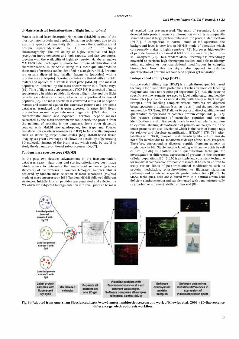
Fig. 3: (Adopted from Amersham Biosciences,http://www1.amershambiosciences.com and work of Knowles et al., 2003.) 2D-fluorescence difference gel electrophoresis workflow.

Katare et al. Int J Pharm Pharm Sci, Vol 5, Issue 3, 14-22
18
Protein samples to be compared are covalently labelled with either Cy3 or Cy5 fluorescent dyes. An internal control, to be run on every single gel in the experiment, is labelled with Cy2. All three samples are combined and separated on the one 2D-gel, thus eliminating gel to gel variation. The single gel is scanned at three different wavelengths to generate an image specific for each CyDyeTM fluore. The DeCyderTM software (Amersham Biosciences, GE Healthcare) normalises the test samples to the internal control, and then overlays the two test samples to identify changes in expression levels of individual protein spots. A 3-dimensional view of matched proteins is generated to ensure correct detection of protein spots. As all gels are run with the same internal standard, multiple gels from numerous experiments can all be compared with statistical confidence.
Imaging mass spectrometry (IMS)
Imaging Mass Spectrometry technique is used for visualization of spatial distribution of different compounds such as proteins, peptides, metabolites, biomarkers etc. by their characteristic molecular masses. This is an emerging technique which combines the specificity and parallel detection of MS with microscopic imaging capabilities [85]. Sample preparation, sensitivity of the ionization step, speed and spatial resolution are important variable in this technique. Emerging technologies in the field of IMS are secondary ion mass spectrometry (SIMS) and matrix-assisted laser desorption/ionization (MALDI) imaging of biosamples. Significant advancements have been at different steps of these techniques [86, 87]. IMS is a promising technique for discovery of biomarkers by MS in the tissue biopsies. Protein expression has been directly visualized by in situ MS analysis of healthy and pathological tissues. Frozen tissue samples are sliced and a small piece are placed on the MALDI plate and examined at regular intervals. The MS data obtained at different time points are evaluated between normal and diseased samples, resulting a spatial distribution of individual masses which reveal the pattern of different protein expression in normal and diseased tissues [88-90].
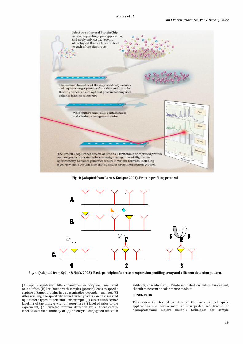
Surface-enhanced laser desorption/ionization is an ionisation technique used for analysis of protein mixtures in time of flight mass spectrometer. In SELDI-TOF-MS modified target is used to achieve biochemical affinity with the protein of interest. In SLEDI, protein mixture is applied on chemically active surface to bind. Binding of protein of interest on SELDI surface acts as a separation step and ease the data analysis. The unbound proteins are washed away and the bound proteins on the surface are detected and analysed in SELDI-TOF-MS mode. The molecular weight and amount of the bound proteins are estimated by TOF-MS. Depending upon properties of target protein, SELDI surface usually includes ion exchange (cation or anion), IMAC, hydrophobic or affinity (antibody, ligand or other proteins) surfaces [91-93]. The SELDI technology is useful in the identification and characterization of proteins involved in the pathogenesis [94]. The SELDI-MS have been used for identification of biomarkers for ALS [95]. SELDI based MS is useful for rapid, sensitive and high-throughput studies of biomarkers.
Procedure for preparing the ProteinChip Arrays with biological samples and for analyzing the retained proteins by SELDI-TOF-MS using a ProteinChip Reader.
Protein microarray
The limitation in the proteomics tools have led to the development of novel miniaturized tools for the proteomic research. Protein microarray is one of the emerging technology in this field [96, 97]. The concept of protein microarray is similar to DNA microarray which allows studies of thousands of genes in a single experiment. Protein microarray can be combined with other techniques such as MS and 2D-DIGE to characterize specific proteins which interact with antibodies, peptides, other ligands coupled on solid surfaces. In neuroproteomics, protein microarrays could be utilized to calculate changes in protein level, their modifications and interactions in the nerve cells. Protein lysate from nerve cells could be simply mixed and incubated with array of antibodies against protein of interest. Using this technique molecular association of large number of proteins could be studied in one run [98].
Surface-enhanced laser desorption/ionization (SELDI)
Katare et al. Int J Pharm Pharm Sci, Vol 5, Issue 3, 14-22
19
Fig. 4: (Adapted from Guru & Enrique 2003). Protein profiling protocol.
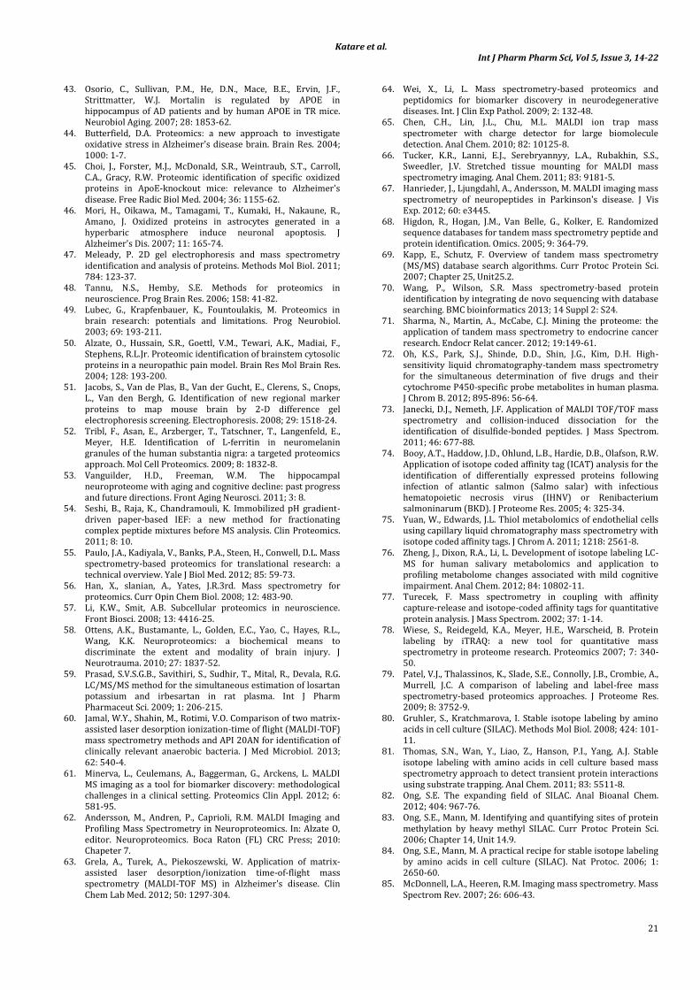
Fig. 4: (Adapted from Sydor & Nock, 2003). Basic principle of a protein expression profiling array and different detection pattern.
(A) Capture agents with different analyte specificity are immobilized on a surface. (B) Incubation with samples (protein) leads to specific capture of target proteins in a concentration dependent manner. (C) After washing, the specificity bound target protein can be visualized by different types of detection, for example (1) direct fluorescence labelling of the analyte with a fluorophore (f) labelled prior to the experiment, (2) targeted protein detection by a fluorescently-labelled detection antibody or (3) an enzyme-conjugated detection
antibody, conceding an ELISA-based detection with a fluorescent, chemiluminescent or colorimetric readout.
CONCLUSION
This review is intended to introduce the concepts, techniques, applications and advancement in neuroproteomics. Studies of neuroproteomics require multiple techniques for sample

Katare et al. Int J Pharm Pharm Sci, Vol 5, Issue 3, 14-22
20
preparation, fractionation, and identification. The combination of these approaches produces gigantic data. To make useful function of each protein must be identified. Therefore, combining highly sensitive and high-throughput techniques will help to understand networks of protein interaction and dynamics.
REFERENCES
1. Wasinger, V.C., Pollack, J.D., Humphery-Smith I. The proteome of Mycoplasma genitalium. Chaps-soluble component. Eur J Biochem. 2000; 267: 1571-82.
2. Watkins, S.M., German, J.B. Metabolomics and biochemical profiling in drug discovery and development. Curr Opin Mol Therapeut. 2002; 4: 224-8.
3. Collins, M.A. Generating 'omic knowledge': the role of informatics in high content screening. Comb Chem High Throughput Screen. 2009; 12: 917-25.
4. Kristensen, T.N., Pedersen, K.S., Vermeulen, C.J., Loeschcke, V. Research on inbreeding in the 'omic' era. Trends Ecol Evol. 2010; 25: 44-52.
5. Drabik, A., Bierczynska-Krzysik, A., Bodzon-Kulakowska, A., Suder, P., Kotlinska, J., Silberring, J. Proteomics in neurosciences. Mass Spectrom Rev. 2007; 26: 432-50.
6. Mak, P., Siwek, M., Pohl, J., Dubin, A. Menstrual hemocidin HbB115-146 is an acidophilic antibacterial peptide potentiating the activity of human defensins, cathelicidin and lysozyme. Am J Reprod Immunol. 2007; 57: 81-91.
7. Dobson, C.M. Protein misfolding, evolution and disease. Trends Biochem Sci. 1999; 24: 329-32.
8. Ideker, T., Thorsson, V., Ranish, J.A., Christmas, R., Buhler, J., Eng, J.K. Integrated genomic and proteomic analyses of a systematically perturbed metabolic network. Science. 2001; 292: 929-34.
9. Johnson, M.D., Yu, L.R., Conrads, T.P., Kinoshita, Y., Uo, T., Matthews, J.D. Proteome analysis of DNA damage-induced neuronal death using high throughput mass spectrometry. J Biol Chem. 2004; 279: 26685-97.
10. Griffin, T.J., Gygi, S.P., Ideker, T., Rist, B., Eng, J., Hood, L. Complementary profiling of gene expression at the transcriptome and proteome levels in Saccharomyces cerevisiae. Mol Cell Proteomics. 2002; 1: 323-33.
11. Pula, J.H., Kim, J., Nichols, J. Visual aspects of neurologic protein misfolding disorders. Curr Opin Ophthalmol. 2009; 20: 482-9.
12. Gregersen, N., Bross, P. Protein misfolding and cellular stress: an overview. Methods Mol Biol. 2010; 648, 3-23.
13. Dunckley, T., Coon, K.D., Stephan, D.A. Discovery and development of biomarkers of neurological disease. Drug Discov Today. 2005; 10: 326-34.
14. Severino, M.E., Dubose, R.F., Patterson, S.D. A strategic view on the use of pharmacodynamic biomarkers in early clinical drug development. IDrugs. 2006; 9: 849-53.
15. Sharma, V. Parkinson's disease and neurotoxic animal model: A mechanistic view. Int J Pharm Pharmaceut Sci. 2012; 4: 55-60.
16. Nikolcheva, T., Jager, S., Bush, T.A., Vargas, G. Challenges in the development of companion diagnostics for neuropsychiatric disorders. Expert Rev Mol Diagnostics. 2011; 11: 829-37.
17. Tropea, D. New challenges and frontiers in the research for neuropsychiatric disorders. Front Psychiatry. 2012; 3: 69.
18. Hulette, C.M., Downey, B.T., Burger, P.C. Macrophage markers in diagnostic neuropathology. Am J Surg Pathol. 1992; 16: 493-9.
19. Shoemaker, L.D., Achrol, A.S., Sethu, P., Steinberg, G.K., Chang, S.D. Clinical neuroproteomics and biomarkers: from basic research to clinical decision making. Neurosurgery. 2012; 70: 518-25.
20. Scholz, B., Skold, K., Kultima, K., Fernandez, C., Waldemarson, S., Savitski, M.M. Impact of temperature dependent sampling procedures in proteomics and peptidomics-a characterization of the liver and pancreas post mortem degradome. Mol Cell proteomics. 2011; 10: M900229MCP200.
21. Ferrer, I., Santpere, G., Arzberger, T., Bell, J., Blanco, R., Boluda, S. Brain protein preservation largely depends on the postmortem storage temperature: implications for study of proteins in human neurologic diseases and management of brain banks: a BrainNet Europe Study. J Neuropathol Exp Neurol. 2007; 66: 35-46.
22. Skold, K., Svensson, M., Norrman, M., Sjogren, B., Svenningsson, P., Andren, P.E. The significance of biochemical and molecular sample integrity in brain proteomics and peptidomics: stathmin 2-20 and peptides as sample quality indicators. Proteomics. 2007; 7: 4445-56.
23. Comabella, M., Racke, M.K. New technologies for biomarker discovery in multiple sclerosis. J Neuroimmunol. 2012; 248: 1.
24. Coon, K.D., Dunckley, T., Stephan, D.A. Biomarker identification in neurologic diseases: improving diagnostics and therapeutics. Expert Rev Mol Diagn. 2004; 4: 361-75.
25. Royall, D.R., Palmer, R.F., Petrovitch, H., Ross, G.W., Masaki, K., White, L.R. Modeling regional vulnerability to Alzheimer pathology. Neurobiol Aging. 2012; 33: 1556-63.
26. Kroksveen, A.C., Opsahl, J.A., Aye, T.T., Ulvik, R.J., Berven, F.S. Proteomics of human cerebrospinal fluid: discovery and verification of biomarker candidates in neurodegenerative diseases using quantitative proteomics. J Proteomics. 2011; 74: 371-88.
27. Shi, M., Caudle, W.M., Zhang, J. Biomarker discovery in neurodegenerative diseases: a proteomic approach. Neurobiol Dis. 2009; 35: 157-64.
28. Schneider, J.A., Arvanitakis, Z., Leurgans, S.E., Bennett, D.A. The neuropathology of probable Alzheimer disease and mild cognitive impairment. Ann Neurol. 2009; 66: 200-8.
29. Schneider, J.A., Bennett, D.A. Where vascular meets neurodegenerative disease. Stroke. 2010; 41: S144-6.
30. Engelborghs, S., Le Bastard, N. The role of CSF biomarkers in the diagnostic work-up of mixed vascular-degenerative dementia. J Neurol Sci. 2012; 322: 197-9.
31. Mastaglia, F.L., Masters, C.L., Beyreuther, K., Kakulas, B.A. Deposition of Alzheimer's disease amyloid (A4) protein in the cerebral cortex in Parkinson's disease. Prog Clin Biol Res. 1989; 317: 475-84.
32. Mastaglia, F.L., Johnsen, R.D., Byrnes, M.L., Kakulas, B.A. Prevalence of amyloid-beta deposition in the cerebral cortex in Parkinson's disease. Mov Disord. 2003; 18: 81-6.
33. Luigetti, M., Quaranta, D., Conte, A., Piccininni, C., Lattante, S., Romano, A. Frontotemporal dementia, Parkinsonism and lower motor neuron involvement in a patient with C9ORF72 expansion. Amyotroph Lateral Scler Frontotemporal Degener. 2013; 14: 66-9.
34. Floris, G., Borghero, G., Cannas, A., Di Stefano, F., Costantino, E., Murru, M.R. Frontotemporal dementia with psychosis, parkinsonism, visuo-spatial dysfunction, upper motor neuron involvement associated to expansion of C9ORF72: a peculiar phenotype? J Neurol. 2012; 259: 1749-51.
35. O'Donovan, J., Watson, R., Colloby, S.J., Firbank, M.J., Burton, E.J., Barber, R. Does posterior cortical atrophy on MRI discriminate between Alzheimer's disease, dementia with Lewy bodies, and normal aging? Int Psychogeriatr. 2013; 25: 111-9.
36. Moreno-Ramos, T., Benito-Leon, J., Villarejo-Galende, A., Bermejo-Pareja, F. Retinal nerve fiber layer thinning in dementia associated with Parkinson's disease, dementia with lewy bodies, and Alzheimer's disease. J Alzheimer's Dis. 2013; 34(3): 659-64.
37. van der Staay, F.J., Arndt, S.S., Nordquist, R.E. Evaluation of animal models of neurobehavioral disorders. Behav Brain Funct. 2009; 5: 11.
38. Stanzione, P., Tropepi, D. Drugs and clinical trials in neurodegenerative diseases. Ann Ist Super Sanita. 2011; 47: 49-54.
39. Tordjman, S., Drapier, D., Bonnot, O., Graignic, R., Fortes, S., Cohen, D. Animal models relevant to schizophrenia and autism: validity and limitations. Behav Genet. 2007; 37: 61-78.
40. O'Farrell, P.H. High resolution two-dimensional electrophoresis of proteins. J Biol Chem. 1975; 250: 4007-21.
41. Van den Bergh, G., Arckens, L. Recent advances in 2D electrophoresis: an array of possibilities. Expert Rev Proteomics. 2005; 2: 243-52.
42. Kim, H., Eliuk, S., Deshane, J., Meleth, S., Sanderson, T., Pinner, A. 2D gel proteomics: an approach to study age-related differences in protein abundance or isoform complexity in biological samples. Methods Mol Biol. 2007; 371: 349-91.
Katare et al. Int J Pharm Pharm Sci, Vol 5, Issue 3, 14-22
21
43. Osorio, C., Sullivan, P.M., He, D.N., Mace, B.E., Ervin, J.F., Strittmatter, W.J. Mortalin is regulated by APOE in hippocampus of AD patients and by human APOE in TR mice. Neurobiol Aging. 2007; 28: 1853-62.
44. Butterfield, D.A. Proteomics: a new approach to investigate oxidative stress in Alzheimer's disease brain. Brain Res. 2004; 1000: 1-7.
45. Choi, J., Forster, M.J., McDonald, S.R., Weintraub, S.T., Carroll, C.A., Gracy, R.W. Proteomic identification of specific oxidized proteins in ApoE-knockout mice: relevance to Alzheimer's disease. Free Radic Biol Med. 2004; 36: 1155-62.
46. Mori, H., Oikawa, M., Tamagami, T., Kumaki, H., Nakaune, R., Amano, J. Oxidized proteins in astrocytes generated in a hyperbaric atmosphere induce neuronal apoptosis. J Alzheimer's Dis. 2007; 11: 165-74.
47. Meleady, P. 2D gel electrophoresis and mass spectrometry identification and analysis of proteins. Methods Mol Biol. 2011; 784: 123-37.
48. Tannu, N.S., Hemby, S.E. Methods for proteomics in neuroscience. Prog Brain Res. 2006; 158: 41-82.
49. Lubec, G., Krapfenbauer, K., Fountoulakis, M. Proteomics in brain research: potentials and limitations. Prog Neurobiol. 2003; 69: 193-211.
50. Alzate, O., Hussain, S.R., Goettl, V.M., Tewari, A.K., Madiai, F., Stephens, R.L.Jr. Proteomic identification of brainstem cytosolic proteins in a neuropathic pain model. Brain Res Mol Brain Res. 2004; 128: 193-200.
51. Jacobs, S., Van de Plas, B., Van der Gucht, E., Clerens, S., Cnops, L., Van den Bergh, G. Identification of new regional marker proteins to map mouse brain by 2-D difference gel electrophoresis screening. Electrophoresis. 2008; 29: 1518-24.
52. Tribl, F., Asan, E., Arzberger, T., Tatschner, T., Langenfeld, E., Meyer, H.E. Identification of L-ferritin in neuromelanin granules of the human substantia nigra: a targeted proteomics approach. Mol Cell Proteomics. 2009; 8: 1832-8.
53. Vanguilder, H.D., Freeman, W.M. The hippocampal neuroproteome with aging and cognitive decline: past progress and future directions. Front Aging Neurosci. 2011; 3: 8.
54. Seshi, B., Raja, K., Chandramouli, K. Immobilized pH gradient-driven paper-based IEF: a new method for fractionating complex peptide mixtures before MS analysis. Clin Proteomics. 2011; 8: 10.
55. Paulo, J.A., Kadiyala, V., Banks, P.A., Steen, H., Conwell, D.L. Mass spectrometry-based proteomics for translational research: a technical overview. Yale J Biol Med. 2012; 85: 59-73.
56. Han, X., slanian, A., Yates, J.R.3rd. Mass spectrometry for proteomics. Curr Opin Chem Biol. 2008; 12: 483-90.
57. Li, K.W., Smit, A.B. Subcellular proteomics in neuroscience. Front Biosci. 2008; 13: 4416-25.
58. Ottens, A.K., Bustamante, L., Golden, E.C., Yao, C., Hayes, R.L., Wang, K.K. Neuroproteomics: a biochemical means to discriminate the extent and modality of brain injury. J Neurotrauma. 2010; 27: 1837-52.
59. Prasad, S.V.S.G.B., Savithiri, S., Sudhir, T., Mital, R., Devala, R.G. LC/MS/MS method for the simultaneous estimation of losartan potassium and irbesartan in rat plasma. Int J Pharm Pharmaceut Sci. 2009; 1: 206-215.
60. Jamal, W.Y., Shahin, M., Rotimi, V.O. Comparison of two matrix-assisted laser desorption ionization-time of flight (MALDI-TOF) mass spectrometry methods and API 20AN for identification of clinically relevant anaerobic bacteria. J Med Microbiol. 2013; 62: 540-4.
61. Minerva, L., Ceulemans, A., Baggerman, G., Arckens, L. MALDI MS imaging as a tool for biomarker discovery: methodological challenges in a clinical setting. Proteomics Clin Appl. 2012; 6: 581-95.
62. Andersson, M., Andren, P., Caprioli, R.M. MALDI Imaging and Profiling Mass Spectrometry in Neuroproteomics. In: Alzate O, editor. Neuroproteomics. Boca Raton (FL) CRC Press; 2010: Chapeter 7.
63. Grela, A., Turek, A., Piekoszewski, W. Application of matrix-assisted laser desorption/ionization time-of-flight mass spectrometry (MALDI-TOF MS) in Alzheimer's disease. Clin Chem Lab Med. 2012; 50: 1297-304.
64. Wei, X., Li, L. Mass spectrometry-based proteomics and peptidomics for biomarker discovery in neurodegenerative diseases. Int. J Clin Exp Pathol. 2009; 2: 132-48.
65. Chen, C.H., Lin, J.L., Chu, M.L. MALDI ion trap mass spectrometer with charge detector for large biomolecule detection. Anal Chem. 2010; 82: 10125-8.
66. Tucker, K.R., Lanni, E.J., Serebryannyy, L.A., Rubakhin, S.S., Sweedler, J.V. Stretched tissue mounting for MALDI mass spectrometry imaging. Anal Chem. 2011; 83: 9181-5.
67. Hanrieder, J., Ljungdahl, A., Andersson, M. MALDI imaging mass spectrometry of neuropeptides in Parkinson's disease. J Vis Exp. 2012; 60: e3445.
68. Higdon, R., Hogan, J.M., Van Belle, G., Kolker, E. Randomized sequence databases for tandem mass spectrometry peptide and protein identification. Omics. 2005; 9: 364-79.
69. Kapp, E., Schutz, F. Overview of tandem mass spectrometry (MS/MS) database search algorithms. Curr Protoc Protein Sci. 2007; Chapter 25, Unit25.2.
70. Wang, P., Wilson, S.R. Mass spectrometry-based protein identification by integrating de novo sequencing with database searching. BMC bioinformatics 2013; 14 Suppl 2: S24.
71. Sharma, N., Martin, A., McCabe, C.J. Mining the proteome: the application of tandem mass spectrometry to endocrine cancer research. Endocr Relat cancer. 2012; 19:149-61.
72. Oh, K.S., Park, S.J., Shinde, D.D., Shin, J.G., Kim, D.H. High-sensitivity liquid chromatography-tandem mass spectrometry for the simultaneous determination of five drugs and their cytochrome P450-specific probe metabolites in human plasma. J Chrom B. 2012; 895-896: 56-64.
73. Janecki, D.J., Nemeth, J.F. Application of MALDI TOF/TOF mass spectrometry and collision-induced dissociation for the identification of disulfide-bonded peptides. J Mass Spectrom. 2011; 46: 677-88.
74. Booy, A.T., Haddow, J.D., Ohlund, L.B., Hardie, D.B., Olafson, R.W. Application of isotope coded affinity tag (ICAT) analysis for the identification of differentially expressed proteins following infection of atlantic salmon (Salmo salar) with infectious hematopoietic necrosis virus (IHNV) or Renibacterium salmoninarum (BKD). J Proteome Res. 2005; 4: 325-34.
75. Yuan, W., Edwards, J.L. Thiol metabolomics of endothelial cells using capillary liquid chromatography mass spectrometry with isotope coded affinity tags. J Chrom A. 2011; 1218: 2561-8.
76. Zheng, J., Dixon, R.A., Li, L. Development of isotope labeling LC-MS for human salivary metabolomics and application to profiling metabolome changes associated with mild cognitive impairment. Anal Chem. 2012; 84: 10802-11.
77. Turecek, F. Mass spectrometry in coupling with affinity capture-release and isotope-coded affinity tags for quantitative protein analysis. J Mass Spectrom. 2002; 37: 1-14.
78. Wiese, S., Reidegeld, K.A., Meyer, H.E., Warscheid, B. Protein labeling by iTRAQ: a new tool for quantitative mass spectrometry in proteome research. Proteomics 2007; 7: 340-50.
79. Patel, V.J., Thalassinos, K., Slade, S.E., Connolly, J.B., Crombie, A., Murrell, J.C. A comparison of labeling and label-free mass spectrometry-based proteomics approaches. J Proteome Res. 2009; 8: 3752-9.
80. Gruhler, S., Kratchmarova, I. Stable isotope labeling by amino acids in cell culture (SILAC). Methods Mol Biol. 2008; 424: 101-11.
81. Thomas, S.N., Wan, Y., Liao, Z., Hanson, P.I., Yang, A.J. Stable isotope labeling with amino acids in cell culture based mass spectrometry approach to detect transient protein interactions using substrate trapping. Anal Chem. 2011; 83: 5511-8.
82. Ong, S.E. The expanding field of SILAC. Anal Bioanal Chem. 2012; 404: 967-76.
83. Ong, S.E., Mann, M. Identifying and quantifying sites of protein methylation by heavy methyl SILAC. Curr Protoc Protein Sci. 2006; Chapter 14, Unit 14.9.
84. Ong, S.E., Mann, M. A practical recipe for stable isotope labeling by amino acids in cell culture (SILAC). Nat Protoc. 2006; 1: 2650-60.
85. McDonnell, L.A., Heeren, R.M. Imaging mass spectrometry. Mass Spectrom Rev. 2007; 26: 606-43.

Katare et al. Int J Pharm Pharm Sci, Vol 5, Issue 3, 14-22
22
86. Grassl, J., Taylor, N.L., Millar, A.H. Matrix-assisted laser desorption/ionisation mass spectrometry imaging and its development for plant protein imaging. Plant methods. 2011; 7: 21.
87. Laskin, J., Heath, B.S., Roach, P.J., Cazares, L., Semmes, O.J. Tissue imaging using nanospray desorption electrospray ionization mass spectrometry. Anal Chem. 2012; 84: 141-8.
88. Gruner, B.M., Hahne, H., Mazur, P.K., Trajkovic-Arsic, M., Maier, S., Esposito, I. MALDI imaging mass spectrometry for in situ proteomic analysis of preneoplastic lesions in pancreatic cancer. PloS one. 2012; 7: e39424.
89. Schone, C., Hofler, H., Walch, A. MALDI imaging mass spectrometry in cancer research: Combining proteomic profiling and histological evaluation. Clin Biochem 2013; 46(6): 539-45.
90. Schwartz, S.A., Weil, R.J., Johnson, M.D., Toms, S.A., Caprioli, R.M. Protein profiling in brain tumors using mass spectrometry: feasibility of a new technique for the analysis of protein expression. Clin Cancer Res. 2004; 10: 981-7.
91. Hu, Q., Huang, Y., Wang, Z., Tao, H., Liu, J., Yan, L. Application of surface-enhanced laser desorption/ionization time-of-flight mass spectrometry coupled with an artificial neural network model for the diagnosis of hepatocellular carcinoma. Hepato-gastroenterology. 2012; 59: 1902-6.
92. Pienaar, I.S., Daniels, W.M., Gotz, J. Neuroproteomics as a promising tool in Parkinson's disease research. J Neural Transm. 2008; 115: 1413-30.
93. Tang, N., Tornatore, P., Weinberger, S.R. Current developments in SELDI affinity technology. Mass Spectrom Rev. 2004; 23: 34-44.
94. Liu, C. The application of SELDI-TOF-MS in clinical diagnosis of cancers. J Biomed Biotechnol. 2011; 2011: 245821.
95. Ranganathan, S., Williams, E., Ganchev, P., Gopalakrishnan, V., Lacomis, D., Urbinelli, L. Proteomic profiling of cerebrospinal fluid identifies biomarkers for amyotrophic lateral sclerosis. J Neurochem. 2005; 95: 1461-71.
96. Joyce, C.W., Murphy, I.G., Rafferty, M., Ryan, D., McDermott, E.W., Gallagher, W.M. Tumor profiling using protein biomarker panels in malignant melanoma: application of tissue microarrays and beyond. Expert Rev Proteomics. 2012; 9: 415-23.
97. Gonzalez, L.C. Protein microarrays, biosensors, and cell-based methods for secretome-wide extracellular protein-protein interaction mapping. Methods. 2012; 57: 448-58.
98. Gonzalez-Gonzalez, M., Jara-Acevedo, R., Matarraz, S., Jara-Acevedo, M., Paradinas, S., Sayagues, J.M. Nanotechniques in proteomics: protein microarrays and novel detection platforms. Eur J Pharm Sci. 2012; 45: 499-506.