abstracts national editor, j. douglas noll, ph.d
TRANSCRIPT

ABSTRACTS
National Editor, J. Douglas Noll, Ph.D.
Franklin L. Ashley, M.D.
Samuel! Berkowitz, D.D.S., M.S.
Bard Cosman, M.D.
Alexander Goldenberg, D.D.S.
John B. Gregg, M.D.
Lawrence S. Harte, D.D.S.
Joseph Luban, D.D.S.
Robert M. Mason, Ph.D.
I. J. Singh, Ph.D.
Charles C. Swoope, D.D.S., M.S.D.
Kenneth C. Troutman, D.D.S.
Paul M. Weeks, M.D.
International Editor, Nicholas G. Georgiade, M.D., D.D.S., Durham, N.C.
Claude Dufourmental, M.D.178 Rue Courcelles
Paris, FRANCE
Paul Fogh-Andersen, M.D.12 Dronningensvelj, Copenhagen
F, Denmark
G. B. Hopkin, F.R.E.D.
31 Chambers StreetEdinburgh, 1, Scotland
Panogiotis Kollipoulos, M.D.
Askilipiou 23
Athens, Greece
Karl E. NordinKGL Tandlakarhogskolan
Box 3207Stockholm 3, Sweden
Bengt Nylen, M.D.
Dept. of Plastic Surgery
Karolinska Hospital
Stockholm, Sweden
Seiichi Ohmori, M.D.
Dept. of Plastic SurgeryTokyo Metropolitan Polce Hospital2-10-41 Fujimi Chiyoda-KuTokyo, Japan
Fernado Ortiz-Monasterio, M.D.
Avenue Chapultepec 384-3Mexico City, Mexico D.F.
Joseph Penkava, M.D.Dept. of Plastic SurgeryBerkova 34
Brno 12, Czechoslovakia
William Henry Reid, F.R.C.S.Consultant Plastic Surgeon
Canniesburn HospitalBearsden, Glasgow
Edward Schmid, M.D.
Marien HospitalStuttgart, West Germany
Victor Spina, M.D.Hospital Das Clinicas Da Facul-dade
De Medicina Da UniversidadeDe S. Paulo
Sao Paulo, Brazil
Raymond Wang, M.D.234 Nathan Road, 3rd Floor,Kowloon, Hong Kong
Bartels, R. J., J. E. O'Malley, W. M.
Douglas, and R. G. Wilson, Varia-
tions of Masters interlocking Z-chei-
lorrhaphy. Plastic and Reconstructive
Surgery, 45, 189-190, 1970.
A technical modification of one of the Z-
plasty techniques for the repair of the uni-
lateral cleft lip is diagrammed and dis-
cussed briefly. (Cosman)
Blaine, H. L., Differential analysis of
cleft palate anomalies. Journal of
Dental Research, 48, 1042-1047, 1969.
Lateral cephalometric radiograms of
children with cleft palate or cleft lip and
palate were statistically analyzed. Selected
landmarks as defined by Krogman and
Sassouni were utilized in addition to a new
base line "ethnic horizontal" as developed
by Walker. Differential analysis using in-
dexes and angular measurements was done
according to age, sex, extent of cleft and
operative history. This study indicated
that in most cases with clefts regardless of
surgery, there is a tendency for vertical
and horizontal maxillary deficiency, retro-
placement of the maxillary complex, and a
general obtuseness of angles relating to the
794
sella-basion line. No sexual dichotomy
could be observed. (Luban)
Bowers, D. G. Jr., Congenital lower lip
sinuses with cleft palate. Plastic and
Reconstructive Surgery, 465, 151-154,
1970.
Lower lip sinuses are relatively rare but
when present are associated with cleft of
the lip and palate in 70 to 80% of patients.
However, less than 1% of all cleft lip and
palate patients have such lip sinuses. The
author reports on 3 families and 12 pa-
tients with such sinuses among whom were
2 in which the sinuses were associated with
submucous cleft palates-the first such as-
sociated occurrences recorded. Some de-
tails of the treatment of these defects are
also discussed. (Cosman)
Converse, J. M., J. Ransohoff, E. S.
Mathews, B. Smith, and H. Mole-
naar, Ocular hypertelorism and pseu-
dohypertelorism. Plastic and Recon-
structive Surgery, 45, 1-18, 1970.
Well conceived and well executed pro-
cedures for this difficult series of problems
are presented. A one stage combined intra-
and extra-cranial approach carried out by
both plastic and neuro-surgeon is detailed.
Those dealing with these complex ceranio-
facial disorders of which these anomalies
are parts will benefit by close study of this
paper. (Cosman)
Converse, J. M., V. M. Hogan, and C.
C. Dupuis, Combined nose-lip repair
in bilateral cleft-lip deformities. Plas-
tic and Reconstructive Surgery, 45,
109-118, 1970.
In the treatment of such secondary de-
fects the prolabium of the lip is, in a
single stage, employed to lengthen the
columella and the resultant defect in the
upper lip is repaired immediately by a
cross-lip flap. In some cases a bone graft
to the nose is performed at the same time.
This one stage nose-lip repair has been
ABSTRACTS 795
carried out over a 10 year period on 16
patients ranging from 5 to 18 years of age.
General anesthesia was used in most cases
with gentle movement of the tube to per-
mit the design of the lip flap. Change of
the tube to the nose after the nasal part
to permit easier lip flap formation can
also be carried out. Division of the Abbé
flap is done under local followed immedi-
ately by general anesthesia in the younger
patients. Presence of hair on the colu-
mella derived in this way from the upper
lip has not been a problem for these au-
thors. Long term follow up of 2 patients
suggests that the improvement obtained
is preserved intact and accordingly the
authors advocate the early repair of such
defects using this combined approach.
(Cosman)
Crikelair, G. F., Kastein, Shulamith,
and B. Cosman, Pharyngeal flap for
post-traumatic palatal paralysis. Plas-
tie and Reconstructive Surgery, 45,
182-185, 1970.
A patient with palatal and upper pha-
ryngeal wall paralysis consequent on cen-
tral nervous system trauma who had strik-
ing improvement in rhinolalia following a
pharyngeal flap is reported. This condition
is added to the growing list of indications
for the pharyngeal flap. Noting the lack
of dynamic tissue in the transplanted flap
and the rapidity of the speech improve-
ment in this and other kinds of cases serves
to demonstrate the importance of the ob-
turator function of the pharyngeal flap.
(Cosman)
Crikelair, G. F., P. Striker, and B.
Cosman, The surgical treatment of
submucous cleft palate. Plastic and
Reconstructive Surgery, 45, 58-65,
1970.
Experiences with the surgical treatment
of 20 palates with significant submucous
defects are presented. The anatomical vari-
ations demonstrated are enumerated. The

796 Abstracts
"classical" submucous cleft description is
shown to be an oversimplification. The
presence of a submucous zone is the single
constant feature and the length of the zone
is the criterion of the empiric definition.
The submucous lesion is but one manifes-
tation of the cleft palate defect and does
not constitute a separate entity. The his-
tory of the discovery and the various treat-
ments of submucous defects are reviewed.
Evidence is presented that, contrary to
present practice, exeision of the submucous
zone is not a necessary part of the therapy
of every submucous cleft. (Cosman)
Gordon, H., D. Davies, and M. Ber-
man, Camptodactyly, cleft palate,
and club foot: A syndrome showing
the autosomal-dominant pattern of in-
heritance. J. Med. Genet., 6, 266-274,
1969.
Camptodactyly refers to one of the
varieties of constitutionally crooked digits,
typically a flexion contracture of the
proximal interphalangeal joint almost al-
ways of the fifth finger. It may be sporadic
or familial. In the latter, there is a simple
autosomal domimant pattern of inherit-
ance, with a high degree of penetrance but
variable phenotypic expression. In these
genetic cases, camptodactyly is often the
only lesion, but it may be associated with
other connective tissue anomalies, or it
may be a major or a minor component of
a variety of complex syndromes. The pres-
ent report describes a family in which
camptodactyly is a major component of a
syndrome which includes cleft of the see-
ondary palate and club foot. The pedigree
shows the inheritance pattern of a single
mutant autosomal gene with dominant
effect and varying phenotypic expression.
The authors have found no record in the
literature of a similar syndrome. (Noll)
Gregg, J. B., Treatment of cleft lip-palate
patients, the surgeon's role. South
Dakota Journal of Medicine. 22, 125-
131, 1969.
This article, directed primarily to gen-
eral practitioners of medicine, outlines and
discusses briefly the many different surgi-
cal procedures which are often necessary
for the total treatment of persons having
congenital facial defects. The viewpoint of
the maxillo-facial surgeon who is trained to
handle all of the problems in the surgical
care of the cleft patients is advanced. II-
lustrations of various common surgical
procedures and photographs of exemplary
patients are presented. A plea is made for
a coordinated or team approach to the care
of persons with cleft lip-palate problems.
(Gregg)
Honjow, I., N. Isshiki, and M. Tanabe,
Objective evaluation of velar mobility.
A double-exposure roentgen tech-
nique. Plastic and Reconstructive Sur-
gery, 44, 597-600, 1969.
A small lead dise 5 mm. in diameter and
1 mm. in thickness is affixed to the soft
palate where velar mobility appears great-
est, using alpha-cyanoacrylate monomer.
Atropine is given to reduce salivary secre-
tion which otherwise limits the adhesive
effect. A four part plastic plate apparatus
immobilizes the head. A double exposure
x-ray is taken with one exposure during
rest and one during sustained phonation of
/s/ or /i/. X-ray factors and tube dig-
tances are detailed. A lead point scale in-
corporated in one of the head immobiliza-
tion bars appears on the film and makes
possible calibration of the measured differ-
ence between the positions of the palate as
determined by the positions of the metal
dise. Ratios of velar mobility can be ar-
rived at. Two normals and 15 cleft palate
patients were the subjects of this initial
exploration of technique. (Cosman)
Horton, C. E., J. E. Adamson, R. A.
Mladick, and R. J. Taddeo, The
upper lip sulcus in cleft lip. Plastic
and Reconstructive Surgery, 45,
31-37, 1970.
Comparatively little attention has been
paid to the deficiencies in the upper lip
sulcus in patients with either bilateral or
unilateral cleft lips. The authors feel it im-
portant to construct a suleus early in those
patients who have adhesion of the lip to
the anterior alveolar ridge so as to allow
unrestricted growth of the lip. Use of the
prolabial vermillion to surface the pre-
maxilla, with the lateral lip vermilion and
mucosa used to cover the under surface of
the prolabium, is described as a primary
procedure for the bilateral cleft lip patient
whose closure is to be accomplished in two
stages. A similar procedure is used for the
occasional unilateral cleft patient whose lip
is adherent to the alveolus. In secondary
bilateral cleft lip repair a V incision be-
neath the lip and V-Y advancement of the
tissue is carried out to release the lip. The
bare alveolar ridge surface is covered by a
small full thickness buccal mucosa mem-
brane graft. (Cosman)
Khar, M. H., and RK. N. Sharma, Meas-
urements of maxillary area in repair of
unilateral clefts. Plastic and Recon-
structive Surgery, 45, 155-159, 1970.
30 cases of unilateral lip/palate clefts
were analyzed by model and tracing tech-
niques which are described. Both bony de-
ficiency in the maxilla and deformity of the
maxilla were found. The maxillary de-
formity involved hypoplasia, medial rota-
tion, and forward drifting with lack of
development of face. Severe degrees of this
defect were in turn associated with less
inter-canine space and more nasal deform-
ity. (Cosman)
Miller, O. J., D. Warburton, and W.
R. Breg, Deletions of Group B chro-
mosomes. Birth Defects Orig. Art.
Ser., 5 (5), 100-105, May 1969.
Patients with a B-group short arm dele-
tion may have a deletion of the longer later
replicating chromosome (4p-) or the
shorter earlier replicating chromosome
(5p-, = eri-du-chat). Both types of pa-
tients share such features as severe mental
ABSTRACTS 797
retardation, microcephaly and antimongo-
lord palpebral fissures. Features distin-
guishing the 4p- syndrome include cleft
palate, coloboma, preauricular sinus, seiz-
ures, dermal ridge hypoplasia and absence
of a cat-like ery. While diagnosis of the
type of deletion is sometimes possible on
clinical grounds, measurement or autora-
diographic identification of the deleted
chromosome is often necessary, especially
in older patients. (23 references.) (This
abstract is from Birth Defects: Abstracts
of Selected Articles. The National Founda-
tion-March of Dimes, 6(11), Nov. 1969,
abstract number 69-913.)
Papangelou, L., Correction of velo-
pharyngeal insufficiency. Arch. Oto-
laryng., 91, 201-2083, 1970.
To aid velopharyngeal closure in a pa-
tient having congenital hypernasality the
result of cleft palate, the author described
a surgical technique in which the tonsils
are utilized to fill the defect. The tonsils
were rotated into a prepared bed in the
posterior pharyngeal wall as a flap having
its base in the superior pole of each tonsil.
The results in a single case are reported to
have shown marked speech improvement
without air leak immediately after the
operation and permanent improvement for
7 months. The author suggests as a possi-
ble disadvantage of the procedure the fact
that the patient could develop tonsillitis
later. (Gregg)
Quigley, L. F. Jr., Comparison of simul-
taneous airflow-pressure measure-
ments and cephalometric technics for
evaluation of normal and cleft palate
patients: III palatopharyngeal com-
petency. Journal of Dental Research,
49, 93-99, 1970.
A group of cleft palate patients and nor-
mal siblings were studied for palato-
pharyngeal competency with cephalome-
tric, manometric, and airflow technics.
There was little correlation between air
pressure measurements and cephalometric

798 Abstracts
data. The nasal anemometer airflow data
correlate well with cephalometric measure-
ments. The nasal anemometer provides an
excellent clinical tool for the determination
of palatopharyngeal competency and it can
be used for this purpose instead of
cephalometric radiographs. (Luban)
Shapira, Y., An autoradiographic study
of °*H Proline uptake in the palate of
normal mice and in the palate of mice
treated with hydrocortisone. Journal
of Dental Research, 48, 1039-1041,
1969.
Normal mice demonstrate a continuous
synthesis of amino acids into protein in the
palatal shelves before and during the pe-
riod of palate closure. In mice treated with
hydrocortisone, there is normal protein
synthesis up to the time of normal palate
closure when there is an abrupt decrease in
protein activity. It is suggested that hydro-
cortisone acts as a teratogen by causing
local alterations of fetal protein metabo-
lism. (Luban)
Smith, N. J. D., and W. P. Heighway,
Patient dose in dental cinefluorog-
raphy. Oral Surgery, Oral Medicine
and Oral Pathology, 27 (3), 349-357,
March 1969.
The purpose of the article was to report
the results of observations on the dose of
radiation received by patients and volun-
teer subjects during cinefluorographic pro-
cedures under taken as part of dental re-
search projects. X-ray and exposure was
measured by means of a 35 c.c. air ioniza-
tion chamber in conjunction with elec-
tronic instruments. Factors which effect
patients dosage were found to be as fol-
lows: Kilovoltage of the x-ray tube need
not necessarily increase the patient dose
since equivalent results can be obtained by
lowering the milliamperage. The greater
the milliamperage the greater the patient
dose therefore the milliamperage should
always be kept as low as practicable in an
effort to reduce the patient dose. Increas-
ing the filtration of the rays will reduce
the skin dose markedly however too much
filtration will reduce the contrast on the
image intensifier screen and will necessitate
the use of higher milliamperage. Where
great contrast was not needed it was often
possible to increase the kilovoltage and to
use copper filtration in addition to the
aluminum filtration. This resulted in a
marked reduction in the patient dose how-
ever it adversely affected the visualization
of the soft tissue outlines. The powter-
Bucky grid necessitates a larger milliam-
perage and thus increases the patient dose.
It was found that the loss of quality when
not using the grid was small and that it
was best to reduce the patient exposure
by not using the grid. The faster the
cinefilm, the smaller the patient dose, since
less brightness is needed on the image in-
tensifier. Two fast a film was found diffi-
cult to handle, and increased the grain size
detracted from the quality of the film. Al-
though increasing the frame speed permits
a more detailed analysis to be made, if
the frame speed is increased then the cam-
era shutter is opened for a shorter period
of time and this requires an increased
brightness on the image intensifier. Thus,
increasing the frame speed will mean an
increase in the patient dose. It was found
that the patient dose is considerably less
with a 16 mm camera where a faster lens
can be used then if a 35 mm camera were
used. The paper stated that the Interna-
tional Commission on Radiological Pro-
tection considered the radiation of the
public from man made environmental ra-
diation and that they recommend that a
whole body dose of 1.5 rems per year
should not be exceeded. The statement was
also made that "at the very low levels of
risk implied, it is likely to be of minor
consequence to their health if the dose
limits is marginally or even substantially
exceeded." The "dose limit" of 1.5 rems a

year to the head and neck only was con-
sidered well within the spirit of the
L.CR.P. recommendation. The study made
the conclusion that at 25 frames per see-
ond, one foot of 16mm film will require 1.6
seconds to expose, and if the filming takes
place with a target-sereen distance of six
feet at 80 Kv. and 5 Ma., a filming time of
85 seconds or fifty feet of film will be
sufficiently low and acceptable in subjects
to whom the dose limits of 1.5 rems is
applied. (Troutman)
Stenstrom, S. J., and Thilander, L.
Birgit, Effects of nasal septal carti-
lage resections on young guinea pigs.
Plastic and Reconstructive Surgery,
465, 160-170, 1970.
The effect of varying degrees of nasal
septal extirpation on the growth of the
midfacial skeleton of guinea pigs is re-
ported here. Contrary to the findings of
other authors (Sarnat, et al, in rabbits)
even very extensive extirpations were as-
sociated with only slight to moderate de-
formity. On the basis of their careful and
well executed studies the authors conclude
that the nasal septal cartilege is not a pri-
mary growth center for the midfacial
skeleton in guinea pigs and seems to serve
mostly as a mechanical support for the
nasal bones and to increase the respiratory
space. The reasons for the discrepancies
between these findings and those of ex-
perimenters employing other animal spe-
cies remains to be elucidated. (Cosman)
Stout, F. W., and W. K. Collett, Etiol-
ogy and incidents of the median max-
illary anterior alveolar cleft. Oral
Surgery, Oral Medicine and Oral
Pathology, 28 (1), 66-72, July 1969.
In incidents of approximately one case
of median maxillary anterior alveolar cleft
in each of 100 patients examined was noted
in a sample population registered at The
University of Maryland Dental Clinic the
data from 66 human people palates indi-
ABSTRACTS 799
cates that an epithelial proliferation in the
area of the maxillary frenum's attachment
to the developing alveolar ridge may serve
as a source of epithelial rest. It is proposed
that these rest may act to interfere me-
chanically with the closure of the premax-
illary suture area, thereby creating a me-
dian maxillary anterior alveolar cleft.
Furthermore, it seems possible that these
rest could provide a source of epithelium
for the formation of the rare median al-
veolar cyst. (Troutman)
Summerfeld, R. M., and J. W.
Schweiter, Cleft palate associated
with Klippel-Feil syndrome. Oral Sur-
gery, Oral Medicine and Oral Pathol-
ogy, 27 (6), 737-739, June 1969.
Klippel-Feil Syndrome is a clinical con-
dition characterized by the congenital fu-
sion of two or more cervical vertebrae.
They hypothesize that the fusion of the
cervical vertebrae cause the mandible to
remain compressed against the chest,
thereby preventing the tongue from drop-
ping, permitting the palatal shelves to meet
and fuse in the middle line. In support of
this hypothesis, persons with Klippel-Felil
syndrome tend to have high palatal vaults
even though they may have no cleft of the
palate. (Troutman)
Van de Mark, T. B., et al, Branchial
cleft cysts. A review and case report.
Oral Surgery, Oral Medicine and Oral
Pathology, 28(2), 149-156, August
1969.
The branchial cleft cyst is described as
"a painless, fluctuant swelling either on the
lateral aspects of the neck or in the floor
of the mouth." Various theories of origins
and etiology are presented. The lesion is
described as a slow growing lesion which
usually becomes apparent in the third
decade as a painless swelling protruding
from beneath the anterior border of the
sternocleidomastord muscle at any level
800 Abstracts
from the hyoid bone to the suprasternal
notch. Pain in the lesion is usually due to
secondary infection. The enlarging cyst
may impinge on neighboring structures,
causing hoarseness, coughing, dyspnea or
dysphagia. A list of differential diagnoses
is presented. Few of these should be con-
fused with the branchial cleft cyst. Surgical
excision is presented as the definitive treat-
ment for branchial cleft eyst with an excel-
lent prognosis. A case report is presented.
(Troutman)
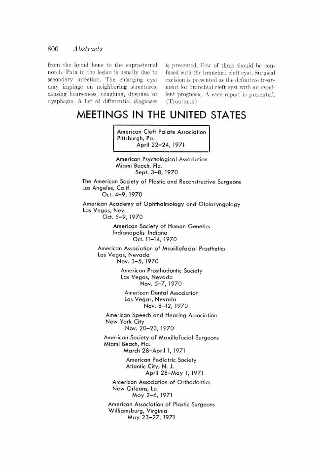
MEETINGS IN THE UNITED STATES
Pittsburgh, Pa.
American Cleft Palate Association
April 22-24, 1971
American Psychological AssociationMiami Beach, Fla.
Sept. 3-8, 1970
The American Society of Plastic and Reconstructive SurgeonsLos Angeles, Calif.
Oct. 4-9, 1970
American Academy of Ophthalmology and OtolaryngologyLas Vegas, Nev.
Oct. 5-9, 1970
American Society of Human GeneticsIndianapolis, Indiana
Oct. 11-14, 1970
American Association of Maxillofacial ProstheticsLas Vegas, Nevada
Nov. 3-5, 1970
American Prosthodontic SocietyLas Vegas, Nevada
Nov. 5-7, 1970
American Dental AssociationLas Vegas, Nevada
Nov. 8-12, 1970
American Speech and Hearing AssociationNew York City
Nov. 20-23, 1970
American Society of Maxillofacial SurgeonsMiami Beach, Fla.
March 28-April 1, 1971
American Pediatric SocietyAtlantic City, N. J.
April 28-May 1, 197]
American Association of OrthodonticsNew Orleans, La.
May 2-6, 197]
American Association of Plastic SurgeonsWilliamsburg, Virginia
May 23-27, 1971
ANNOUNCEMENTS
NEW RESEARCH CENTER AND CLEFT
PALATE CLINIC IN PUERTO RICO
A Combined Research Center and Cleft Palate Clinic has been estab-
lished at the Mayaguez Medical Center in Mayaguez, Puerto Rico. Mem-
bers of the team are: Angela Ramirez-Irizarry, M.D., Plastic Surgery and
Chairman; Luis Nina-Ortega, M.D., Pediatrics; Guillermo Colon-Bonet,
D.D.S., Dentist; Pedro E. Valentin, D.D.S., M.S., Orthodontia; Newton
Martin Ellis, M.D., Otolaryngology; José E. Garcia, M.D., Pediatric
Anesthesia; Raul Maldonado-Sierra, M.D., Physical Medicine and Reha-
bilitation; Miss Margarita Mendez, M.S., Speech Therapy and Audiology ;
Miss Carment Pagin, Medical Social Worker; and Mr. William Perez,
Photographer. Associate members of the team are: Ramon Cabafias,
D.D.S., M.S., Oral Surgery; Donald Steed, D.D.S., Oral Surgery and
William G. Sprague, D.D .S., M.S., Oral Pathology.
14TH ANNUAL SEMINAR TO BE HELD
BY LANCASTER CLEFT PALATE CLINIC
"The Lancaster Cleft Palate Clinic will conduct its Fourteenth Annual
Seminar-HABILITATION-REHABILITATION OF ORAL-FACIAL-
COMMUNICATIVE DISORDERS-October 19-23, 1970. Multidiscipli-
nary diagnostic and treatment procedures will be presented to provide
more comprehensive information about treatment services.
The program will include demonstrations by the staff plastic surgeon,
pediatrician, otolaryngologist, orthodontist, prosthodontist, speech pa-
thologist, audiologist, psychologist and social worker. Emphasis will be
placed upon clinical application and research findings effecting the treat-
ment program.
Discussion periods with each discipline and general meetings will be
held daily regarding orientation and integration of multidisciplinary serv-
ices, parent and patient counseling concerning genetic traits; psychological
behavior of patient within the family environment; role of social case
worker in clinical setting; Pennsylvania's Department of Health, Cleft
Palate Section and Pennsylvania's Bureau of Vocational Rehabilitation
will comprise special sessions.
TUITION: $300.00
Program limited to 30 trainees.
For further information, write to Robert T. Millard, Program Director,
801
802 Announcements
Lancaster Cleft Palate Clinic, 24 N. Lime Street, Lancaster, Pennsylvania
17602."
CLARION STATE COLLEGE OFFERS
BIBLIOGRAPHY ON CLEFT PALATE
A Bibliography on Cleft Palate for the ten-year period (1960-69) has
been compiled as a class project in the graduate course in cleft palate
in the Division of Speech Pathology and Audiology, Clarion State College,
Clarion, Pennsylvania. This bibliography contains listings from journals
and books in the fields of dentistry, plastic surgery, speech pathology and
other disciplines concerned with cleft palate. It is limited to references in
the English language. The bibliography is divided into twenty major divi-
sions and the references in each division are arranged alphabetically. The
major purpose of this bibliography was to organize published material
and to facilitate the task of both the student and clinician in locating this
information. This is a well-organized bibliography and information rela-
tive to it can be obtained by writing to Mary Pannbachet, Associate
Professor, Speech and Hearing Clinic, Clarion State College, Clarion,
Pennsylvania 16214.
GRADUATE-RESIDENCY TRAINING PROGRAM IN
PROSTHODONTICS OFFERED AT MAYO CLINIC
The Mayo Graduate School of Medicine and the Department of
Dentistry and Oral Surgery of the Mayo Clinic offer a graduate-residency
training program in prosthodontics leading to a Master of Science Degree
in Dentistry or Certificate of Achievement. Appointments are made in
December for the 36-month course of study in conventional and maxillo-
facial prosthodontics which begins with the following summer or fall
quarter. Didactic courses, clinical and laboratory experience, and practice
teaching satisfy requirements for certification by the American Board of
Prosthodontics. A stipend is provided with annual increments.
Address inquiries to Director, Mayo Graduate School of Medicine, 200
First Street Southwest, Rochester, Minnesota, 55901.