abstracts and application manualdetection of · pdf fileand application manualdetection of...
TRANSCRIPT

Cytogenetic workshop25/10/2006Brno, Czech RepublicILBIT MU Brno, Kamenice 5
Abstracts and Application ManualDetection of chromosomal aberrations in multiple myeloma
Abstracts and Application Manual

Cytogenetic workshop
25/10/2006Brno, Czech Republic, ILBIT MU Brno, Kamenice 5
Cytogenetic workshop25/10/2006Brno, Czech RepublicILBIT MU Brno, Kamenice 5
Detection of chromosomal aberrations in multiple myeloma
Abstracts and Application Manual
Detection of chromosomal aberrations in multiple myeloma
Edited by prof. MUDr. Roman Hajek, CSc.Published by Masaryk University, 20061 st Edition, 2006, 60 copiesDTP: Studio PORT, BrnoPhotography by A PLUS BRNO a.s.Printed by Masaryk University Press, Brno, Czech Republic
55-956B-2006 02/58 9/LFISBN 80-210-4129-3

Cytogenetic workshop
25/10/2006Brno, Czech Republic, ILBIT MU Brno, Kamenice 5
© Masaryk University, 2006ISBN 80-210-4129-3

Cytogenetic workshop
25/10/2006Brno, Czech Republic, ILBIT MU Brno, Kamenice 5
Notes:
. . . . . . . . . . . . . . . . . . . . . . . . . . . . . . . . . . . . . . . . . . . . . . . . . . . . . . . . . . . . . . . . . . . . . . . . . . . . . . . . . . . . . . . . . . . . . . . . . . . . . . . . . . . . . . . . . . . . . . . . . . . . . . . . . . . . . . . . . . . . . . . . . . . . .
. . . . . . . . . . . . . . . . . . . . . . . . . . . . . . . . . . . . . . . . . . . . . . . . . . . . . . . . . . . . . . . . . . . . . . . . . . . . . . . . . . . . . . . . . . . . . . . . . . . . . . . . . . . . . . . . . . . . . . . . . . . . . . . . . . . . . . . . . . . . . . . . . . . . .
. . . . . . . . . . . . . . . . . . . . . . . . . . . . . . . . . . . . . . . . . . . . . . . . . . . . . . . . . . . . . . . . . . . . . . . . . . . . . . . . . . . . . . . . . . . . . . . . . . . . . . . . . . . . . . . . . . . . . . . . . . . . . . . . . . . . . . . . . . . . . . . . . . . . .
. . . . . . . . . . . . . . . . . . . . . . . . . . . . . . . . . . . . . . . . . . . . . . . . . . . . . . . . . . . . . . . . . . . . . . . . . . . . . . . . . . . . . . . . . . . . . . . . . . . . . . . . . . . . . . . . . . . . . . . . . . . . . . . . . . . . . . . . . . . . . . . . . . . . .
. . . . . . . . . . . . . . . . . . . . . . . . . . . . . . . . . . . . . . . . . . . . . . . . . . . . . . . . . . . . . . . . . . . . . . . . . . . . . . . . . . . . . . . . . . . . . . . . . . . . . . . . . . . . . . . . . . . . . . . . . . . . . . . . . . . . . . . . . . . . . . . . . . . . .
. . . . . . . . . . . . . . . . . . . . . . . . . . . . . . . . . . . . . . . . . . . . . . . . . . . . . . . . . . . . . . . . . . . . . . . . . . . . . . . . . . . . . . . . . . . . . . . . . . . . . . . . . . . . . . . . . . . . . . . . . . . . . . . . . . . . . . . . . . . . . . . . . . . . .
. . . . . . . . . . . . . . . . . . . . . . . . . . . . . . . . . . . . . . . . . . . . . . . . . . . . . . . . . . . . . . . . . . . . . . . . . . . . . . . . . . . . . . . . . . . . . . . . . . . . . . . . . . . . . . . . . . . . . . . . . . . . . . . . . . . . . . . . . . . . . . . . . . . . .
. . . . . . . . . . . . . . . . . . . . . . . . . . . . . . . . . . . . . . . . . . . . . . . . . . . . . . . . . . . . . . . . . . . . . . . . . . . . . . . . . . . . . . . . . . . . . . . . . . . . . . . . . . . . . . . . . . . . . . . . . . . . . . . . . . . . . . . . . . . . . . . . . . . . .
. . . . . . . . . . . . . . . . . . . . . . . . . . . . . . . . . . . . . . . . . . . . . . . . . . . . . . . . . . . . . . . . . . . . . . . . . . . . . . . . . . . . . . . . . . . . . . . . . . . . . . . . . . . . . . . . . . . . . . . . . . . . . . . . . . . . . . . . . . . . . . . . . . . . .
. . . . . . . . . . . . . . . . . . . . . . . . . . . . . . . . . . . . . . . . . . . . . . . . . . . . . . . . . . . . . . . . . . . . . . . . . . . . . . . . . . . . . . . . . . . . . . . . . . . . . . . . . . . . . . . . . . . . . . . . . . . . . . . . . . . . . . . . . . . . . . . . . . . . .
. . . . . . . . . . . . . . . . . . . . . . . . . . . . . . . . . . . . . . . . . . . . . . . . . . . . . . . . . . . . . . . . . . . . . . . . . . . . . . . . . . . . . . . . . . . . . . . . . . . . . . . . . . . . . . . . . . . . . . . . . . . . . . . . . . . . . . . . . . . . . . . . . . . . .
. . . . . . . . . . . . . . . . . . . . . . . . . . . . . . . . . . . . . . . . . . . . . . . . . . . . . . . . . . . . . . . . . . . . . . . . . . . . . . . . . . . . . . . . . . . . . . . . . . . . . . . . . . . . . . . . . . . . . . . . . . . . . . . . . . . . . . . . . . . . . . . . . . . . .
. . . . . . . . . . . . . . . . . . . . . . . . . . . . . . . . . . . . . . . . . . . . . . . . . . . . . . . . . . . . . . . . . . . . . . . . . . . . . . . . . . . . . . . . . . . . . . . . . . . . . . . . . . . . . . . . . . . . . . . . . . . . . . . . . . . . . . . . . . . . . . . . . . . . .
. . . . . . . . . . . . . . . . . . . . . . . . . . . . . . . . . . . . . . . . . . . . . . . . . . . . . . . . . . . . . . . . . . . . . . . . . . . . . . . . . . . . . . . . . . . . . . . . . . . . . . . . . . . . . . . . . . . . . . . . . . . . . . . . . . . . . . . . . . . . . . . . . . . . .
. . . . . . . . . . . . . . . . . . . . . . . . . . . . . . . . . . . . . . . . . . . . . . . . . . . . . . . . . . . . . . . . . . . . . . . . . . . . . . . . . . . . . . . . . . . . . . . . . . . . . . . . . . . . . . . . . . . . . . . . . . . . . . . . . . . . . . . . . . . . . . . . . . . . .
. . . . . . . . . . . . . . . . . . . . . . . . . . . . . . . . . . . . . . . . . . . . . . . . . . . . . . . . . . . . . . . . . . . . . . . . . . . . . . . . . . . . . . . . . . . . . . . . . . . . . . . . . . . . . . . . . . . . . . . . . . . . . . . . . . . . . . . . . . . . . . . . . . . . .
. . . . . . . . . . . . . . . . . . . . . . . . . . . . . . . . . . . . . . . . . . . . . . . . . . . . . . . . . . . . . . . . . . . . . . . . . . . . . . . . . . . . . . . . . . . . . . . . . . . . . . . . . . . . . . . . . . . . . . . . . . . . . . . . . . . . . . . . . . . . . . . . . . . . .
. . . . . . . . . . . . . . . . . . . . . . . . . . . . . . . . . . . . . . . . . . . . . . . . . . . . . . . . . . . . . . . . . . . . . . . . . . . . . . . . . . . . . . . . . . . . . . . . . . . . . . . . . . . . . . . . . . . . . . . . . . . . . . . . . . . . . . . . . . . . . . . . . . . . .
. . . . . . . . . . . . . . . . . . . . . . . . . . . . . . . . . . . . . . . . . . . . . . . . . . . . . . . . . . . . . . . . . . . . . . . . . . . . . . . . . . . . . . . . . . . . . . . . . . . . . . . . . . . . . . . . . . . . . . . . . . . . . . . . . . . . . . . . . . . . . . . . . . . . .
. . . . . . . . . . . . . . . . . . . . . . . . . . . . . . . . . . . . . . . . . . . . . . . . . . . . . . . . . . . . . . . . . . . . . . . . . . . . . . . . . . . . . . . . . . . . . . . . . . . . . . . . . . . . . . . . . . . . . . . . . . . . . . . . . . . . . . . . . . . . . . . . . . . . .
. . . . . . . . . . . . . . . . . . . . . . . . . . . . . . . . . . . . . . . . . . . . . . . . . . . . . . . . . . . . . . . . . . . . . . . . . . . . . . . . . . . . . . . . . . . . . . . . . . . . . . . . . . . . . . . . . . . . . . . . . . . . . . . . . . . . . . . . . . . . . . . . . . . . .
. . . . . . . . . . . . . . . . . . . . . . . . . . . . . . . . . . . . . . . . . . . . . . . . . . . . . . . . . . . . . . . . . . . . . . . . . . . . . . . . . . . . . . . . . . . . . . . . . . . . . . . . . . . . . . . . . . . . . . . . . . . . . . . . . . . . . . . . . . . . . . . . . . . . .
. . . . . . . . . . . . . . . . . . . . . . . . . . . . . . . . . . . . . . . . . . . . . . . . . . . . . . . . . . . . . . . . . . . . . . . . . . . . . . . . . . . . . . . . . . . . . . . . . . . . . . . . . . . . . . . . . . . . . . . . . . . . . . . . . . . . . . . . . . . . . . . . . . . . .
. . . . . . . . . . . . . . . . . . . . . . . . . . . . . . . . . . . . . . . . . . . . . . . . . . . . . . . . . . . . . . . . . . . . . . . . . . . . . . . . . . . . . . . . . . . . . . . . . . . . . . . . . . . . . . . . . . . . . . . . . . . . . . . . . . . . . . . . . . . . . . . . . . . . .
. . . . . . . . . . . . . . . . . . . . . . . . . . . . . . . . . . . . . . . . . . . . . . . . . . . . . . . . . . . . . . . . . . . . . . . . . . . . . . . . . . . . . . . . . . . . . . . . . . . . . . . . . . . . . . . . . . . . . . . . . . . . . . . . . . . . . . . . . . . . . . . . . . . . .
. . . . . . . . . . . . . . . . . . . . . . . . . . . . . . . . . . . . . . . . . . . . . . . . . . . . . . . . . . . . . . . . . . . . . . . . . . . . . . . . . . . . . . . . . . . . . . . . . . . . . . . . . . . . . . . . . . . . . . . . . . . . . . . . . . . . . . . . . . . . . . . . . . . . .
. . . . . . . . . . . . . . . . . . . . . . . . . . . . . . . . . . . . . . . . . . . . . . . . . . . . . . . . . . . . . . . . . . . . . . . . . . . . . . . . . . . . . . . . . . . . . . . . . . . . . . . . . . . . . . . . . . . . . . . . . . . . . . . . . . . . . . . . . . . . . . . . . . . . .
. . . . . . . . . . . . . . . . . . . . . . . . . . . . . . . . . . . . . . . . . . . . . . . . . . . . . . . . . . . . . . . . . . . . . . . . . . . . . . . . . . . . . . . . . . . . . . . . . . . . . . . . . . . . . . . . . . . . . . . . . . . . . . . . . . . . . . . . . . . . . . . . . . . . .
Scientific programme:Detection of chromosomal aberrations in multiple myeloma using simultaneous immunofluorescent labelling of malignant plasma cells and fluorescent in situ hybridization (cIg FISH)
9:00 – 9:15 Chairman’s introduction Roman Hajek, Brno, Czech Republic
9:15 – 10:30 Theoretical part of the workshop9:15 Overview - „Genetic lesions and prognosis in multiple myeloma“.
Johannes Drach, Vienna, Austria9:35 Epigenetic approaches in multiple myeloma
Eva Bartova, Brno, Czech Republic9:55 Experience of Czech Myeloma Group
Zuzana Zemanova, Praha, Czech Republic10:15 Technique of Light chain-specific immunofluorescent staining
of clonal plasma cells and FISH analyses (cIg FISH) Hana Filkova, Brno, Czech Republic
10:30 – 10:45 Coffee break
10:45 – 12:30 Practical part of the workshop (part I) Petr Kuglik, Hana Filkova, Renata Kupska, Brno, Czech Republic
1) Fixation of bone marrow for immunofluorescent labelling of plasma cells, slide preparation 2) Sample slides denaturation3) Incubation at 37°C with I. antibody (anti-IgL, anti-IgK)
12:30 – 13:45 Lunch break
13:45 – 16:45 Practical part of the workshop (part II) Petr Kuglik, Pavel Nemec, Romana Zaoralova, Henrieta Greslikova, Czech Republic
4) Incubation at 37°C with II. antibody (anti-goat) 5) Washing, dehydratation, air drying of slides6) FISH procedure - DNA probe preparation, hybridization at 37°C7) Post-hybridization washing, antifade monting, and scoring of slides prepared one day before8) Examination of abnormal signal pattern with fluorescence microscope,
acquiring of FISH images and image analyses *also continues during the steps 4 - 7
16:45 – 17:00 Chairman’s conclusion Roman Hajek, Brno, Czech Republic

Cytogenetic workshop
25/10/2006Brno, Czech Republic, ILBIT MU Brno, Kamenice 5
Notes:
. . . . . . . . . . . . . . . . . . . . . . . . . . . . . . . . . . . . . . . . . . . . . . . . . . . . . . . . . . . . . . . . . . . . . . . . . . . . . . . . . . . . . . . . . . . . . . . . . . . . . . . . . . . . . . . . . . . . . . . . . . . . . . . . . . . . . . . . . . . . . . . . . . . . .
. . . . . . . . . . . . . . . . . . . . . . . . . . . . . . . . . . . . . . . . . . . . . . . . . . . . . . . . . . . . . . . . . . . . . . . . . . . . . . . . . . . . . . . . . . . . . . . . . . . . . . . . . . . . . . . . . . . . . . . . . . . . . . . . . . . . . . . . . . . . . . . . . . . . .
. . . . . . . . . . . . . . . . . . . . . . . . . . . . . . . . . . . . . . . . . . . . . . . . . . . . . . . . . . . . . . . . . . . . . . . . . . . . . . . . . . . . . . . . . . . . . . . . . . . . . . . . . . . . . . . . . . . . . . . . . . . . . . . . . . . . . . . . . . . . . . . . . . . . .
. . . . . . . . . . . . . . . . . . . . . . . . . . . . . . . . . . . . . . . . . . . . . . . . . . . . . . . . . . . . . . . . . . . . . . . . . . . . . . . . . . . . . . . . . . . . . . . . . . . . . . . . . . . . . . . . . . . . . . . . . . . . . . . . . . . . . . . . . . . . . . . . . . . . .
. . . . . . . . . . . . . . . . . . . . . . . . . . . . . . . . . . . . . . . . . . . . . . . . . . . . . . . . . . . . . . . . . . . . . . . . . . . . . . . . . . . . . . . . . . . . . . . . . . . . . . . . . . . . . . . . . . . . . . . . . . . . . . . . . . . . . . . . . . . . . . . . . . . . .
. . . . . . . . . . . . . . . . . . . . . . . . . . . . . . . . . . . . . . . . . . . . . . . . . . . . . . . . . . . . . . . . . . . . . . . . . . . . . . . . . . . . . . . . . . . . . . . . . . . . . . . . . . . . . . . . . . . . . . . . . . . . . . . . . . . . . . . . . . . . . . . . . . . . .
. . . . . . . . . . . . . . . . . . . . . . . . . . . . . . . . . . . . . . . . . . . . . . . . . . . . . . . . . . . . . . . . . . . . . . . . . . . . . . . . . . . . . . . . . . . . . . . . . . . . . . . . . . . . . . . . . . . . . . . . . . . . . . . . . . . . . . . . . . . . . . . . . . . . .
. . . . . . . . . . . . . . . . . . . . . . . . . . . . . . . . . . . . . . . . . . . . . . . . . . . . . . . . . . . . . . . . . . . . . . . . . . . . . . . . . . . . . . . . . . . . . . . . . . . . . . . . . . . . . . . . . . . . . . . . . . . . . . . . . . . . . . . . . . . . . . . . . . . . .
. . . . . . . . . . . . . . . . . . . . . . . . . . . . . . . . . . . . . . . . . . . . . . . . . . . . . . . . . . . . . . . . . . . . . . . . . . . . . . . . . . . . . . . . . . . . . . . . . . . . . . . . . . . . . . . . . . . . . . . . . . . . . . . . . . . . . . . . . . . . . . . . . . . . .
. . . . . . . . . . . . . . . . . . . . . . . . . . . . . . . . . . . . . . . . . . . . . . . . . . . . . . . . . . . . . . . . . . . . . . . . . . . . . . . . . . . . . . . . . . . . . . . . . . . . . . . . . . . . . . . . . . . . . . . . . . . . . . . . . . . . . . . . . . . . . . . . . . . . .
. . . . . . . . . . . . . . . . . . . . . . . . . . . . . . . . . . . . . . . . . . . . . . . . . . . . . . . . . . . . . . . . . . . . . . . . . . . . . . . . . . . . . . . . . . . . . . . . . . . . . . . . . . . . . . . . . . . . . . . . . . . . . . . . . . . . . . . . . . . . . . . . . . . . .
. . . . . . . . . . . . . . . . . . . . . . . . . . . . . . . . . . . . . . . . . . . . . . . . . . . . . . . . . . . . . . . . . . . . . . . . . . . . . . . . . . . . . . . . . . . . . . . . . . . . . . . . . . . . . . . . . . . . . . . . . . . . . . . . . . . . . . . . . . . . . . . . . . . . .
. . . . . . . . . . . . . . . . . . . . . . . . . . . . . . . . . . . . . . . . . . . . . . . . . . . . . . . . . . . . . . . . . . . . . . . . . . . . . . . . . . . . . . . . . . . . . . . . . . . . . . . . . . . . . . . . . . . . . . . . . . . . . . . . . . . . . . . . . . . . . . . . . . . . .
. . . . . . . . . . . . . . . . . . . . . . . . . . . . . . . . . . . . . . . . . . . . . . . . . . . . . . . . . . . . . . . . . . . . . . . . . . . . . . . . . . . . . . . . . . . . . . . . . . . . . . . . . . . . . . . . . . . . . . . . . . . . . . . . . . . . . . . . . . . . . . . . . . . . .
. . . . . . . . . . . . . . . . . . . . . . . . . . . . . . . . . . . . . . . . . . . . . . . . . . . . . . . . . . . . . . . . . . . . . . . . . . . . . . . . . . . . . . . . . . . . . . . . . . . . . . . . . . . . . . . . . . . . . . . . . . . . . . . . . . . . . . . . . . . . . . . . . . . . .
. . . . . . . . . . . . . . . . . . . . . . . . . . . . . . . . . . . . . . . . . . . . . . . . . . . . . . . . . . . . . . . . . . . . . . . . . . . . . . . . . . . . . . . . . . . . . . . . . . . . . . . . . . . . . . . . . . . . . . . . . . . . . . . . . . . . . . . . . . . . . . . . . . . . .
. . . . . . . . . . . . . . . . . . . . . . . . . . . . . . . . . . . . . . . . . . . . . . . . . . . . . . . . . . . . . . . . . . . . . . . . . . . . . . . . . . . . . . . . . . . . . . . . . . . . . . . . . . . . . . . . . . . . . . . . . . . . . . . . . . . . . . . . . . . . . . . . . . . . .
. . . . . . . . . . . . . . . . . . . . . . . . . . . . . . . . . . . . . . . . . . . . . . . . . . . . . . . . . . . . . . . . . . . . . . . . . . . . . . . . . . . . . . . . . . . . . . . . . . . . . . . . . . . . . . . . . . . . . . . . . . . . . . . . . . . . . . . . . . . . . . . . . . . . .
. . . . . . . . . . . . . . . . . . . . . . . . . . . . . . . . . . . . . . . . . . . . . . . . . . . . . . . . . . . . . . . . . . . . . . . . . . . . . . . . . . . . . . . . . . . . . . . . . . . . . . . . . . . . . . . . . . . . . . . . . . . . . . . . . . . . . . . . . . . . . . . . . . . . .
. . . . . . . . . . . . . . . . . . . . . . . . . . . . . . . . . . . . . . . . . . . . . . . . . . . . . . . . . . . . . . . . . . . . . . . . . . . . . . . . . . . . . . . . . . . . . . . . . . . . . . . . . . . . . . . . . . . . . . . . . . . . . . . . . . . . . . . . . . . . . . . . . . . . .
. . . . . . . . . . . . . . . . . . . . . . . . . . . . . . . . . . . . . . . . . . . . . . . . . . . . . . . . . . . . . . . . . . . . . . . . . . . . . . . . . . . . . . . . . . . . . . . . . . . . . . . . . . . . . . . . . . . . . . . . . . . . . . . . . . . . . . . . . . . . . . . . . . . . .
. . . . . . . . . . . . . . . . . . . . . . . . . . . . . . . . . . . . . . . . . . . . . . . . . . . . . . . . . . . . . . . . . . . . . . . . . . . . . . . . . . . . . . . . . . . . . . . . . . . . . . . . . . . . . . . . . . . . . . . . . . . . . . . . . . . . . . . . . . . . . . . . . . . . .
. . . . . . . . . . . . . . . . . . . . . . . . . . . . . . . . . . . . . . . . . . . . . . . . . . . . . . . . . . . . . . . . . . . . . . . . . . . . . . . . . . . . . . . . . . . . . . . . . . . . . . . . . . . . . . . . . . . . . . . . . . . . . . . . . . . . . . . . . . . . . . . . . . . . .
. . . . . . . . . . . . . . . . . . . . . . . . . . . . . . . . . . . . . . . . . . . . . . . . . . . . . . . . . . . . . . . . . . . . . . . . . . . . . . . . . . . . . . . . . . . . . . . . . . . . . . . . . . . . . . . . . . . . . . . . . . . . . . . . . . . . . . . . . . . . . . . . . . . . .
. . . . . . . . . . . . . . . . . . . . . . . . . . . . . . . . . . . . . . . . . . . . . . . . . . . . . . . . . . . . . . . . . . . . . . . . . . . . . . . . . . . . . . . . . . . . . . . . . . . . . . . . . . . . . . . . . . . . . . . . . . . . . . . . . . . . . . . . . . . . . . . . . . . . .
. . . . . . . . . . . . . . . . . . . . . . . . . . . . . . . . . . . . . . . . . . . . . . . . . . . . . . . . . . . . . . . . . . . . . . . . . . . . . . . . . . . . . . . . . . . . . . . . . . . . . . . . . . . . . . . . . . . . . . . . . . . . . . . . . . . . . . . . . . . . . . . . . . . . .
. . . . . . . . . . . . . . . . . . . . . . . . . . . . . . . . . . . . . . . . . . . . . . . . . . . . . . . . . . . . . . . . . . . . . . . . . . . . . . . . . . . . . . . . . . . . . . . . . . . . . . . . . . . . . . . . . . . . . . . . . . . . . . . . . . . . . . . . . . . . . . . . . . . . .
. . . . . . . . . . . . . . . . . . . . . . . . . . . . . . . . . . . . . . . . . . . . . . . . . . . . . . . . . . . . . . . . . . . . . . . . . . . . . . . . . . . . . . . . . . . . . . . . . . . . . . . . . . . . . . . . . . . . . . . . . . . . . . . . . . . . . . . . . . . . . . . . . . . . .
. . . . . . . . . . . . . . . . . . . . . . . . . . . . . . . . . . . . . . . . . . . . . . . . . . . . . . . . . . . . . . . . . . . . . . . . . . . . . . . . . . . . . . . . . . . . . . . . . . . . . . . . . . . . . . . . . . . . . . . . . . . . . . . . . . . . . . . . . . . . . . . . . . . . .
Abstracts of lectures
CLINICAL IMPLICATIONS OF CYTOGENETIC ABNORMALITIES IN MULTIPLE MYELOMA
Dr. Johannes DrachMedical University Vienna, Department of Medicine I, Clinical Division of Oncology Waehringer Guertel 18-20, A-1090 Vienna, Austria
Phone: +43-1-40400-5457, FAX: +43-1-40400-4461, E-Mail: [email protected]
It is well recognized that multiple myeloma (MM) is a B-cell malignancy with great variability in clinical outcome: Medi-an survival times are approximately 3 years with standard-dose therapy and about 4 to 5 years with intensive treatment programs, but survival may range between only a few months and more than 10 years. Therefore, it has been a relevant issue to identify prognostic indicators for the estimation of the individual patient’s outcome. Knowledge of such factors is critical not only for an improved understanding of disease outcome, but also for the development of strategies to opti-mize treatment, particularly with the aim of risk-adapted therapies. The latter aspect has gained substantial importance due to the availability of “novel” agents for MM therapy.
Standard clinical and laboratory factorsIn 1975, Durie and Salmon proposed a staging system based upon readily available clinical parameters (serum
hemoglobin, size of the paraprotein, serum calcium, and number of osteolytic bone lesions by skeletal radiography).1
The Durie & Salmon staging system, which correlated with tumor burden and survival, was widely used despite its limi-tations, in particular with respect to the definition of bone lesions. Therefore, the search for more accurate prognostic factors continued, and several studies identified serum beta-2-microglobulin (ß2-M) as a powerful prognostic indicator for survival.2-5 However, cut-off levels as well as additional parameters that could be combined with ß2-M remained a matter of controversy. As summarized in Table 1, factors related with demographics, features of the tumor itself, and laboratory abnormalities were associated with poor outcome in patients with MM at presentation.2-9 Combinations of parameters were proposed for staging and prognosis, but none of the models turned out to be superior to the Durie & Salmon staging system.
International Staging System (ISS) for MM. This background provided the basis for an international cooperative project aimed at the identification of a simple
and reliable staging system for MM. Clinical and laboratory parameters from 10750 previously untreated, symptomatic patients with MM were collected (69.1% from clinical trial data). The most powerful classification system was obtained by a combination of serum ß2-M and serum albumin (Table 2).10 This ISS-system was validated in various MM patient populations: It was found to be effective in MM patients independent of age (less or more than 65 years of age), type of therapy (standard dose or autologous transplantation) and geographic region (North America, Europe, and Asia). By now, it is suggested to use the ISS staging system, particularly in the setting of clinical trials. An improved definition of patients at risk is expected in the future by incorporation of genetic and proteomic data.

Cytogenetic workshop
25/10/2006Brno, Czech Republic, ILBIT MU Brno, Kamenice 5
Notes:
. . . . . . . . . . . . . . . . . . . . . . . . . . . . . . . . . . . . . . . . . . . . . . . . . . . . . . . . . . . . . . . . . . . . . . . . . . . . . . . . . . . . . . . . . . . . . . . . . . . . . . . . . . . . . . . . . . . . . . . . . . . . . . . . . . . . . . . . . . . . . . . . . . . . .
. . . . . . . . . . . . . . . . . . . . . . . . . . . . . . . . . . . . . . . . . . . . . . . . . . . . . . . . . . . . . . . . . . . . . . . . . . . . . . . . . . . . . . . . . . . . . . . . . . . . . . . . . . . . . . . . . . . . . . . . . . . . . . . . . . . . . . . . . . . . . . . . . . . . .
. . . . . . . . . . . . . . . . . . . . . . . . . . . . . . . . . . . . . . . . . . . . . . . . . . . . . . . . . . . . . . . . . . . . . . . . . . . . . . . . . . . . . . . . . . . . . . . . . . . . . . . . . . . . . . . . . . . . . . . . . . . . . . . . . . . . . . . . . . . . . . . . . . . . .
. . . . . . . . . . . . . . . . . . . . . . . . . . . . . . . . . . . . . . . . . . . . . . . . . . . . . . . . . . . . . . . . . . . . . . . . . . . . . . . . . . . . . . . . . . . . . . . . . . . . . . . . . . . . . . . . . . . . . . . . . . . . . . . . . . . . . . . . . . . . . . . . . . . . .
. . . . . . . . . . . . . . . . . . . . . . . . . . . . . . . . . . . . . . . . . . . . . . . . . . . . . . . . . . . . . . . . . . . . . . . . . . . . . . . . . . . . . . . . . . . . . . . . . . . . . . . . . . . . . . . . . . . . . . . . . . . . . . . . . . . . . . . . . . . . . . . . . . . . .
. . . . . . . . . . . . . . . . . . . . . . . . . . . . . . . . . . . . . . . . . . . . . . . . . . . . . . . . . . . . . . . . . . . . . . . . . . . . . . . . . . . . . . . . . . . . . . . . . . . . . . . . . . . . . . . . . . . . . . . . . . . . . . . . . . . . . . . . . . . . . . . . . . . . .
. . . . . . . . . . . . . . . . . . . . . . . . . . . . . . . . . . . . . . . . . . . . . . . . . . . . . . . . . . . . . . . . . . . . . . . . . . . . . . . . . . . . . . . . . . . . . . . . . . . . . . . . . . . . . . . . . . . . . . . . . . . . . . . . . . . . . . . . . . . . . . . . . . . . .
. . . . . . . . . . . . . . . . . . . . . . . . . . . . . . . . . . . . . . . . . . . . . . . . . . . . . . . . . . . . . . . . . . . . . . . . . . . . . . . . . . . . . . . . . . . . . . . . . . . . . . . . . . . . . . . . . . . . . . . . . . . . . . . . . . . . . . . . . . . . . . . . . . . . .
. . . . . . . . . . . . . . . . . . . . . . . . . . . . . . . . . . . . . . . . . . . . . . . . . . . . . . . . . . . . . . . . . . . . . . . . . . . . . . . . . . . . . . . . . . . . . . . . . . . . . . . . . . . . . . . . . . . . . . . . . . . . . . . . . . . . . . . . . . . . . . . . . . . . .
. . . . . . . . . . . . . . . . . . . . . . . . . . . . . . . . . . . . . . . . . . . . . . . . . . . . . . . . . . . . . . . . . . . . . . . . . . . . . . . . . . . . . . . . . . . . . . . . . . . . . . . . . . . . . . . . . . . . . . . . . . . . . . . . . . . . . . . . . . . . . . . . . . . . .
. . . . . . . . . . . . . . . . . . . . . . . . . . . . . . . . . . . . . . . . . . . . . . . . . . . . . . . . . . . . . . . . . . . . . . . . . . . . . . . . . . . . . . . . . . . . . . . . . . . . . . . . . . . . . . . . . . . . . . . . . . . . . . . . . . . . . . . . . . . . . . . . . . . . .
. . . . . . . . . . . . . . . . . . . . . . . . . . . . . . . . . . . . . . . . . . . . . . . . . . . . . . . . . . . . . . . . . . . . . . . . . . . . . . . . . . . . . . . . . . . . . . . . . . . . . . . . . . . . . . . . . . . . . . . . . . . . . . . . . . . . . . . . . . . . . . . . . . . . .
. . . . . . . . . . . . . . . . . . . . . . . . . . . . . . . . . . . . . . . . . . . . . . . . . . . . . . . . . . . . . . . . . . . . . . . . . . . . . . . . . . . . . . . . . . . . . . . . . . . . . . . . . . . . . . . . . . . . . . . . . . . . . . . . . . . . . . . . . . . . . . . . . . . . .
. . . . . . . . . . . . . . . . . . . . . . . . . . . . . . . . . . . . . . . . . . . . . . . . . . . . . . . . . . . . . . . . . . . . . . . . . . . . . . . . . . . . . . . . . . . . . . . . . . . . . . . . . . . . . . . . . . . . . . . . . . . . . . . . . . . . . . . . . . . . . . . . . . . . .
. . . . . . . . . . . . . . . . . . . . . . . . . . . . . . . . . . . . . . . . . . . . . . . . . . . . . . . . . . . . . . . . . . . . . . . . . . . . . . . . . . . . . . . . . . . . . . . . . . . . . . . . . . . . . . . . . . . . . . . . . . . . . . . . . . . . . . . . . . . . . . . . . . . . .
. . . . . . . . . . . . . . . . . . . . . . . . . . . . . . . . . . . . . . . . . . . . . . . . . . . . . . . . . . . . . . . . . . . . . . . . . . . . . . . . . . . . . . . . . . . . . . . . . . . . . . . . . . . . . . . . . . . . . . . . . . . . . . . . . . . . . . . . . . . . . . . . . . . . .
. . . . . . . . . . . . . . . . . . . . . . . . . . . . . . . . . . . . . . . . . . . . . . . . . . . . . . . . . . . . . . . . . . . . . . . . . . . . . . . . . . . . . . . . . . . . . . . . . . . . . . . . . . . . . . . . . . . . . . . . . . . . . . . . . . . . . . . . . . . . . . . . . . . . .
. . . . . . . . . . . . . . . . . . . . . . . . . . . . . . . . . . . . . . . . . . . . . . . . . . . . . . . . . . . . . . . . . . . . . . . . . . . . . . . . . . . . . . . . . . . . . . . . . . . . . . . . . . . . . . . . . . . . . . . . . . . . . . . . . . . . . . . . . . . . . . . . . . . . .
. . . . . . . . . . . . . . . . . . . . . . . . . . . . . . . . . . . . . . . . . . . . . . . . . . . . . . . . . . . . . . . . . . . . . . . . . . . . . . . . . . . . . . . . . . . . . . . . . . . . . . . . . . . . . . . . . . . . . . . . . . . . . . . . . . . . . . . . . . . . . . . . . . . . .
. . . . . . . . . . . . . . . . . . . . . . . . . . . . . . . . . . . . . . . . . . . . . . . . . . . . . . . . . . . . . . . . . . . . . . . . . . . . . . . . . . . . . . . . . . . . . . . . . . . . . . . . . . . . . . . . . . . . . . . . . . . . . . . . . . . . . . . . . . . . . . . . . . . . .
. . . . . . . . . . . . . . . . . . . . . . . . . . . . . . . . . . . . . . . . . . . . . . . . . . . . . . . . . . . . . . . . . . . . . . . . . . . . . . . . . . . . . . . . . . . . . . . . . . . . . . . . . . . . . . . . . . . . . . . . . . . . . . . . . . . . . . . . . . . . . . . . . . . . .
. . . . . . . . . . . . . . . . . . . . . . . . . . . . . . . . . . . . . . . . . . . . . . . . . . . . . . . . . . . . . . . . . . . . . . . . . . . . . . . . . . . . . . . . . . . . . . . . . . . . . . . . . . . . . . . . . . . . . . . . . . . . . . . . . . . . . . . . . . . . . . . . . . . . .
. . . . . . . . . . . . . . . . . . . . . . . . . . . . . . . . . . . . . . . . . . . . . . . . . . . . . . . . . . . . . . . . . . . . . . . . . . . . . . . . . . . . . . . . . . . . . . . . . . . . . . . . . . . . . . . . . . . . . . . . . . . . . . . . . . . . . . . . . . . . . . . . . . . . .
. . . . . . . . . . . . . . . . . . . . . . . . . . . . . . . . . . . . . . . . . . . . . . . . . . . . . . . . . . . . . . . . . . . . . . . . . . . . . . . . . . . . . . . . . . . . . . . . . . . . . . . . . . . . . . . . . . . . . . . . . . . . . . . . . . . . . . . . . . . . . . . . . . . . .
. . . . . . . . . . . . . . . . . . . . . . . . . . . . . . . . . . . . . . . . . . . . . . . . . . . . . . . . . . . . . . . . . . . . . . . . . . . . . . . . . . . . . . . . . . . . . . . . . . . . . . . . . . . . . . . . . . . . . . . . . . . . . . . . . . . . . . . . . . . . . . . . . . . . .
. . . . . . . . . . . . . . . . . . . . . . . . . . . . . . . . . . . . . . . . . . . . . . . . . . . . . . . . . . . . . . . . . . . . . . . . . . . . . . . . . . . . . . . . . . . . . . . . . . . . . . . . . . . . . . . . . . . . . . . . . . . . . . . . . . . . . . . . . . . . . . . . . . . . .
. . . . . . . . . . . . . . . . . . . . . . . . . . . . . . . . . . . . . . . . . . . . . . . . . . . . . . . . . . . . . . . . . . . . . . . . . . . . . . . . . . . . . . . . . . . . . . . . . . . . . . . . . . . . . . . . . . . . . . . . . . . . . . . . . . . . . . . . . . . . . . . . . . . . .
. . . . . . . . . . . . . . . . . . . . . . . . . . . . . . . . . . . . . . . . . . . . . . . . . . . . . . . . . . . . . . . . . . . . . . . . . . . . . . . . . . . . . . . . . . . . . . . . . . . . . . . . . . . . . . . . . . . . . . . . . . . . . . . . . . . . . . . . . . . . . . . . . . . . .
. . . . . . . . . . . . . . . . . . . . . . . . . . . . . . . . . . . . . . . . . . . . . . . . . . . . . . . . . . . . . . . . . . . . . . . . . . . . . . . . . . . . . . . . . . . . . . . . . . . . . . . . . . . . . . . . . . . . . . . . . . . . . . . . . . . . . . . . . . . . . . . . . . . . .
Genetics and prognosis in MM
PloidyCytogenetic and molecular genetic investigations of MM cells have provided evidence that virtually all cases of MM
are characterized by chromosomal abnormalities.11 Karyotypes from MM cells are usually very complex, but careful analyses of large series have demonstrated that MM can be subdivided into two cytogenetic categories: The hypo-diploid/pseudodiploid category (which also includes the near-tetraploid karyotypes) and the hyperdiploid category. This observation was extended by recent data obtained by fluorescence in situ hybridization (FISH) indicating presence of hyperdiploid and non-hyperdiploid MM variants. The hyperdiploid subtype is defined by presence of multiple trisomic chromosomes (most commonly chromosomes 3, 5, 7, 9, 11, 15, 19, and 21), but a low frequency of IgH translocations. In contrast, non-hyperdiploid MM is characterized by a high frequency of IgH-translocations and frequent loss of chromo-somes, especially chromosomes 13, 14, 16, and 8.11,12 Recognition of hypodiploid MM is also of clinical significance, since MM patients of this category have a particularly unfavorable prognosis.13
IgH-translocationsOne of the most frequent structural abnormalities observed in MM karyotypes involves the Ig heavy-chain (IgH)
gene locus on 14q32, which is usually part of a translocation. Heterogeneous translocation partners have been described, with 11q13, 4p16.3, 16q23, 20q11 and 6p21 being recurrently involved in 14q32 translocations of primary MM tumor specimens.11 These 5 types of primary IgH-translocations, which are mutually exclusive, comprise about 60% of all IgH-translocations, and are mediated primarily by errors during IgH switch recombination. With respect to biology and prognosis, relevant correlations have emerged: The t(11;14)(q13;q32) resulting in upregulation of cyclin-D1 was origianally thought to characterize a favorable group of patients, in particular when treated with intensive therapy.14
However, most recent results suggest that a t(11;14) does not affect event-free and overall survival,15-17 whereas pre-sence of a t(4;14)(p16;q32) or a t(14;16)(q32;q23) identifies a subset of MM patients with short survival, even in the context of autologous transplantation.14-18 Translocations t(4;14) and t(14;16) are also highly correlated with a deletion of chromosome 13q.
Deletion of chromosome 13qBy metaphase cytogenetics, a chromosome 13q abnormality can be found in about 15% of MM patients at diagnosis,
whereas interphase FISH studies have shown a higher frequency of 13q deletions in MM, occurring in 39 – 54% of newly diagnosed cases. Several studies have reported a strong association of a deletion 13q with an unfavorable prognosis of MM patients (summarized in 19). It appears that chromosome 13 abnormalities are a more powerful predictor of poor outcome when identified by karyotyping.20 The negative prognostic impact of a deletion 13q seems to persist even in the context of allogeneic stem cell transplantation.21

Cytogenetic workshop
25/10/2006Brno, Czech Republic, ILBIT MU Brno, Kamenice 5
List of participants:
Name Centre E-mail
Bartova Eva, RNDr., PhD. Institute of Biophysics AV ČR, Brno, CR [email protected]
Drach Johannes, prof., MD Medical University Vienna, Dept. of Medicina I,Clinical Division of Oncology, Vienna, Austria [email protected]
Filkova Hana, Mgr. Faculty Hospital, Dept. of Medical Genetics, Brno, CR [email protected]
Greslikova Henrieta, RNDr. Campus MU, Myeloma Basic Research Centre, Brno, CR [email protected]
Hanusovska Eva, RNDr.,PhD. National Cancer Institute,Dept. of Cancer Genetics, Bratislava, SK [email protected]
Hajek Roman, prof.,MD, CSc. Medical Faculty and Campus MU,Myeloma Basic Research Centre, Brno, CR [email protected]
Holubova Vladimira, Mgr. Oncology Centre J.G. Mendla, Nový Jičín, CR [email protected]
Ilencikova Denisa, PhD, MD National Cancer Institute,Dept. of Cancer Genetics, Bratislava, SK [email protected]
Kirschnerova Gabriela, PhD. Genetic Laboratory MEDIREX, Bratislava, SK [email protected]
Kupska Renata Faculty Hospital, Dept. of Medical Genetics, Brno, CR
Kuglik Petr, doc., RNDr., CSc. Faculty Hospital, Dept. of Medical Genetics, Brno, CR [email protected]
Lueff Sandor, MD National Medical Centre, BMT Unit, Budapest, Hungary [email protected]
Michalova Kyra, prof., DrSc. Oncology Cytogenetic Centre, Faculty Hospital, Praha, CR [email protected]
Nemec Pavel, Bc. Campus MU, Myeloma Basic Research Centre, Brno, CR [email protected]
Petrovicova Gabriela, Ing. Campus MU, Myeloma Basic Research Centre, Brno, CR [email protected]
Pucek Malgorzata, Mgr. University of Medical Science,Dept. of Hematology, Poznan, Poland [email protected]
Sobotka Jiri, RNDr. Oncology Centre J.G. Mendla, Nový Jičín, CR [email protected]
Tothova Andrea, RNDr. Genetic Laboratory MEDIREX, Bratislava, SK [email protected]
Vigasova Jana, Mgr. Campus MU, Myeloma Basic Research Centre, Brno, CR [email protected]
Zaoralova Romana, Mgr. Campus MU, Myeloma Basic Research Centre, Brno, CR [email protected]
Zemanova Zuzana, RNDr., CSc. Oncology Cytogenetic Centre,Faculty Hospital, Praha, CR [email protected]
Zakovicova Alena, Mgr. National Cancer Institute,Dept. of Cancer Genetics, Bratislava, SK [email protected]
Additional chromosomal aberrationsClinical importance was reported for deletions of 17p13 at the TP53 locus, with similar observations for patients
receiving standard-dose and high-dose therapy.15-17,22 Comprehensive analyses of cytogenetic abnormalities in MMidentified patients with a t(4;14) and/or 17p-deletion as the group of patients with the worst prognosis suggesting thatnovel approaches are required for the treatment of such high-risk patients.
Studies done by the Arkansas group identified a region on chromosome 1, which was linked with an aggressiveclinical course in MM: Global gene expression profiling on plasma cells from newly diagnosed patients treated withautologous transplantation revealed a significant over-representation of chromosome 1 genes in a group of about 70genes whose expression was associated with poor outcome. Further analyses showed that overexpression of CKS1B wasstrongly correlated with a gain of DNA copy numbers at chromosomal region 1q21, and that this abnormality conferreda poor prognosis.23 As a possible mechanism, reduced levels of p27Kip1 protein were observed in cases with 1q21 amplifi-cation, suggesting dysregulated cell cycle control in these cases.
Gene expression profiling in MMToday, genome-wide gene expression profiling based on DNA microarrays represents one of the most powerful tools
in the area of genomics. This technique has become feasible and broadly accessible, and in MM it is a valuable tool toidentify all myeloma-specific genetic abnormalities on a single platform.24 When this technique was used to identifygenes associated with therapeutic outcome in 221 patients with previously untreated MM, unsupervised clustering ledto the identification of four distinct MM subgroups.24 Further studies indicated that three genes of this analysis can beused to predict event-free survival. Furthermore, gene expression profiling provided the basis for a novel molecularclassification of MM because overexpression of one of the cyclin-D genes was found to be universal molecular feature ofMM.25 The so-called TC-classification combines the cytogenetic information about the 14q-translocations with cyclin-Dgene expression as summarized in Table 3. Patients of the TC4 and TC5 categories have shortened survival suggestingthat they should be considered for clinical studies exploring investigational therapies.
Impact of novel agents on prognosisBy now, prognostic factors conferring a poor outcome in MM were defined according to the experience with chemothe-
rapy, with no apparent differences between standard-dose and high-dose therapy (compare all studies referenced above).Recent studies have addressed the question whether or not treatment for high-risk patients may be improved by use ofnovel agents.
Thalidomide. Prognostic information is available mainly in patient populations treated with thalidomide in therelapsed/refractory setting. Among 75 patients treated with single agent thalidomide, advanced age (> 65 years), elevatedserum LDH, and elevated serum creatinine were predictive for inferior outcomes.26 In a similar analysis of relapsed MM pati-ents treated with thalidomide-based regimens, elevated serum LDH, advanced ISS-stage, and reduced performance statuswere independent predictive factors for survival.27 Based on these three variables, a scoring system was developed withsurvival times of 38.1, 28.8, and 5.8 months for scores 0, 1, and 2, respectively. The authors concluded that the addition ofLDH and performance status to the prognostic information provided by the ISS may help select patients who will likely derivebenefit from treatment with thalidomide-based regimens.
Cytogenetic workshop
25/10/2006Brno, Czech Republic, ILBIT MU Brno, Kamenice 5
Reporting resultsThere was strong feeling that it is important to know the proportion of plasma cells with the abnormality (particularly for deletion 13). However, there is insufficient evidence yet for the level that should be set to determine prognostic significance. It is therefore recommended that for the present reported results state the percentage of plasma cells with the abnormality.
The workshop did not endorse the use of ISCN 1995 for reporting results as it was felt; that clinicians find this confusing. If laboratories are required to use this for internal reasons they must ensure that there is a very simple clear interpreta-tion given.
Thus results reported to the clinician should be expressed as clearly as possible and must state the percentage of plasma cells involved and what method was used for plasma cell identification e.g.
“13q14 deleted (90%)4p16 t(4;14) single fusion (96%)11q13 normal17p13 (p53) normal
FISH on purified plasma cells identified a t(4;14) in 96% of plasma cells, but only one fusion was seen using a dual fusion probe, suggesting loss of one half of the translocation. From published results this is most likely to be loss of the derived 14 carrying the IgH/FGFR3 fusion. There was also a deletion of chromosome 13 seen in 90% of the plasma cells, but no abnormality of CCND1 or p53 was detected. Although the t(4;14) and deletion 13 have been associated with a poor prognosis in MM the data is not yet good enough to use these results for treatment decisions outwith the context of a clinical trial.”
Further studiesLaboratories taking part in the workshop will try to pool results obtained using these criteria on an annual basis, to get figures for Europe-wide incidence of these abnormalities. Any other laboratories wishing to take part in this exercise should contact Fiona Ross ([email protected]). It is hoped to send control material to participating laboratories to ensure that results from different labs really are comparable.
According to the experience of the Arkansas-Group (phase 2 trial of single agent thalidomide in 169 patients with pretreated MM), favorable survival rates were observed in patients with normal metaphase cytogenetics, low prolifera-tive activity (plasma cell labeling index < 0.5%) and serum ß2-M below 3 mg/L.28 Overall, these results suggested that prognostic factors for treatment with thalidomide are similar to those observed with chemotherapy.
Bortezomib. In patients enrolled into the SUMMIT-trial, potential association between baseline-characteristics and outcome were explored.29 By multivariate analysis, two parameters emerged as being significantly associated with lower response: Age > 65 years and plasma cell infiltration > 50%. Parameters predicting for shortened overall survival were low serum albumin, bone marrow plasma cell infiltration > 50%, and thrombocytopenia. Of particular note, elevated serum ß2-M and presence of a chromosome 13q deletion (tested in a subset of study patients) were not predictive of poor outcome with bortezomib in this clinical trial.
Among patients treated in the APEX trial, a matched-pair analysis was performed between 21 patients with a dele-tion 13q (metaphase analysis) and 41 patients without this deletion.30 Patients were balanced for other adverse pro-gnostic factors including age, lines of prior therapy, ß2-M, and albumin. Presence of a chromosome 13q-deletion was associated with a markedly decreased survival in the dexamethasone-arm; in contrast, in the bortezomib arm, deletion 13q was not associated with a difference in survival or response rate.
In our own analysis of 51 patients with relapsed/refractory MM, treatment with single agent bortezomib resulted in similar response rates and duration of response in patients with and without a chromosome 13q-deletion.31 Serum ß2-M did not emerge as a relevant parameter associated with treatment outcome after bortezomib (lack of prognostic information for response rate, time to treatment failure, and overall survival). Low serum albumin correlated with short time to treatment failure and poor overall survival, and low albumin identified also those patients with a deletion 13q who did not benefint from treatment with bortezomib.
Thus, although additional data from prospective clinical trials are needed, existing data indicate that prognostic factors established from chemotherapy trials cannot be uniformly applied to patient populations treated with bortezo-mib.
Conclusions and future directionsDuring the past decade, considerable progress has been made in our understanding of the molecular basis and biology of MM. Molecular genetic analyses and gene expression profiling have contributed to the recognition of distinct subtypes of MM with different prognosis. Both cytogenetic and molecular findings are correlated with laboratory and clinical characteristics, and we are beginning to use this information as diagnostic and prognostic indicators for the selection of treatment options. Clinical trials are under way to examine the therapeutic efficacy of agents targeting specific molecu-lar defects in myelomatous plasma cells. It is hoped that novel molecular structures will continue to be discovered for specific therapeutic interventions. Standardization of techniques like gene expression profiling will become helpful in predicting response to therapy and eventually tailoring of therapy to specific molecular MM entities. These advances will hopefully result in further improvements of our therapeutic strategies for patients with MM.

Cytogenetic workshop
25/10/2006Brno, Czech Republic, ILBIT MU Brno, Kamenice 5
Probes to use13qThere was no consensus on which probe to use to detect deletion 13, apart from that it should be in band 13q14. Those who had been using a 13q14 and 13q34 probe confirmed published results that ~90% of deletion cases have lost the whole chromosome. (Peter Leibish: can we quote your results from array CGH? eg: Results of array CGH experiments in Ulm show that in the few cases with deletion rather than monosomy 13, there is no consistent minimal region of deletion). Two groups had a comparison of different 13q14 probes, one had <1% discrepancy between RBI and D13S319 in more than 1100 cases and the other had ~1% discrepancy between RBI, D13S319 and D13S25 in over 350 cases. It was therefore agreed that any of these 3 probes is acceptable for testing for 13q deletion. Abbott/Vysis and Qbiogene commercial 13q14 probes are known to be useful in testing for 13q deletions.
IgH translocation probesMany labs had experience of the Abbott/Vysis probes for IgH break-apart and specific translocations. These were all considered acceptable as they cover large areas on the partner chromosomes. We were unable to endorse any other commercial probes due to lack of experience, but any commercial probes giving consistent strong signals are likely to be acceptable. The difference between the Abbott/Vysis dual fusion t(11;14) and dual fusion TX t(11;14) probes was not considered significant. Laboratories using home grown probes are urged to ensure that they cover a large enough area on the donor chromosome : t(4;14) detection in particular is prone to underestimation if the area on 4 encompasses only FGFR3 and not MMSET due to the frequent loss of the derived chromosome 4. Laboratories may employ a hierarchical approach, attempting t(11;14) FISH first and only performing t(4;14) FISH if that is negative (but see requirement for ploidy estimation for “normal” 13q results above).
Abnormal results for the t(11;14) and t(4;14) should state the number of fusion signals seen.
It should be noted that rearrangement of IgH with something other than CCNDI or FGFR3/MMSET cannot be reliably extrapolated from these results as loss of one or other derived chromosome is common. This would result in an appa-rently normal pattern of 2 IgH signals, despite a translocation being present. The presence of 3 IgH signals but no fusion with the t(11;14) and t(4;14) probes probably indicates an alternative IgH translocation rather than trisomy 14 but no lab had data on this. (Forgot to ask at the meeting. Can anyone make a useful comment?).
p53The majority of labs are using the Abbott/Vysis p53 probe. We had insufficient evidence to know whether any other commercial or home-grown probe would give different results.
Other abnormalitiesSome labs are testing for other things as well, the most common of which are t(6;14)(p21;q32), t(14;16) and t(14;20). The use of the Abbott/Vysis 5, 9, 15 probe set is encouraged to see whether hyperdiploidy assessed from these probes in combination with the other recommended abnormalities is good enough to be used as a prognostic marker.
References1. Durie BGM, Salmon SE. A clinial staging system for multiple myeloma. Cancer 1975; 36: 842-854.2. Kyle RA, Gertz MA, Witzig TE, et al. Review of 1027 patients with newly diagnosed multiple myeloma. Mayo Clinic Proc. 2003;
78:21-33.3. Desikan R, Barlogie B, Sawyer J, et al. Results of high-dose therapy for 1000 patients with multiple myeloma: durable
complete remissions and superior survival in the absence of chromosome 13 abnormalities. Blood 2000; 95:4008-4010.4. Facon T, Avet-Loiseau H, Guillerm G, et al. Cromosome 13 abnormalities identified by FISH analysis
and serum beta-2-microglobulin produce a powerful myeloma staging system for patients receiving high-dosetherapy. Blood 2001; 97:1566-1571.
5. Greipp PR, Lust JA, O’Fallon WM, et al. Plasma cell labelling index and beta-2-microglobulin predict survival independent of thymidine kinase and C-reactive protein in multiple myeloma. Blood 1993;81:3382-3387.
6. San Miguel JF, Garcia-Sanz R, Gonzalez M, et al. A new staging system for multiple myeloma based on the number of S-phase plasma cells. Blood 1995;85:448-455.
7. Rajkumar SV, Mesa RA, Fonseca R, et al. Bone marrow angiogenesis in 400 patients with monoclonalgammopathy of undetermined significance, multiple myeloma, and primary amyloidosis. Clin Cancer Res. 2002;62:715-720.
8. Barlogie B, Smallwood L, Smith T, Alexanian R. High serum levels of LDH identify a high-grade lymphoma-like myeloma.Ann Int Med 1989;110:521-525
9. Bataille R, Boccadoro M, Klein M, et al. C-reactive protein and beta-2-microglobulin produce a simple and powerful myeloma staging system. Blood 1992;80:733-737.
10. Greipp PR, San Miguel J, Durie BGM, Crowley JJ, Barlogie B, Blade J, Boccadoro M, Child JA, Avet-Loiseau H, Kyle RA, Lahuerta JL, Ludwig H, Morgan G, Powles R, Shimizu K, Shustik C, Sonneveld P, Tosi P, Turesson I, Westin J. International Staging System for multiple myeloma. J Clin Oncol 2005; 23:3412-3420.
11. Fonseca R, Barlogie B, Bataille R, et al. Genetics and cytogenetics of multiple myeloma: a workshop report. Cancer Res 2004;64:1546-1558.
12. Fonseca R, Debes-Marun CS, Picken EB, Dewald GW, Bryant SC, Winkler JM, et al. The recurrent IgH translocationsare highly associated with non-hyperdiploid variant of multiple myeloma. Blood 2003;102:2562-2567.
13. Smadja NV, Bastard C, Brigaudeau C, et al. Hypodiploidy is a major prognostic factor in multiple myeloma. Blood 2001;98:2229-223814. Moreau P, Facon T, Leleu X, et al. Recurrent 14q32 translocations determine the prognosis of multiple myeloma,
especially in patients receiving intensive chemotherapy. Blood 2002;100:1579-158315. Gertz MA, Lacy MQ, Dispenzieri A, Greipp PR, Litzow MR, Henderson KJ, Van Wier SA, Ahmann GJ, Fonseca R.
Clinical implications of t(11;14)(q13;q32), t(4;14)(p16.3;q32) and –17p13 in myeloma patients treated with high-dose therapy. Blood 2005;106:2837-2840
16. Chang H, Qi XY, Samie S, et al. Genetic risk identifies multiple myeloma patients who do not benefit from autologous stem cell transplantation. Bone Marrow Transplant 2005;36:793-796
17. Avet Loiseau H, Attal M, Moreau P, et al. A comprehensive analysis of cytogenetic abnormalities in myeloma:results of the FISH analysis of 1000 patients enrolled in the IFM99 trials. Blood 2005;106:185a (abstract # 622).
18. Jaksic W, Trudel S, Chang H, Trieau Y, Qi X, Mikhael J, Reece D, Chen C, Stewart AK. Clinical outcome in t(4;14)multiple myeloma: a chemotherapy-sensitive disease characterized by rapid relapse and alkylating agentresistance. J Clin Oncol 2005;23:7069-7073
19. Seidl S, Kaufmann H, Drach J. New insights into the pathophysiology of multiple myeloma. Lancet Oncol 2003;4:557-56420. Shaughnessy J, Tian E, Sawyer J, et al. Prognostic impact of cytogenetics and interphase FISH-defined chromoso
me 13 deletion in multiple myeloma: early results of total therapy II. Br J Haematol 2003; 120:44-5221. Kroeger N, Schilling G, Einsele H, Liebisch P, Shimoni A, Nagler A, et al. Deletion of chromosome band 13q14
as detected by FISH is a prognostic factor in patients with multiple myeloma who are receiving allogeneic dose-reduced stem cell transplantation. Blood 2004;103:4056-4061

Cytogenetic workshop
25/10/2006Brno, Czech Republic, ILBIT MU Brno, Kamenice 5
Thus the following levels are recommended:For dual fusion, break-apart or numerical gains 10%For deletions or single fusion results with dual fusion probes 20%
(Comments on this, in particular, please. We did not mention the cut-off for gains. We did mention the problems with loss of one of the fusions but I do not remember that we set an acceptable cut-off. Is this right?)
Any laboratory setting up myeloma FISH should ensure that their results are compatible with these cut-off levels.
Laboratories with very low mean+3SD for deletions may wish to consider results in the 10 – 20% range to be borderline for their own records but they should not be reported to clinicians as positive.
Control probesIt is recommended that a control probe is used in all experiments where deletions are expected. The type of this probe should be the same as the type of the probe under test; i.e. a centromere probe is not a suitable control for a locus-spe-cific probe. It is not recommended that 13q14 and p53 probes are used to control for each other because of the difficulty of interpreting cases where both are deleted. It is not felt necessary to use control probes for break-apart, dual fusion or trisomy probes as residual non-plasma cells can be used to assess hybridisation efficiency for these.
Number of analystsProvided the previous recommendations are followed all or most of cells being scored will be plasma cells and most of the important abnormalities are likely to be present in the majority of these. Thus a single experienced analyst is consi-dered adequate to examine the majority of cases. However, results should always be checked where there is an equivocal signal pattern or where purified plasma cells make up less than 30% of the cells. Smaller labs are recommended to use 2 analysts with a third to check any results with a discrepancy of >5%.
Number of cell to scoreIt is recommended that 100 cells be scored wherever possible. In exceptional circumstances an abnormal result as few as 20 confirmed plasma cells is acceptable if at least 15 are abnormal.
Abnormalities to test for It is recommended that all labs should test for deletion 13, t(11;14) and t(4;14) and include p53 deletion wherever possible. Where material is limited it is often possible to re-probe slides to increase the number of results obtained. (In practice, several labs are testing for more than this but these four abnormalities are thought to be most practical in a diagnostic setting). Chromosome 13 results must not be reported as normal in the absence of an indication of the ploidy of the sample, as most near tetraploid karyotypes will have deletion of two copies of 13 and therefore give an apparently normal result. The simplest way to obtain this information is to use the results from the t(4;14) and t(11;14) probes; near tetraploid cases are likely to have at least two copies of each of the 4 and 11 probes, although they may not have four IgH signals. Some laboratories prefer to use an IgH break apart probe to decide whether or not to use the t(11;14) and t(4;14) probes. If this is done, they need to remember also to test all cases that do not have an IgH rearrangement but have two copies of the 13q14 probe for t(4;14) and t(11;14) to establish ploidy status.
22. Drach J, Ackermann J, Fritz E, et al. Presence of a p53 gene deletion in patients wih multiple myeloma predicts for shortsurvival after conventional-dose chemotherapy. Blood 1998;92:802-809
23. Shaughnessy J. Amplification and overexpression of CKS1B at chromosome band 1q21 is associated with reduced levels of p27Kip1 and an aggressive clinical course in multiple myeloma. Hematology 2005;10 Suppl 1: 117-126
24. Zhan F, Hardin J, Kordsmeier B, et al. Global gene expression profiling of multiple myeloma, monoclonal gammopathy of undetermined significance, and normal bone marrow plasma cells. Blood 2002;99:1745-1757
25. Bergsagel PL, Kuehl WM, Zhan F, et al. Cyclin D dysregulation: an early and unifying pathogenetic event in multiple myeloma. Blood 2005;106:296-303.
26. Mileshkin L, Biagi JJ, Mitchell P, Underhill C, Grigg A, Bell R, McKendrick J, Briggs P, Seymour JF, Lillie K, Smith JG, Zeldis JB, Prince HM. Multicenter phase 2 trial of thalidomide in relapsed/refractory multiple myeloma: adverse prognostic impact of advanced age. Blood 2003; 102:69-77
27. Anagnostopoulos A, Gika D, Hamilos G, Zervas K, Zomas A, Pouli A, Zorzou M, Kastritis E, Anagnostopoulos N, Tassidopu A, Anagnostou D, Dimopoulos MA. Treatment of relapsed/refractory multiple myeloma with thalidomide-based regimens: identification of prognostic factors. Leuk Lymphoma 2004; 45:2275-2279
28. Barlogie B, Desikan R, Eddlemon P, et al. Extended survival in advanced and refractory multiple myeloma after single-agentthalidomide: identification of prognostic factors in a phase 2 study of 169 patients. Blood 2001; 98:492-494
29. Richardson PG, Barlogie B, Berenson J, et al. Clinical factors predictive of outcome with bortezomib in patients with relapsed, refractory multiple myeloma. Blood 2005; 106: 2977-2981
30. Jagannath S, Richardson PG, Sonneveld P, et al. Bortezomib appears to overcome poor prognosis conferred by chromosome 13deletion in phase 2 and 3 trials. Proceedings of ASCO 2005; 23: 560s (abstract 6501)
31. Drach J, Küenburg E, Sagaster V, Zojer N, Kaufmann H, Ackermann J, Ludwig H. Short survival, despite promising responserates, after bortezomib treatment of multiple myeloma patients with a 13q deletion. Blood 2005;106:152a (abstract 509)

Cytogenetic workshop
25/10/2006Brno, Czech Republic, ILBIT MU Brno, Kamenice 5
To purify or not.The benefits and disadvantages of plasma cell purification versus cIgFISH primarily apply to associated studies: In gene-ral the expense of purification is best justified in the context of plasma cell banking. Johannes Drach reported on a comparison of the two methods which showed no major difference in the results obtained and therefore we recommend that each laboratory chooses the method that is most suitable for their circumstances.
Choice of purification method.All laboratories at the meeting that had experience of plasma cell purification were using Miltenyi Biotec CD138 mag-netic bead separations. One laboratory also had experience of the StemCell Technologies system and reported that there were differences in yield and purity. However, these should not affect the FISH results therefore the choice of purification method can be left to the individual laboratory. Density gradient separation is generally recommended prior to puri-fication, rather than red cell lysis on the grounds of cost; the majority of mature neutrophils will be lost in the former procedure which reduces the quantity of beads necessary for purification. It is stressed that the purified sample MUST be checked for the proportion of plasma cells, as poor initial samples can lead to relatively low proportions in the final cell suspension. Either morphology or immunostaining (flow) can be used to assess the final plasma cell percentage.
Slide making for purified plasma cellsThe purified plasma cell suspension can either be put directly on to slides by cytospin, can be fixed directly in 3:1 met-hanol:acetic acid and either dropped on to slides or stored as a frozen cell suspension, or can be treated with 0.075M KCl and fixed with methanol acetic acid as for standard cytogenetic preparation. The last method produces bare nuclei which can either be dropped on to slides immediately or stored at -20oC until required. (NB one group had had problems with long term storage of some samples, so individual laboratories should check the reliability of storage). It was not considered that any of these methods would significantly affect the final result.
Simultaneous plasma cell identification + FISH.It is recommended that immunostaining for light chains is used to identify the plasma cells. This gives a much stronger signal than CD138 and is also more likely to identify only the malignant clone if there is contamination with normal plasma cells, although it was stressed that the level of such contamination is extremely low at diagnosis.
Slide making for cIgFISHcIgFISH can be used on marrow aspirate smears but only if these are very fresh. It is therefore recommended that wherever possible the cells are subjected to red cell lysis or density gradient centrifugation and the resultant suspension fixed in 3:1 methanol:acetic acid. This fixed suspension can then be dropped directly on to slides or stored at -20oC until required.
Cut-off levels for a positive result.Myeloma FISH is known to be particularly difficult (ref Paris workshop), thought to be due to the additional problems posed by the paraprotein. Ideal control material is difficult to come by. For these reasons the workshop recommends relatively conservative uniform cut-off levels. These are roughly based on the levels found in a number of laboratories using the mean +3SD of 5-10 controls. In practice, there was considerable concern that results just above the actual mean+3SD were artefactual.
Table 1. Summary of prognostic factors
Demographic factors: Advanced age (> 70 years)2
Standard-dose chemotherapy > 12 months3
Features of the tumor clone: IgA isotype3,4
Increased proliferative activity (high labeling index, high S-phase)1,5,6
Chromosomal abnormalities13-23
High microvessel density7
Laboratory abnormalities: Anemia (hemoglobin < 10 g/dL)2
Elevated creatinine2
High serum LDH8
High serum CRP9
Low serum albumin10
High serum beta-2-microglobulin2-5,10

Cytogenetic workshop
25/10/2006Brno, Czech Republic, ILBIT MU Brno, Kamenice 5
Recommendations for FISHin multiple myeloma
These recommendations arose from a workshop organised for the European Myeloma Network, held at the Royal Mar-sden Hospital, London on March 11th 2005. 31 European laboratories were represented at the meeting.
These recommendations are intended to apply only to newly diagnosed cases of myeloma or frank relapse cases. The use of FISH to monitor response to high dose therapy, or to study diseases such as MGUS or primary amyloidosis where only a small proportion of the plasma cells may belong to the abnormal clone is still considered to be a research tool, and different criteria may need to be used.
The purpose of the workshop was to agree rules for FISH in myeloma but consideration was also given to conventional cytogenetic studies. It was agreed that these should not be discouraged but that, especially in a multi-centre setting, full cytogenetic studies were often impracticable due to the poor quality of samples (see below) and the poor ratio of number of man-hours required for the analysis to the number of patients on whom an abnormal result is obtained.
It was felt very strongly that much still needs to be learned about the significance of chromosome abnormalities in myeloma. For this reason, FISH results should not yet be used to make treatment decisions, except in the context of a clinical trial.
Material for FISH.All laboratories, particularly those involved in multi-centre studies, confirmed that there are major problems with the quality of the bone marrow aspirates received for FISH studies; these frequently contain drastically fewer plasma cells than the corresponding smear used for morphological assessment. It is difficult for clinicians to accept that a normal FISH result from a patient that had 80% plasma cells on the morphology slide could be meaningless, but that is the reality. Modern haematologists should accept that diagnosis and management depends on a multidisciplinary approach so that it is important to ensure that there is suitable material for all necessary tests, and morphology is not supreme. Clinicians should therefore be encouraged to send part of the first draw of the aspirate for FISH studies, and should certainly be told that the needle must be repositioned for further aspiration, rather than simply continuing to withdraw marrow blood from the initial puncture site.
Even if these measures are put in place, many samples will still have relatively low plasma cell percentages. For this reason it is not acceptable to report FISH results in myeloma without either concentrating the plasma cells or employing some means of plasma cell identification so that only these cells are scored.
Timing of samples.It is important that aspirates are processed as soon as possible if FISH is to be acceptable in myeloma. Processing, either by purification or by simultaneous staining of cytoplasmic immunoglobulin with FISH (cIgFISH) is time-consuming. It is therefore strongly recommended that marrow aspirates are not performed on a Friday.
Table 2. International Staging System (ISS) for multiple myeloma10
Stage % of patients Features Median survival
I 28 beta-2-microglobulin < 3.5 mg/L 62 months albumin > 3.5 g/dL
II 33 beta-2-microglobulin < 3.5 mg/L 44 months albumin < 3.5 g/dL or beta-2-microglobulin 3.5 – 5.5 mg/L
III 39 beta-2-microglobulin > 5.5 mg/L 29 months
Table 3. TC molecular classification of MM as proposed by Bergsagel and Kuehl25
Group Translocation Gen(s) CyclinD Ploidya %
TC1 t(11;14)(q13;q32) cyclinD1 D1 NH 15t(6;14)/p21;q32) cyclinD3 D3 NH 3
TC2 None None D1 H 37TC3 None None D2 H = NH 22TC4 t(4;14)(p16;q32) fgfr3/mmset D2 NH > H 16TC5 t(14;16)(q32;q23) c-maf D2 NH 5
t(14;20)(q32;q11) mafB D2 NH 2
a NH, non-hyperdiploid; H, hyperdiploid.

Cytogenetic workshop
25/10/2006Brno, Czech Republic, ILBIT MU Brno, Kamenice 5
• MNC in Carnoy’s fixative for FISH: Centrifuge 500 G / 10 min / laboratory temperature Waste supernatant Add a redundant amount of the Carnoy’s solution Resuspend thoroughly Incubate for 1 hour Centrifuge 500 G / 10 min / laboratory temperature Waste supernatant Add the Carnoy’s solution 1,5 – 2 ml Resuspend thoroughly Label the cryotube by the code due to the evidence paper Freeze to – 20 °C Store in an appropriate box
EPIGENETIC APPROACHES IN MULTIPLE MYELOMA
Eva Bartova1, Jana Krejci1, Roman Hajek2, Jana Smejkalova2, Andrea Harnicarova1, Stanislav Kozubek1
1 Institute of Biophysics, Academy of Sciences of the Czech Republic, Královopolská 135, 612 65 Brno, Czech Republic2 Department of Internal Medicine, Hematologic Oncology, Masaryk University Hospital, Jihlavská 20, 625 00 Brno, Czech Republic
Introduction:DNA methylation and posttranslational modifications of N-terminal histone tails are considered to be importantepigenetic markers of chromatin. Tumour cells are characterized not only by karyotype instability but also by changesin their epigenetic profiles such as histone acetylation, methylation, ubiqutination and phosphorylation. These chroma-tin modifications play an important role in tumour growth-associated gene expression regulation. In our experiments,we are interested in the studies of epigenetic patterns in multiple myeloma (MM) that is considered as a clonal diseaseof human plasma cells.
Methods:In myeloma cell lines affected by selected cytostatic treatments, and in patients suffering from MM, we analysed thelevels of methylation and acetylation of histone H3 at the position of lysine 9 (K9). These epigenetic marks were determi-ned in coding regions and the promoters of the c-myc and CCND1 genes that are considered as a fundamental prognosticfactors of MM. Our analyses were performed using chromatin immunoprecipitation combined with PCR (ChIP-PCR) andchanges in the total nuclear levels of selected histone modifications were determined using Western blots.
Preliminary results:In myeloma cell lines, we have observed a high degree of histone acetylation in selected genomic regions and in themajority of the analysed cases, H3(K9) dimetylation was not found in both the c-myc and CCND1 genes. H3(K9) acetyla-tion was reduced when the genes studied were down regulated. Decreased total nuclear level of H3(K9) acetylation wasaccompanied by an increased level of H3(K9) dimethylation. In selected patient, regions analysed were densely H3(K9)acetylated and dimethylated in CD138+ fraction; however, CD138- fraction lacked both epigenetic markers.
Conclusions:Our results showed that H3(K9) acetylation and H3(K9) dimethylation alter according to the gene expression profiles.In comparison with myeloma cell lines analysed, the histone modifications studied were variable in given genomic regi-ons of selected patients, which is probably a consequence of distinct cytogenetic profiles.

Cytogenetic workshop
25/10/2006Brno, Czech Republic, ILBIT MU Brno, Kamenice 5
A. ISOLATION OF MONONUCLEAR CELLS FROM BONE MARROW (MNC)
Work sterile in a laminar box.1) Put the bone marrow into sterile tube and add the same amount of Hanks’ solution. Mix thoroughly.2) Gradient density centrifugation:
• Layer using 20 ml syringe with needle:1. Histopaque2. Histopaque: sample, proportion 2 :1 vol.
• Max. volume in one 50 ml tube is 30 ml of sample and 15 ml of Histopaque.• Centrifugation 400G / 35 min/ 4 °C or 200 G / 45 min / laboratory temperature
3) After centrifugation aspire the layer containing the MNC (above the Histopaque medium) by the Pasteur pipette and put it into a new sterile tube. Note: The layer could not be visible.
4) Add a redundant amount of the Hanks’ solution to the tube.5) Centrifugation 300 G / 10 min / 4 °C or 400 G / 8 min / laboratory temperature. 6) After centrifugation waste the supernatant and resuspend the sediment in Hanks’ solution.
Final volume should be about 5 ml. Measure the cellularity.
B. THE TREATMENT OF MNC FOR FISH
1. Aliquote the isolated mononuclear cells:• 1 × 10⁶ MNC for DNA analysis 1 tube - 80 °C• 1 × 10⁶ MNC for RNA analysis 1 tube - 80 °C• 2 × 10⁶ MNC in Carnoy’s fixative for FISH 1 tube - 20 °C
2. Process the aliquots: • MNC for DNA analysis: Centrifuge 500 G / 10 min / laboratory temperature Waste supernatant Label the cryotube by the code due to the evidence paper Freeze the sediment in cryotube to – 80 °C Store in an appropriate box
• MNC for RNA analysis: Centrifuge 500 G / 10 min / laboratory temperature Waste supernatant Label the cryotube by the code due to the evidence paper Freeze the sediment in cryotube to – 80 °C Store in an appropriate box
MOLECULAR CYTOGENETIC STUDY OF MULTIPLE MYELOMA. EXPERIENCE OF CZECH MYELOMA GROUP (CMG).
Zemanova Zuzana1, Michalova Kyra1,2, Tajtlova Jana1, Pavlistova Lenka1, Oltova Alexandra3, Filkova Hana3, Kuglik Petr3,Jarosova Marie4, Holzerova Milena4, Rabasova Jana5, Hruba Martina6
1Center of Oncologic cytogenetics, Institute of Clinical Biochemistry and Laboratory Diagnostics, General Faculty Hospital and 1st Medical Faculty of Charles University, Prague; 2Institute of Hematology and Blood Transfusion, Prague; 3Department of Medical Genetics, Faculty Hospital Brno; 4Department of Hematologic Oncology, Faculty Hospital Olomouc; 5Department of Medical Genetics, Faculty Hospital Hradec Králové; 6Institute of Medical Genetics, Faculty Hospital Plzeň; Czech Republic.
Clonal chromosomal abnormalities are one of the most important independent prognostic factors in patients with mul-tiple myeloma (MM). The most frequent and reliable cytogenetic indicators are chromosome 13q deletions (adverse pro-gnosis), and translocations involving the immunoglobulin heavy chain gene (IgH) at 14q32 region. IgH gene is involved in translocations with different partner genes and these rearrangements are mostly related to a very poor prognosis. The only exception is translocation t(11;14)(q13;q32) which is often connected with longer overall survival and therefore considered to be a favorable prognostic factor. The aim of the study was to assess the frequency of the most important chromosomal aberrations in immunofluorescently labeled plasma cells of patients with MM by interphase fluorescence in situ hybridization (I-FISH), and to establish correlation between molecular cytogenetics and other clinical and labo-ratory prognostic factors.We examined 187 newly diagnosed patients with MM by conventional G-banding technique and by I-FISH. All patients were included in the CMG 2002 study. We focused on detection of aberrations of 13q, IgH gene rearrangements, and t(11;14)(q13;q32) translocation. I-FISH analyses were done on plasma cells labeled by the Amca Anti-Human kappa-chain, Amca Anti-Human lambda-chain and Amca Anti-goat IgG monoclonal antibodies (Vector Laboratories). I-FISH was done by locus-specific DNA probes (Abbott-Vysis, Des Plaines, Illinois, USA). G-banding revealed abnormal karyo-types in 21% of patients, I-FISH detected chromosomal abberations in 85% cases. Abberations of chromosome 13 were found in 53.3% of patients. Deletion of 13q14 was found in in 47.4% of all 13 positive cases and monosomy 13 was identified in other 51.5% of them. Combination of of del(13)(q14)/monosomy 13 was proved in 6.2% of patients of this cohort. Aberrations of IgH gene were proved in 62.2% of patients. t(11;14)(q13;q32) was seen in 24.5% of them and other translocations affecting 14q32 region in 26.5% of cases. Besides translocations, different variants of total and/or partial deletions of IgH gene were detected in 37.3% of patients. In 8.8% of patients we found clones with translocation and deletion of 14q32 simultaneously. Method of immunofluorescent labelling of plasma cells allows higher detection of chromosomal changes by I-FISH technique and therefore correct diagnosis and prognosis of the disease can be achie-ved.
Supported by grant IGA NR/8183-4.

Cytogenetic workshop
25/10/2006Brno, Czech Republic, ILBIT MU Brno, Kamenice 5
Cryoconservation of bone marrow sample for subsequent molecular genetic and cytogenetic analyses
Material5 ml of bone marrow heparinized
Solutions• Hanks’ Balanced Salt solution, SIGMA, No. H-6648, 500 ml, store at laboratory
temperature until expiration date• Albumin human 20%, Grifols, No. 59/361/91-C, 100 ml, store at 2 – 8 °C until expiration date• Histopaque-1077 (density 1,077 g.ml-1), SIGMA, No. H-8899, 500 ml, endotoxin tested, store at 2 – 8 °C
until expiration date• Ethanol 70%, 250 ml, store at laboratory temperature• Carnoy’s solution (fixative): Acetic acid 99% : methanol, 1:3 vol.
EQUIPMENT• Refrigerated centrifuge• Haematological analyzer or Bürker cell• Laminar box• Automatic pipettes• Syringes 20 ml
EXPENDABLE SUPPLIES• Sterile syringes, 20 ml and 10 ml• Pasteur pipette sterile, 3 ml• Sterile tubes, 50 ml• Laboratory gloves• Sterile tips for pipettes – 10-200 ml, 200-1000 ml, 1-5 ml
TECHNIQUE OF LIGHT CHAIN-SPECIFIC IMMUNOFLUORESCENT STAINING OF CLONAL PLASMA CELLS AND FISH ANALYSES (CIG FISH)
H. Filkova1, P. Kuglik2, R. Kupska1, A. Oltova1, J. Smejkalova3, R. Hajek3
1 Department of Medical Genetics, University Hospital Brno, Czech Republic2 Department of Genetics and Molecular Biology, Faculty of Science, Masaryk University, Brno, Czech Republic3 Departments of Internal Medicine, Hematologic Oncology, and Clinical Hematology, University Hospital Brno, Czech Republic
MULTIPLE MYELOMA (MM) is characterized by monoclonal proliferation of plasma cells and represents approximately 10% of all hematologic malignancies. The clinical course of patients with MM is highly variable. Reliable genetic indica-tors are therefore required for their stratification and for more adequate therapy. The metaphase cytogenetic studies in MM are hampered by a low proliferative activity of myeloma cells in vitro. The-refore, interphase fluorescent in situ hybridization (I-FISH) using specific DNA probes is the technique of choice for the determination of genomic aberrations associated with this disease. However, I-FISH on bone marrow specimens do not distinguish malignant plasma cells. In order to solve this problem, various novel techniques have been designated for selection of myeloma cells. Immunofluorescent labelling of malignant plasma cells is suitable approach for the diagnosis of chromosomal abnorma-lities in patients with MM. This method allows selection of monotypic plasma cells by monoclonal antibody fluorescence (anti-kappa or anti- lambda) and detection of chromosomal abnormalities by interphase FISH (cIg FISH). Another possible way of detection plasma cells is their purification on magnetic activated cell sorting (MACS). In our presentation we show single steps of immunofluorescent labelling procedure and we compare the results of cyto-genetic analyses performed by cIg-FISH method with FISH analyses performed on MACS.According to our experience, based on our research and present conditions of our laboratory, the cIg FISH represents cheaper and more effective method than MACS.
Supported by NR 8183/4 and MSM0021622415 grants.

Cytogenetic workshop
25/10/2006Brno, Czech Republic, ILBIT MU Brno, Kamenice 5
Abnormal malignant plasma cell hybridized with LSI IGH (14q32) probe. This signal pattern indicates 14q32 translocation.
Abnormal malignant plasma cell hybridized with the LSI IGH 14q32 (green) /CCND 11q13 (red) probe. The cell shows the one red (CCND1), one green (IgH), two-fusion (der (11) and der (14)) signal pattern indicative of a t(11;14).
Methodology
Protocol for the identification of malignant plasma cells in bone marrow samples using simultaneous staining of cytoplasmic immunoglobulin with FISH (cIg FISH)
Ahmann, G.J., Syed, M.J. at al.: A Novel Three-Color, Clone-Specific Fluorescence In Situ Hybridization Procedure for Monoclonal Gammopathies. Cancer Genet. Cytogenet. 101, 7-11, 1998.Johannes Drach and Jutta Ackermann – personal communication.
Reagents and equipment:
Solutions: 3 : 1 methanol-acetic acid fix, 96 % ethanol fix, 10 mM citrate buffer - citric acid (1g/500ml H2O) / natrium citrat (1,45g/500ml H2O) mix 1:1; pH 6,0, PBS solution, ethanol (70 %, 80%, 96 %), 0,5 x SSC, antifade solution (0,01 % p-phenylendiammine dissolved in glycerol/PBS buffer pH=8,0, 1:1)
Antibodies used (Vector Laboratories)• Amca Anti-Human Kappa Chain, Catalog No. CI-3060, 1 : 10 (diluted in PBS)• Amca Anti-Human Lambda Chain, Catalog No. CI-3070, 1 : 10 (diluted in PBS)• Amca Anti-Goat IgG (H + L), Catalog No. CI-5000, 1 : 20 (diluted in PBS)
DNA Probe used:• del(13q14)/monosomy 13 (Vysis-Abbott LSI RB1 DNA Probe/ Vysis – Abbott LSI 13q34 DNA Probe)
Digital Image Analysis• Fluorescence Microscope Olympus BX-61• CCD Camera Cohu 4910• Software: Lucia 4.80 – KARYO/FISH/CGH, Laboratory Imaging, s.r.o., Prague, Czech Republic

Cytogenetic workshop
25/10/2006Brno, Czech Republic, ILBIT MU Brno, Kamenice 5
Examples of cIg - FISH
Illustrations of clonal plasma cells in bone marrow sample labelled using light chain-specific immunofluorescent detection (cIg). The malignant plasma cells can be distinguished by the intense blue fluorescence of the cytoplasma.
Abnormal malignant plasma cell hybridized with the LSI 13q14 (red)/13q34 (green) probes.The cell shows deletions of one copy of the 13q14 regions of chromosome 13 as indicated by the single red signal.Green signals enable the differentiation of the interstitial deletions from loss of the entire q arms.
Method: (this protocol is for use with Vysis-Abbott DNA probes)
cIg FISH is carried out on bone marrow (BM) aspirate. BM samples are cultured in Panserin 441 medium for 24 hours at 37 0C in 5% CO2. The cells are harvested using hypotonic potassium chloride, fixed by methanol/glacial acetic acid (3:1), and then stored at -20 0C.
1. Collects the fixed bone marrow cells by centrifugation at 1500 rpm for 10 min2. Remove the supernatant, and add 8 ml of 96 % ethanol. Resuspend sediment and incubate 15 min
at room temperature 3. Repeat step 1 and 24. Resuspend the pellet in 96 % ethanol and place a drop of fixed cells onto a cleaned microscope slide.
Let slides air dry, and examine under phase contrast microscope to check cell density5. Incubate the slides 10 min in glass Coplin jar with 10 mM citrate buffer prewarmed at 95 0C (water bath)6. After incubation, remove Coplin jar from water bath and leave cool at room temperature for 30 min7. Wash slides twice for 2 min in PBS solution (at room temperature)8. Apply 150 ml of diluted Amca Anti-Human Kappa Antibody or Amca Anti-Human Lambda Antibody onto each
slide, and incubate under plastic cover slip in humified chamber for 1 hour (1 – 1,5 hour)9. Wash slides twice for 2 min in PBS solution (at room temperature)10. Apply 150 ml of diluted Amca Anti-Goat Antibody and incubate under glass cover slip in wet chamber for 1 hour
(1 – 1,5 hour)11. Wash slides twice for 2 min in PBS solution (at room temperature)12. View slides under fluorescent microscope to check staining of cytoplasmic immunoglobulin13. Dehydrate slides by serial ethanol washing (70 %, 80 %, 96 %) for 2 min14. DNA Probe Preparation: denatured probe - manufacturer ´s protocol is recommended15. Hybridisation: add 10 ml of DNA Probe per 22 x 22 mm slide area.
Apply glass cover slip and seal with rubber cement. Incubate 16 hour in a humified chamber.16. Post-hybridisation wash: wash slides in 0,5 x SSC for 4 min at 72 0C, and leave slides in the dark to dry17. Pipette 10 ml of antifade solution onto each slide, overlay with cover slip and analyze under fluorescent
microscope18. Score 100 Amca positive plasma cells for each patients

Cytogenetic workshop
25/10/2006Brno, Czech Republic, ILBIT MU Brno, Kamenice 5
Abnormal malignant plasma cell hybridized with LSI IGH (14q32) probe. This signal pattern indicates 14q32 translocation.
Abnormal malignant plasma cell hybridized with the LSI IGH 14q32 (green) /CCND 11q13 (red) probe. The cell shows the one red (CCND1), one green (IgH), two-fusion (der (11) and der (14)) signal pattern indicative of a t(11;14).
Methodology
Protocol for the identification of malignant plasma cells in bone marrow samples using simultaneous staining of cytoplasmic immunoglobulin with FISH (cIg FISH)
Ahmann, G.J., Syed, M.J. at al.: A Novel Three-Color, Clone-Specific Fluorescence In Situ Hybridization Procedure for Monoclonal Gammopathies. Cancer Genet. Cytogenet. 101, 7-11, 1998.Johannes Drach and Jutta Ackermann – personal communication.
Reagents and equipment:
Solutions: 3 : 1 methanol-acetic acid fix, 96 % ethanol fix, 10 mM citrate buffer - citric acid (1g/500ml H2O) / natrium citrat (1,45g/500ml H2O) mix 1:1; pH 6,0, PBS solution, ethanol (70 %, 80%, 96 %), 0,5 x SSC, antifade solution (0,01 % p-phenylendiammine dissolved in glycerol/PBS buffer pH=8,0, 1:1)
Antibodies used (Vector Laboratories)• Amca Anti-Human Kappa Chain, Catalog No. CI-3060, 1 : 10 (diluted in PBS)• Amca Anti-Human Lambda Chain, Catalog No. CI-3070, 1 : 10 (diluted in PBS)• Amca Anti-Goat IgG (H + L), Catalog No. CI-5000, 1 : 20 (diluted in PBS)
DNA Probe used:• del(13q14)/monosomy 13 (Vysis-Abbott LSI RB1 DNA Probe/ Vysis – Abbott LSI 13q34 DNA Probe)
Digital Image Analysis• Fluorescence Microscope Olympus BX-61• CCD Camera Cohu 4910• Software: Lucia 4.80 – KARYO/FISH/CGH, Laboratory Imaging, s.r.o., Prague, Czech Republic

Cytogenetic workshop
25/10/2006Brno, Czech Republic, ILBIT MU Brno, Kamenice 5
Cryoconservation of bone marrow sample for subsequent molecular genetic and cytogenetic analyses
Material5 ml of bone marrow heparinized
Solutions• Hanks’ Balanced Salt solution, SIGMA, No. H-6648, 500 ml, store at laboratory
temperature until expiration date• Albumin human 20%, Grifols, No. 59/361/91-C, 100 ml, store at 2 – 8 °C until expiration date• Histopaque-1077 (density 1,077 g.ml-1), SIGMA, No. H-8899, 500 ml, endotoxin tested, store at 2 – 8 °C
until expiration date• Ethanol 70%, 250 ml, store at laboratory temperature• Carnoy’s solution (fixative): Acetic acid 99% : methanol, 1:3 vol.
EQUIPMENT• Refrigerated centrifuge• Haematological analyzer or Bürker cell• Laminar box• Automatic pipettes• Syringes 20 ml
EXPENDABLE SUPPLIES• Sterile syringes, 20 ml and 10 ml• Pasteur pipette sterile, 3 ml• Sterile tubes, 50 ml• Laboratory gloves• Sterile tips for pipettes – 10-200 ml, 200-1000 ml, 1-5 ml
TECHNIQUE OF LIGHT CHAIN-SPECIFIC IMMUNOFLUORESCENT STAINING OF CLONAL PLASMA CELLS AND FISH ANALYSES (CIG FISH)
H. Filkova1, P. Kuglik2, R. Kupska1, A. Oltova1, J. Smejkalova3, R. Hajek3
1 Department of Medical Genetics, University Hospital Brno, Czech Republic2 Department of Genetics and Molecular Biology, Faculty of Science, Masaryk University, Brno, Czech Republic3 Departments of Internal Medicine, Hematologic Oncology, and Clinical Hematology, University Hospital Brno, Czech Republic
MULTIPLE MYELOMA (MM) is characterized by monoclonal proliferation of plasma cells and represents approximately 10% of all hematologic malignancies. The clinical course of patients with MM is highly variable. Reliable genetic indica-tors are therefore required for their stratification and for more adequate therapy. The metaphase cytogenetic studies in MM are hampered by a low proliferative activity of myeloma cells in vitro. The-refore, interphase fluorescent in situ hybridization (I-FISH) using specific DNA probes is the technique of choice for the determination of genomic aberrations associated with this disease. However, I-FISH on bone marrow specimens do not distinguish malignant plasma cells. In order to solve this problem, various novel techniques have been designated for selection of myeloma cells. Immunofluorescent labelling of malignant plasma cells is suitable approach for the diagnosis of chromosomal abnorma-lities in patients with MM. This method allows selection of monotypic plasma cells by monoclonal antibody fluorescence (anti-kappa or anti- lambda) and detection of chromosomal abnormalities by interphase FISH (cIg FISH). Another possible way of detection plasma cells is their purification on magnetic activated cell sorting (MACS). In our presentation we show single steps of immunofluorescent labelling procedure and we compare the results of cyto-genetic analyses performed by cIg-FISH method with FISH analyses performed on MACS.According to our experience, based on our research and present conditions of our laboratory, the cIg FISH represents cheaper and more effective method than MACS.
Supported by NR 8183/4 and MSM0021622415 grants.

Cytogenetic workshop
25/10/2006Brno, Czech Republic, ILBIT MU Brno, Kamenice 5
A. ISOLATION OF MONONUCLEAR CELLS FROM BONE MARROW (MNC)
Work sterile in a laminar box.1) Put the bone marrow into sterile tube and add the same amount of Hanks’ solution. Mix thoroughly.2) Gradient density centrifugation:
• Layer using 20 ml syringe with needle:1. Histopaque2. Histopaque: sample, proportion 2 :1 vol.
• Max. volume in one 50 ml tube is 30 ml of sample and 15 ml of Histopaque.• Centrifugation 400G / 35 min/ 4 °C or 200 G / 45 min / laboratory temperature
3) After centrifugation aspire the layer containing the MNC (above the Histopaque medium) by the Pasteur pipette and put it into a new sterile tube. Note: The layer could not be visible.
4) Add a redundant amount of the Hanks’ solution to the tube.5) Centrifugation 300 G / 10 min / 4 °C or 400 G / 8 min / laboratory temperature. 6) After centrifugation waste the supernatant and resuspend the sediment in Hanks’ solution.
Final volume should be about 5 ml. Measure the cellularity.
B. THE TREATMENT OF MNC FOR FISH
1. Aliquote the isolated mononuclear cells:• 1 × 10⁶ MNC for DNA analysis 1 tube - 80 °C• 1 × 10⁶ MNC for RNA analysis 1 tube - 80 °C• 2 × 10⁶ MNC in Carnoy’s fixative for FISH 1 tube - 20 °C
2. Process the aliquots: • MNC for DNA analysis: Centrifuge 500 G / 10 min / laboratory temperature Waste supernatant Label the cryotube by the code due to the evidence paper Freeze the sediment in cryotube to – 80 °C Store in an appropriate box
• MNC for RNA analysis: Centrifuge 500 G / 10 min / laboratory temperature Waste supernatant Label the cryotube by the code due to the evidence paper Freeze the sediment in cryotube to – 80 °C Store in an appropriate box
MOLECULAR CYTOGENETIC STUDY OF MULTIPLE MYELOMA. EXPERIENCE OF CZECH MYELOMA GROUP (CMG).
Zemanova Zuzana1, Michalova Kyra1,2, Tajtlova Jana1, Pavlistova Lenka1, Oltova Alexandra3, Filkova Hana3, Kuglik Petr3,Jarosova Marie4, Holzerova Milena4, Rabasova Jana5, Hruba Martina6
1Center of Oncologic cytogenetics, Institute of Clinical Biochemistry and Laboratory Diagnostics, General Faculty Hospital and 1st Medical Faculty of Charles University, Prague; 2Institute of Hematology and Blood Transfusion, Prague; 3Department of Medical Genetics, Faculty Hospital Brno; 4Department of Hematologic Oncology, Faculty Hospital Olomouc; 5Department of Medical Genetics, Faculty Hospital Hradec Králové; 6Institute of Medical Genetics, Faculty Hospital Plzeň; Czech Republic.
Clonal chromosomal abnormalities are one of the most important independent prognostic factors in patients with mul-tiple myeloma (MM). The most frequent and reliable cytogenetic indicators are chromosome 13q deletions (adverse pro-gnosis), and translocations involving the immunoglobulin heavy chain gene (IgH) at 14q32 region. IgH gene is involved in translocations with different partner genes and these rearrangements are mostly related to a very poor prognosis. The only exception is translocation t(11;14)(q13;q32) which is often connected with longer overall survival and therefore considered to be a favorable prognostic factor. The aim of the study was to assess the frequency of the most important chromosomal aberrations in immunofluorescently labeled plasma cells of patients with MM by interphase fluorescence in situ hybridization (I-FISH), and to establish correlation between molecular cytogenetics and other clinical and labo-ratory prognostic factors.We examined 187 newly diagnosed patients with MM by conventional G-banding technique and by I-FISH. All patients were included in the CMG 2002 study. We focused on detection of aberrations of 13q, IgH gene rearrangements, and t(11;14)(q13;q32) translocation. I-FISH analyses were done on plasma cells labeled by the Amca Anti-Human kappa-chain, Amca Anti-Human lambda-chain and Amca Anti-goat IgG monoclonal antibodies (Vector Laboratories). I-FISH was done by locus-specific DNA probes (Abbott-Vysis, Des Plaines, Illinois, USA). G-banding revealed abnormal karyo-types in 21% of patients, I-FISH detected chromosomal abberations in 85% cases. Abberations of chromosome 13 were found in 53.3% of patients. Deletion of 13q14 was found in in 47.4% of all 13 positive cases and monosomy 13 was identified in other 51.5% of them. Combination of of del(13)(q14)/monosomy 13 was proved in 6.2% of patients of this cohort. Aberrations of IgH gene were proved in 62.2% of patients. t(11;14)(q13;q32) was seen in 24.5% of them and other translocations affecting 14q32 region in 26.5% of cases. Besides translocations, different variants of total and/or partial deletions of IgH gene were detected in 37.3% of patients. In 8.8% of patients we found clones with translocation and deletion of 14q32 simultaneously. Method of immunofluorescent labelling of plasma cells allows higher detection of chromosomal changes by I-FISH technique and therefore correct diagnosis and prognosis of the disease can be achie-ved.
Supported by grant IGA NR/8183-4.

Cytogenetic workshop
25/10/2006Brno, Czech Republic, ILBIT MU Brno, Kamenice 5
• MNC in Carnoy’s fixative for FISH: Centrifuge 500 G / 10 min / laboratory temperature Waste supernatant Add a redundant amount of the Carnoy’s solution Resuspend thoroughly Incubate for 1 hour Centrifuge 500 G / 10 min / laboratory temperature Waste supernatant Add the Carnoy’s solution 1,5 – 2 ml Resuspend thoroughly Label the cryotube by the code due to the evidence paper Freeze to – 20 °C Store in an appropriate box
EPIGENETIC APPROACHES IN MULTIPLE MYELOMA
Eva Bartova1, Jana Krejci1, Roman Hajek2, Jana Smejkalova2, Andrea Harnicarova1, Stanislav Kozubek1
1 Institute of Biophysics, Academy of Sciences of the Czech Republic, Královopolská 135, 612 65 Brno, Czech Republic2 Department of Internal Medicine, Hematologic Oncology, Masaryk University Hospital, Jihlavská 20, 625 00 Brno, Czech Republic
Introduction:DNA methylation and posttranslational modifications of N-terminal histone tails are considered to be importantepigenetic markers of chromatin. Tumour cells are characterized not only by karyotype instability but also by changesin their epigenetic profiles such as histone acetylation, methylation, ubiqutination and phosphorylation. These chroma-tin modifications play an important role in tumour growth-associated gene expression regulation. In our experiments,we are interested in the studies of epigenetic patterns in multiple myeloma (MM) that is considered as a clonal diseaseof human plasma cells.
Methods:In myeloma cell lines affected by selected cytostatic treatments, and in patients suffering from MM, we analysed thelevels of methylation and acetylation of histone H3 at the position of lysine 9 (K9). These epigenetic marks were determi-ned in coding regions and the promoters of the c-myc and CCND1 genes that are considered as a fundamental prognosticfactors of MM. Our analyses were performed using chromatin immunoprecipitation combined with PCR (ChIP-PCR) andchanges in the total nuclear levels of selected histone modifications were determined using Western blots.
Preliminary results:In myeloma cell lines, we have observed a high degree of histone acetylation in selected genomic regions and in themajority of the analysed cases, H3(K9) dimetylation was not found in both the c-myc and CCND1 genes. H3(K9) acetyla-tion was reduced when the genes studied were down regulated. Decreased total nuclear level of H3(K9) acetylation wasaccompanied by an increased level of H3(K9) dimethylation. In selected patient, regions analysed were densely H3(K9)acetylated and dimethylated in CD138+ fraction; however, CD138- fraction lacked both epigenetic markers.
Conclusions:Our results showed that H3(K9) acetylation and H3(K9) dimethylation alter according to the gene expression profiles.In comparison with myeloma cell lines analysed, the histone modifications studied were variable in given genomic regi-ons of selected patients, which is probably a consequence of distinct cytogenetic profiles.

Cytogenetic workshop
25/10/2006Brno, Czech Republic, ILBIT MU Brno, Kamenice 5
Recommendations for FISHin multiple myeloma
These recommendations arose from a workshop organised for the European Myeloma Network, held at the Royal Mar-sden Hospital, London on March 11th 2005. 31 European laboratories were represented at the meeting.
These recommendations are intended to apply only to newly diagnosed cases of myeloma or frank relapse cases. The use of FISH to monitor response to high dose therapy, or to study diseases such as MGUS or primary amyloidosis where only a small proportion of the plasma cells may belong to the abnormal clone is still considered to be a research tool, and different criteria may need to be used.
The purpose of the workshop was to agree rules for FISH in myeloma but consideration was also given to conventional cytogenetic studies. It was agreed that these should not be discouraged but that, especially in a multi-centre setting, full cytogenetic studies were often impracticable due to the poor quality of samples (see below) and the poor ratio of number of man-hours required for the analysis to the number of patients on whom an abnormal result is obtained.
It was felt very strongly that much still needs to be learned about the significance of chromosome abnormalities in myeloma. For this reason, FISH results should not yet be used to make treatment decisions, except in the context of a clinical trial.
Material for FISH.All laboratories, particularly those involved in multi-centre studies, confirmed that there are major problems with the quality of the bone marrow aspirates received for FISH studies; these frequently contain drastically fewer plasma cells than the corresponding smear used for morphological assessment. It is difficult for clinicians to accept that a normal FISH result from a patient that had 80% plasma cells on the morphology slide could be meaningless, but that is the reality. Modern haematologists should accept that diagnosis and management depends on a multidisciplinary approach so that it is important to ensure that there is suitable material for all necessary tests, and morphology is not supreme. Clinicians should therefore be encouraged to send part of the first draw of the aspirate for FISH studies, and should certainly be told that the needle must be repositioned for further aspiration, rather than simply continuing to withdraw marrow blood from the initial puncture site.
Even if these measures are put in place, many samples will still have relatively low plasma cell percentages. For this reason it is not acceptable to report FISH results in myeloma without either concentrating the plasma cells or employing some means of plasma cell identification so that only these cells are scored.
Timing of samples.It is important that aspirates are processed as soon as possible if FISH is to be acceptable in myeloma. Processing, either by purification or by simultaneous staining of cytoplasmic immunoglobulin with FISH (cIgFISH) is time-consuming. It is therefore strongly recommended that marrow aspirates are not performed on a Friday.
Table 2. International Staging System (ISS) for multiple myeloma10
Stage % of patients Features Median survival
I 28 beta-2-microglobulin < 3.5 mg/L 62 months albumin > 3.5 g/dL
II 33 beta-2-microglobulin < 3.5 mg/L 44 months albumin < 3.5 g/dL or beta-2-microglobulin 3.5 – 5.5 mg/L
III 39 beta-2-microglobulin > 5.5 mg/L 29 months
Table 3. TC molecular classification of MM as proposed by Bergsagel and Kuehl25
Group Translocation Gen(s) CyclinD Ploidya %
TC1 t(11;14)(q13;q32) cyclinD1 D1 NH 15t(6;14)/p21;q32) cyclinD3 D3 NH 3
TC2 None None D1 H 37TC3 None None D2 H = NH 22TC4 t(4;14)(p16;q32) fgfr3/mmset D2 NH > H 16TC5 t(14;16)(q32;q23) c-maf D2 NH 5
t(14;20)(q32;q11) mafB D2 NH 2
a NH, non-hyperdiploid; H, hyperdiploid.
Cytogenetic workshop
25/10/2006Brno, Czech Republic, ILBIT MU Brno, Kamenice 5
To purify or not.The benefits and disadvantages of plasma cell purification versus cIgFISH primarily apply to associated studies: In gene-ral the expense of purification is best justified in the context of plasma cell banking. Johannes Drach reported on a comparison of the two methods which showed no major difference in the results obtained and therefore we recommend that each laboratory chooses the method that is most suitable for their circumstances.
Choice of purification method.All laboratories at the meeting that had experience of plasma cell purification were using Miltenyi Biotec CD138 mag-netic bead separations. One laboratory also had experience of the StemCell Technologies system and reported that there were differences in yield and purity. However, these should not affect the FISH results therefore the choice of purification method can be left to the individual laboratory. Density gradient separation is generally recommended prior to puri-fication, rather than red cell lysis on the grounds of cost; the majority of mature neutrophils will be lost in the former procedure which reduces the quantity of beads necessary for purification. It is stressed that the purified sample MUST be checked for the proportion of plasma cells, as poor initial samples can lead to relatively low proportions in the final cell suspension. Either morphology or immunostaining (flow) can be used to assess the final plasma cell percentage.
Slide making for purified plasma cellsThe purified plasma cell suspension can either be put directly on to slides by cytospin, can be fixed directly in 3:1 met-hanol:acetic acid and either dropped on to slides or stored as a frozen cell suspension, or can be treated with 0.075M KCl and fixed with methanol acetic acid as for standard cytogenetic preparation. The last method produces bare nuclei which can either be dropped on to slides immediately or stored at -20oC until required. (NB one group had had problems with long term storage of some samples, so individual laboratories should check the reliability of storage). It was not considered that any of these methods would significantly affect the final result.
Simultaneous plasma cell identification + FISH.It is recommended that immunostaining for light chains is used to identify the plasma cells. This gives a much stronger signal than CD138 and is also more likely to identify only the malignant clone if there is contamination with normal plasma cells, although it was stressed that the level of such contamination is extremely low at diagnosis.
Slide making for cIgFISHcIgFISH can be used on marrow aspirate smears but only if these are very fresh. It is therefore recommended that wherever possible the cells are subjected to red cell lysis or density gradient centrifugation and the resultant suspension fixed in 3:1 methanol:acetic acid. This fixed suspension can then be dropped directly on to slides or stored at -20oC until required.
Cut-off levels for a positive result.Myeloma FISH is known to be particularly difficult (ref Paris workshop), thought to be due to the additional problems posed by the paraprotein. Ideal control material is difficult to come by. For these reasons the workshop recommends relatively conservative uniform cut-off levels. These are roughly based on the levels found in a number of laboratories using the mean +3SD of 5-10 controls. In practice, there was considerable concern that results just above the actual mean+3SD were artefactual.
Table 1. Summary of prognostic factors
Demographic factors: Advanced age (> 70 years)2
Standard-dose chemotherapy > 12 months3
Features of the tumor clone: IgA isotype3,4
Increased proliferative activity (high labeling index, high S-phase)1,5,6
Chromosomal abnormalities13-23
High microvessel density7
Laboratory abnormalities: Anemia (hemoglobin < 10 g/dL)2
Elevated creatinine2
High serum LDH8
High serum CRP9
Low serum albumin10
High serum beta-2-microglobulin2-5,10

Cytogenetic workshop
25/10/2006Brno, Czech Republic, ILBIT MU Brno, Kamenice 5
Thus the following levels are recommended:For dual fusion, break-apart or numerical gains 10%For deletions or single fusion results with dual fusion probes 20%
(Comments on this, in particular, please. We did not mention the cut-off for gains. We did mention the problems with loss of one of the fusions but I do not remember that we set an acceptable cut-off. Is this right?)
Any laboratory setting up myeloma FISH should ensure that their results are compatible with these cut-off levels.
Laboratories with very low mean+3SD for deletions may wish to consider results in the 10 – 20% range to be borderline for their own records but they should not be reported to clinicians as positive.
Control probesIt is recommended that a control probe is used in all experiments where deletions are expected. The type of this probe should be the same as the type of the probe under test; i.e. a centromere probe is not a suitable control for a locus-spe-cific probe. It is not recommended that 13q14 and p53 probes are used to control for each other because of the difficulty of interpreting cases where both are deleted. It is not felt necessary to use control probes for break-apart, dual fusion or trisomy probes as residual non-plasma cells can be used to assess hybridisation efficiency for these.
Number of analystsProvided the previous recommendations are followed all or most of cells being scored will be plasma cells and most of the important abnormalities are likely to be present in the majority of these. Thus a single experienced analyst is consi-dered adequate to examine the majority of cases. However, results should always be checked where there is an equivocal signal pattern or where purified plasma cells make up less than 30% of the cells. Smaller labs are recommended to use 2 analysts with a third to check any results with a discrepancy of >5%.
Number of cell to scoreIt is recommended that 100 cells be scored wherever possible. In exceptional circumstances an abnormal result as few as 20 confirmed plasma cells is acceptable if at least 15 are abnormal.
Abnormalities to test for It is recommended that all labs should test for deletion 13, t(11;14) and t(4;14) and include p53 deletion wherever possible. Where material is limited it is often possible to re-probe slides to increase the number of results obtained. (In practice, several labs are testing for more than this but these four abnormalities are thought to be most practical in a diagnostic setting). Chromosome 13 results must not be reported as normal in the absence of an indication of the ploidy of the sample, as most near tetraploid karyotypes will have deletion of two copies of 13 and therefore give an apparently normal result. The simplest way to obtain this information is to use the results from the t(4;14) and t(11;14) probes; near tetraploid cases are likely to have at least two copies of each of the 4 and 11 probes, although they may not have four IgH signals. Some laboratories prefer to use an IgH break apart probe to decide whether or not to use the t(11;14) and t(4;14) probes. If this is done, they need to remember also to test all cases that do not have an IgH rearrangement but have two copies of the 13q14 probe for t(4;14) and t(11;14) to establish ploidy status.
22. Drach J, Ackermann J, Fritz E, et al. Presence of a p53 gene deletion in patients wih multiple myeloma predicts for shortsurvival after conventional-dose chemotherapy. Blood 1998;92:802-809
23. Shaughnessy J. Amplification and overexpression of CKS1B at chromosome band 1q21 is associated with reduced levels of p27Kip1 and an aggressive clinical course in multiple myeloma. Hematology 2005;10 Suppl 1: 117-126
24. Zhan F, Hardin J, Kordsmeier B, et al. Global gene expression profiling of multiple myeloma, monoclonal gammopathy of undetermined significance, and normal bone marrow plasma cells. Blood 2002;99:1745-1757
25. Bergsagel PL, Kuehl WM, Zhan F, et al. Cyclin D dysregulation: an early and unifying pathogenetic event in multiple myeloma. Blood 2005;106:296-303.
26. Mileshkin L, Biagi JJ, Mitchell P, Underhill C, Grigg A, Bell R, McKendrick J, Briggs P, Seymour JF, Lillie K, Smith JG, Zeldis JB, Prince HM. Multicenter phase 2 trial of thalidomide in relapsed/refractory multiple myeloma: adverse prognostic impact of advanced age. Blood 2003; 102:69-77
27. Anagnostopoulos A, Gika D, Hamilos G, Zervas K, Zomas A, Pouli A, Zorzou M, Kastritis E, Anagnostopoulos N, Tassidopu A, Anagnostou D, Dimopoulos MA. Treatment of relapsed/refractory multiple myeloma with thalidomide-based regimens: identification of prognostic factors. Leuk Lymphoma 2004; 45:2275-2279
28. Barlogie B, Desikan R, Eddlemon P, et al. Extended survival in advanced and refractory multiple myeloma after single-agentthalidomide: identification of prognostic factors in a phase 2 study of 169 patients. Blood 2001; 98:492-494
29. Richardson PG, Barlogie B, Berenson J, et al. Clinical factors predictive of outcome with bortezomib in patients with relapsed, refractory multiple myeloma. Blood 2005; 106: 2977-2981
30. Jagannath S, Richardson PG, Sonneveld P, et al. Bortezomib appears to overcome poor prognosis conferred by chromosome 13deletion in phase 2 and 3 trials. Proceedings of ASCO 2005; 23: 560s (abstract 6501)
31. Drach J, Küenburg E, Sagaster V, Zojer N, Kaufmann H, Ackermann J, Ludwig H. Short survival, despite promising responserates, after bortezomib treatment of multiple myeloma patients with a 13q deletion. Blood 2005;106:152a (abstract 509)
Cytogenetic workshop
25/10/2006Brno, Czech Republic, ILBIT MU Brno, Kamenice 5
Probes to use13qThere was no consensus on which probe to use to detect deletion 13, apart from that it should be in band 13q14. Those who had been using a 13q14 and 13q34 probe confirmed published results that ~90% of deletion cases have lost the whole chromosome. (Peter Leibish: can we quote your results from array CGH? eg: Results of array CGH experiments in Ulm show that in the few cases with deletion rather than monosomy 13, there is no consistent minimal region of deletion). Two groups had a comparison of different 13q14 probes, one had <1% discrepancy between RBI and D13S319 in more than 1100 cases and the other had ~1% discrepancy between RBI, D13S319 and D13S25 in over 350 cases. It was therefore agreed that any of these 3 probes is acceptable for testing for 13q deletion. Abbott/Vysis and Qbiogene commercial 13q14 probes are known to be useful in testing for 13q deletions.
IgH translocation probesMany labs had experience of the Abbott/Vysis probes for IgH break-apart and specific translocations. These were all considered acceptable as they cover large areas on the partner chromosomes. We were unable to endorse any other commercial probes due to lack of experience, but any commercial probes giving consistent strong signals are likely to be acceptable. The difference between the Abbott/Vysis dual fusion t(11;14) and dual fusion TX t(11;14) probes was not considered significant. Laboratories using home grown probes are urged to ensure that they cover a large enough area on the donor chromosome : t(4;14) detection in particular is prone to underestimation if the area on 4 encompasses only FGFR3 and not MMSET due to the frequent loss of the derived chromosome 4. Laboratories may employ a hierarchical approach, attempting t(11;14) FISH first and only performing t(4;14) FISH if that is negative (but see requirement for ploidy estimation for “normal” 13q results above).
Abnormal results for the t(11;14) and t(4;14) should state the number of fusion signals seen.
It should be noted that rearrangement of IgH with something other than CCNDI or FGFR3/MMSET cannot be reliably extrapolated from these results as loss of one or other derived chromosome is common. This would result in an appa-rently normal pattern of 2 IgH signals, despite a translocation being present. The presence of 3 IgH signals but no fusion with the t(11;14) and t(4;14) probes probably indicates an alternative IgH translocation rather than trisomy 14 but no lab had data on this. (Forgot to ask at the meeting. Can anyone make a useful comment?).
p53The majority of labs are using the Abbott/Vysis p53 probe. We had insufficient evidence to know whether any other commercial or home-grown probe would give different results.
Other abnormalitiesSome labs are testing for other things as well, the most common of which are t(6;14)(p21;q32), t(14;16) and t(14;20). The use of the Abbott/Vysis 5, 9, 15 probe set is encouraged to see whether hyperdiploidy assessed from these probes in combination with the other recommended abnormalities is good enough to be used as a prognostic marker.
References1. Durie BGM, Salmon SE. A clinial staging system for multiple myeloma. Cancer 1975; 36: 842-854.2. Kyle RA, Gertz MA, Witzig TE, et al. Review of 1027 patients with newly diagnosed multiple myeloma. Mayo Clinic Proc. 2003;
78:21-33.3. Desikan R, Barlogie B, Sawyer J, et al. Results of high-dose therapy for 1000 patients with multiple myeloma: durable
complete remissions and superior survival in the absence of chromosome 13 abnormalities. Blood 2000; 95:4008-4010.4. Facon T, Avet-Loiseau H, Guillerm G, et al. Cromosome 13 abnormalities identified by FISH analysis
and serum beta-2-microglobulin produce a powerful myeloma staging system for patients receiving high-dosetherapy. Blood 2001; 97:1566-1571.
5. Greipp PR, Lust JA, O’Fallon WM, et al. Plasma cell labelling index and beta-2-microglobulin predict survival independent of thymidine kinase and C-reactive protein in multiple myeloma. Blood 1993;81:3382-3387.
6. San Miguel JF, Garcia-Sanz R, Gonzalez M, et al. A new staging system for multiple myeloma based on the number of S-phase plasma cells. Blood 1995;85:448-455.
7. Rajkumar SV, Mesa RA, Fonseca R, et al. Bone marrow angiogenesis in 400 patients with monoclonalgammopathy of undetermined significance, multiple myeloma, and primary amyloidosis. Clin Cancer Res. 2002;62:715-720.
8. Barlogie B, Smallwood L, Smith T, Alexanian R. High serum levels of LDH identify a high-grade lymphoma-like myeloma.Ann Int Med 1989;110:521-525
9. Bataille R, Boccadoro M, Klein M, et al. C-reactive protein and beta-2-microglobulin produce a simple and powerful myeloma staging system. Blood 1992;80:733-737.
10. Greipp PR, San Miguel J, Durie BGM, Crowley JJ, Barlogie B, Blade J, Boccadoro M, Child JA, Avet-Loiseau H, Kyle RA, Lahuerta JL, Ludwig H, Morgan G, Powles R, Shimizu K, Shustik C, Sonneveld P, Tosi P, Turesson I, Westin J. International Staging System for multiple myeloma. J Clin Oncol 2005; 23:3412-3420.
11. Fonseca R, Barlogie B, Bataille R, et al. Genetics and cytogenetics of multiple myeloma: a workshop report. Cancer Res 2004;64:1546-1558.
12. Fonseca R, Debes-Marun CS, Picken EB, Dewald GW, Bryant SC, Winkler JM, et al. The recurrent IgH translocationsare highly associated with non-hyperdiploid variant of multiple myeloma. Blood 2003;102:2562-2567.
13. Smadja NV, Bastard C, Brigaudeau C, et al. Hypodiploidy is a major prognostic factor in multiple myeloma. Blood 2001;98:2229-223814. Moreau P, Facon T, Leleu X, et al. Recurrent 14q32 translocations determine the prognosis of multiple myeloma,
especially in patients receiving intensive chemotherapy. Blood 2002;100:1579-158315. Gertz MA, Lacy MQ, Dispenzieri A, Greipp PR, Litzow MR, Henderson KJ, Van Wier SA, Ahmann GJ, Fonseca R.
Clinical implications of t(11;14)(q13;q32), t(4;14)(p16.3;q32) and –17p13 in myeloma patients treated with high-dose therapy. Blood 2005;106:2837-2840
16. Chang H, Qi XY, Samie S, et al. Genetic risk identifies multiple myeloma patients who do not benefit from autologous stem cell transplantation. Bone Marrow Transplant 2005;36:793-796
17. Avet Loiseau H, Attal M, Moreau P, et al. A comprehensive analysis of cytogenetic abnormalities in myeloma:results of the FISH analysis of 1000 patients enrolled in the IFM99 trials. Blood 2005;106:185a (abstract # 622).
18. Jaksic W, Trudel S, Chang H, Trieau Y, Qi X, Mikhael J, Reece D, Chen C, Stewart AK. Clinical outcome in t(4;14)multiple myeloma: a chemotherapy-sensitive disease characterized by rapid relapse and alkylating agentresistance. J Clin Oncol 2005;23:7069-7073
19. Seidl S, Kaufmann H, Drach J. New insights into the pathophysiology of multiple myeloma. Lancet Oncol 2003;4:557-56420. Shaughnessy J, Tian E, Sawyer J, et al. Prognostic impact of cytogenetics and interphase FISH-defined chromoso
me 13 deletion in multiple myeloma: early results of total therapy II. Br J Haematol 2003; 120:44-5221. Kroeger N, Schilling G, Einsele H, Liebisch P, Shimoni A, Nagler A, et al. Deletion of chromosome band 13q14
as detected by FISH is a prognostic factor in patients with multiple myeloma who are receiving allogeneic dose-reduced stem cell transplantation. Blood 2004;103:4056-4061

Cytogenetic workshop
25/10/2006Brno, Czech Republic, ILBIT MU Brno, Kamenice 5
Reporting resultsThere was strong feeling that it is important to know the proportion of plasma cells with the abnormality (particularly for deletion 13). However, there is insufficient evidence yet for the level that should be set to determine prognostic significance. It is therefore recommended that for the present reported results state the percentage of plasma cells with the abnormality.
The workshop did not endorse the use of ISCN 1995 for reporting results as it was felt; that clinicians find this confusing. If laboratories are required to use this for internal reasons they must ensure that there is a very simple clear interpreta-tion given.
Thus results reported to the clinician should be expressed as clearly as possible and must state the percentage of plasma cells involved and what method was used for plasma cell identification e.g.
“13q14 deleted (90%)4p16 t(4;14) single fusion (96%)11q13 normal17p13 (p53) normal
FISH on purified plasma cells identified a t(4;14) in 96% of plasma cells, but only one fusion was seen using a dual fusion probe, suggesting loss of one half of the translocation. From published results this is most likely to be loss of the derived 14 carrying the IgH/FGFR3 fusion. There was also a deletion of chromosome 13 seen in 90% of the plasma cells, but no abnormality of CCND1 or p53 was detected. Although the t(4;14) and deletion 13 have been associated with a poor prognosis in MM the data is not yet good enough to use these results for treatment decisions outwith the context of a clinical trial.”
Further studiesLaboratories taking part in the workshop will try to pool results obtained using these criteria on an annual basis, to get figures for Europe-wide incidence of these abnormalities. Any other laboratories wishing to take part in this exercise should contact Fiona Ross ([email protected]). It is hoped to send control material to participating laboratories to ensure that results from different labs really are comparable.
According to the experience of the Arkansas-Group (phase 2 trial of single agent thalidomide in 169 patients with pretreated MM), favorable survival rates were observed in patients with normal metaphase cytogenetics, low prolifera-tive activity (plasma cell labeling index < 0.5%) and serum ß2-M below 3 mg/L.28 Overall, these results suggested that prognostic factors for treatment with thalidomide are similar to those observed with chemotherapy.
Bortezomib. In patients enrolled into the SUMMIT-trial, potential association between baseline-characteristics and outcome were explored.29 By multivariate analysis, two parameters emerged as being significantly associated with lower response: Age > 65 years and plasma cell infiltration > 50%. Parameters predicting for shortened overall survival were low serum albumin, bone marrow plasma cell infiltration > 50%, and thrombocytopenia. Of particular note, elevated serum ß2-M and presence of a chromosome 13q deletion (tested in a subset of study patients) were not predictive of poor outcome with bortezomib in this clinical trial.
Among patients treated in the APEX trial, a matched-pair analysis was performed between 21 patients with a dele-tion 13q (metaphase analysis) and 41 patients without this deletion.30 Patients were balanced for other adverse pro-gnostic factors including age, lines of prior therapy, ß2-M, and albumin. Presence of a chromosome 13q-deletion was associated with a markedly decreased survival in the dexamethasone-arm; in contrast, in the bortezomib arm, deletion 13q was not associated with a difference in survival or response rate.
In our own analysis of 51 patients with relapsed/refractory MM, treatment with single agent bortezomib resulted in similar response rates and duration of response in patients with and without a chromosome 13q-deletion.31 Serum ß2-M did not emerge as a relevant parameter associated with treatment outcome after bortezomib (lack of prognostic information for response rate, time to treatment failure, and overall survival). Low serum albumin correlated with short time to treatment failure and poor overall survival, and low albumin identified also those patients with a deletion 13q who did not benefint from treatment with bortezomib.
Thus, although additional data from prospective clinical trials are needed, existing data indicate that prognostic factors established from chemotherapy trials cannot be uniformly applied to patient populations treated with bortezo-mib.
Conclusions and future directionsDuring the past decade, considerable progress has been made in our understanding of the molecular basis and biology of MM. Molecular genetic analyses and gene expression profiling have contributed to the recognition of distinct subtypes of MM with different prognosis. Both cytogenetic and molecular findings are correlated with laboratory and clinical characteristics, and we are beginning to use this information as diagnostic and prognostic indicators for the selection of treatment options. Clinical trials are under way to examine the therapeutic efficacy of agents targeting specific molecu-lar defects in myelomatous plasma cells. It is hoped that novel molecular structures will continue to be discovered for specific therapeutic interventions. Standardization of techniques like gene expression profiling will become helpful in predicting response to therapy and eventually tailoring of therapy to specific molecular MM entities. These advances will hopefully result in further improvements of our therapeutic strategies for patients with MM.
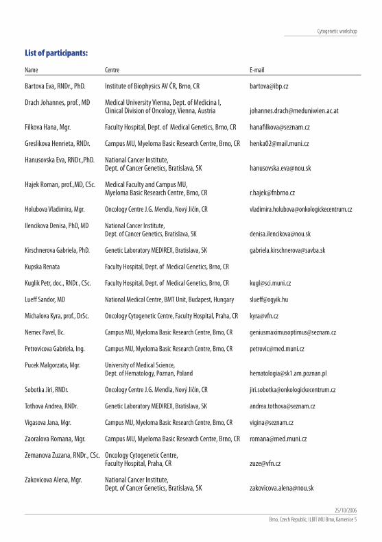
Cytogenetic workshop
25/10/2006Brno, Czech Republic, ILBIT MU Brno, Kamenice 5
List of participants:
Name Centre E-mail
Bartova Eva, RNDr., PhD. Institute of Biophysics AV ČR, Brno, CR [email protected]
Drach Johannes, prof., MD Medical University Vienna, Dept. of Medicina I,Clinical Division of Oncology, Vienna, Austria [email protected]
Filkova Hana, Mgr. Faculty Hospital, Dept. of Medical Genetics, Brno, CR [email protected]
Greslikova Henrieta, RNDr. Campus MU, Myeloma Basic Research Centre, Brno, CR [email protected]
Hanusovska Eva, RNDr.,PhD. National Cancer Institute,Dept. of Cancer Genetics, Bratislava, SK [email protected]
Hajek Roman, prof.,MD, CSc. Medical Faculty and Campus MU,Myeloma Basic Research Centre, Brno, CR [email protected]
Holubova Vladimira, Mgr. Oncology Centre J.G. Mendla, Nový Jičín, CR [email protected]
Ilencikova Denisa, PhD, MD National Cancer Institute,Dept. of Cancer Genetics, Bratislava, SK [email protected]
Kirschnerova Gabriela, PhD. Genetic Laboratory MEDIREX, Bratislava, SK [email protected]
Kupska Renata Faculty Hospital, Dept. of Medical Genetics, Brno, CR
Kuglik Petr, doc., RNDr., CSc. Faculty Hospital, Dept. of Medical Genetics, Brno, CR [email protected]
Lueff Sandor, MD National Medical Centre, BMT Unit, Budapest, Hungary [email protected]
Michalova Kyra, prof., DrSc. Oncology Cytogenetic Centre, Faculty Hospital, Praha, CR [email protected]
Nemec Pavel, Bc. Campus MU, Myeloma Basic Research Centre, Brno, CR [email protected]
Petrovicova Gabriela, Ing. Campus MU, Myeloma Basic Research Centre, Brno, CR [email protected]
Pucek Malgorzata, Mgr. University of Medical Science,Dept. of Hematology, Poznan, Poland [email protected]
Sobotka Jiri, RNDr. Oncology Centre J.G. Mendla, Nový Jičín, CR [email protected]
Tothova Andrea, RNDr. Genetic Laboratory MEDIREX, Bratislava, SK [email protected]
Vigasova Jana, Mgr. Campus MU, Myeloma Basic Research Centre, Brno, CR [email protected]
Zaoralova Romana, Mgr. Campus MU, Myeloma Basic Research Centre, Brno, CR [email protected]
Zemanova Zuzana, RNDr., CSc. Oncology Cytogenetic Centre,Faculty Hospital, Praha, CR [email protected]
Zakovicova Alena, Mgr. National Cancer Institute,Dept. of Cancer Genetics, Bratislava, SK [email protected]
Additional chromosomal aberrationsClinical importance was reported for deletions of 17p13 at the TP53 locus, with similar observations for patients
receiving standard-dose and high-dose therapy.15-17,22 Comprehensive analyses of cytogenetic abnormalities in MMidentified patients with a t(4;14) and/or 17p-deletion as the group of patients with the worst prognosis suggesting thatnovel approaches are required for the treatment of such high-risk patients.
Studies done by the Arkansas group identified a region on chromosome 1, which was linked with an aggressiveclinical course in MM: Global gene expression profiling on plasma cells from newly diagnosed patients treated withautologous transplantation revealed a significant over-representation of chromosome 1 genes in a group of about 70genes whose expression was associated with poor outcome. Further analyses showed that overexpression of CKS1B wasstrongly correlated with a gain of DNA copy numbers at chromosomal region 1q21, and that this abnormality conferreda poor prognosis.23 As a possible mechanism, reduced levels of p27Kip1 protein were observed in cases with 1q21 amplifi-cation, suggesting dysregulated cell cycle control in these cases.
Gene expression profiling in MMToday, genome-wide gene expression profiling based on DNA microarrays represents one of the most powerful tools
in the area of genomics. This technique has become feasible and broadly accessible, and in MM it is a valuable tool toidentify all myeloma-specific genetic abnormalities on a single platform.24 When this technique was used to identifygenes associated with therapeutic outcome in 221 patients with previously untreated MM, unsupervised clustering ledto the identification of four distinct MM subgroups.24 Further studies indicated that three genes of this analysis can beused to predict event-free survival. Furthermore, gene expression profiling provided the basis for a novel molecularclassification of MM because overexpression of one of the cyclin-D genes was found to be universal molecular feature ofMM.25 The so-called TC-classification combines the cytogenetic information about the 14q-translocations with cyclin-Dgene expression as summarized in Table 3. Patients of the TC4 and TC5 categories have shortened survival suggestingthat they should be considered for clinical studies exploring investigational therapies.
Impact of novel agents on prognosisBy now, prognostic factors conferring a poor outcome in MM were defined according to the experience with chemothe-
rapy, with no apparent differences between standard-dose and high-dose therapy (compare all studies referenced above).Recent studies have addressed the question whether or not treatment for high-risk patients may be improved by use ofnovel agents.
Thalidomide. Prognostic information is available mainly in patient populations treated with thalidomide in therelapsed/refractory setting. Among 75 patients treated with single agent thalidomide, advanced age (> 65 years), elevatedserum LDH, and elevated serum creatinine were predictive for inferior outcomes.26 In a similar analysis of relapsed MM pati-ents treated with thalidomide-based regimens, elevated serum LDH, advanced ISS-stage, and reduced performance statuswere independent predictive factors for survival.27 Based on these three variables, a scoring system was developed withsurvival times of 38.1, 28.8, and 5.8 months for scores 0, 1, and 2, respectively. The authors concluded that the addition ofLDH and performance status to the prognostic information provided by the ISS may help select patients who will likely derivebenefit from treatment with thalidomide-based regimens.

Cytogenetic workshop
25/10/2006Brno, Czech Republic, ILBIT MU Brno, Kamenice 5
Cytogenetic workshop25/10/2006Brno, Czech RepublicILBIT MU Brno, Kamenice 5
Detection of chromosomal aberrations in multiple myeloma
Abstracts and Application Manual
Detection of chromosomal aberrations in multiple myeloma
Edited by prof. MUDr. Roman Hajek, CSc.Published by Masaryk University, 20061 st Edition, 2006, 60 copiesDTP: Studio PORT, BrnoPhotography by A PLUS BRNO a.s.Printed by Masaryk University Press, Brno, Czech Republic
55-956B-2006 02/58 9/LFISBN 80-210-4129-3
Cytogenetic workshop25/10/2006Brno, Czech RepublicILBIT MU Brno, Kamenice 5
Abstracts and Application ManualDetection of chromosomal aberrations in multiple myeloma
Abstracts and Application Manual