abstract stain-style transfer network implement fully convolutional network (fcns)long et al.(2015)...
TRANSCRIPT

JMLR: Workshop and Conference Proceedings 80:1–10, 2017 ACML 2017
Neural Stain-Style Transfer Learning using GAN forHistopathological Images
Hyungjoo Cho∗ [email protected] National University
Sungbin Lim∗ [email protected] University
Gunho Choi [email protected] University
Hyunseok Min [email protected]
KAIST
Abstract
Performance of data-driven network for tumor classification varies with stain-style ofhistopathological images. This article proposes the stain-style transfer (SST) model basedon conditional generative adversarial networks (GANs) which is to learn not only the cer-tain color distribution but also the corresponding histopathological pattern. Our modelconsiders feature-preserving loss in addition to well-known GAN loss. Consequently ourmodel does not only transfers initial stain-styles to the desired one but also prevent thedegradation of tumor classifier on transferred images. The model is examined using theCAMELYON16 dataset.
Keywords: Deep Learning, Stain Normalization, Domain Adaptation, Neural Style Trans-fer, Generative Adversarial Network
1. Introduction
Deep learning based image recognition receives a lot of attention due to its notable ap-plication to digital histopathology including automatic tumor classification. Convolutionalneural networks(CNNs) have recently achieved state-of-the-art performance in the taskof image classification and detection, especially, replaced the traditional rule-based meth-ods in the several contests of medical image diagnosis LeCun et al. (2015); Wang et al.(2016). Such data-driven approach especially depends on quality of training dataset henceit requires sensible preprocesses. In histopathology, staining e.g. haematoxylin and eosin(H&E) is essential to examine the microscopic presence and characteristics of disease notonly for pathologists but also for neural networks. For digital histopathology, several stainnormalization preprocesses are well-known Ruifrok et al. (2001); Reinhard et al. (2001);Ruifrok et al. (2003); Annadurai (2007); Magee et al. (2009); Macenko et al. (2009); Khanet al. (2014); Li and Plataniotis (2015); Bejnordi et al. (2016).
Standard stain normalization algorithms are based on stain-specific color deconvolutionRuifrok et al. (2001). Stain deconvolution requires prior knowledge of reference stain vec-
∗ Equal contribution
c© 2017 H. Cho, S. Lim∗, G. Choi & H. Min.
arX
iv:1
710.
0854
3v2
[cs
.CV
] 2
5 O
ct 2
017
Cho Lim∗ Choi Min
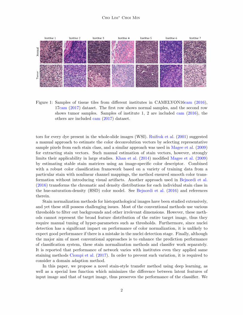
Figure 1: Samples of tissue tiles from different institutes in CAMELYON16cam (2016),17cam (2017) dataset. The first row shows normal samples, and the second rowshows tumor samples. Samples of institute 1, 2 are included cam (2016), theothers are included cam (2017) dataset.
tors for every dye present in the whole-slide images (WSI). Ruifrok et al. (2001) suggesteda manual approach to estimate the color deconvolution vectors by selecting representativesample pixels from each stain class, and a similar approach was used in Magee et al. (2009)for extracting stain vectors. Such manual estimation of stain vectors, however, stronglylimits their applicability in large studies. Khan et al. (2014) modified Magee et al. (2009)by estimating stable stain matrices using an image-specific color descriptor. Combinedwith a robust color classification framework based on a variety of training data from aparticular stain with nonlinear channel mappings, the method ensured smooth color trans-formation without introducing visual artifacts. Another approach used in Bejnordi et al.(2016) transforms the chromatic and density distributions for each individual stain class inthe hue-saturation-density (HSD) color model. See Bejnordi et al. (2016) and referencestherein.
Stain normalization methods for histopathological images have been studied extensively,and yet these still possess challenging issues. Most of the conventional methods use variousthresholds to filter out backgrounds and other irrelevant dimensions. However, these meth-ods cannot represent the broad feature distribution of the entire target image, thus theyrequire manual tuning of hyper-parameters such as thresholds. Furthermore, since nucleidetection has a significant impact on performance of color normalization, it is unlikely toexpect good performance if there is a mistake in the nuclei detection stage. Finally, althoughthe major aim of most conventional approaches is to enhance the prediction performanceof classification system, these stain normalization methods and classifer work separately.It is reported that performance of network varies with institutes even they applied samestaining methods Ciompi et al. (2017). In order to prevent such variation, it is required toconsider a domain adaption method.
In this paper, we propose a novel stain-style transfer method using deep learning, aswell as a special loss function which minimizes the difference between latent features ofinput image and that of target image, thus preserves the performance of the classifier. We
2

Neural Stain-Style Transfer Network
implement fully convolutional network (FCNs) Long et al. (2015) in proposed stain-stylegenerator that learns the color distribution of dataset which is used to train the tumorclassifier.
Our contributions in this paper are of two areas. First, we replace the color normalizationmethods with a generative model which learns certain stain-style distribution of dataset.Second, we introduce feature-preserving loss to induce the classifier to extract better featuresthan different methods.
2. Stain-Style Transfer with GAN
2.1. Stain-Style of Dataset
In this section, we summarize relevant material on our model. Let I be a set of institutesand let x ∈ X be the dataset of histological sample and the corresponding label y ∈ Y.The class of stained images or color images with RGB channels, denoted by C =Md(R3),is defined to be the set of d × d-matrix with R3 entries. Under this setting, we define thestain-style of institute i ∈ I to be a random variable φ(i) : X → C with a probabilitydistribution
P(i)(c) := P(φ(i) = c), c ∈ CSince φ(i) admits a certain conditional probability distribution P(i)(·|y), the definition ofdifferent stain-style with the same label makes sense.
2.2. Tumor Classifier Network
Suppose we trained tumor classifier network f : X → Y which infers histological pattern ofinput image x ∈ X . We write f = f (i) if the classifier is especially trained on dataset whichfollows stain-style φ(i). We estimate the performance of f (i) by
L(i)tumor := E(x,y)∼P (x,y)
[`(f (i)(x), y)
](1)
where ` is a loss function for classification e.g. cross-entropy. Practically, we make classifierto learn stained images φ(i)(x) ∈ C rather than x ∈ X . Hence one can decompose theclassifier by f (i) = f (i) ◦ φ(i) where f (i) : C → Y is an actual network which is trained ondataset with stain-style φ(i). In this case, we estimate (1) by
L(i)tumor ≈ Ec∼P(i)(c|y)
[`(f (i)(c), y)
](2)
2.3. Stain-Style Transfer Network
Since the stain-styles of each institute are dissimilar, the histological pattern in image fromdifferent institute would break up in the view of classifier network. Consequently, it wouldshow degraded performance Ciompi et al. (2017):
Ec∼P(j)(c|y)
[`(f (i)(c), y)
]≥ Ec∼P(i)(c|y)
[`(f (i)(c), y)
], i 6= j
3

Cho Lim∗ Choi Min
C G C
G⇣
�(1)
�(2)
�(3)
�(1)
⌧ = ⇣ � G
Figure 2: Overview of the stain-style transfer network. The network τ is composed of twotransformations: Gray-normalization G and style-generator ζ. G standardizeseach stain-style of color images from different institutes and ζ colorizes gray im-ages following the stain-style of certain institute.
To overcome this problem, we propose stain-style transfer (SST) network which transfersstain-style φ(j) to the initial φ(i). Precisely, our aim is to find a network τ : C → C whichsatisfies
P(τ ◦ φ(j) = c|y) = P(j)(τ−1(c)|y) ≈ P(i)(·|y), ∀c ∈ C (3)
Due to the change of variable formula (Durrett, 2010, Theorem 1.6.9), (3) implies
Ec∼P(j)(τ−1(·)|y)
[`(f (i)(c), y)
]≈ Eτ(c)∼P(i)(·|y)
[`(f (i)(τ(c)), y)
](4)
= Ec∼P(i)(·|y)
[`(f (i)(c), y)
]hence the tumor classifier recovers its performance (2).
We emphasize that our SST network does not require the dataset of institute j to trainboth f (i) and τ . To make τ independent of institute j ∈ I, we employ the gray normalizationG : C → G and train stain-style generator ζ : G → C such that τ = ζ ◦ G, as illustrated inFigure 2.
2.4. Stain-Style Generator by Conditional GAN
To train the style-generator ζ, we introduce three loss functions (a) reconstruction loss, (b)GAN loss, and (c) feature-preserving loss.
2.4.1. Reconstruction Loss
Restricted to the initial φ(i), SST network τ should be an reconstruction map i.e. τ ◦φ(i) =φ(i). Hence we apply a reconstruction loss to minimize the L2-distance between τ ◦φ(i) and
4

Neural Stain-Style Transfer Network
Figure 3: Illustration of feature-preserving loss. We use the global average pooled layers ofinput and generated image as the input of feature preserving loss.
its original image φ(i) using the architecture from Quan et al. (2016) which has very deepstructure with short-cut and skip connections. The reconstruction loss is denoted by
LRecon(τ, φ(i)) := E‖τ(φ(i))− φ(i)‖2
2.4.2. Conditional GAN Loss
As Pathak et al. showed in Pathak et al. (2016), mixing GAN loss Goodfellow et al. (2014)with some traditional loss, such as Lrecon, improves the performance of generator. Since wehave labeled images, conditional GAN Mirza and Osindero (2014) was applied instead ofGoodfellow et al. (2014). By means of GAN, ζ is to learn a mapping from G to C and totrick the discriminator D. Here D is to distinguish between fake and real images using thearchitecture from DCGAN Radford et al. (2015). We use the following GAN loss
LGAN(τ, φ(i)) := E[logD(ζ, φ(i))] + E[log(1−D(ζ, φ(i)))]
While D learns to maximize LGAN, ζ tries to minimize it until both arrives at its optimalstate. Through the above procedure, every stained image might be transferred to havethe desired stain-style. However, this approach often tend to make frequent color imagesindependent of histological pattern. This phenomena is called mode collapse (of GANs)which possibly interrupt achieving (3). Therefore we need an additional loss function.
5

Cho Lim∗ Choi Min
2.4.3. Feature-preserving Loss
As in (3), in the optimal state τ∗, an output of SST network τ∗ ◦ φ(j) should approximatetarget φ(i). By the means of Kullback-Leibler divergence, (3) can be restated by
τ∗ = arg minτ
KL[P(i)(c|y)
∥∥∥P(j)(τ−1(c)|y)]
To obtain τ∗, having (4) in mind, we employ the feature-presearving loss
LFP(τ, φ(i)) := KL[F(f (i)(c)
)∥∥∥F (f (i)(τ(c)))]
where F(f (i)(·)) indicates the feature of given color image extracted from the classifier f (i).As illustrated in Figure 3, the final layer before the activation function is used to examinefeature vector, precisely, global average pooled layer.
Consequently, the overall loss function is
L(τ, φ(i)) := λReconLRecon(τ, φ(i)) + LGAN(τ, φ(i)) + λFPLFP(τ, φ(i))
where λRecon, λFP are the weights which are used to balance the update between differentloss functions.
3. Experiment
We perform quantitative experiment in tumor classification to evaluate the SST network. Toshow the general performance of our method, we apply the extensions to vanilla models aswell as conventional method. We have 4 baseline methods: Reinhard et al. (2001), Macenkoet al. (2009), Histogram specification (HS) Annadurai (2007) and WSI color standardization(WSICS) Bejnordi et al. (2016).
3.1. Dataset
The Camelyon16 dataset is composed of 400 slides from two different institutes, Radboundand Utrecht. We use 180,000 patches for training, 20,000 for validation from Radbound and140,000 patches for testing from Utrecht. The number of tumor and normal are the same.Hypothesizing the training and validation dataset belong to a certain institue and the testset is from another one, we can merge every stain-style into the same space by applying thegray normalization. Both training and validation dataset are labeled, supervised learningcan be applied to train the mapping from gray image to the colored one. We used graynormalization based on Pillow package of python which uses this formula L = 0.299×R+0.587×G+ 0.144×B.
3.2. Network Architecture
In this part, we explain each network structure of classifier network and stain-style generatorwhich constitute SST network.
6

Neural Stain-Style Transfer Network
Figure 4: Comparison between SST and other stain normalization method: (a) Target im-age for transfer (b) Original input image to be transferred (c) SST (d) WSICS(e) HS (f) Marcenko (g) Reinhard
3.2.1. Classifier Network
Classifier network carries out two tasks in experiment. Firstly, it is a discriminator whichevaluates the performance of stain-style generator ζ. Secondly, as already explained insubsection 2.4.3, it works as a feature-extractor which is used in feature preserving lossLFP . We use ResNet-34 from torchvision library in PyTorch as a framework.
3.2.2. Stain-Style Generator
The generator network ζ is provided an image as an input instead of a noise vector. There-fore we can use FCN type architectures and U-Net is one of the most famous network amongthem. However, because of its limit of performance, we use FusionNet which has combinedthe advantages of U-Net and that of ResNet. Hyperparameters of network are set as sameas Quan et al. (2016). We adapt our discriminator architectures from Radford et al. (2015)which is based on VGG-Net without pooling layer. The hyperparameters of discriminatorare the same as those in Radford et al. (2015).
3.3. Result
Figure 4 illustrates the result of each stain normalization method on a sample image. Tar-get image comes from Radbound which is used for training the tumor classifier. Originalimage is sampled from Utrecht, used for testing the tumor classifier. Although there is novisual difference between outputs of each method, the classification performance on thesecolor images varies significantly. Given the experiment results in Table 1, SST networksuccessfully avoids the performance degradation. SST achieves the highest performance onoriginal images on tumor classification with Area Under Curve(AUC) = 0.9185. This resultshows that there are difference between visual judgment and the result of classifier. In caseof WSICS’s result, which is most visually similar to SST’s, the AUC score is worse thanthat of SST by about 30%. On the other hand, Macenko, which was visually the worst,performs better than other methods except for SST. Conventional methods consider onlythe physical features of input images and lose patterns which are key features for classifier’sdecision making process. In contrast, SST maintains those key features, input image’s ownpatterns, and also consider the color distribution of target images as well as the contextualinformation of original images.
7

Cho Lim∗ Choi Min
Table 1: Performance of tumor classifier network on different stain normalization methods.SST network shows significant improvement compared to direct application tooriginal (untransferred image) and outperforms the others.
Model Target Original SST WSICS HS Macenko Reinhard
AUC 0.9760 0.8900 0.9185 0.6408 0.4245 0.7169 0.5611
Precision 0.9114 0.8098 0.8440 0.5989 0.4987 0.6983 0.6114
Recall 0.9126 0.8111 0.8460 0.5957 0.4986 0.6956 0.6119
Specificity 0.9583 0.8014 0.8371 0.6010 0.4162 0.6500 0.5471
4. Conclusion
In this work, we have presented a stain style transfer approach to stain normalization forhistopathological images. To that end, we replace the stain normalization models with agenerative model which learns certain stain-style distribution of training dataset. This stainstyle transfer network is considerably simpler than contemporaneous work, and producesmore realistic results without any additional labeling or annotation for training as wellas prior knowledge. Further, unlike conventional stain normalization, which acts indepen-dently of the tumor classifier, the proposed feature-preserving loss induces our colorationin a direction that does not affect the tumor classifier. We demonstate that our modelis optimized for the performance of the tumor classifier and allows successful stain-styletransfer.
The style of chemical cell staining is mainly affected by structural information andmorphology of cells rather than factors such as cell brightness. Based on these observationpoints, we converted the test image into a gray image and performed a stain style transferprocess. While this method has the advantage of making the process simpler, it has alsolost some information. To resolve the limitation, further investigation will assess directstain style transfer approach from color image to color image. In addition, we hope to moreclosely examine parameters of our deep learning approach. Further, we will perform morerounds of hard negative mining and consider the reliability and reproducibility of the deepCNN models.
References
http://camelyon16.grand-challenge.org/, 2016.
http://camelyon17.grand-challenge.org/, 2017.
S Annadurai. Fundamentals of digital image processing. Pearson Education India, 2007.
Babak Ehteshami Bejnordi, Geert Litjens, Nadya Timofeeva, Irene Otte-Holler, AndreHomeyer, Nico Karssemeijer, and Jeroen AWM van der Laak. Stain specific standardiza-tion of whole-slide histopathological images. IEEE transactions on medical imaging, 35(2):404–415, 2016.
8

Neural Stain-Style Transfer Network
Francesco Ciompi, Oscar Geessink, Babak Ehteshami Bejnordi, Gabriel Silva de Souza,Alexi Baidoshvili, Geert Litjens, Bram van Ginneken, Iris Nagtegaal, and Jeroen van derLaak. The importance of stain normalization in colorectal tissue classification with con-volutional networks. arXiv preprint arXiv:1702.05931, 2017.
Rick Durrett. Probability: theory and examples. Cambridge university press, 2010.
Ian Goodfellow, Jean Pouget-Abadie, Mehdi Mirza, Bing Xu, David Warde-Farley, SherjilOzair, Aaron Courville, and Yoshua Bengio. Generative adversarial nets. In Advances inneural information processing systems, pages 2672–2680, 2014.
Adnan Mujahid Khan, Nasir Rajpoot, Darren Treanor, and Derek Magee. A nonlinearmapping approach to stain normalization in digital histopathology images using image-specific color deconvolution. IEEE Transactions on Biomedical Engineering, 61(6):1729–1738, 2014.
Yann LeCun, Yoshua Bengio, and Geoffrey Hinton. Deep learning. Nature, 521(7553):436–444, 2015.
Xingyu Li and Konstantinos N Plataniotis. A complete color normalization approach tohistopathology images using color cues computed from saturation-weighted statistics.IEEE Transactions on Biomedical Engineering, 62(7):1862–1873, 2015.
Jonathan Long, Evan Shelhamer, and Trevor Darrell. Fully convolutional networks forsemantic segmentation. In Proceedings of the IEEE Conference on Computer Vision andPattern Recognition, pages 3431–3440, 2015.
Marc Macenko, Marc Niethammer, JS Marron, David Borland, John T Woosley, XiaojunGuan, Charles Schmitt, and Nancy E Thomas. A method for normalizing histology slidesfor quantitative analysis. In Biomedical Imaging: From Nano to Macro, 2009. ISBI’09.IEEE International Symposium on, pages 1107–1110. IEEE, 2009.
Derek Magee, Darren Treanor, Doreen Crellin, Mike Shires, Katherine Smith, Kevin Mo-hee, and Philip Quirke. Colour normalisation in digital histopathology images. In ProcOptical Tissue Image analysis in Microscopy, Histopathology and Endoscopy (MICCAIWorkshop), volume 100. Citeseer, 2009.
Mehdi Mirza and Simon Osindero. Conditional generative adversarial nets. arXiv preprintarXiv:1411.1784, 2014.
Deepak Pathak, Philipp Krahenbuhl, Jeff Donahue, Trevor Darrell, and Alexei A Efros.Context encoders: Feature learning by inpainting. In Proceedings of the IEEE Conferenceon Computer Vision and Pattern Recognition, pages 2536–2544, 2016.
Tran Minh Quan, David GC Hilderbrand, and Won-Ki Jeong. Fusionnet: A deep fullyresidual convolutional neural network for image segmentation in connectomics. arXivpreprint arXiv:1612.05360, 2016.
9

Cho Lim∗ Choi Min
Alec Radford, Luke Metz, and Soumith Chintala. Unsupervised representation learning withdeep convolutional generative adversarial networks. arXiv preprint arXiv:1511.06434,2015.
Erik Reinhard, Michael Adhikhmin, Bruce Gooch, and Peter Shirley. Color transfer betweenimages. IEEE Computer graphics and applications, 21(5):34–41, 2001.
Arnout C Ruifrok, Dennis A Johnston, et al. Quantification of histochemical staining bycolor deconvolution. Analytical and quantitative cytology and histology, 23(4):291–299,2001.
Arnout C Ruifrok, Ruth L Katz, and Dennis A Johnston. Comparison of quantifica-tion of histochemical staining by hue-saturation-intensity (hsi) transformation and color-deconvolution. Applied Immunohistochemistry & Molecular Morphology, 11(1):85–91,2003.
Dayong Wang, Aditya Khosla, Rishab Gargeya, Humayun Irshad, and Andrew H Beck.Deep learning for identifying metastatic breast cancer. arXiv preprint arXiv:1606.05718,2016.
10