abstract microencapsulation of probiotic - drum
TRANSCRIPT
ABSTRACT
Title of dissertation: MICROENCAPSULATION OF PROBIOTICBACTERIA IN XANTHAN-CHITOSANPOLYELECTROLYTE COMPLEX GELS
Sanem Argin, Doctor of Philosophy, 2007
Dissertation directed by: Professor Y. Martin LoDepartment of Nutrition and Food Science
In recent years, increasing evidence indicating numerous health benefits asso-
ciated with the intake of probiotic bacteria has created a big market of probiotic
foods worldwide. However, maintaining high numbers of viable cells in probiotic
food products during the shelf life of the product and during gastrointestinal transit
is a challenge. The goal of this research is to develop a novel microencapsulation
system using xanthan gum and chitosan polyelectrolyte complex gels in order to
protect the probiotic cells against adverse environmental conditions, and to increase
their recovery rates.
The extrusion method was used to form the xanthan-chitosan microcapsules.
The effects of initial polymer concentration and chitosan solution pH on the crosslink
density of the capsule network were investigated by swelling studies and modulated
differential scanning calorimetry (MDSC) analysis. Once the capsule formulations
resulting in a highly crosslinked network structure were determined, P. acidilactici
cells were successfully encapsulated using these formulations. Efforts were made to
study the release kinetics of probiotic cells from these capsules in gastrointestinal
conditions. Cell release was found to be negligible in simulated gastric juice. In
simulated intestinal conditions, the release was relaxation controlled and complete
cell release was achieved in at least 5 hours. After exposure to simulated gastric fluid
(pH=2.0), encapsulation with xanthan gum and chitosan provided up to six-log and
four-log preservation of the freeze-dried probiotic cells over free suspending cells for
1 and 2 hours, respectively.
These results suggest that xanthan-chitosan capsules have a good potential
for delivery of probiotic cells to the intestines in high numbers where the cells can
release and colonize to benefit the consumer.

MICROENCAPSULATION OF PROBIOTIC BACTERIAIN XANTHAN-CHITOSAN
POLYELECTROLYTE COMPLEX GELS
by
Sanem Argın
Dissertation submitted to the Faculty of the Graduate School of theUniversity of Maryland, College Park in partial fulfillment
of the requirements for the degree ofDoctor of Philosophy
2007
Advisory Committee:Professor Y. Martin Lo, Chair/AdvisorProfessor Peter Kofinas, Co-AdvisorProfessor Srinivasa R. RaghavanProfessor Mark KantorProfessor Mickey Parish
© Copyright bySanem Argın
2007
DEDICATION
To my daughter, Kayla Soysal, and my husband, Alkan Soysal.
ii

Acknowledgments
First and foremost, I would like to thank my husband, Alkan Soysal, for his
endless support, unconditional love, continuous encouragement, and tremendous
patience. Without him, I would never have been able to come this far. He has been
my coach, my best friend, and the shoulder I cried on throughout these years. Thank
you for believing in me and for always being there for me. Thank you for finding
a solution to every problem I had and not complaining about it even once. Many,
many thanks for the millions of small things you have done to make it possible for
me to write this dissertation.
I would like to thank my advisor, Dr. Y. Martin Lo, for giving me the oppor-
tunity to pursue my Ph.D. in his laboratory and for his support during my studies.
From my work with him I learned more about myself and what I am capable of
achieving.
I am extremely thankful to my co-advisor, Dr. Peter Kofinas, for offering his
help at the time that I needed it the most and for welcoming me into his group. His
guidance and support greatly impacted the progress of my research. Dr. Kofina sas
eyxaristw poly gia ola.
I would also like to thank Dr. Srinivasa Raghavan for not only being in my
dissertation committee but also for everything I learned from him about polymers
and for the valuable discussions he had with me about my research. I am also
thankful to the other members of my former and final examination committee, Dr.
Mickey Parish, Dr. Mark Kantor, Dr. Dallas Hoover, Dr. Inder Vijay, and Dr.
iii
Yang Tao, for their time and suggestions.
I would like to thank my parents, Fusun and Ibrahim Argın, and my brother,
Erman Argın, for their love and support throughout my life.
I also owe thanks to all my friends in Dr. Lo’s and Dr. Kofinas’ groups for their
friendship, support, and for all the fun they brought into my life: Linden Bolisay,
Brendan Casey, Arthur von Wald Cresce, Angela Fu, Ayan Ghosh, Daniel Janiak,
Peter Machado, Daniel Reese, Karen Silagyi, Josh Silverstein, Pavan Kumar Soma,
Patrick Williams, Ta-I Yang, and Afra Yeh.
Finally, my special thanks go to my 10-week old daughter Kayla for being the
biggest joy of my life and for giving me the motivation to finish my work.
iv
TABLE OF CONTENTS
List of Tables vii
List of Figures viii
1 Introduction 11.1 Significance . . . . . . . . . . . . . . . . . . . . . . . . . . . . . . . 11.2 Objectives . . . . . . . . . . . . . . . . . . . . . . . . . . . . . . . . 2
2 Literature Review 32.1 Microencapsulation of probiotic bacteria . . . . . . . . . . . . . 32.2 Xanthan Gum . . . . . . . . . . . . . . . . . . . . . . . . . . . . . . 6
2.2.1 Source, structure and applications . . . . . . . . . . . . . 62.2.2 Rheological properties . . . . . . . . . . . . . . . . . . . . 8
2.3 Chitosan . . . . . . . . . . . . . . . . . . . . . . . . . . . . . . . . . 92.3.1 Source, structure and applications . . . . . . . . . . . . . 92.3.2 Rheological properties . . . . . . . . . . . . . . . . . . . . 11
2.4 Microencapsulation with xanthan gum and chitosan . . . . . . 122.4.1 Polyelectrolyte complex (PEC) gels by xanthan gum
and chitosan . . . . . . . . . . . . . . . . . . . . . . . . . . . 122.4.2 pH-sensitive swelling characteristics of xanthan-chitosan
gels . . . . . . . . . . . . . . . . . . . . . . . . . . . . . . . . 14
3 Effects of Complexation Conditions on Xanthan-Chitosan Polyelec-trolyte Complex Gels 173.1 Introduction . . . . . . . . . . . . . . . . . . . . . . . . . . . . . . . 173.2 Materials and methods . . . . . . . . . . . . . . . . . . . . . . . . 18
3.2.1 Preparation of chitosan and xanthan solutions . . . . . 183.2.2 Capsule formation and Inverted microscopy . . . . . . . 193.2.3 Determination of the swelling degree . . . . . . . . . . . 193.2.4 Differential Scanning Calorimetry (DSC) measurements 203.2.5 Statistical analysis . . . . . . . . . . . . . . . . . . . . . . . 21
3.3 Results and discussion . . . . . . . . . . . . . . . . . . . . . . . . 213.3.1 Capsule formation . . . . . . . . . . . . . . . . . . . . . . . 21
v

3.3.2 Effect of complexation conditions on the swelling de-gree of xanthan-chitosan capsules . . . . . . . . . . . . . 22
3.3.3 DSC analysis of xanthan-chitosan capsules . . . . . . . . 263.4 Conclusions . . . . . . . . . . . . . . . . . . . . . . . . . . . . . . . 32
4 The Release Kinetics and Swelling Behavior of Xanthan-ChitosanCapsules 334.1 Introduction . . . . . . . . . . . . . . . . . . . . . . . . . . . . . . . 334.2 Materials and Methods . . . . . . . . . . . . . . . . . . . . . . . . 34
4.2.1 Calibration curve . . . . . . . . . . . . . . . . . . . . . . . 344.2.2 Microencapsulation of P. acidilactici . . . . . . . . . . . 354.2.3 Kinetics of cell release . . . . . . . . . . . . . . . . . . . . 354.2.4 Dynamic swelling behavior . . . . . . . . . . . . . . . . . 37
4.3 Results and Discussion . . . . . . . . . . . . . . . . . . . . . . . . 374.3.1 Calibration curve . . . . . . . . . . . . . . . . . . . . . . . 374.3.2 Kinetics of cell release . . . . . . . . . . . . . . . . . . . . 384.3.3 Dynamic swelling behavior . . . . . . . . . . . . . . . . . 43
4.4 Conclusions . . . . . . . . . . . . . . . . . . . . . . . . . . . . . . . 46
5 Protective effects of Xanthan-Chitosan Encapsulation on P. acidi-lactici cells 475.1 Introduction . . . . . . . . . . . . . . . . . . . . . . . . . . . . . . . 475.2 Materials and Methods . . . . . . . . . . . . . . . . . . . . . . . . 485.3 Results and Discussion . . . . . . . . . . . . . . . . . . . . . . . . 495.4 Conclusions . . . . . . . . . . . . . . . . . . . . . . . . . . . . . . . 54
6 Conclusions 56
Bibliography 58
vi
LIST OF TABLES
2.1 Microorganisms considered as probiotics. . . . . . . . . . . . . . . . . 4
3.1 Swelling degrees of xanthan-chitosan capsules in DI water . . . . . . . 24
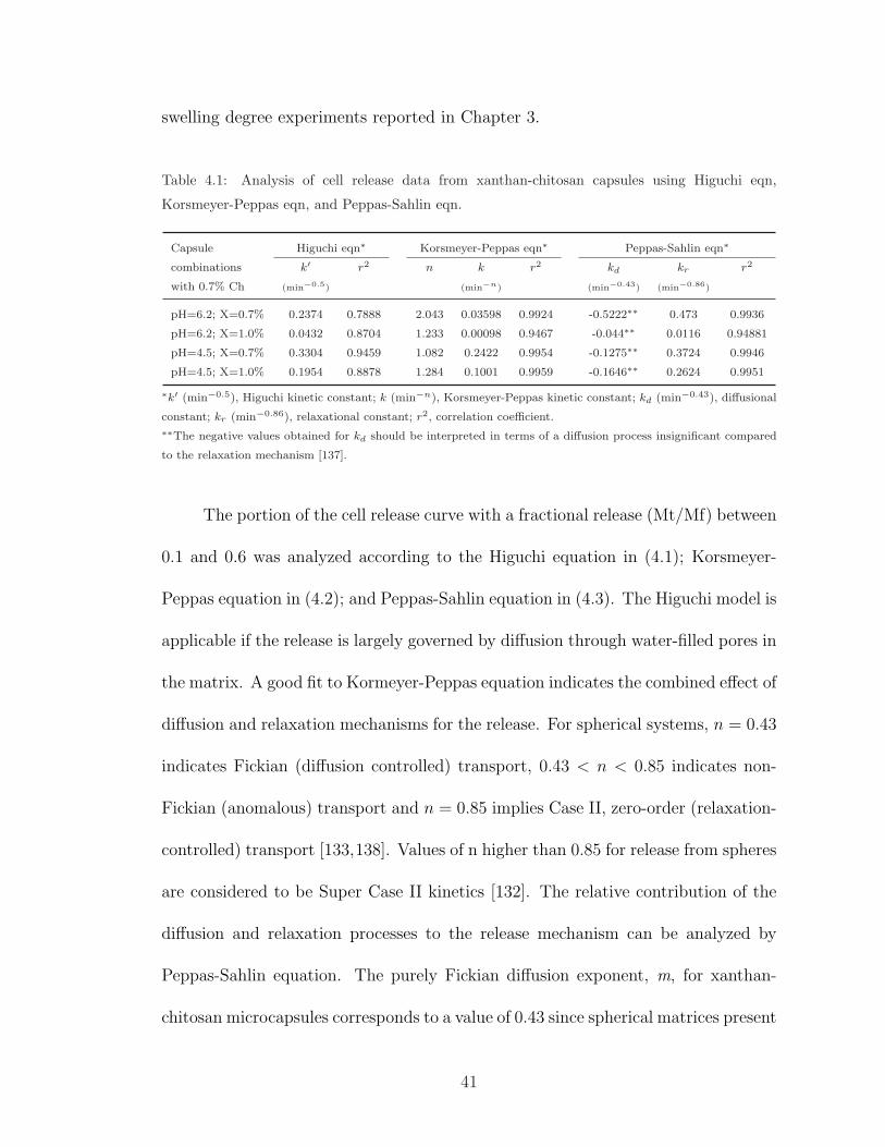
4.1 Analysis of cell release data from xanthan-chitosan capsules . . . . . 41
4.2 Analysis of dynamic swelling data for xanthan-chitosan capsules . . . 45
vii
LIST OF FIGURES
2.1 Pentasaccharide repeating unit of xanthan gum. . . . . . . . . . . . . 7
2.2 Chemical structure of chitosan. . . . . . . . . . . . . . . . . . . . . . 10
2.3 pH-sensitive swelling of xanthan-chitosan complex . . . . . . . . . . . 16
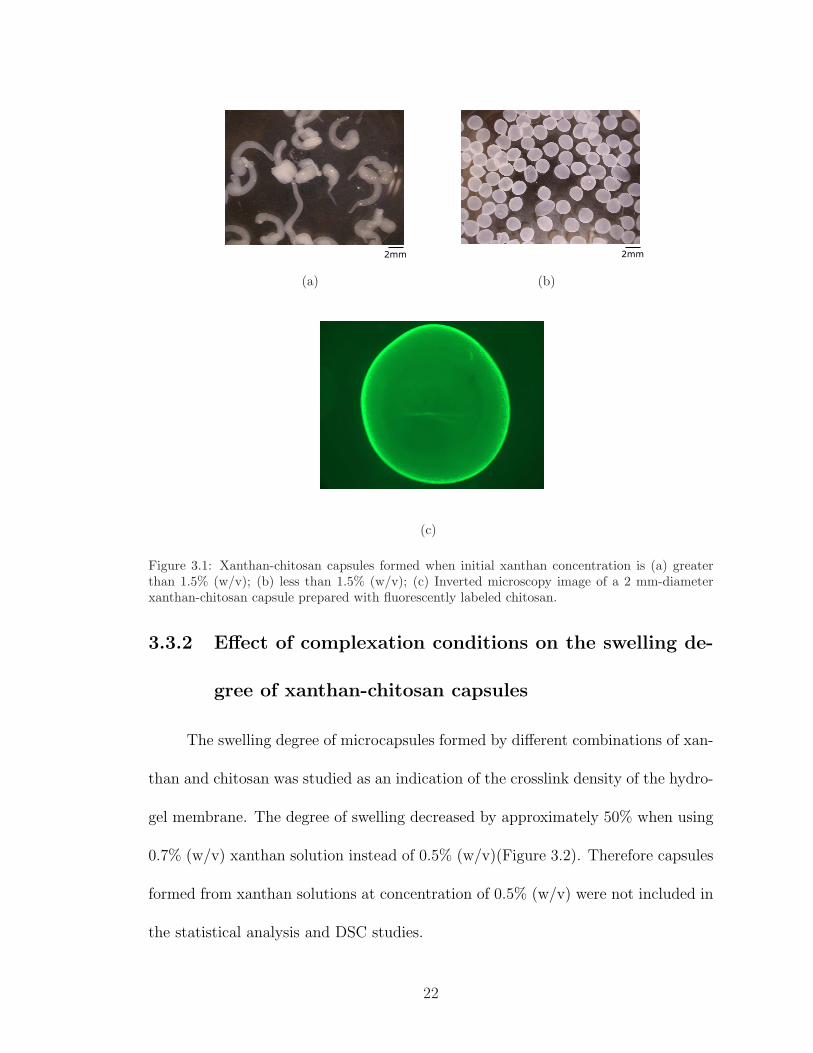
3.1 Xanthan-chitosan capsules formed when initial xanthan concentrationis (a) greater than 1.5% (w/v); (b) less than 1.5% (w/v); (c) Invertedmicroscopy image of a 2 mm-diameter xanthan-chitosan capsule pre-pared with fluorescently labeled chitosan. . . . . . . . . . . . . . . . . 22
3.2 Swelling degree of xanthan-chitosan capsules formed by using 0.5%(w/v) and 0.7% (w/v) xanthan solutions. . . . . . . . . . . . . . . . . 23
3.3 MDSC curves of (a) chitosan; (b) xanthan gum. . . . . . . . . . . . . 28
3.4 MDSC curves of xanthan-chitosan capsules showing the effect of ini-tial xanthan concentration on the resulting capsule network structure. 29
3.5 MDSC curves of xanthan-chitosan capsules showing the effect of ini-tial chitosan solution pH on the resulting capsule network structure. . 31
4.1 Calibration curve of P. acidilactici. . . . . . . . . . . . . . . . . . . . 38
4.2 The release of P. acidilactici cells from xanthan- chitosan capsules. . 40
4.3 Fractional release of P. acidilactici cells from xanthan-chitosan cap-sules in SGF at pH=2.0 and in SIF at pH 6.8. . . . . . . . . . . . . . 42
4.4 (a) Dynamic swelling ; (b) water absorption curve of xanthan-chitosancapsules in SGF at pH=2.0 and in SIF at pH 6.8. . . . . . . . . . . . 44
viii
5.1 Protective effects of encapsulation on the viability of P.acidilacticicells against freeze-drying and SGF (pH=2.0) exposure (n=2) using(a) 0.7% and 1.0% chitosan. . . . . . . . . . . . . . . . . . . . . . . . 51
5.2 Relative size of capsules prepared by using a syringe and a nozzle. . . 52
5.3 Protective effect of xanthan-chitosan capsules prepared by using a0.7-mm nozzle on the viability of P. acidilactici cells against freeze-drying and SGF exposure for 1 hr. . . . . . . . . . . . . . . . . . . . 53
ix

Chapter 1
Introduction
1.1 Significance
Life style and eating habits play an important role in overall health of in-
dividuals. In recent years, increasing evidence indicating numerous health benefits
associated with the intake of probiotic bacteria has created a big market of probiotic
foods worldwide [1]. The biggest challenge in the development of probiotic prod-
ucts is to maintain the adequate number of viable cells during the shelf life of the
product as well as during the gastrointestinal (GI)-tract transit after consumption,
so that the claimed health benefits can be delivered to the consumer [2–5]. Con-
sequently, there has been a growing interest in developing techniques to enhance
the survival of probiotic bacteria particularly during the GI-tract transit of the
cells [6–14]. Microencapsulation of probiotic bacteria in hydrocolloid beads is one of
the recent techniques studied to improve the viability and activity of the cells under
unfavorable conditions by entrapping the bacteria within a bead matrix [15–18].
1
Na-Alginate and κ-carrageenan, alone or combined with other polymers and cry-
oprotectants, are the most commonly used hydrocolloids for the encapsulation of
microorganisms [17, 19–21]. However, each system has its own limitations such as
susceptibility to ions, lack of mechanical strength and scale-up difficulties [22, 23].
For this reason, there remains an important need for an effective capsule design
that ensures the proper delivery of probiotics to the target destination and enables
the complete release of the cells at desired conditions to benefit from the health
promoting effects of probiotic bacteria.
1.2 Objectives
The ultimate goal of this work was to develop a novel microencapsulation
system using polyelectrolyte complex gels formed by xanthan gum and chitosan
to effectively sustain the viability of probiotics that are known to provide health
benefits to human and animals. The purpose was to protect the encapsulated cells
against harsh environmental conditions, particularly against the highly acidic gastric
conditions, and to establish a release mechanism so that the bacteria can colonize
in the gut to improve the intestinal microbiota. Specifically, emphasis was placed
on the effects of complexation conditions on the capsule network structure, the
release kinetics of the cells under GI-tract conditions, and the protective effects of
the xanthan-chitosan capsules on probiotics during GI-tract transit.
2
Chapter 2
Literature Review
2.1 Microencapsulation of probiotic bacteria
Probiotics are defined as live microbial food supplements that benefit the host
by improving its intestinal microbial balance [24]. Lactic acid bacteria are among
the most important probiotic microorganisms (Table 2.1) associated with the human
gastrointestinal (GI)-tract [25]. Known health benefits of probiotic bacteria include
suppressing the growth of undesirable microorganisms in the colon and small in-
testine [26, 27], controlling serum cholesterol levels [28], reducing the risk of colon
cancer [29], stimulating the immune system [30], improving lactose utilization [31],
and controlling the allergic inflammation associated with food allergy [32].
To act as probiotics, bacteria must arrive in intestines alive and in sufficient
numbers which is suggested at 106-107 cfu/g product [26, 33]. However, signifi-
cant reduction of the number of viable and active cells in a food product occurs
inevitably due to the environmental changes such as pH, temperature, and dis-
3
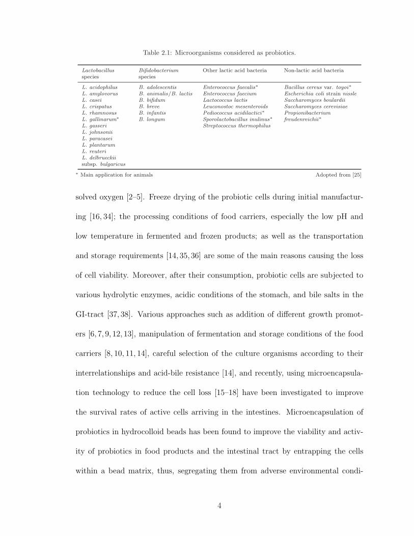
Table 2.1: Microorganisms considered as probiotics.
Lactobacillusspecies
Bifidobacteriumspecies
Other lactic acid bacteria Non-lactic acid bacteria
L. acidophilus B. adolescentis Enterococcus faecalis∗ Bacillus cereus var. toyoi∗L. amylovorus B. animalis/B. lactis Enterococcus faecium Escherichia coli strain nissleL. casei B. bifidum Lactococcus lactis Saccharomyces boulardiiL. crispatus B. breve Leuconostoc mesenteroids Saccharomyces cerevisiaeL. rhamnosus B. infantis Pediococcus acidilactici∗ PropionibacteriumL. gallinarum∗ B. longum Sporolactobacillus inulinus∗ freudenreichii∗L. gasseri Streptococcus thermophilusL. johnsoniiL. paracaseiL. plantarumL. reuteriL. delbrueckiisubsp. bulgaricus
∗ Main application for animals Adopted from [25]
solved oxygen [2–5]. Freeze drying of the probiotic cells during initial manufactur-
ing [16, 34]; the processing conditions of food carriers, especially the low pH and
low temperature in fermented and frozen products; as well as the transportation
and storage requirements [14, 35, 36] are some of the main reasons causing the loss
of cell viability. Moreover, after their consumption, probiotic cells are subjected to
various hydrolytic enzymes, acidic conditions of the stomach, and bile salts in the
GI-tract [37, 38]. Various approaches such as addition of different growth promot-
ers [6, 7, 9, 12, 13], manipulation of fermentation and storage conditions of the food
carriers [8, 10, 11, 14], careful selection of the culture organisms according to their
interrelationships and acid-bile resistance [14], and recently, using microencapsula-
tion technology to reduce the cell loss [15–18] have been investigated to improve
the survival rates of active cells arriving in the intestines. Microencapsulation of
probiotics in hydrocolloid beads has been found to improve the viability and activ-
ity of probiotics in food products and the intestinal tract by entrapping the cells
within a bead matrix, thus, segregating them from adverse environmental condi-
4
tions, as well as protecting them against bacteriophages [21, 33, 39]. There are two
common techniques applied to the microencapsulation of probiotic bacteria: The
extrusion (droplet) method [40–43] and emulsion (two-phase) system [16,21,42,44].
Both systems achieve survival rates as high as 80-95%, but the extrusion method is
simpler and cheaper [33]. Alginate is the most commonly used hydrocolloid for the
entrapment of cells due to its simple and low cost gelling mechanism [19], excellent
biocompatibility [20], and the reversibility of the immobilization [21]. However, al-
ginate gels are unstable with phosphate and lactate ions as these ions can displace
the calcium ions that stabilize the alginate gels [44]. Recent studies showed that
Ca-alginate encapsulation does not significantly improve the survival of the cells in
gastric conditions [15,38,45]. On the other hand, κ-carrageenan capsules have been
reported to provide better protection against refrigeration temperatures than Ca-
alginate beads [17]. However, with κ-carrageenan, flexible gels can only be formed in
the presence of locust bean gum [44]. The formation of κ-carrageenan-locust bean
gum capsules requires potassium ions, which can damage the probiotic cells [46].
Another approach was to use Ca-pectate gels as carriers since they are less suscep-
tible to decalcifying and acidification than Ca-alginate gels [47]. Combining other
polymers (starch, pectin, etc) and/or whey proteins with these common hydrocol-
loids has been shown to offer better protection of cells under low temperature and
pH than when these gums are used alone for the encapsulation [41, 48, 49]. More-
over, different coating materials to coat the beads as well as some additives such as
cryoprotectants were used to overcome the aforementioned disadvantages [39, 50].
However, all of these approaches have had varying degrees of success and the costs
5
of operation remain a concern. The main problems of bacteria encapsulation with
Ca-alginate and κ-carrageenan gels can be summarized as susceptibility to ions,
lack of mechanical strength and scale-up difficulties [22, 23]. Some other methods
to encapsulate bacteria such as spray drying with starch reportedly did not offer
any protection against acidic conditions [15]. Sun and Griffiths [51] reported that
a mixture of gellan and xanthan gum, both of which are microbial exopolysaccha-
rides, serves as a better encapsulating agent than Ca-alginate when the cells are
exposed to the same environmental conditions. Produced by microorganisms and
secreted to the environment to serve as a protection against desiccation, bacterio-
phage attacks, variations in temperature, cell wall degrading enzymes, and osmotic
stress [52–56], microbial exopolysaccharides may be suggested as promising agents
for encapsulation of probiotic bacteria.
2.2 Xanthan Gum
2.2.1 Source, structure and applications
Xanthan gum (Figure 2.1) is a microbial exopolysaccharide consisting of a
cellulosic backbone with side chains of two mannose and one glucuronic acid on
every second glucose residue [57, 58]. In nature, the plant pathogenic bacterium
Xanthomonas campestris produce highly viscous xanthan gum which attaches to
cabbage and protects itself against dehydration and access of harmful substances, or
as a way of binding and neutralizing bacteriophages [52,54]. Presently, commercial
6

Figure 2.1: Pentasaccharide repeating unit of xanthan gum.
xanthan gum is produced from glucose by batch fermentation of X. campestris. The
molecular weight of xanthan gum can reach up to 6 million Daltons, which makes
it possible to create extremely viscous solutions at very low concentrations [59, 60].
Pyruvate and acetate substitution may vary strongly depending on the par-
ent bacterial strain and the growth conditions of the bacteria [61]. Side chains
of xanthan gum represent a very high proportion of the molecule (60%). Due to
the side chains, the polymer completely hydrates in water [62]. In addition, enzy-
matic resistance of xanthan gum is thought to be due to the arrangement of the
side chains which prevents the enzymes from attacking the β-(1-4) linkages in the
backbone, thereby preventing depolymerization by enzymes, acid and alkali [63].
In aqueous solution, the stable conformation of xanthan is double helical below a
transition temperature [64]. Solution studies on the conformation of xanthan gum
suggest a rod-like structure with some degree of flexibility, depending on the molec-
ular weight [65–67]. In addition to its enzymatic resistance, xanthan gum is stable
7
over a wide range of temperatures and pH, which finds many applications in food,
pharmaceutical, cosmetic, and oil-drilling industries [68–72]. Xanthan gum was ap-
proved by the U.S. Food and Drug Administration (FDA) in 1965 and became the
most widely used thickening agent in food products.
2.2.2 Rheological properties
In aqueous solution, high molecular weight xanthan molecules form a semi-
flexible wormlike structure and develop a weak-gel network due to intermolecular
interactions [66,67,73,74]. These intermolecular associations such as hydrogen bond-
ing, electrostatic and hydrophobic interactions play a crucial role in the changes of
rheological properties of the xanthan solutions [74,75]. Since the junctions between
the ordered chain sequences in the gel network of xanthan gum are weaker than
those in the true gel networks, they can be easily broken down under stress allowing
the system to flow which gives xanthan the shear-thinning behavior [76]. Due to the
presence of glucuronic acid and pyruvate in its side chains, xanthan is considered
to be an anionic polyelectrolyte [77]. However, the response of xanthan solutions to
increasing ionic strength differs from other polyelectrolytes in that they maintain a
high solution viscosity as ionic strength increases due to the increase in hydrody-
namic radius [78,79]. At low salt concentrations (up to 0.05 M), the viscosity of the
xanthan gum solutions slightly decreases and beyond this, no significant effect on
the steady shear and dynamic properties is observed [79, 80]. In addition, xanthan
viscosity does not change significantly with temperature in the presence of salt and
8
the ordered conformation exists at room or elevated temperatures [77,78,81].
2.3 Chitosan
2.3.1 Source, structure and applications
Chitosan (Figure 2.2), poly-β-(1 4)-D-glucosamine, is the only natural cationic
polysaccharide and is produced by alkaline deacetylation of chitin [82, 83]. Chitin
is the second most abundant natural polysaccharide which is present in crustacea,
insects and yeasts. The molecular weight of chitin and chitosan ranges between
several hundred thousands to 1 million Daltons [84]. As one of the major by-products
of the crabbing and shrimp canning industry, chitin is also the major source of
surface pollution in coastal areas [85]. Studies on chitin and chitosan have increased
since the 1990’s to find value-added uses of these polysaccharides that show excellent
biological properties such as biocompatibility, biodegradability, lack of toxicity, and
adsorption, as well as relatively high percentage of nitrogen [82,86–88]. The degree
of acetylation (DA) is a very important property of chitosan molecules since DA
affects biodegradation capability, solubility, gelling and reactivity of chitosan [89].
The unique properties of chitosan arise from its amino groups that carry pos-
itive charges at pH values below 6.5, enabling it to bind to negatively charged
materials such as enzymes, cells, polysaccharides, nucleic acids, hair and skin [90].
Being a linear polysaccharide, chitosan has both reactive amino groups and hydroxyl
groups that can be used to alter its physical and solution properties [90]. Chitosan
9
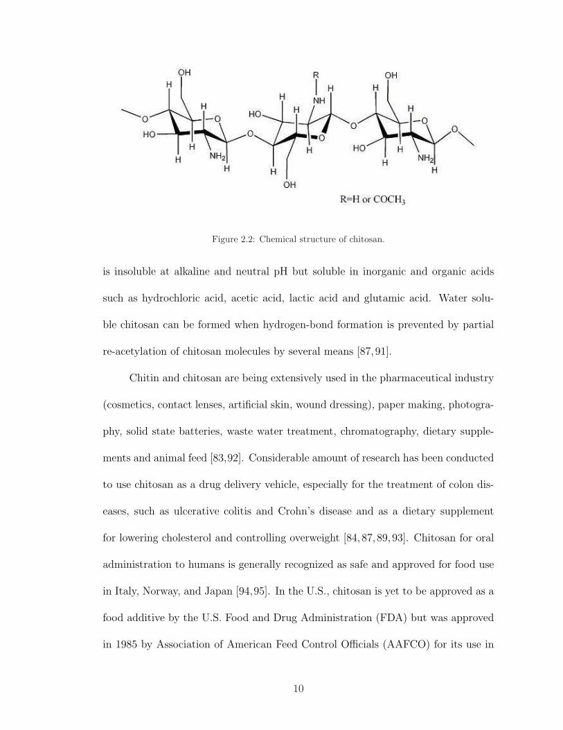
Figure 2.2: Chemical structure of chitosan.
is insoluble at alkaline and neutral pH but soluble in inorganic and organic acids
such as hydrochloric acid, acetic acid, lactic acid and glutamic acid. Water solu-
ble chitosan can be formed when hydrogen-bond formation is prevented by partial
re-acetylation of chitosan molecules by several means [87,91].
Chitin and chitosan are being extensively used in the pharmaceutical industry
(cosmetics, contact lenses, artificial skin, wound dressing), paper making, photogra-
phy, solid state batteries, waste water treatment, chromatography, dietary supple-
ments and animal feed [83,92]. Considerable amount of research has been conducted
to use chitosan as a drug delivery vehicle, especially for the treatment of colon dis-
eases, such as ulcerative colitis and Crohn’s disease and as a dietary supplement
for lowering cholesterol and controlling overweight [84, 87, 89, 93]. Chitosan for oral
administration to humans is generally recognized as safe and approved for food use
in Italy, Norway, and Japan [94,95]. In the U.S., chitosan is yet to be approved as a
food additive by the U.S. Food and Drug Administration (FDA) but was approved
in 1985 by Association of American Feed Control Officials (AAFCO) for its use in
10
animal feed, as long as the level does not exceed 0.1% of the feed [96,97].
2.3.2 Rheological properties
Concentrated chitosan solutions exhibit shear-thinning behavior and the vis-
cosity of dilute chitosan solutions are independent of shear rate [98]. Due to strong
intermolecular hydrogen bonding even at low concentration, chitosan molecules have
a tendency to entangle and form a network [99]. As a polyelectrolyte, the confor-
mation of the chitosan molecule is affected by the electric charge density [100]. For
this reason, the rheological properties of chitosan depend on factors such as pH,
ionic strength, solvent selection, concentration, molecular weight, DA, and the dis-
tribution of the acetyl groups [101]. When the pH of the solution is increased, the
intermolecular and intramolecular electrostatic repulsions between cationic charges
are reduced and chitosan chains come closer together, lowering the hydrodynamic
volume of the chitosan molecules. Such an effect may enhance the interchain and
intrachain hydrogen bonding. Similarly, as the ionic strength increases, the intrinsic
viscosity decreases due to shielding effect of counterions [85]. The apparent vis-
cosities of chitosan solutions were found to be different even at the same pH when
different types of acids were used to adjust the pH of the solution. The apparent
viscosity of chitosan was higher when dissolved in organic acids than it was when
hydrochloric acid was used. This can be attributed to the overall effect of screening
and steric effects exerted by different anions on the conformation and chain stiffness
of the chitosan molecules [98,102].
11
2.4 Microencapsulation with xanthan gum and
chitosan
2.4.1 Polyelectrolyte complex (PEC) gels by xanthan gum
and chitosan
Polyelectrolytes are macromolecules having many ionizable groups such as pro-
teins and biopolymers. They fall apart into charged polyions and many oppositely
charged counterions when dissolved in polar solvents like water [103]. Their size
and shape depends on the charge and interaction with counterions. With increas-
ing charge, the flexible chain changes its shape from a contracted random coil to a
fully extended one since the charges attached to the polymer repel each other [104].
When the polyelectrolyte chain stretches out, it occupies more space and the solu-
tion becomes more viscous. On the other hand, addition of low-molecular weight
salt causes a decrease in the range of the intramolecular coulomb force and polymer
becomes a random coil again [103]. In general, the configuration of the polyelec-
trolytes depends on several factors such as pH, ionic strength, type of the solvent,
concentration, molecular weight and distribution of the charged group. This con-
figuration determines the dynamic response of the polyelectrolyte solution and the
resultant rheology [105].
Polyelectrolyte complex (PEC) gels formed by the electrostatic attractions be-
tween two oppositely charged polyelectrolytes mixed in an aqueous solution [106]
are known to exhibit unique physical and chemical properties as the electrostatic in-
12
teractions are considerably stronger than most secondary binding interactions [107].
A variety of polyelectrolyte complexes can be obtained by changing the chemical
structure of the components such as molecular weight, flexibility, functional group
structure, charge density, hydrophilicity, hydrophobicity, and stereoregularity, as
well as changing reaction conditions such as pH, ionic strength, polymer concentra-
tion, mixing ratio, and temperature [85].
In the last decade, there has been an increasing interest in the use of PEC
gels formed by chitosan and polyanions, such as alginate, carrageenan, pectin,
poly(acrylic acid), carboxymethylcellulose (CMC), and xanthan gum as carriers for
drug delivery and in immobilized systems [108–113]. Xanthan gum and chitosan
form a three dimensional network formed by reversible ionic linkages that can ab-
sorb much more water than their own weight [113,114]. As polyelectrolyte hydrogels,
xanthan-chitosan complexes were suggested as promising candidates for targeted de-
livery and controlled release of encapsulated products for oral administration due
to the fact that only nontoxic metabolites are produced during degradation and the
complex has relatively high enzymatic resistance and pH-sensitive swelling charac-
teristics [115–117]. Xanthan-chitosan PEC gels were studied as microcarriers mostly
for encapsulation of enzymes [116–119] and the studies on the applicability of the
system for bacterial cells are scarce [116].
13
2.4.2 pH-sensitive swelling characteristics of xanthan-chitosan
gels
Chitosan-xanthan hydrogels are suggested as a potential candidate for targeted
delivery since the pH-sensitive swelling allows the slow release of the encapsulant
in the intestines while providing protection against acidic GI-tract conditions [117].
The swelling behavior of the xanthan-chitosan hydrogels is influenced by polymer
properties such as molecular weight and DA of chitosan as well as the complexa-
tion conditions such as chitosan solution pH and complexation time, all of which
determines the crosslink density of the complex [113, 118]. In weak acid-weak base
polyelectrolyte complexes as in the case of xanthan-chitosan, the number of elec-
trostatic bonds varies with the ambient pH because of the change in the degree
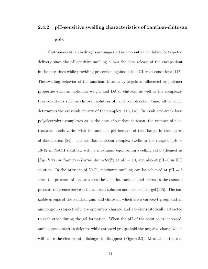
of dissociation [85]. The xanthan-chitosan complex swells in the range of pH =
10-12 in NaOH solution, with a maximum equilibrium swelling ratio (defined as
[Equilibrium diameter/Initial diameter]3) at pH = 10, and also at pH=0 in HCl
solution. In the presence of NaCl, maximum swelling can be achieved at pH = 8
since the presence of ions weakens the ionic interactions and increases the osmotic
pressure difference between the ambient solution and inside of the gel [115]. The ion-
izable groups of the xanthan gum and chitosan, which are a carboxyl group and an
amino group respectively, are oppositely charged and are electrostatically attracted
to each other during the gel formation. When the pH of the solution is increased,
amino groups start to deionize while carboxyl groups hold the negative charge which
will cause the electrostatic linkages to disappear (Figure 2.3). Meanwhile, the car-
14
boxyl groups attract Na+ ions and water diffuses into the complex. Both of these
phenomena increase the osmotic pressure of the gel and consequently, the complex
swells. However, equilibrium swelling ratio decreases in NaOH solution at pH values
higher than 10 since too much increase in Na+ concentration decreases the difference
of osmotic pressure between the gel and ambient solution. Similarly, the swelling
in acidic solutions can be explained by the neutralization of the negative charges of
xanthan while the amino groups of chitosan hold their positive charge, resulting in
the swelling of the gel complex [115].
Swelling capsules formed by xanthan-chitosan complexation can be described
as hollow spheres that expand until equilibrium is reached in the release solution.
Determination of the effective diffusion coefficient of hydrogels is very complex since
their properties are affected by many parameters. Due to the swelling and destruc-
tion of the linkages between two polymers, the radius of the capsules and the amount
of the impermeable segments (obstruction effect) change with time which makes the
calculations more complex. However, effective diffusion coefficient is expected to
increase with the increased swelling and decreased polymer fraction [120].
15
NaOH
+
+
-
-
-
-
+
+
+
+
-
-
-
-
Chitosan Xanthan
+ :NH:NH 33++ - :COO:COO -- :NH:NH 22
Am ino groups are deionized
HH 22 OO
HH 22 OO
(a)
+
+
+
+
-
-
-
-
Chitosan Xanthan
NaOH
NaCl
Na+
Na+
Na+
Na+
-
-
-
-
+ :NH:NH33
++ - :COO:COO -- :NH:NH22
(b)
Figure 2.3: pH-sensitive swelling of xanthan-chitosan complex: (a) In alkaline pH (10 to 12), aminogroups deionize and electrostatic linkages disappear; whereas (b) attraction of Na+ ions increasesosmotic pressure difference and causes swelling of the gel where addition of salt helps achieve theswelling at a lower pH such as pH 8.
16
Chapter 3
Effects of Complexation
Conditions on Xanthan-Chitosan
Polyelectrolyte Complex Gels
3.1 Introduction
Polyelectrolyte hydrogels formed by xanthan gum and chitosan can be used
for encapsulation and controlled release of food ingredients, cells, enzymes, and
therapeutic agents. A variety of xanthan-chitosan PEC gels can be obtained by
changing the molecular properties of the xanthan and chitosan polymers, such as
molecular weight, degree of acetylation of chitosan, and pyruvic acid content of
xanthan, as well as changing the complexation conditions, such as chitosan solution
pH, polymer concentration, complexation time, and mixing ratio [113,118,119,121].
This study addresses the importance of polymer concentration and chitosan
17
solution pH in the complexation of xanthan gum and chitosan in the form of cap-
sules. The swelling degree of the microcapsules formed by different combinations of
xanthan and chitosan was studied as an indication of the crosslink density of the
hydrogel membrane. Crosslink density is an important factor determining the sta-
bility, pH-sensitive swelling behavior (thus the release properties), as well as the me-
chanical strength of hydrogel networks. [113, 122–124]. An increase in the crosslink
density restricts the degree of swelling and reduces the pH-sensitivity by improv-
ing the stability of the network. Hence, by controlling the amount of crosslinking,
xanthan-chitosan microcapsules suitable for targeted release of probiotics can be
prepared.
3.2 Materials and methods
3.2.1 Preparation of chitosan and xanthan solutions
Chitosan from crab shells with a minimum deacetylation amount of 85% and a
molecular weight of 370 000 (reported by the supplier) was purchased from Sigma-
Aldrich Chemicals (St. Louis, MO). A known amount of chitosan was dissolved
in 1 N HCl by heating and agitating. The desired solution pH was adjusted by 1
M NaOH and DI water was added to bring it to the final volume. Xanthan gum
with a molecular weight of 1.02 million (TICAXAN r) was kindly supplied by TIC
Gums (Belcamp, MD). A predetermined amount of xanthan gum was dissolved in
DI water under heating and agitation. Both solutions were autoclaved before use.
18
3.2.2 Capsule formation and Inverted microscopy
In this study, the extrusion (complex coacervation) method was used. Capsules
were formed by dropwise addition of a solution of xanthan (50 mL) into a solution
of chitosan (300 mL) using a manually operated syringe with a 0.7-mm cannula
(Becton-Dickinson, Franklin Lakes, NJ). The chitosan solution was agitated con-
tinuously for 40 min to allow crosslinking and avoid coalescence of capsules. The
capsules were filtered through a 160 µm Millipore nylon filter, washed twice with
DI water, and then freeze-dried for 24 hours.
NHS-Fluorescein labeled chitosan, prepared according to Yi et al. [125], was
kindly obtained from Dr. Payne’s lab in the Center of Biosystems Research at
University of Maryland, College Park. Fluorescently-labeled capsules were examined
under Axiovert 200 Inverted microscope (Carl Zeiss, Inc, Oberkochen, Germany).
3.2.3 Determination of the swelling degree
The effect of three complexation parameters, namely initial xanthan solution
concentration (0.5, 0.7, 1.0, and 1.5% w/v), initial chitosan solution concentration
(0.7 and 1.0% w/v) and chitosan solution pH (4.5, 5.5, and 6.2) on the degree
of swelling of the resulting capsules were studied. Ten freeze-dried capsules were
weighed and suspended in DI water overnight for each combination. The capsules
were filtered, blotted to remove surface water, and weighed. The swelling degree
(SD) values were calculated using the following equation:
19
SD (%) =Ws −Wd
Wd
× 100 (3.1)
where Ws and Wd are the weight of swollen capsules and that of dry capsules,
respectively. The averages of four replicates for each combination were reported.
3.2.4 Differential Scanning Calorimetry (DSC) measurements
Differential scanning calorimetry (DSC) measurements were carried out on
a TA Instruments Q100 DSC device (New Castle, DE). The cell resistance and
capacitance calibrations were performed in two steps. The first step was heating
an empty cell and the second step was heating the cell with equal weight sapphire
disks on the sample and reference platforms. The cell constant and temperature
calibrations were performed with an indium standard.
Standard DSC was used for the first heating and cooling runs. Approximately
5 mg of dry capsules were placed in sealed aluminum pans in small pieces. Each
sample was heated up to 160� at a rate of 10�/min and cooled back to 40� at
a rate of 5�/min to erase the thermal history of the polymers and eliminate the
effect of moisture. Modulated DSC curves were obtained from the second heating
run at 2�/min. Samples were heated from 100� to 175� with a modulation period
of 60 seconds and modulation amplitude of 0.318�. A nitrogen purge was applied
for all experiments. The reversing signal was used to compare the glass transitions
of the samples. In addition, chitosan in flakes and xanthan in powder form were
subjected to the same experimental procedure to determine their glass transition
20
temperatures.
3.2.5 Statistical analysis
Statistical analyses were conducted using SAS 9.1.2 Software (Cary, NC). Fac-
torial analysis of variance was used to analyze the effect of xanthan concentration,
chitosan concentration, and chitosan solution pH on the swelling degree of the cap-
sules. Differences in least square means were used for pairwise mean comparison.
Analyses were performed using mixed procedure of SAS.
3.3 Results and discussion
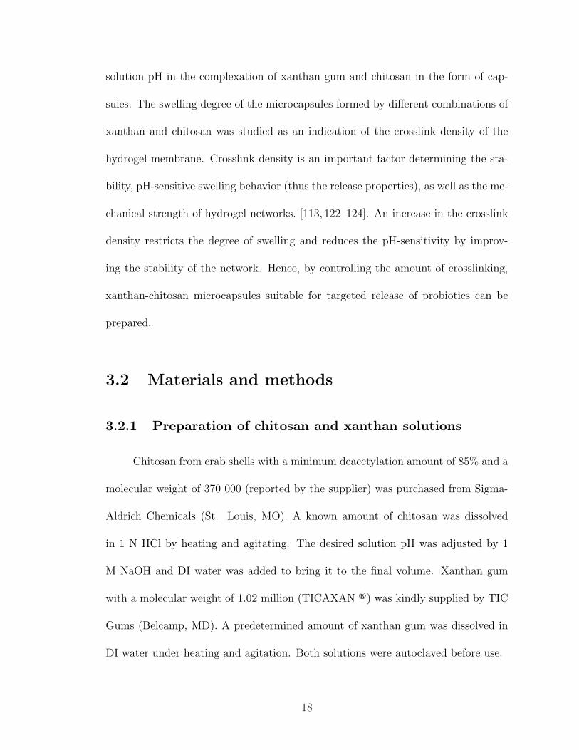
3.3.1 Capsule formation
By extruding the xanthan solution into moderate concentrations of chitosan
(0.7 and 1.0% [w/v]), no stable capsules could be formed while xanthan concen-
trations were below 0.5% (w/v). Xanthan concentrations exceeding 1.5% (w/v)
resulted in formation of amorphous capsules (Figure 3.1-a, Figure 3.1-b). The pH
of chitosan solution was controlled, ranging from 4.5, where the ionization degree of
chitosan is unity, to 6.2, since chitosan precipitates above its pKa value of approxi-
mately 6.3.
Capsules formed by xanthan-chitosan complexation can be described as hol-
low spheres. Figure 3.1-c shows a xanthan-chitosan capsule prepared by NHS-
Fluorescein labeled chitosan under inverted microscope.
21
2mm
(a)
2mm
(b)
(c)
Figure 3.1: Xanthan-chitosan capsules formed when initial xanthan concentration is (a) greaterthan 1.5% (w/v); (b) less than 1.5% (w/v); (c) Inverted microscopy image of a 2 mm-diameterxanthan-chitosan capsule prepared with fluorescently labeled chitosan.
3.3.2 Effect of complexation conditions on the swelling de-
gree of xanthan-chitosan capsules
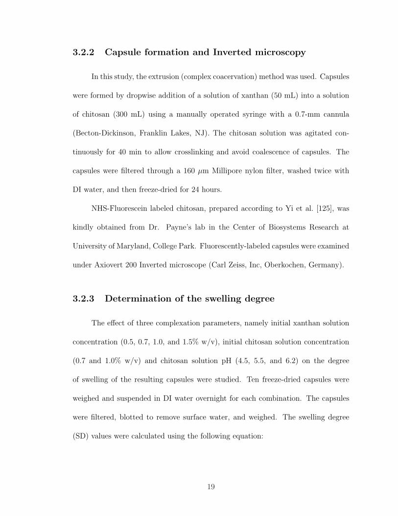
The swelling degree of microcapsules formed by different combinations of xan-
than and chitosan was studied as an indication of the crosslink density of the hydro-
gel membrane. The degree of swelling decreased by approximately 50% when using
0.7% (w/v) xanthan solution instead of 0.5% (w/v)(Figure 3.2). Therefore capsules
formed from xanthan solutions at concentration of 0.5% (w/v) were not included in
the statistical analysis and DSC studies.
22
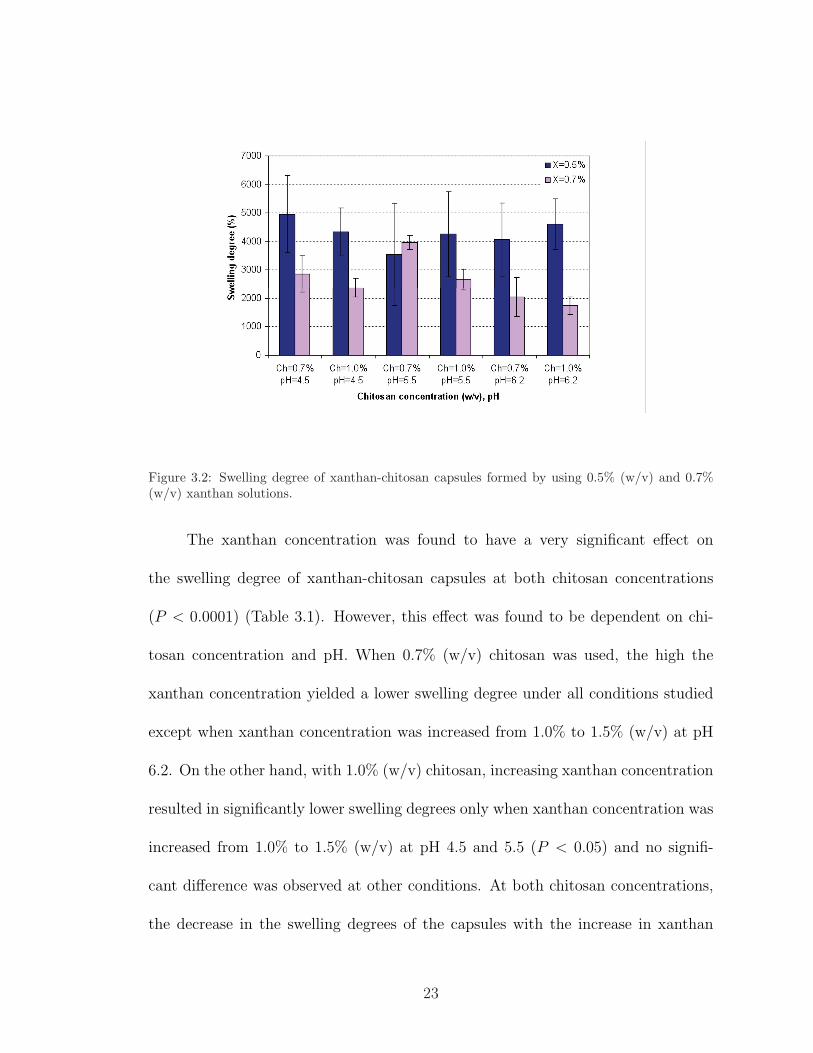
Figure 3.2: Swelling degree of xanthan-chitosan capsules formed by using 0.5% (w/v) and 0.7%(w/v) xanthan solutions.
The xanthan concentration was found to have a very significant effect on
the swelling degree of xanthan-chitosan capsules at both chitosan concentrations
(P < 0.0001) (Table 3.1). However, this effect was found to be dependent on chi-
tosan concentration and pH. When 0.7% (w/v) chitosan was used, the high the
xanthan concentration yielded a lower swelling degree under all conditions studied
except when xanthan concentration was increased from 1.0% to 1.5% (w/v) at pH
6.2. On the other hand, with 1.0% (w/v) chitosan, increasing xanthan concentration
resulted in significantly lower swelling degrees only when xanthan concentration was
increased from 1.0% to 1.5% (w/v) at pH 4.5 and 5.5 (P < 0.05) and no signifi-
cant difference was observed at other conditions. At both chitosan concentrations,
the decrease in the swelling degrees of the capsules with the increase in xanthan
23
concentration was more pronounced when chitosan solution pH was 5.5 and least
significant at pH 6.2.
Table 3.1: Swelling degrees of xanthan-chitosan capsules in DI water
Chitosan (w/v)
0.7% 1.0%
Xanthan (w/v) pH 4.5 pH 5.5 pH 6.2 pH 4.5 pH 5.5 pH 6.2
0.7% ∗29i 40 21m 24bgi 27cg 17dm
1.0% 21j 30 13an 22bhj 26ch 12dn
1.5% 13ek 16el 15aeo 16fk 11fl 15dfo
Mean values with same letter are not significantly different at P=0.05 level.
* Swelling degree/100
The effect of chitosan solution pH on the degree of swelling was more significant
when the chitosan solution concentration was 0.7% (w/v) (P < 0.0001) than when
it was 1.0% (w/v) (P < 0.005) (Table 3.1). With 1.5% (w/v) xanthan concentra-
tion, the increases in pH had no significant effect on the swelling degree (P > 0.05),
whereas at other xanthan concentrations, the degree of swelling decreased signifi-
cantly when chitosan pH was increased from 4.5 to 6.2. Such significant decreases
in swelling degree with increasing chitosan pH could be attributed to the changes
in the chain flexibility of chitosan polymer with the changes in the solution pH.
The ionization degree of chitosan decreases from 1.0 to 0.5 as pH increases from 4.5
to 6.2 [126], which means that amino groups become less charged as pH increases.
As a result, one may expect that fewer ionic linkages would occur between the two
24
polymers, resulting in higher swelling degrees as the pH approaches to its pKa value.
However, since the charge density of the chitosan molecule is reduced by almost 50%
as pH approaches 6.2 from a value of 4.5, the polymer chains become less extended
with a smaller radius of gyration. This might result in a higher diffusion coefficient
for chitosan chains at pH 6.2, consequently enhancing diffusion of chitosan into the
xanthan-chitosan network and forming more linkages during the specified reaction
time. This result suggests that since the formation of the xanthan-chitosan network
is instantaneous upon contact, the diffusion of chitosan chains in the bulk solu-
tion through this network plays an important role to achieve a highly crosslinked
structure with a small swelling degree.
Intriguingly, when 0.7 and 1.0% (w/v) xanthan solutions were extruded into
0.7% (w/v) chitosan solution, increases of chitosan pH from 4.5 to 5.5, first signifi-
cantly increased the degree of swelling before it reached the lowest swelling degree at
pH 6.2. This significant increase in the swelling degree when chitosan pH was raised
from 4.5 to 5.5 might be associated with the slight decrease in the charge density of
chitosan (ionization degree ca. 0.9) that lessens the ionic linkages between the two
polymers, rendering higher swelling degrees. Magnin et al. [113] have demonstrated
similar results where the swelling degree of xanthan-chitosan matrix continued to
increase with increasing chitosan solution pH from 3.5 to 5.8 in bulk form by us-
ing 0.65 wt% chitosan and 0.65 wt% xanthan solutions. Moreover, our results also
showed that when 1.0% (w/v) chitosan concentration was used, pH change from 4.5
to 5.5 chitosan solution had no significant effect on the swelling degree, suggesting
that the swelling degree was less affected by the decrease in the charge density of the
25
chitosan chains as pH approaches to 5.5 when chitosan concentration is increased.
The effect of chitosan solution concentration on the swelling degree of capsules
was less pronounced than the effect of xanthan solution concentration and chitosan
solution pH. Increasing chitosan concentration from 0.7 to 1.0% (w/v) significantly
decreased the degree of swelling only when chitosan pH was 5.5 and at xanthan
concentrations of 0.7 or 1.0% (w/v). No significant difference was observed at other
conditions. These findings indicate that the parameters studied cannot be viewed
as independent parameters, as the effect of one parameter on the degree of swelling
depends on the other two parameters. While swelling studies were capable of iden-
tifying the combined effect of polymer concentration and chitosan solution pH on
the crosslink densities of xanthan-chitosan hydrogel capsules, further investigation
is needed to understand the differences in the membrane structure as influenced by
the xanthan-chitosan hydrogel preparation conditions. Therefore, DSC analysis was
performed to compare the changes in thermal transitions in order to elucidate the
changes in the crosslinking density of the capsule network.
3.3.3 DSC analysis of xanthan-chitosan capsules
Conventional DSC was used for the first heating and cooling runs. The first
heating run of each sample gave single endothermic peak at about 100�, which
was attributed to the absorbed water. Samples were cooled back to 40� before the
second heating. The modulated differential scanning calorimetry (MDSC) technique
was used for the second heating. MDSC applies two simultaneous heating rates to
26
the sample. The linear heating rate provides total heat flow as conventional DSC and
sinusoidal (modulated) heating rate provides the heat capacity-related (reversing)
component of the total heat flow. The reversing signal was used to quantify the glass
transition since it separates the glass transition completely from other non-reversing
processes [127,128]. The transition enthalpies were calculated by integration of the
peaks on the reversing heat capacity (Rev Cp) curves as is usually done for first-order
phase transitions [129].
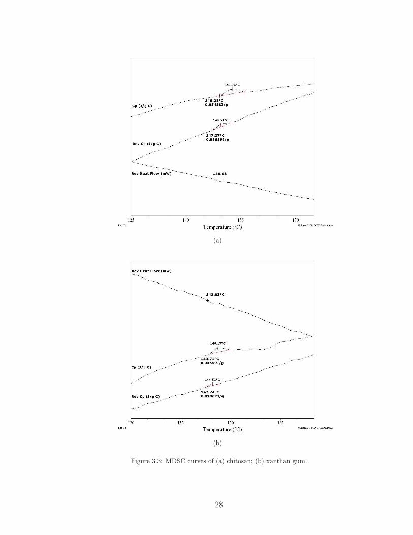
The small glass transitions observed for both chitosan and xanthan can be
explained by the fact that both polymers are partially crystalline (Figure 3.3). The
inflection points of the peaks on the Rev Cp curves correspond to the glass transition
temperature (Tg) of the samples. The Tg of chitosan was determined to be approx-
imately 148� and the enthalpy of this transition (H) was calculated as 0.016 J/g.
The Tg for xanthan gum was found to be at approximately 143� with a transition
enthalpy of H=0.01 J/g.
Representative reversing heat flow curves from the second heating runs of
the freeze-dried hydrogel capsules are shown in Figures 3.4 and 3.5. The transition
enthalpies and the glass transition temperatures were determined as described above.
It is evident from these data that the polymer complex shows weak transitions as
expected in physically crosslinked networks. For this reason, the transition enthalpy
of xanthan gum, 0.01 J/g, was selected as the threshold enthalpy to differentiate
the noise from the actual transitions appearing on the MDSC curves of the hydrogel
capsules.
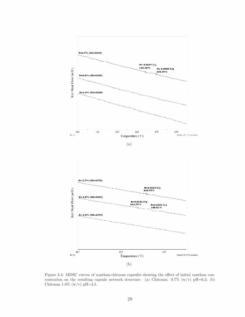
Figure 3.4 shows the effect of initial xanthan concentration on the capsule
27
(a)
(b)
Figure 3.3: MDSC curves of (a) chitosan; (b) xanthan gum.
28
(a)
(b)
Figure 3.4: MDSC curves of xanthan-chitosan capsules showing the effect of initial xanthan con-centration on the resulting capsule network structure. (a) Chitosan: 0.7% (w/v) pH=6.2; (b)Chitosan 1.0% (w/v) pH=4.5.
29
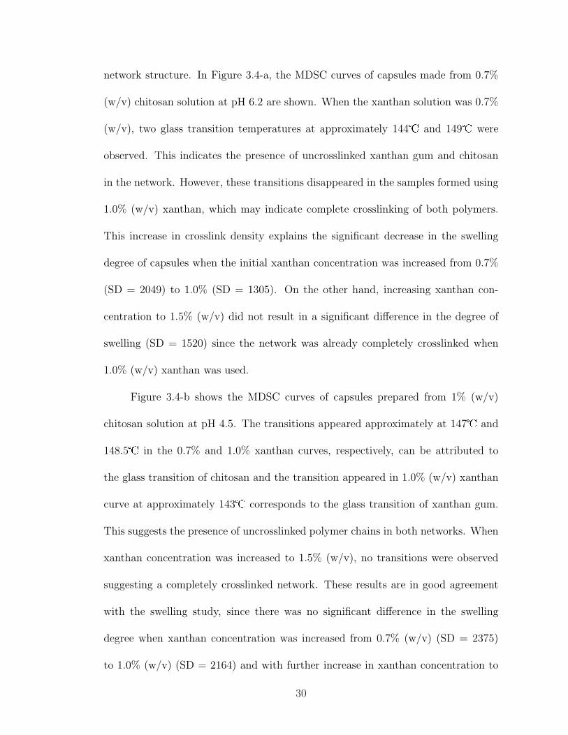
network structure. In Figure 3.4-a, the MDSC curves of capsules made from 0.7%
(w/v) chitosan solution at pH 6.2 are shown. When the xanthan solution was 0.7%
(w/v), two glass transition temperatures at approximately 144� and 149� were
observed. This indicates the presence of uncrosslinked xanthan gum and chitosan
in the network. However, these transitions disappeared in the samples formed using
1.0% (w/v) xanthan, which may indicate complete crosslinking of both polymers.
This increase in crosslink density explains the significant decrease in the swelling
degree of capsules when the initial xanthan concentration was increased from 0.7%
(SD = 2049) to 1.0% (SD = 1305). On the other hand, increasing xanthan con-
centration to 1.5% (w/v) did not result in a significant difference in the degree of
swelling (SD = 1520) since the network was already completely crosslinked when
1.0% (w/v) xanthan was used.
Figure 3.4-b shows the MDSC curves of capsules prepared from 1% (w/v)
chitosan solution at pH 4.5. The transitions appeared approximately at 147� and
148.5� in the 0.7% and 1.0% xanthan curves, respectively, can be attributed to
the glass transition of chitosan and the transition appeared in 1.0% (w/v) xanthan
curve at approximately 143� corresponds to the glass transition of xanthan gum.
This suggests the presence of uncrosslinked polymer chains in both networks. When
xanthan concentration was increased to 1.5% (w/v), no transitions were observed
suggesting a completely crosslinked network. These results are in good agreement
with the swelling study, since there was no significant difference in the swelling
degree when xanthan concentration was increased from 0.7% (w/v) (SD = 2375)
to 1.0% (w/v) (SD = 2164) and with further increase in xanthan concentration to
30
1.5% (w/v), the degree of swelling decreased significantly (SD = 1573).
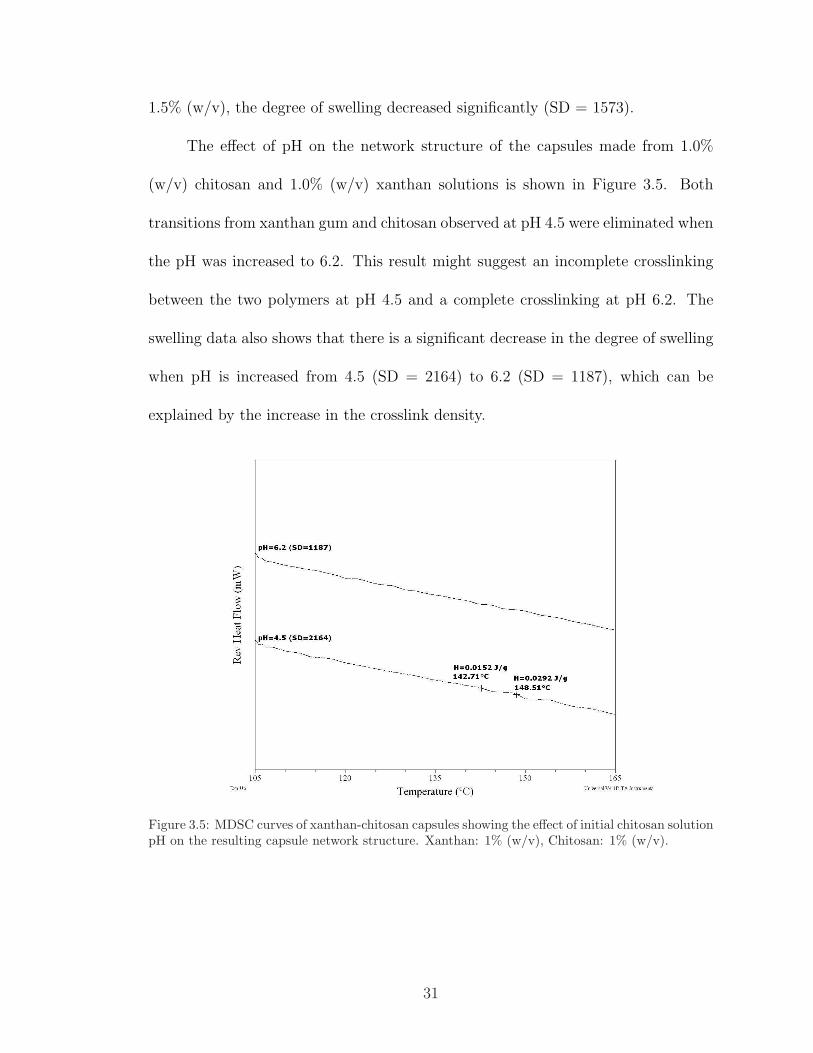
The effect of pH on the network structure of the capsules made from 1.0%
(w/v) chitosan and 1.0% (w/v) xanthan solutions is shown in Figure 3.5. Both
transitions from xanthan gum and chitosan observed at pH 4.5 were eliminated when
the pH was increased to 6.2. This result might suggest an incomplete crosslinking
between the two polymers at pH 4.5 and a complete crosslinking at pH 6.2. The
swelling data also shows that there is a significant decrease in the degree of swelling
when pH is increased from 4.5 (SD = 2164) to 6.2 (SD = 1187), which can be
explained by the increase in the crosslink density.
Figure 3.5: MDSC curves of xanthan-chitosan capsules showing the effect of initial chitosan solutionpH on the resulting capsule network structure. Xanthan: 1% (w/v), Chitosan: 1% (w/v).
31
3.4 Conclusions
Characterization of factors contributing to the crosslink density of xanthan-
chitosan network is important in developing hydrogels with particular mechanical
and controlled release properties. Results from the swelling degree and DSC experi-
ments showed that the crosslink density of xanthan-chitosan network was dependent
on the complexation conditions employed in the present study. Xanthan concen-
tration was found to be the most critical parameter in xanthan-chitosan network
formation. The hydrogel capsules were completely crosslinked at all conditions stud-
ied when initial xanthan solution concentration was at 1.5% (w/v). The increase in
xanthan concentration significantly affected the degree of swelling of the hydrogel at
both chitosan concentrations. On the other hand, the effect of chitosan solution pH
on the degree of swelling was more pronounced at 0.7% (w/v) than at 1.0% (w/v)
chitosan concentration. The swelling degree was less dependent on chitosan con-
centration than xanthan concentration and chitosan solution pH. Conformational
changes of chitosan polymer chains, which is dependent on the solution pH, were
critical in determining the crosslinked network structure that affects the swelling
degrees of resulting gels. Results from this study showed that pH and concentration
effects on the xanthan-chitosan network properties are dependent on each other.
It can be concluded that, the xanthan-chitosan network properties can be easily
modulated by changing operationally controllable parameters, especially xanthan
concentration and chitosan solution pH.
32

Chapter 4
The Release Kinetics and Swelling
Behavior of Xanthan-Chitosan
Capsules
4.1 Introduction
Xanthan-chitosan hydrogels swell in response to changes in pH, temperature,
and ionic strength of the aqueous environment. In swelling controlled systems,
the molecular structure as well as the nature of the polymer system control the
mechanism by which a solute may be released from a polymer network [130] . At
high or very low pH values, the electrostatic linkages between xanthan gum and
chitosan start to disappear, allowing the network to expand and imbibe water [115].
The release of probiotic cells by such a pH-sensitive swelling-controlled mechanism
is related to the macromolecular chain relaxation and to the diffusion of the cells
33
through the polymer membrane under countercurrent diffusion of water or biological
fluids into the capsules.
It is important that the encapsulation system developed in this study keeps the
cell release to a minimum until the capsules reach the small intestines where the rapid
release of the cells is desirable for their colonization. For this reason, the kinetics of
cell release under different conditions need to be studied in order to understand the
response of the system to environmental changes, particularly variations in ambient
pH. The main goal of this study was to characterize the swelling and release behavior
under simulated GI-tract conditions.
4.2 Materials and Methods
4.2.1 Calibration curve
Pediococcus acidilactici (MA18/5M, National Collection of Microorganism
Culture, Pasteur-France) was kindly provided by Imagilin Technology LLC (Po-
tomac, MD). One gram of bacterial powder was hydrated in 9 mL of DI water for
30 min by shaking at 260 rpm. One mL hydrated bacteria was inoculated to 99
mL MRS broth and incubated for 24 hours at 35�. To construct the calibration
curve, a 25 mL sample was taken from MRS broth, centrifuged at 3000 rpm for 30
minutes and washed with sterile distilled water under the same conditions. After
necessary dilutions, OD values at 600 nm were recorded by a Heβ IOS Spectropho-
tometer (ThermoSpectronic, Rochester, NY) and the corresponding samples were
34

oven dried overnight for mass determination.
4.2.2 Microencapsulation of P. acidilactici
One milliliter of hydrated P. acidilactici cells was inoculated into 99 mL MRS
broth and incubated at 35� for 24 hours. Actively growing cells were recovered from
MRS broth by centrifuging at 10000 rpm for 10 minutes and then were washed twice
with sterile phosphate buffered saline (PBS) solution under the same centrifugation
conditions. DI water was added to the cell pellet and vortexed. Xanthan and
chitosan solutions were prepared as described in section 3.2.1. Encapsulation was
achieved by dropwise addition of xanthan and P. acidilactici mixture (9:1 v/v)
into chitosan solution using a manually operated syringe with 0.7-mm cannula. The
chitosan solution was agitated continuously for 40 min to allow crosslinking and to
avoid coalescence of capsules. The capsules were filtered through a 160µm Millipore
nylon filter, washed twice with DI water, and then freeze-dried for 24 hours.
4.2.3 Kinetics of cell release
A known amount of freeze-dried capsules were suspended in release solutions,
namely simulated gastric fluid (Fisher Chemicals, Suwanee, GA), simulated intesti-
nal fluid (Fisher Chemicals, Suwanee, GA), and DI-water. The cell concentration
in the solutions was monitored over time. A 1 mL sample was taken periodically
in order to monitor the changes in OD values at 600 nm by using a HeβIOS Spec-
trophotometer (ThermoSpectronic, Rochester, NY). Consequently, 1 mL of release
35
solution was added after each sample is taken in order to avoid any errors in calcula-
tions. Dilutions were performed when necessary. The calibration curve of the strain
was used to relate the OD values to cell concentration. Averages of two replicates
for each sample were reported. Cell release kinetics were studied in deionized water
for 72 hours at room temperature (under 150 rpm); in simulated intestinal fluid
(composed of pancreatin, KH2PO4 and NaOH ) at pH=6.8 for 24 hours at room
temperature (under 150 rpm); in simulated gastric fluid (composed of pepsin, NaCl
and HCl) at pH=1.0 and pH=2.0 (37� under 150 rpm); and finally in simulated
intestinal fluid after 2 hour exposure to simulated gastric fluid at pH=2.0 (37�
under 150 rpm).
The cell release data were analyzed according to the Higuchi equation [131],
eqn (4.1); Korsmeyer-Peppas equation [132], eqn (4.2); and Peppas-Sahlin equation
[133], eqn (4.3) using MATLAB version 6 R13 (MathWorks Inc, Natick, MA). Non-
linear least squares fitting method was used to determine the parameters in each
equation.
Mt
Mf
= k′t1/2 (4.1)
Mt
Mf
= ktn (4.2)
Mt
Mf
= kdtm + krt
2m (4.3)
where Mt is the concentration of the cells released at time t, Mf is the concentra-
tion of the cells released at equilibrium, k and k′ are constants incorporating the
structural and geometric characteristics of the hydrogel, and n is the release expo-
nent describing the mode of the transport mechanism, and m is the purely Fickian
36
diffusion exponent for a system of any geometrical shape.
4.2.4 Dynamic swelling behavior
Ten freeze-dried capsules were weighed and placed in simulated gastric fluid
followed by simulated intestinal fluid. The capsules were carefully removed from
the solutions, blotted for the removal of surface water and weighed at specified time
intervals. The swelling ratio, q, was calculated as follows:
q =Ws
Wd
(4.4)
where Ws is the weight of swollen capsules and Wd is the weight of the dried capsules.
The averages of three replicates for each combination were reported.
The water absorption data was analyzed according to the Higuchi equation,
eqn (4.1); Korsmeyer-Peppas equation, eqn (4.2); and Peppas-Sahlin equation, eqn
(4.3) using MATLAB version 6 R13. Non-linear least squares fitting method was
used to determine the parameters in each equation.
4.3 Results and Discussion
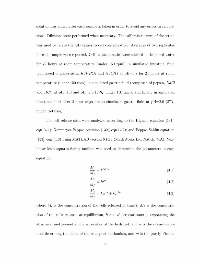
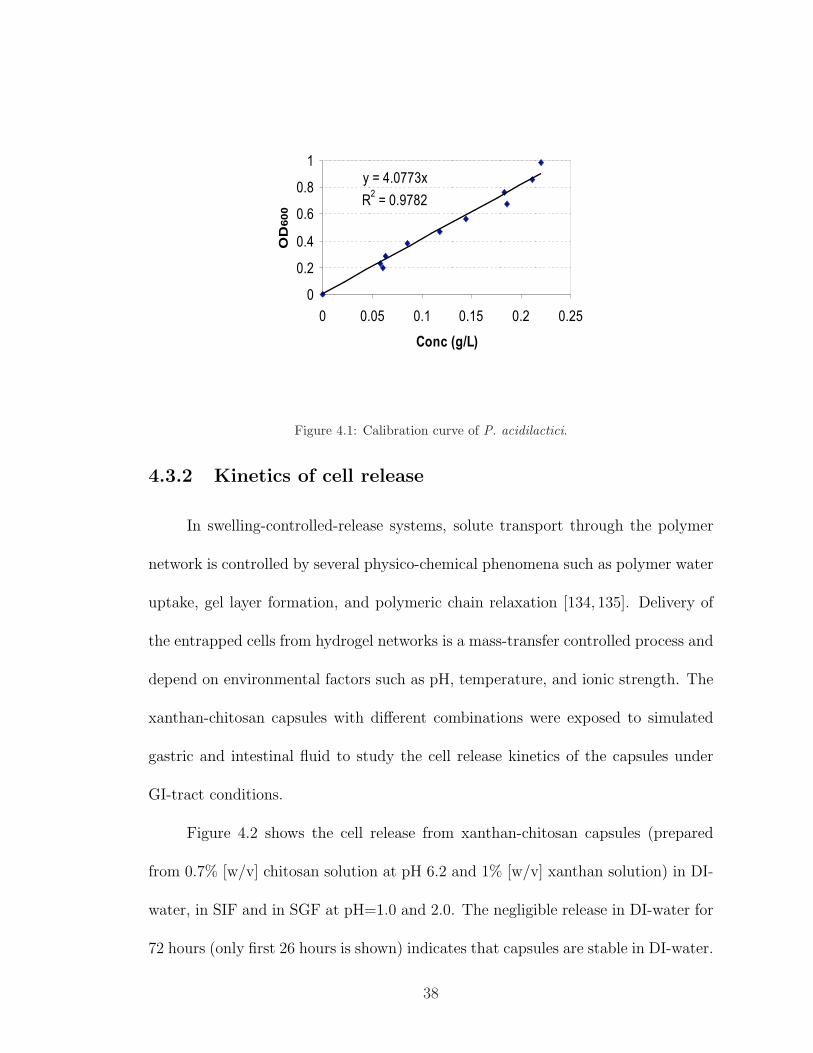
4.3.1 Calibration curve
The optical density of the cells in suspension was found to be related to cell
concentration by a factor of 4.0773 (Figure 4.1).
37
y = 4.0773x
R2 = 0.9782
0
0.2
0.4
0.6
0.8
1
0 0.05 0.1 0.15 0.2 0.25
Conc (g/L)
OD
600
Figure 4.1: Calibration curve of P. acidilactici.
4.3.2 Kinetics of cell release
In swelling-controlled-release systems, solute transport through the polymer
network is controlled by several physico-chemical phenomena such as polymer water
uptake, gel layer formation, and polymeric chain relaxation [134, 135]. Delivery of
the entrapped cells from hydrogel networks is a mass-transfer controlled process and
depend on environmental factors such as pH, temperature, and ionic strength. The
xanthan-chitosan capsules with different combinations were exposed to simulated
gastric and intestinal fluid to study the cell release kinetics of the capsules under
GI-tract conditions.
Figure 4.2 shows the cell release from xanthan-chitosan capsules (prepared
from 0.7% [w/v] chitosan solution at pH 6.2 and 1% [w/v] xanthan solution) in DI-
water, in SIF and in SGF at pH=1.0 and 2.0. The negligible release in DI-water for
72 hours (only first 26 hours is shown) indicates that capsules are stable in DI-water.
38
In SIF, the release from the capsules was very slow in the beginning and the release
rate increased after 8 hours. Maximum release of the cells from the capsules was
achieved after 24 hours in SIF. When capsules were suspended in SGF at pH=2.0,
the cell release was in negligible amounts for 2 hours. However, cells were released
rapidly in SGF at pH=1.0 and the OD values started to decrease after 2.5 hours,
indicating cell death due to low pH. These results suggest that the pH of the SGF
solution is more critical in determining the release properties of xanthan-chitosan
capsules than the presence of enzyme (pepsin). The enzymatic resistance of the
complex can be explained by the conformational changes that occur in chitosan after
complexation with enzymatically resistant xanthan gum. These changes might make
the sensitive sites of chitosan less accessible to hydrolytic action of the media [117].
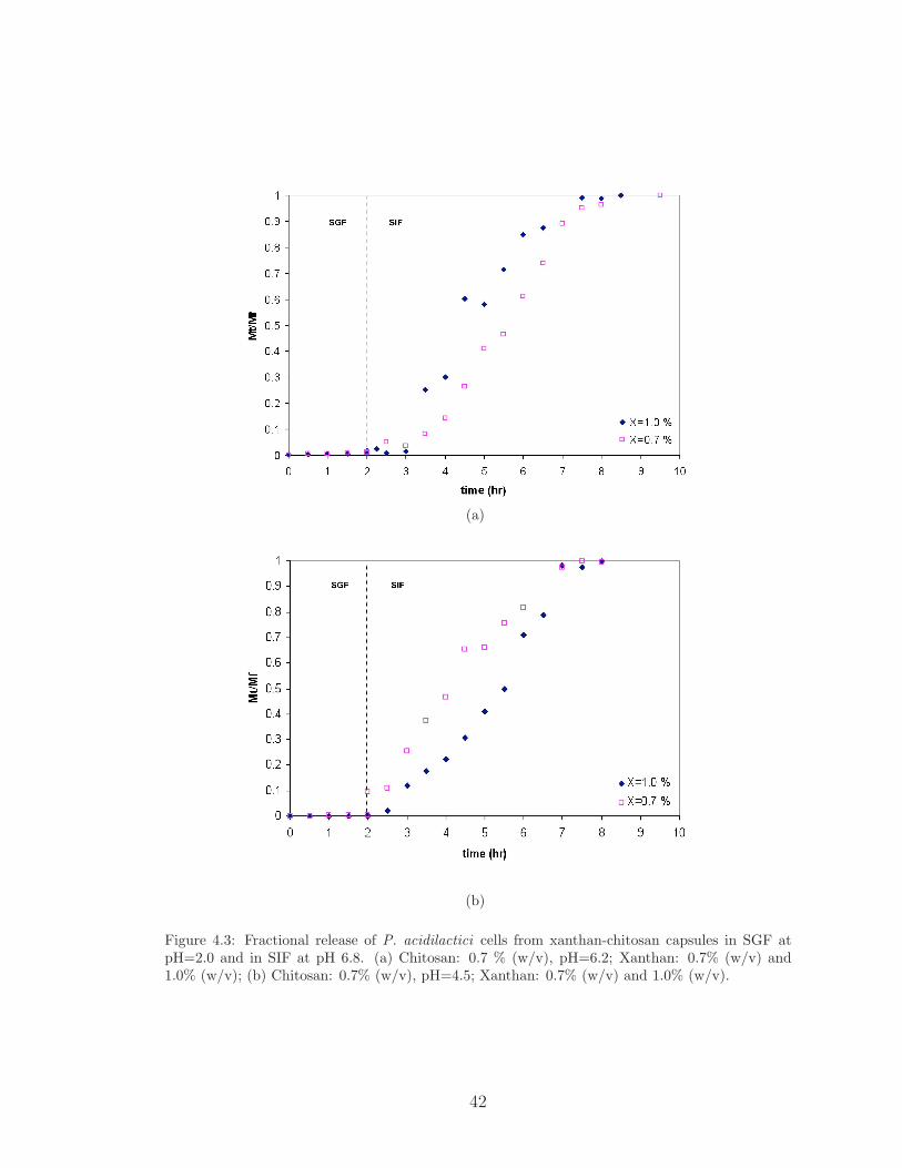
Figure 4.3 shows the cell release profiles from xanthan-chitosan capsules for
four different matrix formulations. Capsules were kept in SGF at pH 2.0 for 2 hours
prior to be suspended in SIF at pH 6.8. For all combinations, the cell release in SGF
was negligible. A time lag was observed in the early phase of the cell release from
the capsules in SIF, indicating a highly-crosslinked network structure [136]. The
time lag was longer for the capsules prepared from chitosan solution at pH 6.2 (60
min) than at pH 4.5 (30 min). Complete release of the encapsulated cells in SIF was
achieved after 5.5 hours and 5 hours of exposure when initial chitosan solution pH
was controlled at 6.2 and 4.5, respectively. Longer time lag and longer release time
observed in the case of capsules prepared from chitosan solution at pH 6.2 suggests a
more crosslinked hydrogel network compared to the membrane network of capsules
prepared from chitosan solution at pH 4.5. These results are in agreement with the
39

(a)
(b)
Figure 4.2: The release of P. acidilactici cells from xanthan- chitosan capsules (Chitosan: 0.7%(w/v), pH=6.2; Xanthan: 1.0% (w/v)) in (a) DI water and SIF; (b) in SGF at pH=1.0 and pH=2.0.
40
swelling degree experiments reported in Chapter 3.
Table 4.1: Analysis of cell release data from xanthan-chitosan capsules using Higuchi eqn,
Korsmeyer-Peppas eqn, and Peppas-Sahlin eqn.
Capsule Higuchi eqn∗ Korsmeyer-Peppas eqn∗ Peppas-Sahlin eqn∗
combinations k′ r2 n k r2 kd kr r2
with 0.7% Ch (min−0.5) (min−n) (min−0.43) (min−0.86)
pH=6.2; X=0.7% 0.2374 0.7888 2.043 0.03598 0.9924 -0.5222∗∗ 0.473 0.9936
pH=6.2; X=1.0% 0.0432 0.8704 1.233 0.00098 0.9467 -0.044∗∗ 0.0116 0.94881
pH=4.5; X=0.7% 0.3304 0.9459 1.082 0.2422 0.9954 -0.1275∗∗ 0.3724 0.9946
pH=4.5; X=1.0% 0.1954 0.8878 1.284 0.1001 0.9959 -0.1646∗∗ 0.2624 0.9951
∗k′ (min−0.5), Higuchi kinetic constant; k (min−n), Korsmeyer-Peppas kinetic constant; kd (min−0.43), diffusional
constant; kr (min−0.86), relaxational constant; r2, correlation coefficient.
∗∗The negative values obtained for kd should be interpreted in terms of a diffusion process insignificant compared
to the relaxation mechanism [137].
The portion of the cell release curve with a fractional release (Mt/Mf) between
0.1 and 0.6 was analyzed according to the Higuchi equation in (4.1); Korsmeyer-
Peppas equation in (4.2); and Peppas-Sahlin equation in (4.3). The Higuchi model is
applicable if the release is largely governed by diffusion through water-filled pores in
the matrix. A good fit to Kormeyer-Peppas equation indicates the combined effect of
diffusion and relaxation mechanisms for the release. For spherical systems, n = 0.43
indicates Fickian (diffusion controlled) transport, 0.43 < n < 0.85 indicates non-
Fickian (anomalous) transport and n = 0.85 implies Case II, zero-order (relaxation-
controlled) transport [133,138]. Values of n higher than 0.85 for release from spheres
are considered to be Super Case II kinetics [132]. The relative contribution of the
diffusion and relaxation processes to the release mechanism can be analyzed by
Peppas-Sahlin equation. The purely Fickian diffusion exponent, m, for xanthan-
chitosan microcapsules corresponds to a value of 0.43 since spherical matrices present
41
(a)
(b)
Figure 4.3: Fractional release of P. acidilactici cells from xanthan-chitosan capsules in SGF atpH=2.0 and in SIF at pH 6.8. (a) Chitosan: 0.7 % (w/v), pH=6.2; Xanthan: 0.7% (w/v) and1.0% (w/v); (b) Chitosan: 0.7% (w/v), pH=4.5; Xanthan: 0.7% (w/v) and 1.0% (w/v).
42

an aspect ratio of 1 [133]. The main parameter values from different equations are
summarized in Table 4.1 to describe the mode of transport mechanism.
A good fit to Korsmeyer-Peppas (n > 0.85) and Peppas-Sahlin (kr À kd)
equations suggests that the cell release from xanthan-chitosan capsules in SIF (af-
ter 2 hrs exposure to SGF) is controlled by polymer relaxation (Table 4.1). Since
Higuchi model is only applicable to diffusion-controlled release mechanism, the re-
lease data showed a poor fit to this equation. The n values higher than 0.85 reveals
a Super Case II transport mechanism for the release of cells from xanthan-chitosan
capsules regardless of the combination used. Super Case II transport mechanism
might result from an increased plasticization at the relaxing boundary (gel layer)
due to a reduction of the attractive forces among polymeric chains [139,140].
4.3.3 Dynamic swelling behavior
Xanthan-chitosan hydrogels exhibit pH-sensitive swelling characteristics due to
the deionization of the functional groups in the hydrogel which significantly affects
the penetrant transport mechanism of the polymer networks [115, 130]. Highly
swollen hydrogels contain large amounts of unbound water which allows greater
solute release [135]. The objective was to study the dynamic swelling behavior of
xanthan-chitosan capsules under simulated GI-tract conditions.
Figure 4.4 shows the dynamic swelling of xanthan-chitosan capsules, prepared
from 0.7% (w/v) chitosan at pH=6.2 and 0.7% (w/v) or 1.0% (w/v) xanthan in SGF
at pH=2.0 and in SIF. The swelling ratio, q, was constant in SGF and increased
43

(a)
(b)
Figure 4.4: (a) Dynamic swelling ; (b) water absorption curve of xanthan-chitosan capsules (pre-pared from 0.7% [w/v] chitosan at pH=6.2 in combination with 0.7% and 1.0% [w/v] xanthan) inSGF at pH=2.0 and in SIF at pH 6.8.
44
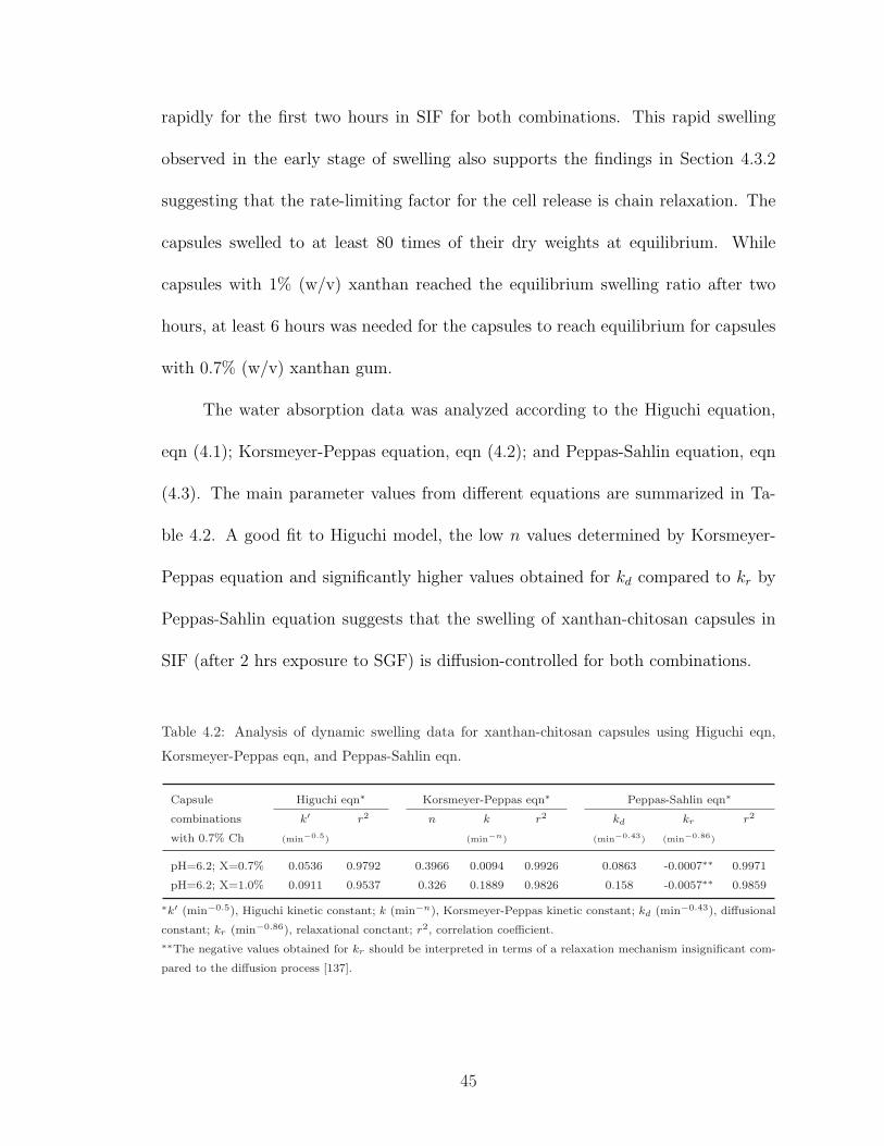
rapidly for the first two hours in SIF for both combinations. This rapid swelling
observed in the early stage of swelling also supports the findings in Section 4.3.2
suggesting that the rate-limiting factor for the cell release is chain relaxation. The
capsules swelled to at least 80 times of their dry weights at equilibrium. While
capsules with 1% (w/v) xanthan reached the equilibrium swelling ratio after two
hours, at least 6 hours was needed for the capsules to reach equilibrium for capsules
with 0.7% (w/v) xanthan gum.
The water absorption data was analyzed according to the Higuchi equation,
eqn (4.1); Korsmeyer-Peppas equation, eqn (4.2); and Peppas-Sahlin equation, eqn
(4.3). The main parameter values from different equations are summarized in Ta-
ble 4.2. A good fit to Higuchi model, the low n values determined by Korsmeyer-
Peppas equation and significantly higher values obtained for kd compared to kr by
Peppas-Sahlin equation suggests that the swelling of xanthan-chitosan capsules in
SIF (after 2 hrs exposure to SGF) is diffusion-controlled for both combinations.
Table 4.2: Analysis of dynamic swelling data for xanthan-chitosan capsules using Higuchi eqn,
Korsmeyer-Peppas eqn, and Peppas-Sahlin eqn.
Capsule Higuchi eqn∗ Korsmeyer-Peppas eqn∗ Peppas-Sahlin eqn∗
combinations k′ r2 n k r2 kd kr r2
with 0.7% Ch (min−0.5) (min−n) (min−0.43) (min−0.86)
pH=6.2; X=0.7% 0.0536 0.9792 0.3966 0.0094 0.9926 0.0863 -0.0007∗∗ 0.9971
pH=6.2; X=1.0% 0.0911 0.9537 0.326 0.1889 0.9826 0.158 -0.0057∗∗ 0.9859
∗k′ (min−0.5), Higuchi kinetic constant; k (min−n), Korsmeyer-Peppas kinetic constant; kd (min−0.43), diffusional
constant; kr (min−0.86), relaxational conctant; r2, correlation coefficient.
∗∗The negative values obtained for kr should be interpreted in terms of a relaxation mechanism insignificant com-
pared to the diffusion process [137].
45
4.4 Conclusions
In swelling-controlled systems, the coupling of diffusion and macromolecular
relaxation control the release mechanism. The xanthan-chitosan capsules were found
to be stable in DI water. The pH of SGF was critical in determining the release
properties of the capsules. The cell release in SGF at pH 2.0 was negligible suggesting
that xanthan-chitosan capsules have a good potential for delivery of probiotic cells
to intestines. The cell release from xanthan-chitosan capsules in SIF after 2 hr
exposure to SGF at pH 2.0, exhibited a Super Case II transport mechanism (n >
0.85) regardless of the formulation used. The complete release of the cells from
the capsules was achieved in at least 5 hours. Xanthan-chitosan capsules were
found to swell by a diffusion-controlled mechanism. The rapid swelling observed
also supported the findings that the cell release is relaxation-controlled (kr À kd).
46
Chapter 5
Protective effects of
Xanthan-Chitosan Encapsulation
on P. acidilactici cells
5.1 Introduction
For probiotic bacteria to be beneficial to the host, they should be able to
survive gastric transit and reach the small intestine in sufficient numbers [26, 33].
The harsh environment of the GI-tract (the low pH conditions of the stomach and the
presence of bile in the intestines) adversely affect the viability of probiotic cultures
[37, 38]. In this study, Pediococcus acidilactici cells were encapsulated in xanthan-
chitosan PEC gels to increase the survival rates during GI-tract transit.
Among the available techniques for encapsulating bacterial cells, entrapment
in xanthan-chitosan capsules has not been used for the encapsulation of probiotic
47

bacteria. The only application of xanthan-chitosan PEC gels as microcarriers for
bacterial cells was reported by Chu et al. [116] for the encapsulation of Corynebac-
terium glutamicum, a Gram (+) soil bacterium. The immobilization was carried out
by mixing xanthan-chitosan PEC solution with the cell suspension and adding this
mixture to distilled water through a syringe.
In this work, a simple and cost-effective extrusion method was used to en-
capsulate the probiotic cells. The goal was to determine the protective effects of
encapsulation with xanthan gum and chitosan on P. acidilactici cells against freeze-
drying and simulated gastric fluid at pH 2.0.
5.2 Materials and Methods
P. acidilactici cells were encapsulated as described in Section 4.2.2. Fifty
milliliters of cell-xanthan mixture were dropped into 200 mL of chitosan solution
by using a manually operated syringe to form the capsules. Freeze-dried capsules
were subjected to simulated gastric fluid (SGF) at pH 2.0 for 1 hour and 2 hours
at 37� under 150 rpm. Two different combinations (chitosan: 0.7% [w/v], pH 6.2;
xanthan: 1.0% [w/v] and chitosan: 1.0% [w/v], pH 6.2; xanthan: 0.7% [w/v]) were
used to encapsulate the bacteria. Capsules were filtered from SGF solution and
suspended in SIF solution for 5 hours at 37� under 150 rpm to release the cells
from the capsules. One milliliter aliquots from the release solutions were serially
diluted and 3 replicates for each dilution were plated on MRS agar and incubated
for 48 hours at 35�. The experiments were repeated twice for each combination.
48
Smaller capsules were prepared by spraying the xanthan gum solution at 0.7%
(w/v) through a 0.7 mm nozzle (Buchi B-290 Spray Dryer, Flawil, Switzerland)
into chitosan solutions at 3 different concentrations (0.4, 0.7, and 1.0 % [w/v]) and
2 different pH values (4.5 and 6.2). Moreover, the effect of increasing xanthan
concentration from 0.7 to 1.5% (w/v) on the protective properties of the capsules
was studied. Capsules were subjected to SGF at pH 2.0 for 1 hour and cells were
released from capsules in SIF solution. One milliliter aliquots from the release
solutions were serially diluted and 3 replicates for each dilution were plated on MRS
agar and incubated for 48 hours at 35�. The experiments were repeated three times
for each combination.
Free cells were mixed with 10% (w/v) skim milk (9:1 v/v), transferred to
freeze-drying flask and frozen in dry ice before connected to the freeze dryer (Ther-
moSavant, Holbrook, NY). Freeze-dried cells were suspended in SGF for 1 hour and
2 hours under 150 rpm. One milliliter samples from free-cell solution were serially
diluted and 3 replicates for each dilution were plated on MRS agar and incubated
for 48 hours at 35�.
5.3 Results and Discussion
P. acidilactici are used in dietary supplements and in animal feed for their
probiotic effects. They can produce large amounts of lactic acid that helps keep a
proper balance of microflora in the digestive system [141]. Nevertheless, to act as
a probiotic, the bacteria must survive in acidic stomach conditions and arrive in
49
the intestines in high numbers. Gastric juice has a very low pH of 0.9 in healthy
adults. However, the presence of food may raise the pH value to 3.0 and it takes 2
to 4 hours for stomach to empty after ingestion of food [142]. In this work, free and
encapsulated P. acidilactici cells were subjected to SGF at pH=2.0 for 1 hr or 2
hours at 37� with moderate shaking to evaluate the effect of encapsulation on the
survival of P. acidilactici cells in stomach conditions.
Two different combinations based on the swelling degree and DSC studies
(Chapter 3) were chosen to encapsulate the bacteria in the capsules prepared by us-
ing a syringe. The capsules formed with these combinations were highly crosslinked
with relatively small swelling degrees (Table 3.1). Xanthan-chitosan capsules pre-
pared from 0.7% (w/v) chitosan at pH 6.2 and 1.0% (w/v) xanthan showed six-log
preservation of P. acidilactici cells over free suspending cells after exposed to SGF
(pH=2.0) for 1 hr and over four-log retention when exposure time was 2 hours (Fig-
ure 5.1-a). The protection effect of the capsules prepared from 1.0 (w/v) chitosan
at pH 6.2 and 0.7% (w/v) xanthan was more than five logs and three logs after 1hr
and 2 hours in SGF, respectively (Figure 5.1-b).
The resistance of lactic acid bacteria to freeze-drying and low pH is strain
specific. It has been reported that survival rates of P. acidilactici in simulated
gastric juice (SGF) is similar to the survival rates of Lactococcus lactis subsp. lactis
and slightly lower than Lactobacillus casei subsp. casei [143]. Xanthan-chitosan
microcapsules were found to provide better protection to P. acidilactici cells against
freeze-drying and SGF than the protection provided by alginate capsules to L. casei
cells [144,145].
50

(a)
(b)
Figure 5.1: Protective effects of encapsulation on the viability of P.acidilactici cells against freeze-drying and SGF (pH=2.0) exposure (n=2) using (a) 0.7% and 1.0% chitosan.
51
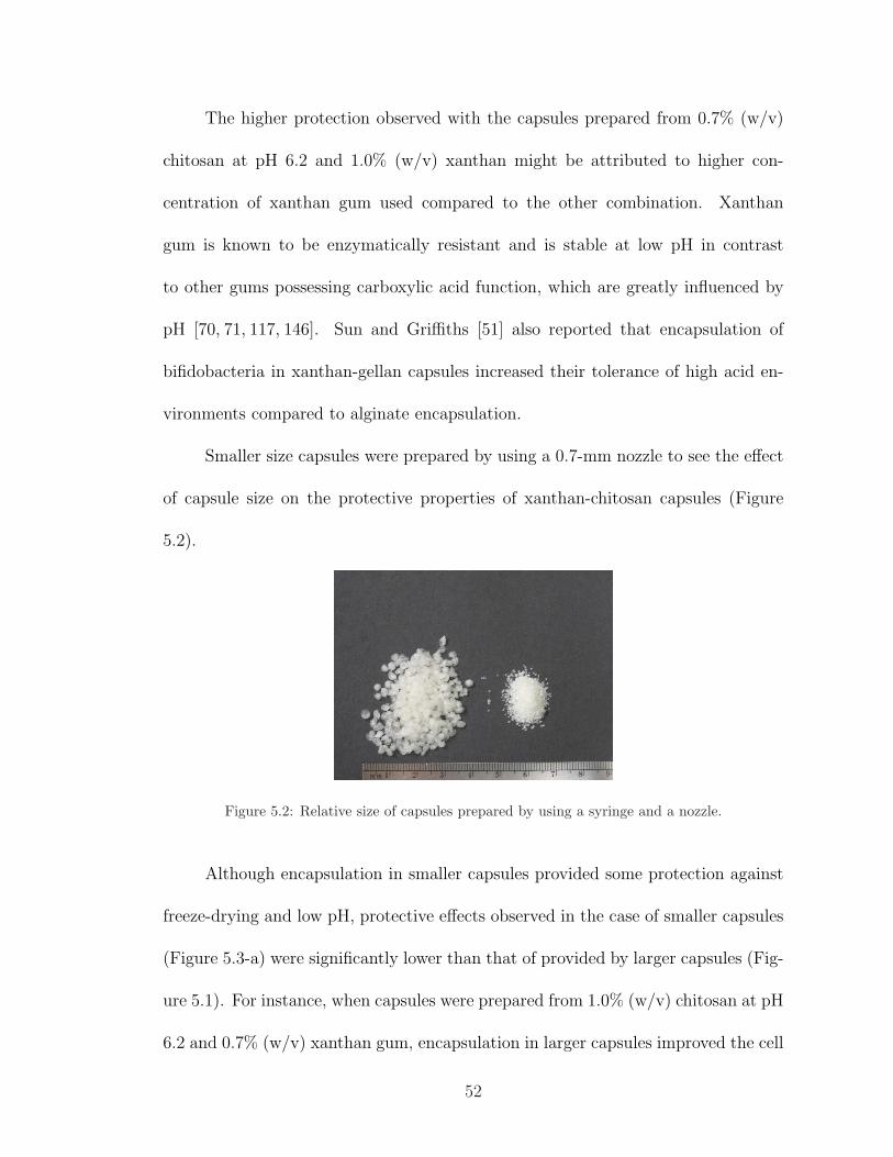
The higher protection observed with the capsules prepared from 0.7% (w/v)
chitosan at pH 6.2 and 1.0% (w/v) xanthan might be attributed to higher con-
centration of xanthan gum used compared to the other combination. Xanthan
gum is known to be enzymatically resistant and is stable at low pH in contrast
to other gums possessing carboxylic acid function, which are greatly influenced by
pH [70, 71, 117, 146]. Sun and Griffiths [51] also reported that encapsulation of
bifidobacteria in xanthan-gellan capsules increased their tolerance of high acid en-
vironments compared to alginate encapsulation.
Smaller size capsules were prepared by using a 0.7-mm nozzle to see the effect
of capsule size on the protective properties of xanthan-chitosan capsules (Figure
5.2).
Figure 5.2: Relative size of capsules prepared by using a syringe and a nozzle.
Although encapsulation in smaller capsules provided some protection against
freeze-drying and low pH, protective effects observed in the case of smaller capsules
(Figure 5.3-a) were significantly lower than that of provided by larger capsules (Fig-
ure 5.1). For instance, when capsules were prepared from 1.0% (w/v) chitosan at pH
6.2 and 0.7% (w/v) xanthan gum, encapsulation in larger capsules improved the cell
52
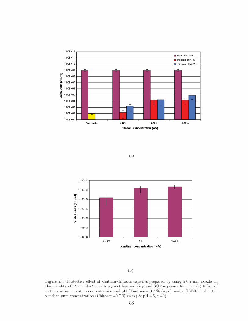
(a)
(b)
Figure 5.3: Protective effect of xanthan-chitosan capsules prepared by using a 0.7-mm nozzle onthe viability of P. acidilactici cells against freeze-drying and SGF exposure for 1 hr. (a) Effect ofinitial chitosan solution concentration and pH (Xanthan= 0.7 % (w/v), n=3), (b)Effect of initialxanthan gum concentration (Chitosan=0.7 % (w/v) & pH 4.5, n=3).
53

viability at least three logs compared to the encapsulation in small capsules. Simi-
lar results have been reported by other researchers. Lee and Heo [45] reported that
survival of bacterial cells under in vitro gastric conditions decreased with decreasing
capsule size (diameter 1-2.6 mm). Sheu and Marshall [21] indicated that larger bead
diameters provided more protection for Lactobacillus bulgaricus in frozen desserts.
Moreover, it has been reported that small beads of size less than 100 µm did not
significantly protect the probiotic bacteria in simulated gastric juice compared to
free cells [147]. Finally, the effect of increasing initial xanthan concentration on
the protective properties of xanthan-chitosan capsules was studied for the capsules
prepared by nozzle. It was found that increasing initial xanthan gum concentra-
tion from 0.7 to 1.5% (w/v) only slightly improved the cell viability when chitosan
solution concentration and pH were 0.7% (w/v) and 4.5, respectively (Figure 5.3-b).
These results suggest that xanthan-chitosan hydrogels have a promising pro-
tective mechanism for probiotic preservation.
5.4 Conclusions
Encapsulation of probiotic bacteria in capsules composed of xanthan gum and
chitosan increased the survival rate of the cells during freeze-drying and in simulated
gastric fluid compared to non-encapsulated cells. The viability of freeze-dried P.
acidilactici cells in simulated gastric conditions was found to increase with increased
capsule size. This approach might be useful in delivery of probiotic cultures to the
GI-tract in high numbers to benefit from the health promoting effects of probiotic
54
cells.
55
Chapter 6
Conclusions
In this dissertation, a microencapsulation system using polyelectrolyte com-
plex gels formed by xanthan gum and chitosan was developed to sustain the viability
of probiotic bacteria in simulated gastrointestinal tract conditions. First, the effects
of initial polymer concentration and chitosan solution pH on the crosslinking density
of the capsule network were investigated by swelling studies and modulated differen-
tial scanning calorimetry analysis. Xanthan concentration was found to be the most
critical parameter in xanthan-chitosan network formation. Conformational changes
of chitosan polymer chains as pH approaches to 6.2 were critical in determining
the crosslinked network structure. Swelling degree was less dependent on chitosan
concentration than xanthan concentration and chitosan solution pH. Results from
this study showed that when initial xanthan concentration was 1.5% (w/v) complete
crosslinking can be achieved at all conditions studied. However, it was also possi-
ble to obtain a completely crosslinked network at 1% (w/v) xanthan when chitosan
solution pH was 6.2.
56
In the second part of this dissertation, kinetics of cell release from highly
crosslinked xanthan-chitosan capsules was studied under different conditions. It
was found that xanthan-chitosan capsules were stable in DI water and cell release
was negligible in simulated gastric fluid (SGF) at pH 2.0 for 2 hrs. These findings
suggest that xanthan-chitosan capsules have a good potential for the delivery of
probiotic cells to intestines with minimum damage to the cells in stomach conditions.
The cell release from xanthan-chitosan capsules in simulated intestinal fluid (after 2
hrs exposure to SGF at pH 2.0) was found to be relaxation-controlled and exhibited
Super Case II transport mechanism. The maximum cell release was achieved at least
in 5 hrs. This rapid release in intestinal conditions was desirable for the colonization
of the cells in the intestines.
Finally, the protection effects of capsules on probiotic cells against freeze-
drying and simulated gastric fluid at pH 2.0 were investigated. Capsules with high
crosslinking densities were chosen to encapsulate P. acidilactici cells. Xanthan-
chitosan capsules showed up to six-log and four-log preservation of probiotic cells
in SGF after 1 hr and 2 hrs, respectively. The viability of the cells was found to
increase with the increase in capsule size.
In conclusion, encapsulation of probiotic bacteria in xanthan-chitosan capsules
might be useful in delivery of probiotic cultures to the intestines in high numbers
to benefit from their health promoting effects. Based on the results reported in
this study, capsules prepared from 0.7% (w/v) chitosan at pH 6.2 and 1.0% (w/v)
xanthan gum might be suggested as the optimum combinations to encapsulate the
probiotic cells.
57

BIBLIOGRAPHY
[1] R. Agheyisi. Ga-121 probiotics: Ingredients, supplements, foods. Technicalreport, Business Communication Company, Inc, Norwalk, CT, 2005.
[2] S. K. Hood and E. A. Zottola. Effect of low pH on the ability of Lactobacillus-acidophilus to survive and adhere to human intestinal-cells. Journal of FoodScience, 53(5):1514–1516, 1988.
[3] F. A. M. Klaver, F. Kingma, and A. H. Weerkamp. Growth and survival ofbifidobacteria in milk. Netherlands Milk Dairy Journal, 47:151–164, 1993.
[4] N. P. Shah and W. E. V. Lankaputhra. Improving viability of Lactobacillusacidophilus and Bifidobacterium spp. in yogurt. International Dairy Journal,7:349–356, 1997.
[5] D. B. Hughes and D. G. Hoover. Bifidobacteria: Their potential for use inamerican dairy products. Food Technology, 45(4):74–83, 1991.
[6] B. Ahmed and B. K. Mital. Effect of magnesium and manganese ions on thegrowth of Lactobacillus acidophilus. Journal of Food Technology, 27:228–229,1990.
[7] V. Babu, B. K. Mital, and S. K. Garg. Effect of tomato juice addition onthe growth and activity of Lactobacillus acidophilus. International Journal ofFood Microbiology, 17:67–70, 1992.
[8] V. M. Marshall. Inoculated ecosystems in a milk environment. Journal ofApplied Bacteriology, 73:S127–S135 Suppl. S, 1992.
[9] T. Mitsuoka. Role of intestinal flora in health with special reference to dietarycontrol of intestinal flora. In B. .H Hga and Y. K. Lee, editors, Microbiologyapplications in food biotechnology, pages 135–148. Elsevier Science PublishersLtd, London, 1992.
[10] T. Tanaka and K. Hatanaka. Application of hydrostatic pressure to yogurtto prevent its after acidification. Journal of the Japanese Society for FoodScience and Technology, 39:173–177, 1992.
58
[11] D. Supriadi, K. Kailasapathy, and J. A. Hourigan. Effect of partial replace-ment of skim milk powder with whey protein concentrate on buffering capacityof yogurt. In B. Dixion and Muller L., editors, The XXIV International DairyCongress, Melbourne, 1994. The Australian National Committee of the Inter-national Dairy Federation.
[12] R. I. Dave and N. P. Shah. Ingredient supplementation effects on viability ofprobiotic bacteria in yogurt. Journal of Dairy Science, 81:2804–2816, 1998.
[13] A. M. P. Gomes, F. X. Malcata, and F. A. M. Klaver. Growth enhance-ment of Bifidobacterium lactis Bo and Lactobacillus acidophilus Ki by milkhydrolyzates. Journal of Dairy Science, 81:2817–2825, 1998.
[14] N. P. Shah. Probiotic bacteria: Selective enumeration and survival in dairyfoods. Journal of Dairy Science, 83:894–907, 2000.
[15] K. O’Riordan, D. Andrews, K. Buckle, and P. Conway. Evaluation of mi-croencapsulation of a bifidobacterium strain with starch as an approach toprolonging viability during storage. Journal of Applied Microbiology, 91:1059–1066, 2001.
[16] C. Desmond, R. P. Ross, E. O’Callaghan, G. Fitzgerald, and C. Stanton.Improved survival of Lactobacillus paracasei NFBC 338 in spray-dried powderscontaining gum acacia. Journal of Applied Microbiology, 93:1003–1011, 2002.
[17] K. Adhikari, A. Mustapha, and I. U. Grun. Survival and metabolic activity ofmicroencapsulated Bifidobacterium longum in stirred yogurt. Journal of FoodScience, 68(1):275–280, 2003.
[18] V. Chandramouli, K. Kailasapathy, P. Peiris, and M. Jones. An improvedmethod of microencapsulation and its evaluation to protect Lactobacillus spp.in simulated gastric conditions. Journal of Microbiological Methods, 56:27–35,2004.
[19] P. Brodelius and K. Nilsson. Entrapment of plant-cells in different matrices-a comparative study. FEBS Letters, 122:312–316, 1980.
[20] H. Prevost and C. Divies. Cream fermentation by a mixed culture of lacto-cocci entrapped in two-layer calcium alginate gel beads. Biotechnology Letters,14:583–588, 1992.
[21] T. Y. Sheu and R. T. Marshall. Microentrapment of Lactobacilli in calciumalginate gels. Journal of Food Science, 54(3):557–561, 1993.
[22] D. Roy, J. Goulet, and A. LeDuy. Continuous production of lactic acid fromwhey permeate by free and calcium alginate entrapped Lactobacillus helveti-cus. Journal of Dairy Science, 70:506–513, 1987.
59

[23] A. F. Groboillot, C. P. Champagne, G. D. Darling, D. Poncelet, and R. J.Neufeld. Membrane formation by interfacial cross-linking of chitosan formicroencapsulation of Lactococcus lactis. Biotechnology and Bioengineering,42:1157–1163, 1993.
[24] R. Fuller. Probiotics in man and animals. Journal of Applied Bacteriology,66:365–378, 1989.
[25] W. H. Holzapfel, P. Haberer, R. Geisen, J. Bjorkroth, and U. Schilinger. Tax-onomy and important features of probiotic microorganisms in food and nutri-tion. American Journal of Clinical Nutrition, 73 (suppl):365S–373S, 2001.
[26] K. Kailasapathy and J. Chin. Survival and therapeutic potential of probioticorganisms with reference to Lactobacillus acidophilus and Bifidobacterium spp.Immunology and Cell Biology, 78:80–88, 2000.
[27] K. Orrhage and C.E. Nord. Bifidobacteria and Lactobacilli in human health.Drugs Under Expperimental and Clinical Resesarch, 26(3):95–111, 2000.
[28] H. Nakajima, Y. Suzuki, H. Kaizu, and T. Hirota. Cholesterol lowering activityof ropy fermented milk. Journal of Food Science, 57(6):1327–1329, 1992.
[29] J. Singh, A. Rivenson, M. Tomita, S. Shimamura, N. Ishibashi, and B. S.Reddy. Bifidobacterium longum, a lactic acid-producing intestinal bacteriuminhibits colon cancer and modulates the intermediate biomarkers of colon car-cinogenesis. Carcinogenesis, 18(4):833–841, 1997.
[30] A. C. Ouwehand and S. J. Salminen. The health effects of cultured milkproducts with viable and non-viable bacteria. International Dairy Journal,8:749–758, 1998.
[31] H. S. Kim and S. Giliand. Lactobacillus acidophilus as a dietary adjunct formilk to aid lactose digestion in humans. Journal of Dairy Science, 3(4):253–257, 1983.
[32] P.V. Kirjavainen, E. Apostolou, S.J. Salminen, and E. Isolauri. New aspectsof probiotics-a novel approach in the management of food allergy. Allergy,54:909–951, 1999.
[33] W. Krasaekoopt, B. Bhandari, and H.F Deeth. Evaluation of encapsulationtechniques of probiotics for yoghurt. International Dairy Journal, 13:3–13,2003.
[34] C. P. Champagne, N. Morin, R. Couture, C. Gagnon, P. Jelen, and C. Lacroix.The potential of immobilized cell technology to produce freeze-dried, phage-protected cultures of Lactococcus lactis. Food Research International, 25:417–427, 1992.
60
[35] S. E. Gilliand and M. L. Speck. Instability of Lactobacillus acidophilus inyogurt. Journal of Dairy Science, 60:1394–1398, 1977.
[36] H. Iwana, H. Masuda, T. Fujisawa, H. Suzuki, and T. Mitsuoka. Isolationand identification of bifidobacterium ssp in commercial yogurt sold in Europe.Bifidobacteria microflora, 12:39–45, 1993.
[37] W.E.V. Lankaputhra and N.P. Shah. Survival of Lactobacillus acidophilusand Bifidobacterium spp in the preence of acid and bile salts. Cultured DairyProducts Journal, 30(3):2–7, 1995.
[38] K. Sultana, G. Godward, N. Reynolds, R. Arumugaswamy, P. Peiris, andK. Kailasapathy. Encapsulation of probiotic bacteria with alginate-starch andevaluation of survival in simulated gastrointestinal conditions and in yoghurt.International Journal of Food Microbiology, 62:47–55, 2000.
[39] L. Kearney, M. Upton, and A. McLoughlin. Enhancing the viability of Lac-tobacillus plantarum inoculum by immobilizing the cells in calcium-alginatebeads incorporating cryoprotectants. Applied and Environmental Microbiol-ogy, 56(10):3112–3116, 1990.
[40] C. P. Champagne, C. Gaudy, D. Poncelet, and R. J. Neufeld. Lactococcus lactisrelease from calcium alginate beads. Applied and Environmental Microbiology,58(5):1429–1434, 1992.
[41] T. Jankowski, M. Zielinska, and A. Wysakowska. Encapsulation of lactic acidbacteria with alginate/starch capsules. Biotechnology Techniques, 11(1):31–34,1997.
[42] B. F. McNamee, E. D. O’Riordan, and M. O’Sullivan. Emulsification andmicroencapsulation properties of gum arabic. Journal of Agricultural andFood Chemistry, 46:4551–4555, 1998.
[43] E. Selmer-Olsen, S. E. Birkeland, and T. Sorhaug. Effect of protective soluteson leakage from and survival of immobilized Lactobacillus subjected to drying,storage and rehydration. Journal of Applied Microbiology, 87:429–437, 1999.
[44] P. Audet, C. Paquin, and C. Lacroix. Sugar utilization and acid productionby free and entrapped cells of Streptococcus salivarius subsp. thermophilus,Lactobacillus delbrueckii subsp. bulgaricus, and Lactococcus lactis subsp. lac-tis in a whey permeate medium. Applied and Environmental Microbiology,55(1):185–189, 1989.
[45] K. Y Lee and T. R. Heo. Survival of Bifidobacterium longum immobilizedin calcium alginate beads in simulated gastric juices and bile salt solution.Applied and Environmental Microbiology, 66(2):869–873, 2000.
61
[46] C. Paquin, M. Leroy, and C. Lacroix. Bifidobacterium longum ATCC 15707production using free and immobilized fermentation in a whet permeaste basedmedium. In Proceedings of the 23rd International Dairy Congress, page 32,Brussels, belgium, 1990. International Dairy Federation.
[47] P. Gemeiner, J. Nahalka, A. Vikartovska, J. Nahalkova, M. Tomaska, E. Stur-dik, O. Markovic, A. Malovikova, I. Zatkova, and M. Ilavsky. Calcium pectategel could be a better alternative to calcium alginate gel in multiple applicationsof immobilized cells. In RH Wijffels, RM Buitelaar, C Bucke, and J Tram-per, editors, Immobilized cells: Basics and Applications. Elsevier, Netherlands,1996.
[48] G. Godward and K. Kailasapathy. Viability and survival of free, encapsu-lated and co-encapsulated probiotic bacteria in yoghurt. Milchwissenschaft,58(7/8):396–399, 2003.
[49] D. Guerin, J-C. Vuillemard, and M. Subirade. Protection of Bifidobacteriaencapsulated in polysaccharide-protein gel beads against gastric juice and bile.Journal of Food Protection, 66(11):2076–2084, 2003.
[50] W. Krasaekoopt, B. Bhandari, and H. Deeth. The influence of coating materi-als on some properties of alginate beads and survivability of microencapsulatedprobiotic bacteria. International Dairy Journal, 14:737–743, 2004.
[51] W. Sun and M. W. Griffiths. Survival of Bifidobacteria in yogurt and simulatedgastric juice following immobilization in gellan-xanthan beads. InternationalJournal of Food Micobiology, 61:17–25, 2000.
[52] R. Weiner, S. Langille, and E. Quintero. Structure, function and immuno-chemistry of bacterial exopolysaccharides. Journal of Industrial Microbiology,15:339–346, 1995.
[53] P. L. Looijesteijn, L. Trapet, E. deVries, T. Abee, and J. Hugenholtz. Physi-ological function of exopolysaccharides produced from Lactococcus lactis. In-ternational Journal of Food Microbiology, 64:71–80, 2001.
[54] I. W. Sutherland. Microbial exopolysaccharides from gram-negative bacteria.International Dairy Journal, 11:663–674, 2001.
[55] M. Akcelik and P. Sanlıbaba. Characterisation of an exopolysaccharide pre-venting phage adsorption in Lactococcus lactis subsp. cremoris MA39. TurkishJournal of Veterinary and Animal Sciences, 26:1151–1156, 2002.
[56] P. Ruas-Madiedo, J. Hugenholtz, and P. Zoon. An overview of the functional-ity of exopolysaccharides produced by lactic acid bacteria. International DairyJournal, 12:163–171, 2002.
62

[57] P. Jannson, L. Kenne, and B. Lindberg. Structure of extracellular polysac-charide from Xanthomonas campestris. Carbohydrate Research, 45:275–282,1975.
[58] L. D. Melton, L. Mindt, D. A. Rees, and G. R. Sanderson. Covalent structureof the polysaccharide from Xanthomonas campestris : evidence from partialhydrolysis. Carbohydrate Research, 46(2):245–257, 1976.
[59] I. Cotrell, K. Kang, and P. Kovacs. Handbook of water-soluble gums and resins,chapter Xanthan gum, pages 24 (1)–24 (31). McGraw-Hill, New York, 1980.
[60] Y. M. Lo, S. T. Yang, and D. B. Min. Effects of yeast extract and glu-cose on xanthan production and cell growth in batch culture of Xanthomonascampestris. Applied Microbiology and Biotechnology, 47:689–694, 1997.
[61] B. T. Stokke, B. E. Christensen, and O. Smidsrod. Macromolecular propertiesof xanthan. In S Dumitriu, editor, Polysaccharides: Structural diversity andfunctional versatility, pages 433–472. Marcel Dekker, Inc, USA, 1998.
[62] B. Urlacher and O. Noble. Xanthan gum. In A. Imeson, editor, Thickeningand gelling agents for food, pages 284–311. second edition, 1997.
[63] G. Sworn. Handbook of hydrocolloids, chapter Xanthan gum, pages 103–115.CRC Press, Boca Raton, FL, 2000.
[64] G. Holzwarth and E. B. Prestridge. Multistranded helix DNA in xanthanpolysaccharide. Science, 197:757–759, 1977.
[65] T. Sato, T. Norisuye, and H. Fujita. Double-stranded helix of xanthan: Di-mensional and hydrodynamic properties in 0.1 m aqueous sodium chloride.Macromolecules, 17(12):2696–2700, 1984.
[66] H. Lee and D.A. Brant. Rheology of concentrated isotropic and anisotropicxanthan solutions.1. a rodlike low molecular weight sample. Macromolecules,35:2212–2222, 2002a.
[67] H. Lee and D.A. Brant. Rheology of concentrated isotropic and anisotropicxanthan solutions.2. a semiflexible wormlike intermediate molecular weightsample. Macromolecules, 35:2223–2234, 2002b.
[68] E.L. Meyer, G.G. Fuller, R.C. Clark, and W.M. Kulicke. Investigation ofxanthan gum solution behavior under shear flow using rheooptical techniques.Macromolecules, 26(3):504–511, 1993.
[69] G. R. Sanderson. Gums and their use in food systems. Food Technology,50(3):81–84, 1996.
[70] A. Nussinovitch. Xanthan gum. In A. Nussinovitch, editor, Hydrocolloidapplications: Gum technology in the food and other industries, pages 154–168.1997.
63
[71] F. Garcia-Ochoa, V.E. Santos, J.A. Casas, and E.Gomez. Xanthan gum:production, recovery, and properties. Biotechnology Advances, 18:549–579,2000.
[72] K. S. Kang and D. J. Pettitt. Industrial Gums: Polysaccharides and theirderivatives, chapter Xanthan, gellan,wellan and rhamsan, pages 341–371. Aca-demic Press: San Diego, CA, 1993.
[73] M. Milas and M. Rinaudo. Properties of xathan gum in aqueous solutions:Role of the conformational transition. Carbohydrate Research, 158:191–204,1986.
[74] W.E. Rochefort and S. Middleman. Rheology of xanthan gum: Salt, temper-ature, and strain effects in oscillatory and steady shear experiments. Journalof Rheology, 31(4):337–369, 1987.
[75] J.A. LopesdaSilva and M.A. Rao. Rheological behavior of food gel systems.In M.A. Rao, editor, Rheology of Fluid and Semisolid Foods: Principles andApplications. Aspen, Gaithersburg, 1999.
[76] R. Lapasin and S. Pricl. Rheology of Industrial Polysaccharides: Theory andApplications. Blackie Academic & Professional Glasgow, 1995.
[77] R.K. Richardson and S.B. Ross-Murphy. Non-linear viscoelasticity of polysac-charide solutions. 2: Xanthan polysaccharide solutions. International Journalof Biological Macromolecules, 9:257–264, 1987.
[78] C.S.H. Chen and E.W. Sheppard. Conformation and shear stability of xanthangum in solution. Polymer Engineering and Science, 20(7):512–516, 1980.
[79] J.G. Southwick, A.M. Jamieson, and J. Blackwell. Conformation of xanthandissolved in aqueous urea and sodium chloride solutions. Carbohydrate Re-search, 99(2):117–127, 1982.
[80] K.C. Tam and C. Tiu. Rheology of water-soluble polymers: A comparativestudy on the effect of monovalent salt. Polymer-Plastics Technology and En-gineering, 32(1&2):123–138, 1993.
[81] H. Lee and D.A. Brant. Rheology of concentrated isotropic and anisotropicxanthan solutions: 3. temperature dependence. Biomacromolecules, 3:742–753, 2002c.
[82] R. A. A. Muzzarelli. Chitin. Pergamon, Oxford, 1977.
[83] P. Sandford. Chitosan: Commercial uses and potential applications. InG. Skjak-Braek, T. Anthonsen, and P. Sandford, editors, Chitin and Chitosan,pages 51–69. Elsevier Science Publishers Ltd, Northern Ireland, 1989.
64
[84] L. K. Han, Y. Kimura, and H. Okuda. Reduction in fat storage during chitin-chitosan treatment in mice fed a high-fat diet. International Journal of Obe-sity, 23:174–179, 1999.
[85] N. Kubota and Y. Kikuchi. Macromolecular complexes of chitosan. In S. Du-mitriu, editor, Polysaccharides: Structural diversity and functional versatility.Marcel Dekker Inc., USA, 1998.
[86] P. K. Dutta, J. Dutta, and V. S. Tripathi. Chitin and chitosan: Chemistry,properties and applications. Journal of Scientific and Industrial Research,63:20–31, 2004.
[87] M. N. V. R. Kumar, R. A. A. Muzzarelli, C. Muzzarelli, H. Sashiwa, andA. J. Domb. Chitosan chemistry and pharmaceutical perspectives. ChemicalReviews, 104:6017–6084, 2004.
[88] H. Sashiwa and S. I. Aiba. Chemically modified chitin and chitosan as bio-materials. Progress in Polymer Science, 29:887–908, 2004.
[89] E. Kohr. Chitin: Fulfilling a Biomaterials Promise. Elsevier, Netherlands,2001.
[90] P. Sandford. High purity chitosan and alginate: Preparation, analysis and ap-plications. In R. Chandrasekaran, editor, Frontiers in Carbohydrate Research-2, pages 250–269. Elsevier Science Publishers Ltd, 1992.
[91] S. Lu, X. Song, D. Cao, Y. Chen, and K. Yao. Preparation of water-solublechitosan. Journal of Applied Polymer Science, 91:3497–3503, 2004.
[92] P. A. Felse and T. Panda. Studies on applications of chitin and its derivatives.Bioprocess Engineering, 20:505–512, 1999.
[93] H. Bokura and S. Kobayashi. Chitosan decreases total cholesterol in women:a randomized, double-blind, placebo-controlled trial. European Journal ofClinical Nutrition, 57:721–725, 2003.
[94] L.W. Myra. An overview of the regulatory status and of the safety of chitinand chitosan as food and pharmaceutical ingredients. In C. J. Brine, P. A.Sandford, and J. P. Zikakis, editors, Advances in Chitin and Chitosan, pages663–670. Elsevier Science Publishers Ltd, Oxford, 1992.
[95] R. A. A. Muzzarelli. Chitosan-based dietary foods. Carbohydrate Polymers,29:309–316, 1996.
[96] J. D. McCurdy. FDA and the use of chitin and chitosan derivatives. In C. J.Brine, P. A. Sandford, and J. P. Zikakis, editors, Advances in Chitin andChitosan, pages 659–662. Elsevier Science Publishers Ltd, Oxford, 1992.
65
[97] M. L. Weiner. An overview of the regulatory status and of the safety of chitinand chitosan as food and pharmaceutical ingredients. In C. J. Brine, P. A.Sandford, and J. P. Zikakis, editors, Advances in Chitin and Chitosan, pages663–672. Elsevier Science Publishers Ltd, Oxford, 1992.
[98] A. Martınez, E. Chornet, and D. Rodrigue. Steady-shear rheology of concen-trated chitosan solutions. Journal of Texture Studies, 35:53–74, 2004.
[99] M. Mucha. Rheological characteristics of semi-dilute chitosan solutions.Macromolecular Chemistry and Physics, 198:471–484, 1997.
[100] R. H. Chen, M. L. Tsaih, and W-C. Lin. Effects of chain flexibility of chi-tosan molecules on the preparation, physical, and release characteristics of theprepared capsule. Carbohydrate Polymers, 31:141–148, 1996.
[101] M. W. Anthonsen, K. M. Varum, and O. Smidsrød. Solution properties ofchitosan: conformation and chain stiffness of chitosans with different degreesof n-acetylation. Carbohydrate Polymers, 22:193–201, 1993.
[102] R. H. Chen, W-C. Lin, and J. H. Lin. Effects of pH, ionic strength, and typeof anion on the rheological properties of chitosan solutions. Acta Polymerica,45:41–46, 1994.
[103] D. Braun, H. Cherdron, M. Rehahn, H. Ritter, and B. Voit. Polymer Synthesis:Theory and Practice. Springer, Germany, 4th edition, 2005.
[104] F. Oosawa. Polyelectrolytes. Marcel Dekker, Inc, New York, 1971.
[105] W. E. Krause, J. S. Tan, and R. H. Colby. Semidilute solution rheology ofpolyelectrolytes with no added salt. Journal of Polymer Science: Part B:Polymer Physics, 37:3429–3437, 1999.
[106] A. S. Michaels and R. G. Miekka. Polycation-polyanion complexespreparation and properties of poly-(vinylbenzyltrimethylammonium) poly-(styrenesulfonate). Journal of Physical Chemistry, 65:1765–1773, 1961.
[107] J. W. Lee, S. Y. Kim, S. S. Kim, Y. M. Lee, K. H. Lee, and S. J. Kim. Synthesisand characteristics of interpenetrating polymer network hydrogel composed ofchitosan and poly(acrylic acid). Journal of Applied Polymer Science, 73:113–120, 1999.
[108] D. Thacharodi and K. P. Rao. Development and in vitro evaluation ofchitosan-based transdermal drug delivery systems for the controlled deliveryof propanolol hydrochloride. Biomaterials, 16:145–148, 1995.
[109] V. R. Patel and M. M. Amiji. Preparation and characterization of freeze-driedchitosan-poly(ethylene oxide) hydrogels for site-specific antibiotic delivery inthe stomach. Pharmaceutical Research, 13:588–593, 1996.
66
[110] M. Risbud, A. A. Hardikar, S. V. Bhat, and R. R. Bhonde. pH-sensitive freeze-dried chitosan-polyvinyl pyrrolidone hydrogels as controlled release system forantibiotic delivery. Journal of Controlled Release, 68:23–30, 2000.
[111] M. V. Risbud and R. R. Bhonde. Polyacrylamide-chitosan hydrogels: In vitrobiocompatibility and sustained antibiotic release studies. Drug Delivery, 7:69–75, 2000.
[112] K. C. Gupta and M. N. V. R. Kumar. Studies on semi-interpenetrating poly-mer network beads of chitosan-poly(ethylene glycol) for the controlled releaseof drugs. Journal of Applied Polymer Science, 80:639–649, 2001.
[113] D. Magnin, J. Lefebvre, E. Chornet, and S. Dumitriu. Physicochemical andstructural characerization of a polyionic matrix of interest in biotechnology, inthe pharmaceutical and biomedical fields. Carbohydrate Polymers, 55:437–453,2004.
[114] J. Berger, M. Reist, J. M. Mayer, O. Felt, N. A. Peppas, and R. Gurny.Structure and interactions in covalently and ionically crosslinked chitosan hy-drogels for biomedical applications. European Journal of Pharmaceutics andBiopharmaceutics, 57:19–34, 2004.
[115] C. H. Chu, T. Sakiyama, and T. Yano. pH-sensitive swelling of a polyelec-trolyte complex gel prepared from xanthan and chitosan. Bioscience Biotech-nology and Biochemistry, 59(4):717–719, 1995.
[116] C. H. Chu, H. Kumagai, and K. Nakamura. Application of polyelectrolytecomplex gel composed of xanthan and chitosan to the immobilization ofCorynebacterium glutamicum. Journal of Applied Polymer Science, 60:1041–1047, 1996.
[117] F. Chellat, M. Tabrizian, S. Dumitriu, S. Chornet, C. H. Rivard, and L. Yahia.Study of biodegradation behavior of chitosan-xanthan microspheres in simu-lated physiological media. Journal of Biomedical Materials Research, 53:592–599, 2000.
[118] S. Dumitriu, P. Magny, D. Montane, P. F. Vidal, and E. Chornet. Polyionichydrogels obtained by complexation betweeen xanthan and chitosan: Theirproperties as supports for enzyme immobilization. Journal of Bioactive andCompatible Polymers, 9:184–209, 1994.
[119] S. Dumitriu and S. Chornet. Immobilization of xylanase in chitosan-xanthanhydrogels. Biotechnology Progress, 13:539–545, 1997.
[120] A. Axelsson, C. Sisak, B. A. Westrin, and B. Szajani. Diffusional character-istics of a swelling gel and its consequences for bioreactor performance. TheChemical Engineering Journal, 55:B35–B39, 1994.
67
[121] S. Dumitriu. Polymeric biomaterials, chapter Polysaccharides as biomaterials,pages 1–61. Marcel Dekker Inc,New York, 2002.
[122] N. A. Peppas, P. Bures, W. Leobandung, and H. Ichikawa. Hydrogels inpharmaceutical formulations. European Journal of Pharmaceutics and Bio-pharmaceutics, 50:27–46, 2000.
[123] N. A. Peppas, M. W. Wood, and J. O. Blanchette. Hydrogels for oral deliveryof therapeutic proteins. Expert Opinion on Biological Therapy, 4:881–887,2004.
[124] J. Mao, S. Kondu, H-F. Ji, and M. J. McShane. Study of the near-neutral pH-sensitivity of chitosan/gelatin hydrogels by turbidimetry and microcantileverdeflection. Biotechnology and Bioengineering, 85:333–341, 2006.
[125] H. Yi, L-Q Wu, R. Ghodssi, G. W. Rubloff, G. F. Payne, and W. E. Bent-ley. Signal-directed sequential assembly of biomolecules on patterned surfaces.Langmuir, 21:2104–2107, 2005.
[126] S. Ikeda, H. Kumagai, T. Sakiyama, C. H. Chu, and K. Nakamura. Methodfor analyzing pH-sensitive swelling of amphoteric hydrogels-application to apolyelectrolyte complex gel prepared from xanthan and chitosan. BioscienceBiotechnology and Biochemistry, 59(8):1422–1427, 1995.
[127] P. S. Gill, S. R. Sauerbrunn, and M. Reading. Modulated differential scanningcalorimetry. Journal of Thermal Analysis, 40:931–939, 1993.
[128] M. Reading, D. Elliott, and V. L. Hill. A new approach to the calorimetric in-vestigation of physical and chemical transitions. Journal of Thermal Analysis,40:949–955, 1993.
[129] K Kohler, H Mohwald, and GB Sukhorukov. Thermal behavior of polyelec-trolyte multilayer microcapsules: 2.insight into molecular mechanisms for thepdadmac/pss system. Journal of Physical Chemistry B, 110:24002–24010,2006.
[130] L. Brannon-Peppas and N. A. Peppas. Solute and penetrant diffusion inswellable polymers. IX. the mechanism of drug release from ph-sensitiveswelling-controlled systems. Journal of Controlled Release, 8:267–274, 1989.
[131] T. Higuchi. Theoretical analysis of rate of release of solid drugs dispersed insolid matrices. Journal of Pharmaceutical Sciences, 52:1145–1149, 1963.
[132] R. W. Korsmeyer, R. Gurny, E. Doelker, P Buri, and N. A. Peppas. Mechanismof solute release from porous hydrophilic polymers. Internationa Journal ofPharmaceutics, 15:25–35, 1983.
68
[133] N. A. Peppas and J. J. Sahlin. A simple equation for the description of soluterelease. III: Coupling of diffusion and relaxation. International Journal ofPharmaceutics, 57:169–172, 1989.
[134] J. M. Llabot, R. H. Manzo, and D. A. Allemandi. Drug release from car-bomer:carbomer sodium salt matrices with potential use as mucoadhesive drugdelivery system. Internationa Journal of Pharmaceutics, 276:59–66, 2004.
[135] B. Kim, K. La Flamme, and N. A. Peppas. Dynamic swelling behavior ofpH-sensitive anionic hydrogels used for protein delivery. Journal of AppliedPolymer Science, 89:1606–1613, 2003.
[136] V. Pillay and R. Fassihi. In vitro release modulation from crosslinked pelletsfor site-specific drug delivery to the gastrointestinal tract. I. comparison of pH-responsive drug release and associated kinetics. Journal of Controlled Release,59:229–242, 1999.
[137] C. Ferrero, A. Munoz Ruiz, and M. R. Jimenez-Catellanos. Fronts move-ment as a useful tool for hydrophilic matrix release mechanism elucidation.Internationa Journal of Pharmaceutics, 202:21–28, 2000.
[138] Jr. T. Alfrey, E. F. Gurnee, and W. G. Lloyd. Diffusion in glassy polymers.Journal of Polymer Science: Part C, 12:249–261, 1966.
[139] H. B. Hopfenberg and K. C. Hsu. Swelling-controlled, constant rate deliverysystems. Polymer Engineering and Science, 18:1186–1191, 1978.
[140] P. L. Ritger and N. A. Peppas. A simple equation for description of soluterelease. II. fickian and anomalous release from swellable devices. Journal ofControlled Release, 5:37–42, 1987.
[141] W. J. Simpson and H. Taguchi. The genera of lactic acid bacteria, chapter Thegenus Pediococcus, with notes on the genera Tetragenococcus and Aerococcus,pages 125–172. Blackie Academic and Professional, London, 1995.
[142] S. Erkkila and Petaja E. Screening of commercial meat starter cultures at lowph and in the presence of bile salts for potential probiotic use. Meat Science,55:297–300, 2000.
[143] N. P. Guerra, P. F. Bernardez, J. Mendez, P. Cachaldora, and L. P. Castro.Production of four potentially probiotic lactic acid bacteria and their evalua-tion as feed additives for weaned piglets. Animal Feed Science and Technology,134:89–107, 2007.
[144] S. Mandal, A. K. Puniya, and K. Singh. Effect of alginate concentrationson survival of microencapsulated Lactobacillus casei ncdc-298. InternationalDairy Journal, 16:1190–1195.
69
[145] S. M. Koo, Y. H. Cho, C. S. Huh, Y. J. Baek, and J. Park. Improvement of thestability of Lactobacillus casei YIT 9018 by microencapsulation using alginateand chitosan. Journal of Microbiology and Biotechnology, 11:376–383, 2001.
[146] G.R. Sanderson, V.L. Bell, and D. Ortega. A comparison of gellan gum, agar,κ-carrageenan and algin. Cereal Foods World, 34:991, 994–995,997–998, 1989.
[147] L. Truelstrup Hansen, P. M. Allan-Wojtas, Y. L. Jin, and A. T. Paulson.Survival of ca-alginate microencapsulated Bifidobacterium spp. in milk andsimulated gastrointestinal conditions. Food Microbiology, 19:35–45, 2002.
70