absar ahmad, m.i.khan and s. anil kumar...
TRANSCRIPT

IN VITROIN VITRO SYNTHESIS OF NANOMATERIALS SYNTHESIS OF NANOMATERIALS USING ENZYMES AND PROTEINS FROM FUNGI USING ENZYMES AND PROTEINS FROM FUNGI
AND ACTINOMYCETESAND ACTINOMYCETES
ABSAR AHMAD, M.I.KHAN AND S. ANIL KUMARABSAR AHMAD, M.I.KHAN AND S. ANIL KUMARBIOCHEMICAL SCIENCES DIVISIONBIOCHEMICAL SCIENCES DIVISION
NATIONAL CHEMICAL LABORATORYNATIONAL CHEMICAL LABORATORYPUNEPUNE--411 008411 008

OUTLINEOUTLINEWhat are Endophytic Fungi and Actinomycetes.
Biosynthesis of Intracellular/Extracellular Inorganic
Nanoparticles Using Fungi and Actinomycetes .
Biosynthesis of Inorganic nanoparticles Using Plants
and their endophytic fungi.
Biosynthesis of Oxides Nanoparticles using Fungi
and Actinomycetes.
Biosynthesis of Carbonate Biominerals using Fungi
and Actinomycetes.
Conclusions

BIOLOGICALLY ACTIVE SECONDARY METABOLITE ISOLATED FROM ENDOPHYTIC FUNGI
1. TAXOL2. JASMONIC ACID 3. ERGOT ALKALOIDS
LYSERGIC ACID-TYPEERGOPEPTIDE-TYPECLAVINE-TYPE
4. PLANT GROWTH REGULATORSAUXINSGIBBERELLINS
5. PEPTIDE LEUCINOSTATINS6. PERAMINE ALKALOIDS7. SUBGLUTINOL A & B8. ERGOSTEROLS

APPLICATIONS OF INORGANIC APPLICATIONS OF INORGANIC NANOPARTICLESNANOPARTICLES
CATALYSIS (HOMOGENEOUS AND HETEROGENEOUS)AS PRECURSORS FOR THE SYNTHESIS OF COATING FOR ELECTRONIC APPLICATIONSHYPERTHERMIA (TUMOUR DESTRUCTION VIA HEATING)SENSORSDRUG DELIVERYDIAGNOSTICSQUANTUM DOTS AS LUMINESCENT PROBES IN BIOLOGICAL SYSTEMSSEMICONDUCTOR TECHNOLOGYCdS NANOPARTICLE AS A FLUOROSCENT LABEL IN FLUOROSCENCE MICROSCOPYUSED AS A SPECIFIC STAINING AGENT IN BIOLOGICAL ELECTRON MICROSCOPY SUCH AS SEM AND TEM
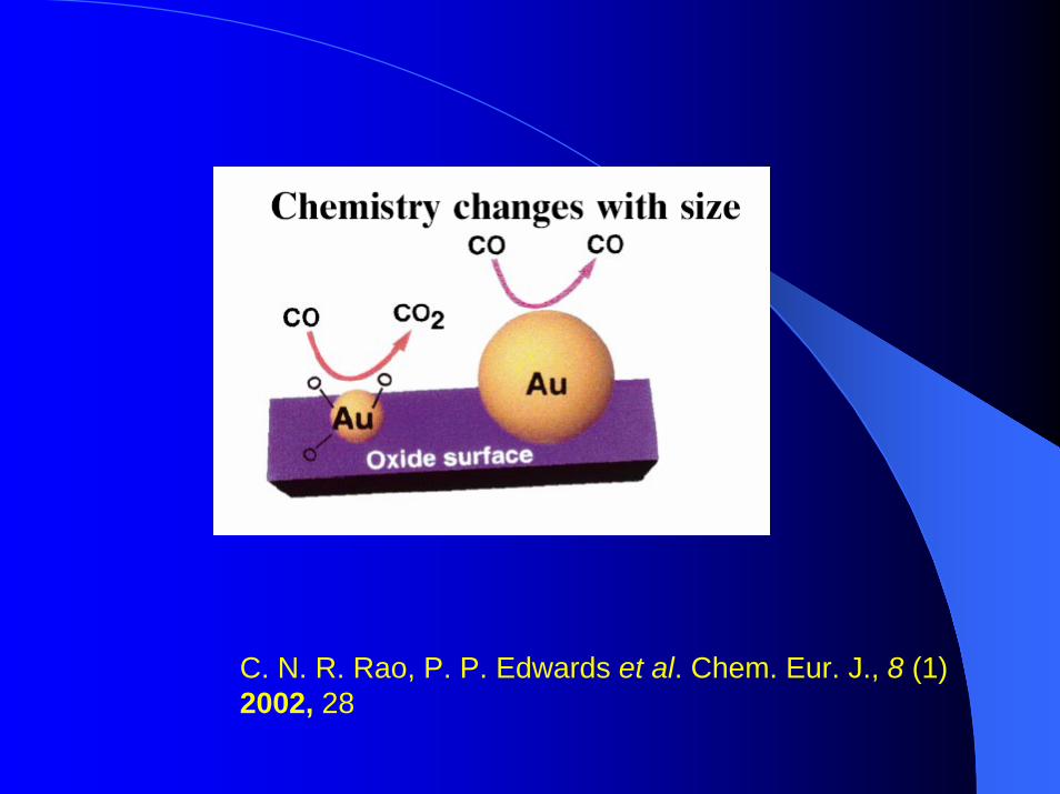
C. N. R. Rao, P. P. Edwards et al. Chem. Eur. J., 8 (1) 2002, 28

WHY BIOSYNTHETIC APPROACH TO WHY BIOSYNTHETIC APPROACH TO NANOMATERIAL SYNTHESIS ?NANOMATERIAL SYNTHESIS ?
Physical (vapour deposition, lithography, radiolysis) and Chemical (reduction by borohydrate or citrate) methods of Nanoparticles synthesis are Hazardous, Eco-unfriendly, Cumbersome and yield big particles.
Need for reliable Ecofriendly and Economic synthesis of Nanomaterials.
Screening of fungi and actinomycetes (more than 200 cultures for metal salt reduction).
Fungi and actinomycetes have also been found to carry out oxidation reduction reactions..

Fusarium oxysporumBIOTRANSFORMATION OF CYCLOHEXANONE TO CAPROLACTONE ::
O OH
O
O
Cyclohexanone Cyclohexanol Caprolactone
24h 48h
J. Mol. J. Mol. CatalCatal. A: Chem. A: Chem. . 181181 ,,20022002,, 237.237.
Biotransformation of Acetophenone to 1-Phenylethanol
C
H
(OH)
C H 3
C
O
C H 3
C
O
C H 3 O OH
C
H
(OH)
C H 3
C
O
C H 3
C
O
C H 3 O OH
C
H
(OH)
C H 3
C
O
C H 3
C
O
C H 3 O OH
J. Mol. J. Mol. CatalCatal. B: Enzym. B: Enzym. . 2727 ,,20042004,, 6161
Acetophenone
Phenyl acetate Phenol
R(+) 1-Phenylethanol ee>99%
Verticillium sp.
Rhodococcus Thermomonospora

Intracellular biosynthesis of inorganic nanoparticles Intracellular biosynthesis of inorganic nanoparticles (Au,Ag,CdS) using Endophytic fungus, (Au,Ag,CdS) using Endophytic fungus, VerticilliumVerticillium sp. sp. and Alkalotolerant Actinomycete, and Alkalotolerant Actinomycete, RhodococcusRhodococcus sp. sp.
Mycelial (cell) mass, separated from culture broth, is suspended in sterile water along with aq. Solution of metal salt ( HAuCl4, AgNO3 CdSO4 ) under shaking at ambient conditions.After few hours, the fungal mycelial (cell) mass develops distinct characteristic colour & solution remains colourless after filtration.Biomass-nanoparticle composite is characterized by XRD, UV-Vis, SEM, EDAX, TEM and SAED.These nanoparticles are suitable for bio-inorganic composite film, in catalysis and other electronic applications.*Angew. Chem. Int. Ed. Engl., 40, 2001, 3585 (Au nanoparticles).*Nano. Lett. 1, 2001, 515 (Ag nanoparticles).Nanotechnology, 14, 2003, 824 (Au Nanoparticles).

Intracellular Intracellular Verticillium Verticillium -- Au Nanoparticles:Au Nanoparticles:Colour of Biomass in Reaction Flask after Colour of Biomass in Reaction Flask after
Addition of HAuClAddition of HAuCl44BeforeBefore AfterAfter
Angew. Chem. Int. Ed. EnglAngew. Chem. Int. Ed. Engl., ., 4040, 2001, , 2001, 3585.3585.

SEM / EDAX of SEM / EDAX of VerticilliumVerticillium--Au NanoparticlesAu Nanoparticles
Angew. Chem. Int. Ed. Engl.,Angew. Chem. Int. Ed. Engl., 4040, 2001, , 2001, 3585.3585.
UVUV--Vis / XRD of Vis / XRD of VerticilliumVerticillium –– Au nanoparticlesAu nanoparticles

Intracellular Intracellular Rhodococcus Rhodococcus -- Au Nanoparticles:Au Nanoparticles:Colour of Biomass in Reaction Flask after Colour of Biomass in Reaction Flask after
Addition of HAuClAddition of HAuCl44
BeforeBefore AfterAfter
Nanotechnology.14: 2003, 824Nanotechnology.14: 2003, 824
6 9 1 2 1 50
1 5
3 0
4 5
No.
of p
artic
les
P a r t i c l e S i z e ( n m )
(111
)(2
00)
3 0 4 0 5 0 6 0 7 0 8 0
Inte
nsit
y (a
.u.)
2 θ ( ° )
(220
) (311
)
(111
)(2
00)
3 0 4 0 5 0 6 0 7 0 8 0
Inte
nsit
y (a
.u.)
2 θ ( ° )
(220
) (311
)

Transmission electron microscopy (TEM) studies of ultra thin section of Au nano Verticillium mycelial mass
A small portion of the mycelial mass which showed the formation of gold nano particles was taken and fixed in 2.5 % gluteraldehyde for 24 h at R. T.
After fixation, mycelial mass was centrifuged, washed several times in D. W.
Mycelial mass was dehydrated with 30, 50, 70, 90% and finally with absolute ethanol
After dehydration mycelial mass was kept in propylene oxide and finally kept in 1:1 mixture of propylene oxide and Epon 812 and kept over night at R. T.
Then kept for embedding in Epon 812 + doddecynyl succunic anhydride (DDSA) + methyl nadic anhydride (MNA) in the ratio of 1:1:5 and added two drops of tridimethyl amino methyl phenol (DMP30) to accelerate polymerization process and kept three days at 60 °C
Ultrathin sections were cut on utramicrotome and were taken on copper TEM grids
Sections were stained with uranyl acetate and lead citrate prior to TEM analysis

TEM OF THIN SECTIONS OF TEM OF THIN SECTIONS OF VERTICILLIUM VERTICILLIUM -- Au Au AND AND RHODOCOCCUSRHODOCOCCUS-- Au NANOPARTICLESAu NANOPARTICLES
AuAu
Particle size = 20±8 A
AuAu
B
Verticillium sp.
Rhodococcus sp.
Particle size= 9

PROBABLE MECHANISM OF PROBABLE MECHANISM OF
INTRACELLULAR BIOSYNTHESISINTRACELLULAR BIOSYNTHESIS
Nanoparticles are formed on the inner surface of the fungal/actinomycete cell and not in solution.Trapping of ions on the surface of fungal/ actinomycete cell.Ions are reduced by enzyme (reductases) present in the cell wall or in the cytoplasmic membrane.

Extracellular Biosynthesis of Inorganic(Au,Ag Extracellular Biosynthesis of Inorganic(Au,Ag and CdS) Nanoparticles Using Plant pathogenic and CdS) Nanoparticles Using Plant pathogenic fungus fungus Fusarium oxysporumFusarium oxysporum and Extremophilic and Extremophilic actinomycete,actinomycete,ThermomonosporaThermomonospora sp.sp.
Mycelial (cell) mass, separated from culture broth, is suspended in sterile water along with aq. Solution of metal salt (HAuCl4, AgNO3 CdSO4) under shaking at ambient conditions.After few hours, the fungal mycellial (cell) mass develops distinct characteristic colour. Biomass remains colourless after filtrationSolution containing metal nanoparticle is then characterized by UV-Vis, FT-IR, TEM, SAED.These nanoparticles are suitable for homogeneous catalysis and can be immobilized in different matrices or in thin film form for optoelectronic and other electronic application.
Langmuir, 19, 2003, 3550.ChemBioChemChemBioChem,, 33, 2002,, 2002, 461461 (Au nanoparticles).(Au nanoparticles).

Extracellular Extracellular Fusarium oxysporumFusarium oxysporum--Au Au Nanoparticles:Colour of Biomass in Reaction Nanoparticles:Colour of Biomass in Reaction
Flask after Addition of HAuClFlask after Addition of HAuCl44
Before After
ChemBioChem ChemBioChem 33, 2002, , 2002, 461461..
Upon filtration, aqueous solution of the control flask was colourless
Upon filtration, aqueous solution of treated flask showed vivid purple colour

TEM micrograph of a drop coated film of Au nanoparticles
synthesized extracellularly
Selected area electron diffraction pattern of the nanoparticles shown
in TEM micrograph
50 nm
ChemBioChemChemBioChem 33, 2002, , 2002, 461461..
(111)(200)
(220)(311)
Particle Size = 20-40 nm
TEM/ SAED of Fusarium oxysporum- Au Nanoparticles

Extracellular Extracellular ThermomonosporaThermomonospora--Au Au Nanoparticles:Colour of Biomass in Reaction Nanoparticles:Colour of Biomass in Reaction
Flask after Addition of HAuClFlask after Addition of HAuCl44Before After
Langmuir, Langmuir, 1919, 2003, , 2003, 35503550
Upon filtration, aqueous solution of the control flask was colourless
Upon filtration, aqueous solution of treated flask showed vivid purple colour

TEM micrograph of a drop coated film of Au nanoparticles
synthesized extracellularly
Selected area electron diffraction pattern of the nanoparticles shown in TEM micrograph
Langmuir,Langmuir, 1919, 2003, , 2003, 35503550..
TEM/ SAED of Thermomonospora - Au Nanoparticles

IntraIntra--/ Extracellular Biosynthesis of Gold / Extracellular Biosynthesis of Gold Nanoparticles by an Nanoparticles by an AlkalotolerantAlkalotolerant Fungus, Fungus,
TrichotheciumTrichothecium sp.sp.Extracellular- Stationary Condition
Intracellular- Shaking ConditionA B CA B C
J. BioMed. Nanotech, 1,2005,47

Immobilization of Immobilization of Fusarium oxysporumFusarium oxysporumsynthesized gold nanoparticles in thermally synthesized gold nanoparticles in thermally evaporated fatty acid and amine thin filmsevaporated fatty acid and amine thin films
Representative TEM micrographs of Fusarium oxysporumsynthesized gold nanoparticles immobilized in a 500-A0 - thick StA (Stearicacid, anionic lipid) film at pH 4.5 (A) and in a 500-A0 - thick ODA (Octadecylamine, cationic lipid) film at pH 6.6(B)
J- Colloid and Interface Science 274,2004,69-75

AERIAL OXIDATION OF CYCLOHEXANE TO ADIPIC ACID USING NANO GOLD AS CATALYST IN A
SOLVENT FREE SYSTEM
Adipic acid ⇒ production of nylon-6 and nylon-66 polymers, fibers, plastics, lubricant additive as well as important intermediate ⇒pharmaceuticals & insecticides industries.
Industrially, First step ⇒ cyclohexane oxidation ⇒ cyclohexanol-cyclohexanone(KA oil) using a soluble cobalt catalyst at 150 °C & 1–2 MPa pressure.
Second step, this, KA oil is further oxidized to adipic acid using 40–60 % HNO3 in presence of copper and vanadium catalysts.
The use of nitric acid as oxidant causes environmental constraints since it generates NOx effluent ⇒ global warming & ozone layer depletion ⇒ present commercial hazardous processes with a more effective catalytic process.

EXPERIMENTAL SECTION
Synthesis of Gold Nanoparticles Supported on Fumed Silica
20 g Fusarium oxysporum ⇒ 1 mM HAuCl4 aqueous solution ⇒ 500 mg of amorphous (fumed) silica was added ⇒ shaker at 28 °C (200 rpm) for 48 h.
After reduction of AuCl4– ions ⇒ nanoparticle solution was subjected to filtration.
The solid Au-SiO2-FO along with biomass ⇒washed several times ⇒ heated at 150 °C overnight to remove water and fungal biomass.

Catalytic reaction ⇒ in a high pressure autoclave ⇒ 30 mL of cyclohexane ⇒ 25 mg of Au-SiO2-FO in a solvent free condition at 120 °C and at 4.3 MPa. For 8 hoursSolid product was esterified to test the presence of acids ⇒ analyzed by GC and GC-MS.
CATALYTIC REACTION OF CYCLOHEXANE TO ADIPIC ACID
OH O
+O
O
OO
OH
O
COOH
COOH
O
COOH
COOH
-CO2
COOH
COOH
OH
OH
O
O
ODimer
Succinic Acid Glutaric Acid Adipic Acid
1,2-CyclohexanedioneO

Before After
Extracellular Extracellular Fusarium oxysporumFusarium oxysporum--Ag Ag Nanoparticles:Colour of Biomass in Reaction Flask Nanoparticles:Colour of Biomass in Reaction Flask
after Addition of AgNOafter Addition of AgNO33
Coll. Surf. B: BiointerfaceColl. Surf. B: Biointerface 2828, 2003, , 2003, 313313..
Extracellular silver nanoparticles before centrifugation
Extracellular silver nanoparticles after centrifugation (15,000 rpm)

TEM micrograph of a drop coated film of Ag
nanoparticles synthesized extracellularly
Selected area electron diffraction pattern of the nanoparticles shown in TEM micrograph
Coll. Surf. B: BiointerfaceColl. Surf. B: Biointerface 2828, 2003, , 2003, 313313..
(200)
(220)
(311)
100 nm
Particle Size = 5-15 nm
TEM/SAED of Fusarium oxysporum - Ag nanoparticles
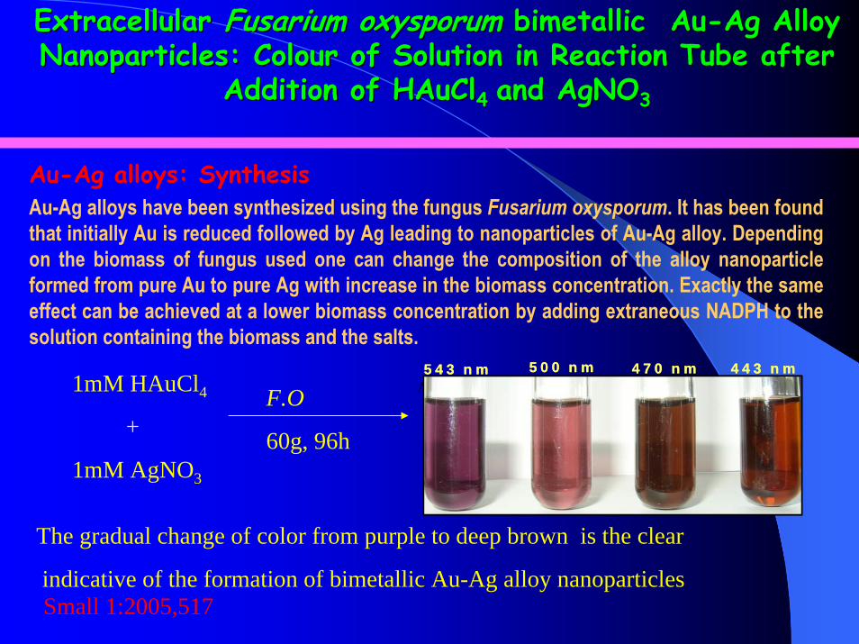
Extracellular Extracellular Fusarium oxysporum Fusarium oxysporum bimetallicbimetallic AuAu--Ag Alloy Ag Alloy Nanoparticles: Colour of Solution in Reaction Tube after Nanoparticles: Colour of Solution in Reaction Tube after
Addition of HAuClAddition of HAuCl44 and AgNOand AgNO33
Au-Ag alloys have been synthesized using the fungus Fusarium oxysporum. It has been found that initially Au is reduced followed by Ag leading to nanoparticles of Au-Ag alloy. Depending on the biomass of fungus used one can change the composition of the alloy nanoparticle formed from pure Au to pure Ag with increase in the biomass concentration. Exactly the same effect can be achieved at a lower biomass concentration by adding extraneous NADPH to the solution containing the biomass and the salts.
Au-Ag alloys: Synthesis
A B C D
Small 1:2005,517
5 4 3 n m 4 4 3 n m5 0 0 n m 4 7 0 n m5 4 3 n m 4 4 3 n m5 0 0 n m 4 7 0 n m1mM HAuCl4
+
1mM AgNO3
F.O
60g, 96h
The gradual change of color from purple to deep brown is the clear
indicative of the formation of bimetallic Au-Ag alloy nanoparticles

Particle Size 6-14 nm
UVUV-- Vis /TEM of Extracellular Vis /TEM of Extracellular Fusarium Fusarium oxysporumoxysporum--AuAu--Ag Alloy Nanoparticles:Ag Alloy Nanoparticles:
TEM micrograph of Au- Ag alloy nanoparticles synthesized extracellulary by reaction of a mixture of 1mM of HAuCl4 and 1mM AgNO3 with 60g F.O wet biomass for 96hrs
Small 1:2005,517
543nm500nm
470nm443nm
The inset shows the picture of test tubes (1-4) of diluted colloidal solution and the corresponding UV-Vis spectra of Au- Ag alloy nanopartilces with respect to time

Extracellular Extracellular Fusarium oxysporumFusarium oxysporum-- Semiconductor Semiconductor CdSCdS Nanoparticles:Colour of Solution in Reaction Nanoparticles:Colour of Solution in Reaction
Tube after Addition of CdSOTube after Addition of CdSO44
J. Am. Chem. Soc.J. Am. Chem. Soc. 124124 ,,20022002, 12108., 12108.
Upon filtration, aqueous solution of the control flask was colourless
Upon filtration, aqueous solution of treated flask showed yellowcolour
20 25 30 35 40 45 50
(220)(002)
(101)
(111)
Inte
nsity
(a.u
.)
2 θ (degree)
TEM micrograph/ XRD of a drop coated film of CdS nanoparticles synthesized extracellularly

Extracellular Synthesis of other Metal Sulfides
PbS
MnS
ZnS
NiS

TEM /SAED/XRD of Fusarium oxysporum- Pt nanoparticles
30 40 50 60 70 80
0
200
400
600
800
1000
1200
1400
(220
)
(200
)
(111
)
Inte
nsity
(a.u
.)
2θ

MECHANISM OF EXTRACELLULAR MECHANISM OF EXTRACELLULAR BIOSYNTHESISBIOSYNTHESIS
A) FT-IR, B) UV-Vis spectra and C) Polyacrylamide gel electrophoresis at pH 4.3 of the aqueous extract of the fungus.
1300 1400 1500 1600 1700 1800
20
30
40
50
60
70
80
Tran
smitt
ance
(%)
Wavenumber (cm )
1
2
1
2
-1
A BC

Purification of the proteins and enzymes involved in extra cellular biosynthesis of nano
particles by Fusarium oxysporum
The fungus Fusarium oxysporum was cultured in MGYP media under shaking condition for 5 days.
The mycelia was harvested by centrifugation; washed three times with sterile distilled water and suspended in sterile distilled water in the ratio of 1:10 (w/v).
After 72 hrs the mycelia was removed by centrifugation and the supernatant containing the enzymes and the proteins was used for the purification of the proteins.

Purification protocol•Concentrated supernatant dialysed against 20 mM, Phosphate buffer (pH 7.2) was loaded on DEAE-Sephadex equilibrated with the same buffer.• 1/3 of total protein comes out of the DEAE column in the breakthrough.• The bound protein on batch elution get eluted at 100 mM NaCl; 200 mM NaCl; 300 mM NaCl and 500 mM NaCl.• 100 mM NaCl fraction contains two proteins, one of them is a reductase, which was separated by Gel filtration on sephacrylS300.• The enzyme obtained after gel filtration was found to be sulphite reductase with molecular mass of 35,000 D.

Separation of the DEAE unbound proteins
•The unbound proteins from DEAE column were concentrated and dialyzed against 20 m M acetate buffer, pH 5.0 and loaded on CM- Sephadex equilibrated with the same buffer.•The bound protein eluted at 100m M NaCl; 200m M NaCl and 300m M NaCl .•The 100 m M NaCl fraction was identified as sulfur lyase with the molecular mass of 38000D •The 200 m M NaCl fraction was identified as nitrate reductase.

Proteins Present in the water Proteins Present in the water suspension of suspension of Fusarium oxysporumFusarium oxysporum
100 mM DEAE Fraction (i) Sulfite Reductase(ii) Capping Protein
200 mM DEAE Fraction (i) Y- Protein(ii) Z- Protein
300 mM DEAE Fraction (i) a- Protein(ii) b- Protein
100 mM CM Fraction (i) ATP Sulfurlyase200 mM CM Fraction (i) Nitrate Reductase
(ii) X- Protein

SDS PAGE profile of Sulphite Reductase, nitrate reductase and capping protein
66
20.1
97.4KD
44.1 4329.1
14.3
Sulphite reductaseNitrate reductase
43
14.3
97.4KD66
29.120.1
15.1
Capping protein
205116978465554536292420
36.5 KD

In vitro Synthesis of Au, Ag and CdS Nanoparticles In vitro Synthesis of Au, Ag and CdS Nanoparticles using purified sulphite reductase and nitrate reductaseusing purified sulphite reductase and nitrate reductase
1: Capping protein + X-Reductase + NADPH + HAuCl4/ AgNO3 / CdSO4 NADP + Au/Ag/CdS nanoparticles + X-Reductase2: X-Reductase+ NADPH + HAuCl4 /AgNO3 / CdSO4 NADP + X-
Reductase + Au/Ag/CdS (Reduced Metal) No nanoparticles.
InvitroInvitro Synthesized Au, Ag and CdS nanoparticlesSynthesized Au, Ag and CdS nanoparticles
AuAg CdS

ConclusionsConclusions
Novel, rational, Ecofriendly and simple Biosynthetic method is developed for nanomaterials of different compositions.Eukaryotic microorganisms like Fungi are used for the first time for preparing extracellular nanomaterials.Metal ions are not toxic to Biomass and Fungus grows after the reaction.Extracellular method is suitable for entrapment and immobilization of nanomaterials on desired support.Intracellular method may be suitable for bio inorganic compositefilms.The use of enzymes secreted by the fungus and subsequent formation of nanoparticles invitro opens up the exciting possibility of extending the protocol to nanoparticles of different chemical composition and developing a rational, eco-friendly fungal enzyme based process for nanoparticle synthesis.

Biosynthesis Of Inorganic Nanoparticles Using Biosynthesis Of Inorganic Nanoparticles Using Plants and their endophytic fungiPlants and their endophytic fungi
Why this Approach to Nanomaterial Synthesis ???
Barring the fungus Fusarium oxysporum and the actinomycete Thermomonospora sp., all other microorganisms and plants investigated thus far when challenged with metal ions have resulted in their reduction and growth of nanoparticles intracellularly.
Screened and identified a number of plants and their endophytic fungi for extracellular biosynthesis of inorganic nanoparticles.
These plants have been found to reduce the metal ions and subsequent formation of nanoparticles extracellularly as fast as the chemical methods.

Extracellular Biosynthesis of Inorganic Nanoparticles using Plants and their endophytic
fungiThe Leaf broth, used for reduction of Ag+ and AuCl4- ions and synthesis of Silver and Gold nanoparticles was prepared by taking 20 g of thoroughly washed and finally cut leaves (Lemon grass, Geranium and Neem) in a separate 500 ml conical flask.
To the biomass, 100 ml of sterile distilled water was added and then boiled for 5 min. After boiling, the solution was decanted and filtered, and 5 ml each of the broth was added to 100 ml of 1mM aq. AgNO3 solution and 1mM aq. HAuCl4 solution .
After few minutes, the solution develops distinct characteristiccolour.
The solution containing the nanoparticles is then characterized by colour, UV-Vis, XRD, FTIR, EDAX, XPS, AFM, SAED etc.

Extracellular Lemongrass-Au/Ag and Neem-Au/Ag: Colour of Broth in Reaction Flask After Addition of
HAuCl4/AgNO3
Lemongrass and its endophytic fungus (F.O)
Neem and its endophyticfungus (Colletotrichum sp) Nature Materials, 3, (2004), 482
J. Mater. Chem. 13, 2003, 1822

Representative TEM images of gold nanoparticles synthesized by the reduction of 5 mLof 10-3 M aqueous HAuCl4 solution with a) 0.2 mL, b) 0.3 mL, c) 0.5 mL and d)1.0 mL of lemon grass extract.Nature Materials, 3, (2004), 482 Chem. Mater, 17:2005,566
Extracellular biosynthesis of AuExtracellular biosynthesis of Au--Nanoparticles using lemongrass extract.Nanoparticles using lemongrass extract.

UV-vis-NIR spectra of gold nanoparticles synthesized by adding different amount of lemongrass leaf extract to 5 mL of 10-3 M HAuCl4 solution. Curves 1 – 10 correspond to solutions with 0.2, 0.3, 0.4, 0.5, 0.6, 0.7, 0.8, 1.0, 1.2 and 1.6 mL of lemongrass leaf extract in 5 mL of 10-3 M HAuCl4 solution respectively.
AFM image of one of the truncated triangles formed by reduction of Au3+ ions using lemongrass leaf extract.
TEM image and electron diffraction pattern of Gold nanoparticle
Nature Materials, 3, (2004), 482
UV-VIS-NIR /AFM/TEM of lemongrass Gold nanoparticle

Extracellular Ag/Au nanoparticles using plantsExtracellular Ag/Au nanoparticles using plants
16-14 nm20-40 nm
11-34 nm
J. Mater. Chem 13, 2003, 1822Biotecnol. Prog. 19, 2003, 1624
A C
D
B
TEM micrographs of Au nanoparticles synthesized by various plants

Why biosynthetic approach to oxides nanoparticle synthesis?
•Current physico-chemical methods (Sol gel technique, Chemical vapor deposition, Hydrothermal synthesis, Precipitation method, Micro emulsion method) of oxide nanoparticles synthesis are hazardous, eco-unfriendly, cumbersome, costly and required very high temperature , pH and pressure for synthesis )
•Need for reliable eco-friendly and economic synthesis of oxide nanoparticles at ambient temperatures, pressures and neutral pH.
•Screening of microbes (more than hundred cultures) for oxide nanoparticle synthesis
•Microbes has been found to carry out oxidation-reduction reactions.
Biosynthesis of Oxides Nanoparticles using Microbes

Fungi positive for oxides nanoparticles synthesisFungi positive for oxides nanoparticles synthesis
NCL-MNPF1 NCL-MNPV1 NCL-MNPB1
O OH
O
O
Cyclohexanone Cyclohexanol Caprolactone
24h 48h
BIOTRANSFORMATION OF CYCLOHEXANONE TO CAPROLACTONE :BIOTRANSFORMATION OF CYCLOHEXANONE TO CAPROLACTONE :
J. Mol. J. Mol. CatalCatal. A: Chem. A: Chem. . 181181 ,,20022002,, 237.237.
J. Mol. J. Mol. CatalCatal. B: Enzymatic. B: Enzymatic. 27 . 27 ,,20042004,, 61.61.

Extracellular Biosynthesis of oxides nanoparticles(FeExtracellular Biosynthesis of oxides nanoparticles(Fe33OO44, , ZrOZrO22, SiO, SiO22, TiO, TiO22) using fungus ) using fungus Fusarium oxysporumFusarium oxysporum
and and VerticilliumVerticillium sp.sp.
•Mycelial mass separated from culture broth, is suspended in sterile water along with aqueous solution of metal salt, (K2SiF6, K2TiF6, K2ZrF6 K3Fe(CN)6 and K4Fe(CN)6under shaking at ambient condition.
•After few hours, the fungal mycelial mass does not develop distinct characteristic colour. Biomass remains colourless after filtration.
•Solution containing oxides nanoparticle is then characterized by FTIR, TEM, SEM, EDAX, TGA, XPS etc.

Extracellular oxides nanoparticles using fungus Extracellular oxides nanoparticles using fungus Fusarium oxysporumFusarium oxysporumand and VerticilliumVerticillium sp.: colour of biomass in reaction flask after sp.: colour of biomass in reaction flask after addition of Kaddition of K22SiFSiF66, K, K22TiFTiF66, K, K22ZrFZrF6 6 KK33Fe(CN)Fe(CN)66 and Kand K44Fe(CN)Fe(CN)66
BeforeBefore AfterAfter

Extracellular biosynthesis of Iron oxide (Magnetite)Nanoparticles using fungus Fusarium oxysporum
TEM micrographs of iron oxide nanoparticles synthesized using fungus Fusarium oxysporum before (A & B) and after calcination at 400o C for 3 h (C & D).
22-50 nm
Small ( In Press )

TEM micrographs of iron oxide nanoparticles synthesized using fungus Verticillium sp.before (A & B) and after calcination at 400o
C for 3 h (C & D). Small ( In Press )
Extracellular biosynthesis of Iron oxide (Magnetite) Nanoparticles using fungus Verticillium sp.
100-400 nm 10-40 nm

FT-IR XRD
500 600 700
683
568
627
522
4
3
2
1
Tran
smitt
ance
(a.u
.)
Wavenumber (cm-1)1400 1500 1600 1700
163815
4043
2
1
Tr
ansm
ittan
ce (a
.u.)
Wavenumber (cm-1)15 20 25 30 35 40
2
1
*o****
o*
o**
*
**
o*
Inte
nsity
(a.u
.)
2θ (degrees)
* - Fe3O4o - γFe2O3
CA B
FT-IR and XRD Spectra of Iron oxide nanoparticles synthesized using fungus Fusarium oxysporum and
Verticillium sp.
SDS PAGE data showing the extracellular protein profile of V. sp
Small (In Press )

Bioleaching of sand by the fungus, Bioleaching of sand by the fungus, Fusarium oxysporumFusarium oxysporumas a means of producing extracellular silica nanoparticles:as a means of producing extracellular silica nanoparticles:
Color of biomass in reaction flask after addition of sandColor of biomass in reaction flask after addition of sand
Before After
Adv. Mater.17:2005,889

Extracellular biosynthesis of Silica Nanoparticles synthesized by the exposure of sand to the fungus Fusarium oxysporum
2-5 nm
Fig.1. TEM micrographs at different magnifications of silica nanoparticles synthesized bythe exposure of sand to the fungus, Fusarium oxysporum before (A and B) and aftercalcination at 400o C for 2 h (C and D). The insets in B and C are SAED patternsrecorded from representative silica nanoparticles
TEM micrographs at different magnifications of silica nanoparticles synthesized by the exposure of sand to the fungus, Fusarium oxysporum before (A and B) and after calcination at 400 C for 2hrs (C and D)
Adv. Mater.17:2005,889

FTIR spectrum of FTIR spectrum of Fusarium oxysporumFusarium oxysporum: Silica nanoparticles: Silica nanoparticles
Fig. 4. SEM micrograph of a single grain of sand before (A) and after (B) exposure to thefungus, Fusarium oxysporum for 24 h. The insets in A and B show magnified views of aselected area from the respective grains.
SEM micrograph of a single grain of sand before (A) and after (B) exposure to the fungus, Fusarium oxysporum for 24 h. The insets in A and B show magnified views of a selected area from the respective grains.

FUNGUS-MEDIATED BIOTRANSFORMATION OF AMORPHOUS SILICA IN RICE HUSK TO NANOCRYSTALLINE SILICA.
Color of biomass in reaction flasks after addition of rice husk
Before After

NANOCRYSTALLINE SILICA FROM RICE HUSK.
Crystalline silica nanoparticles obtained by reacting the rice husk with the fungus Fusarium oxysporum which biotransforms the amorphous silica present in rice husk to crystalline silica nanoparticles at room temperature. This leads to an exciting possibility of an energy-conserving and economically viable green approach towards the large-scale synthesis of nanomaterials.
A. SEM micrograph showing an edge of a single rice husk flake.
B. TEM micrograph of silica nanoparticles synthesized by the exposure of rice husk to the fungus, Fusarium oxysporum.
C. SAED patterns recorded from silica nanoparticle shown in image D.
D. TEM micrograph of silica nanoparticles synthesized by the exposure of rice husk to the fungus Fusarium oxysporum after calcination at 400 o C for 2 hour.
A B
C D
2-6 nm

FTIR / XRD of Fusarium oxysporum : Silica nanoparticle
A) FTIR spectra recorded from the filtrate containing silica particles synthesized by exposing rice husk to the fungus, Fusarium oxysporum for 24 h (curve 1) and from the filtrate obtained by exposing rice husk to water of pH 4.5 for 15 days (curve 2).
B) XRD patterns recorded from silica particles synthesized by the exposure of rice husk to the fungus, Fusarium oxysporum before (curve 1) and after calcination at 400 °C for 2h (curve 2).

Extracellular biosynthesis of Extracellular biosynthesis of ZirconiaZirconia (Ceramic Steel) (Ceramic Steel) Nanoparticles using the fungus Nanoparticles using the fungus Fusarium oxysporumFusarium oxysporum
TEM micrographs at different magnifications of Zirconia nanoparticles synthesized using the fungus F.O before (A and B) and after calcination at 600 C for 3 h (C and D)
20-60 nm
SDS PAGE data showing the extracellular protein profile of
F. oxysporumJ. Mater. Chem- 14:2004,3303

A B
C D
Extracellular biosynthesis of Titania Nanoparticles using Extracellular biosynthesis of Titania Nanoparticles using fungus fungus Fusarium oxypsorumFusarium oxypsorum
TEM micrograph of Titania nanoparticles synthesized using fungus Fusarium oxysporum after 12 hrs(A) and 24 hrs (B and C) of reaction before (A-C) and after calcination at 300 C for 3 h (D)
2-10 nm 50-100 nm
SDS-PAGE data showing the
extracelluar protein profile of
Fusarium oxysporum
J.Mater.Chem 15:2005,2583

Biosynthesis of Carbonate Biominerals using microbes.
Why the living organisms generate biominerals
The mineral crystals that are formed by the organisms are called biominerals. These are essentially inorganic salts, which serve a variety of biological purposes specially protection.
Organisms are capable of forming a diverse array of crystalline minerals with complex morphologies specific size and shape at ambient temperature through specific proteins by the process of biomineralization.
Biomineralization process occurs in many different organisms and their tissues including bones, tooth enamel, egg shell formation and in marine shells.

Biominerals of dimensions nano-submicrones are extremely important
material for:
1. Pigments in paints2. Cosmetics3. Ceramics4. Pharmaceuticals5. Electronics6. Paper, rubber and polymer industries7. Manufacture of cement iron, steel8. A constituent of antacids9. To neutralizing soil acidity10. Plastic
Carbonate biominerals
11. Calcium carbonate. Calcium carbonate2. Strontium carbonate 2. Strontium carbonate 3. Barium carbonate3. Barium carbonate
Calcium carbonate has three stable polymorphs 1. Calcite (thermodynamically most stable)2. Aragonite (slightly less stable than calcite)3. Vaterite (the most unstable polymorph)
Uses of Carbonate biomineral

Why fungal approach to biomineral Why fungal approach to biomineral synthesis ?synthesis ?
Chemical methodsBiomimetic templates such as Langmuir monolayers at the air-water interface, self assembled monolayers (SAMs),Lipid bilayer stacks, Functionalized polymer surfaces have been developed for CaCo3 synthesis. These methods are hazardous eco-unfriendly and yields less stable biominerals.
Biological MethodsSpecific proteins extracted from CaCO3 rich organisms as a template and an external source of CO2 for reaction with suitable Ca2+ ions to produce CaCO3.crystals. These methods are very costly and laborious.
Need for reliable ecofreindly and economic synthesis of biominerals:Many fungi are known to release reasonable amount of carbon dioxide and characteristic proteins during their growth,Screening of fungi (more than 100 culture for biomineral synthesis)

Verticillium sp. Fusarium oxysporum
J.Am. Chem. Soc. 125: 14656J.Am. Chem. Soc. 125: 14656--57, 57, 20032003
CaCl2 Ca2+ + 2Cl-
Proteins
CO2 + H2O + Ca2+ CaCO3 + 2H+
HCO3- + Ca2+ CaCO3 + H+
Biosynthesis of carbonate biominerals
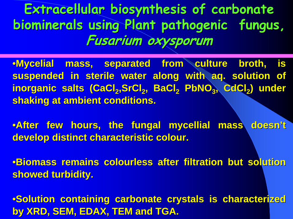
Extracellular biosynthesis of carbonate Extracellular biosynthesis of carbonate biominerals using Plant pathogenic fungus, biominerals using Plant pathogenic fungus,
Fusarium oxysporumFusarium oxysporum••Mycelial mass, separated from culture broth, is Mycelial mass, separated from culture broth, is suspended in sterile water along with aq. solution of suspended in sterile water along with aq. solution of inorganic salts (CaClinorganic salts (CaCl22,SrCl,SrCl22, BaCl, BaCl22 PbNOPbNO33, CdCl, CdCl22) under ) under shaking at ambient conditions.shaking at ambient conditions.
••After few hours, the fungal mycellial mass doesnAfter few hours, the fungal mycellial mass doesn’’t t develop distinct characteristic colour.develop distinct characteristic colour.
••Biomass remains colourless after filtration but solution Biomass remains colourless after filtration but solution showed turbidity.showed turbidity.
••Solution containing carbonate crystals is characterized Solution containing carbonate crystals is characterized by XRD, SEM, EDAX, TEM and TGA.by XRD, SEM, EDAX, TEM and TGA.

Extracellular Extracellular Fusarium oxysporumFusarium oxysporum--carbonate carbonate biomineralsbiominerals:Colour of Biomass in Reaction :Colour of Biomass in Reaction Flask after Addition ofCaClFlask after Addition ofCaCl22, SrCl, SrCl22, BaCl, BaCl2, 2,
Pb(NOPb(NO33))22 and CdCland CdCl22Before After

Upon filtration, aqueous solution
of the control flask was clear (no
turbidity)
Upon filtration, aqueous solution
of treated flask (CaCl2,BaCl2,SrCl2) showed turbidity
Extracellular Fusarium oxysporum–carbonate biominerals.

Extracelluar biosynthesis of CaCOExtracelluar biosynthesis of CaCO33 crystals after crystals after reaction of Careaction of Ca2+ 2+ ions with ions with Fusarium oxysporumFusarium oxysporum
SEM micrographs of CaCO3 crystals after, A) 1day; B) 2 days and C and D) 3 days of reaction of aqueous Ca2+ ions with Fusarium oxysporum. The inset in D shows a magnified image of one of the circular CaCO3 crystal super structures.
Advance Funct. Materials 14:2004,1075

EDAX/XRD/FTEDAX/XRD/FT--IR of IR of Fusarium oxysporumFusarium oxysporumCaCOCaCO33 biomineralbiomineral
EDAX/XRD/FT-IR spectra of CaCO3 crystals synthesized using Fusarium oxysporum.
EDAX XRD FT-IR
Advance Funct. Materials 14:2004,1075

SEM micrographs (A SEM micrographs (A __ C) at different magnifications of SrCOC) at different magnifications of SrCO33 crystals formed crystals formed after 3 days of reaction of aqueous Srafter 3 days of reaction of aqueous Sr2+ 2+ ions with ions with Fusarium oxysporumFusarium oxysporum..
12 µm length
Pore size10-15 nm
BaCO3
CD
Langmuir 20, 2004, 6827
Extracellular biosynthesis of SrCOExtracellular biosynthesis of SrCO33 crystals crystals formed after reaction of aqueous Srformed after reaction of aqueous Sr2+ 2+ ions with ions with Fusarium oxysporumFusarium oxysporum

SEM micrographs at different magnifications of biogenic PbCO3 / CdCO3crystals formed after 1 day of reaction of aqueous Pb2+/Cd2+
ions with the fungus, Fusarium oxysporum.
CdCO3
PbCO3
Heavy metal remediation by Heavy metal remediation by Fusarium oxysporumFusarium oxysporum as a as a means of production of lead(Pb) and cadmium (Cd)means of production of lead(Pb) and cadmium (Cd)
carbonate crystalscarbonate crystals
Size 100-200 nm
SDS-PAGE of protein fraction
responsible for CdCO3 crystals
Langmuir 21,2005,7220

SEM micrograph of CaCO3 crystals of different morphology using different fungi
J. Materials. Chem 14, 2004 , 2333

SEM micrograph/XRD of CaCO3(vaterite)crystals using germinating chick pea seeds.
Biological Synthesis of Stable Vaterite Crystals by the Reaction of Calcium Ions with Germinating Chickpea Seeds
Cryst.Growth Des.5(2) 399-402,2005
Treated Control SEM XRD

Conclusions:
The total biological synthesis of CaCO3, SrCO3 and BaCO3 PbCO3 CdCO3of interesting morphology and polymorph selectivity by challenging the whole cells (not pure enzyme) of endophytic fungus Verticillium sp. and plant pathogenic fungus Fusarium oxysporum with aqueous Ca2+, Sr2+ and Ba2+ Pb2+ Cd2+ have been described.
The source of carbonate ions are fungi themselves, thus significantly enhancing the application potential of this approach.
The action of specific proteins secreted by the fungi and carbon dioxide released by the fungi play a very important role in directing the morphology and crystal structure of the minerals grown in solution.
Our finding that minerals of such complex morphology can be grown by completely biological processes using fungi that are not normally (if ever) exposed to metal ions is exciting with important implications in crystal engineering and associated applications.

AcknowledgementsAcknowledgements
Financial assistanceDBT, New Delhi, IndiaNCL, Pune, IndiaDST, New Delhi,IndiaCollaboratorsDr. M. Sastry Dr. R. KumarTechnical assistance Dr. S.R. SainkarDr. R. RamaniMrs. R. PasrichaDr. M. SanyalDr. S. M. Yusuf
Graduate StudentsDr. D. MandalDr. P. MukherjeeMr. S. SenapatiMr. S. ShankarMr. D. RautarayMr. A. SanyalMr. F. KhanMr. V. Bansal