about - mica – mutual insurance company of arizona aspect of the rules allowing crnas to practice...
TRANSCRIPT

O
Issue 9 | Summer 2016Nevada Issue
PROVIDING MEDICAL PROFESSIONAL LIABILIT Y INSURANCE IN ARIZONA, COLORADO, NEVADA AND UTAH
Low-Dose Chest CT Lung Cancer Screeningpage 3
Ransomware: Don't Be a Victimpage 5
Counsel’s Cornerpage 6
Case History Delay in Diagnosing Lung Cancerpage 6
MICA was founded in 1976 after the largest medical professional liability (MPL) insurance carrier announced it was no longer underwriting MPL insurance coverage for the physicians of Arizona. MICA off ers stability through the peaks and valleys of the insurance cycle.
Our mission is to protect and defend the practice of medicine in Arizona, Colorado, Nevada and Utah. Our vi-sion is to be the insurance company of choice for physicians and their groups, medical facilities and nurse practitioners.
Our values include outstanding service to our mem-bers, aff ordable insurance coverage, prudent underwrit-ing principles, relevant risk management programs to educate and protect our members, aggressive defense against claims, and paying dividends to our members when fi nancial conditions warrant. In fact, over the past ten years, we have paid $337 million in dividends to our members*.
Our mission is to protect and defend the practice of medicine in Arizona, Colorado, Nevada and Utah.
About us
* Dividends declared in a given policy year reflect the Company’s financial performance during that year. Past performance does not guarantee future dividends.
2 | MICA Risk Rundown | Spring 2016
Executive Staff : James F. Carland, III, M.D. President & Chief
Executive Offi cer Ronald E. Malpiedi, Vice President & Chief
Operating Offi cerEdward G. Marley, MBA, Vice President &
Chief Financial Offi cer Darren J. Palmer, Vice President & Chief
Information Offi cerRobin L. Charles, MBA, CIC Vice President,
Marketing & Corporate Communication Philip J. Smith, Vice President, Claims
Mary K. Hedin, MBA, RPLU, Vice President, Underwriting
Leon W. Kochan, M.C.Ed, Vice President, Human Resources
Julie A. Ritzman, MBA, Vice President, Risk Management Service
Board of Trustees: James F. Carland, III, M.D., Chairman
Marc L. Leib, M.D., Vice ChairmanJoseph W. Hanss, Jr., M.D., Secretary
Douglas P. Jensen, M.D., Treasurer Phyllis I. Biedess
James G. Leiferman, M.D. Steven P. Matteucci, J.D. Jeff rey W. Morgan, D.O. Karen J. Nichols, D.O.
David A. Pedersen, M.D. J. Greg Rula, M.D.
Amy A. Silverthorn, M.D. Walter K. Sosey, M.D.
Charles W. Swetnam, M.D.Michael A. Trainor, D.O.
Summer 2016 | MICA Risk Rundown | 3
LDCT | Continued on page 4
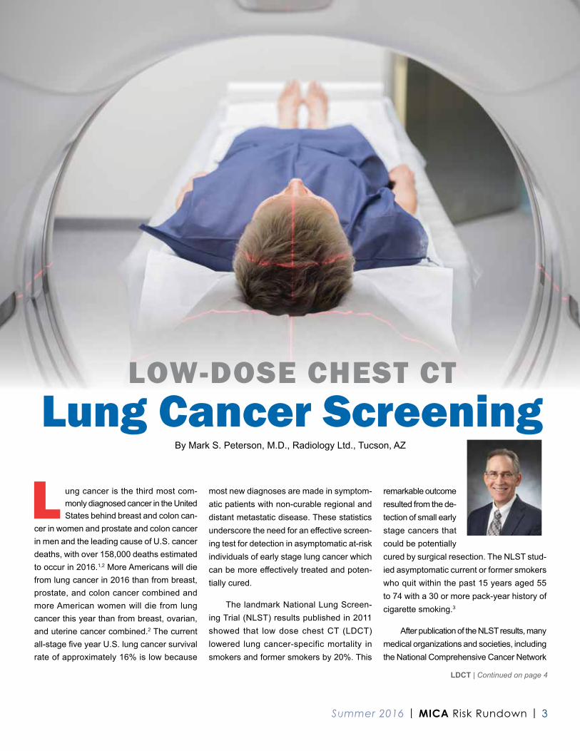
L ung cancer is the third most com-monly diagnosed cancer in the United States behind breast and colon can-
cer in women and prostate and colon cancer in men and the leading cause of U.S. cancer deaths, with over 158,000 deaths estimated to occur in 2016.1,2 More Americans will die from lung cancer in 2016 than from breast, prostate, and colon cancer combined and more American women will die from lung cancer this year than from breast, ovarian, and uterine cancer combined.2 The current all-stage fi ve year U.S. lung cancer survival rate of approximately 16% is low because
most new diagnoses are made in symptom-atic patients with non-curable regional and distant metastatic disease. These statistics underscore the need for an eff ective screen-ing test for detection in asymptomatic at-risk individuals of early stage lung cancer which can be more eff ectively treated and poten-tially cured.
The landmark National Lung Screen-ing Trial (NLST) results published in 2011 showed that low dose chest CT (LDCT) lowered lung cancer-specific mortality in smokers and former smokers by 20%. This
remarkable outcome resulted from the de-tection of small early stage cancers that could be potentially cured by surgical resection. The NLST stud-ied asymptomatic current or former smokers who quit within the past 15 years aged 55 to 74 with a 30 or more pack-year history of cigarette smoking.3
After publication of the NLST results, many medical organizations and societies, including the National Comprehensive Cancer Network
LOW-DOSE CHEST CTLung Cancer Screening
By Mark S. Peterson, M.D., Radiology Ltd., Tucson, AZ

4 | MICA Risk Rundown | Summer 2016
LDCT | Continued from page 3
(NCCN), the American College of Radiol-ogy (ACR), the American Cancer Society, the American Thoracic Society, and the Society of Thoracic Surgeons, advocated for Medicare and private insurance coverage of LDCT lung cancer screenings. In December 2013, the United States Preventive Services Task Force (USPSTF) issued a Grade B recommendation, indicating more potential benefits than potential harms for LDCT lung cancer screening. In Janu-ary 2015, under provisions of the Affordable Care Act, screening tests given a USPSTF Grade B or higher recommendation must be covered by private insurers, which led to private insurer coverage of annual LDCT lung cancer screening, without a copay or deductible. Subsequently, the Centers for Medicare and Medicaid Services (CMS) issued a national coverage decision in February 2015, approving annual LDCT lung cancer screening for Medicare beneficiaries who meet the original inclusion criteria of the NLST.
In an effort to ensure that the outcomes of widespread LDCT lung cancer screening
equal those of the NLST, CMS has estab-lished requirements for physicians, physician assistants, nurse practitioners, and clinical nurse specialists who refer Medicare benefi-ciaries for screening, imaging sites perform-ing LDCT scans, and radiologists who inter-pret LDCT scans. Among the most important CMS requirements for clinicians is the need to provide a written order for the examination with attestation, for the initial LDCT scan only, that beneficiary lung cancer screening counseling with shared decision-making was performed. The lung cancer screening coun-seling session may be billed (2016 Medicare Physician Fee Schedule code G0296) the same day as a medically necessary evalu-ation and management services visit or an annual wellness visit. Screening counseling should include discussions of the benefits of screening including decreased lung cancer mortality, quality of life benefits from screen-ing and early detection, and detection of thoracic diseases other than lung cancer,
and harms, such as false positive and false negative results, futile detection of aggres-sive cancer that has already metastasized or of indolent disease. Use of online patient education sources, such as Lung Cancer CT Screening at http://www.shouldiscreen.com/, may also be helpful for screening counseling.
Smoking cessation counseling must be provided during lung cancer screening counseling, and smoking cessation inter-ventions and materials must also be pro-vided by the imaging center performing the LDCT scan. While tobacco use remains the leading risk factor for lung cancer, causing 85% of lung cancer deaths, 50% of deaths are former smokers and 35% are current smokers, 15% of the people who die from lung cancer have never smoked. Smoking cessation remains the primary prevention measure for lung cancer, while screening is the secondary prevention measure. An LDCT scan cannot prevent cancer, but can contribute to early detection and treatment.

Summer 2016 | MICA Risk Rundown | 5
Among CMS requirements for imaging centers is the use of low radiation dose CT scan technique, with a volumetric CT dose index of less than or equal to three milligray for a standard size patient. This LDCT radia-tion dose is approximately 1/4 of the radiation dose of a standard diagnostic chest CT scan, approximately the dose of 15 chest radio-graphs, or approximately equivalent to six months of natural background radiation. The estimated additional risk of a lethal cancer induced from LDCT scan radiation is very low, estimated to be 1 in 100,000 to 1 in 10,000 (RadiologyInfo.org).3 Given the estimated one in three to one in four lifetime risk for the general public of dying of cancer and the high risk for heavy smokers of dying of lung cancer, this additional risk of develop-ing cancer from the low radiation dose of an LDCT scan is insignificant.
CMS also requires the submission of beneficiary data and scan metrics by the screening center to a CMS-approved
screening registry, of which the only one currently approved is sponsored by the ACR. A standardized reporting and data system is required, for which most sites use the ACR Lung-RADS system, which assigns scan re-sults based on lung nodule size, morphology, and interval growth to assessment categories ranging in most cases from “1=Negative” to “4=Suspicious” for lung cancer. Lung-RADS goals include standardization of reporting and management recommendations, facilitation of outcome monitoring, and minimization of false positive results.
LDCT lung cancer screening is an on-going multidisciplinary process, with the success of screening programs requiring the input of multiple stakeholders, includ-ing screened individuals, ordering clinicians, radiology personnel, and subspecialty physi-cians. Together we can use LDCT, the only currently recommended screening test for lung cancer, to substantially reduce mortality from this disease.
1 U.S. Cancer Statistics Working Group. United States Cancer Statistics: 1999–2012 Incidence and Mortality Web-based Report. Atlanta (GA): Department of Health and Human Services, Cen-ters for Disease Control and Prevention, and Na-tional Cancer Institute; 2015. Available at: www.cdc.gov/uscs
2 Cancer Facts and Figures 2016. American Cancer Society. Available at: http://bit.ly/232SULI
3 Reduced Lung-Cancer Mortality with Low-Dose Computed Tomographic Screening. The National Lung Screening Trial Research Team. N Engl J Med 2011;365:395-409.
4 RadiologyInfo.org for Patients. April 5, 2016 Available at: http://bit.ly/1KxEnvD
For additional information and resources please see:
• http://bit.ly/2aZBe0j (Lung Cancer Screening Resources)
• http://bit.ly/2boWY7H (NCCN Guideline for Patients)

6 | MICA Risk Rundown | Summer 2016
Counsel’s Corner

Summer 2016 | MICA Risk Rundown | 7
A
QWhat liability risks do Nevada physicians/surgeons have when working with certifi ed registered nurse anesthetists (CRNAs)?
As a preliminary matter, CRNAs are permitted to
practice without any written practice agreement with a
physician. Generally, then, any physician choosing to
work with a CRNA would experience the same liability
risks they would by working with an anesthesiologist.
Ultimately, there is no special, additional requirement
placed on a physician/surgeon in this circumstance.
Nevada Administrative Code section 632.500-550
governs CRNAs in Nevada. The Code outlines what
specifi c functions a CRNA is permitted to undertake fol-
lowing appropriate determination by a patient’s physician/
surgeon. One aspect of the rules allowing CRNAs to
practice independently that might implicate a physician/
surgeon’s liability is that CRNAs are allowed to act when
“it has been determined by a patient’s physician . . .” that
a given treatment is necessary. See NAC § 632.500.
A physician/surgeon who breached their own stan-
dard of care by ordering improper/unnecessary treatment
might be found liable for that order. However, this is
identical in all relevant respects to a physician/surgeon
order to be carried out by a nurse or other physician. If,
hypothetically, a physician or surgeon properly ordered
a given treatment but the CRNA followed that order in a
way resulting in medical negligence, liability would likely
stop at the CRNA. Working with an independent CRNA
in Nevada does not impose any unique or additional
obligation or liability.
BY JUSTIN SHIROFFSnell & Wilmer L.L.P.

8 | MICA Risk Rundown | Summer 2016
CASE HISTORY Delay in Diagnosing Lung Cancer
A t age 73, a female patient saw her primary care physician with complaints of shortness of breath
over the last month; chest pain, worse on right side; low-grade fever; night sweats; and fatigue. The patient had a 40 pack year history of smoking but quit when she was 61. Over the years the patient had several chest x-rays which were negative. The pri-mary care physician clinically diagnosed right upper lobe pneumonia. He ordered a chest x-ray, started the patient on Amoxi-cillin, and referred her to the Emergency Department for admission to the hospital.
In the hospital, the hospitalist consulted with a pulmonologist who noted the chest x-ray showed a right upper lobe mass-like infiltrate but incorrectly documented in the “Impressions” it was the right lower lobe that was involved. He diagnosed the pa-tient with right lower lobe pneumonia or mass with prolonged symptoms of pleurisy; eosinophilia, most likely from coccidioido-mycosis (cocci); coronary artery disease, status post coronary artery bypass graft; mild renal insufficiency; and hypertension. His plan was to administer IV fluids, get a cocci serology and CT scan of the chest.
He started fluconazole and continued her medications from home.
The cocci serology was negative, but the CT scan revealed a 7.5 cm right perihilar mass with probable adjacent lymphangetic spread versus focal consolidation of the mid-right lung with adjacent inflammatory change and large mediastinal lymph nodes. A subsequent comparison of images from a previous chest x-ray led the radiologist to conclude the mass was new. This was documented in an addendum to the original report.

Summer 2016 | MICA Risk Rundown | 9
The pulmonologist’s differential diag-nosis was cancer versus valley fever. He spoke with the patient regarding her treat-ment options which included a fiber optic bronchoscopy with a biopsy of the mass or continue the anti-fungal medication for 6 weeks and repeat the chest x-ray. The patient decided to wait on the biopsy. She was discharged with prescriptions for flu-conazole and levofloxacin and instructed to follow up with the pulmonologist in his office.
The patient was seen by the pulmo-nologist in his office six weeks later. The encounter note indicated the patient had a positive cocci serology during her hos-pitalization. He recommended the patient continue the fluconazole, repeat the chest x-ray and cocci serology, and return in two months. The patient had the cocci serology drawn the next day and it was reported negative. The chest x-ray was read as right upper lobe pneumonia, there was no men-tion in the report regarding a right upper lobe mass.
The patient returned for her two-month visit with the pulmonologist who ordered a repeat cocci serology and chest x-ray. The cocci serology was negative and the fluconazole was discontinued. Since her last visit with the pulmonologist, the pa-tient had seen her primary care physician several times with complaints of bilateral rib pain. The repeat chest x-ray revealed mass-like consolidation in the right upper lobe raising the suspicion of a neo-plasm. A CT scan identified the mass that was evident in the previous CT scan and noted that it had increased in size and there was pre-tracheal adenopathy. A right lung biopsy was performed and the diagnosis was non-small cell lung cancer, Stage 3A, but a later bone scan established the cancer as Stage four. The patient was not a surgical candi-date so chemotherapy and radiation were recommended as palliative care. The patient expired approximately three months later.
The patient was survived by four adult children who filed a lawsuit naming the pul-monologist, the hospitalists, the radiologist who interpreted the first CT scan in the hospital and the hospital. All the named defendants were dismissed except the pul-monologist and the hospital. This wrongful death case was eventually settled.
Risk Management IssuesThe pulmonologist relied upon his recol-
lection that the patient had a positive cocci serology during her hospitalization. In fact, the hospital reported a negative serology but the hospital lab results were not reviewed by the pulmonologist when the patient was seen in the office, thus perpetuating a di-agnostic error.
The imaging studies consistently re-ported the patient’s mid to upper right lung was the primary area of concern. However, the pulmonologist repeatedly referenced the right lower lung.
DiscussionThe lawsuit alleged a delay in diagnosis
of lung cancer. Lung cancer is one of the most common cancers in the world and is a leading cause of cancer death in men and women in the United States. The pul-monologist conceded during his deposition that he should have done more to make or rule-out a diagnosis of lung cancer.
Cognitive errors often play a major role in cases where there is a missed or de-layed diagnosis. Anchoring is the cognitive disposition to respond by focusing on key aspects of the patient’s presentation too early in the diagnostic process and failing to adjust the initial impression in spite of additional information received. In this case the pulmonologist included cancer in his differential diagnosis, but failed to follow through and order the additional testing nec-essary to eliminate lung cancer as a possible diagnosis. Adding to the momentum of the diagnosis of coccidioidomycosis was the
pulmonologist’s incorrect recollection that the patient’s cocci serology in the hospital was positive instead of negative.
The patient’s medical records spoke volumes in this case. The pulmonologist’s impression following his initial consultation was that the patient’s symptoms were most likely from cocci. However, the report does not give any insight into the pulmonologist’s thought process that lead to this diagnosis. In both the hospital and office records the pulmonologist documented the x-ray reports indicated a right lower lobe pneumonia, but the chest radiographs and CT scan indicated the area of concern was the right mid to upper lobe. Additionally, the pulmonolo-gist documented in his office note that the cocci serology performed in the hospital was positive when it was negative. Finally, the pulmonologist asserted he never saw the report for the CT scan performed in the hospital but it had been scanned into the office electronic record.
On the surface, these documentation omissions and errors might seem like harm-less inaccuracies when, in fact, they could lead a jury to conclude that the physician was not attentive to the details of the pa-tient’s condition. Complete and accurate documentation weaves a precise picture of the patient’s care and their progress through the healthcare continuum. Without adequate documentation the healthcare team has lim-ited information to effectively evaluate and appropriately adjust the patient’s plan of care. In addition, should the clinicians be called upon months or years later to rec-reate the patterns of care provided to the patient, they will look to the medical record to reconstruct a comprehensive picture of the care rendered. If the information is not available in the record or the credibility of the information in the record is questioned, the physician or other healthcare practitioner may be left in a difficult position defending the care they provided.

10 | MICA Risk Rundown | Summer 2016
WHAT IS RANSOMWARE?According to the Microsoft Malware Protection Center, ran-
somware prevents you from using your PC; for example, you cannot access Windows, your files will be encrypted or certain applications will stop running. In other words, ransomware can render your entire computer system, including your electronic medical records (EMR), unusable and locked down (both in en-tering and retrieving data) until the files are unencrypted or the ransom is paid. According to Microsoft, the ransomware often asks you to do something before you can use your PC such as paying money in bitcoins (discussed below).
HOW IS IT PAID?The ransom is often paid with an online currency called
bitcoin. Bitcoin is digital or virtual "currency" that allows people to conduct and pay for legal, online transactions. In just five years this digital currency has gone from being worth pennies to nearly $500-600 per bitcoin; moreover, the online transactions and users are untraceable, making them a favorable form of payment for those conducting illegal activities (Goldman, 2013).
WHAT SHOULD YOU DO?First, shut down the infected computer immediately and
disconnect it from the network. Then, contact your information
DON'T BE A VICTIM

Summer 2016 | MICA Risk Rundown | 11
technology (IT) department. If you do not have an IT department or employed or contracted IT professional, it is recommended that you call a reputable company to assist you.
Contact your local FBI Field Office to file a report and lo-cal law enforcement authorities. In addition, file a complaint with the Internet Crime Complaint Center (IC3) and include as much information as possible in your complaint.
Call your insurance company as soon as possible to inform them of the situation. Coverage by NAS Insurance (available to MICA insureds), with certain provisions, may cover the ran-som payment.
Based on the information provided by an IT professional, local authorities and your insurance carrier, you can make an informed decision on whether or not to pay the ransom.
HOW TO PROTECT AGAINST RANSOMWARE? In most cases the ransomware “infection” occurs when
someone clicks on a link or opens an attachment in an email that spreads the infection. It is important to educate your staff that the best ransomware prevention is NOT opening links or attachments from recipients that you do not know.
Install and utilize an antivirus program on all of your comput-ers. Run the updates on the antivirus app daily or as instructed. Microsoft also recommends having a pop-up blocker running to identify and block potential internet threats.
Make sure to backup your computer files on a regular ba-sis. This would allow you to restore data from a recent backup instead of having to pay to have your data unencrypted. More-over, keep the backup files “offline” and inaccessible to user accounts, otherwise your backups could be encrypted as well.
It is also a good idea to develop a downtime policy which sets forth manual documentation and scheduling process in case your computer system and/or EMR is unusable. The downtime policy should include the process for updating the computer system and the EMR when the system comes back up.
ReferencesGoldman, R. (2013, Nov. 18). What are bitcoins? Virtual currency ex-plained (like you’re an idiot). Retrieved from http://abcnews.go.com/Technology/bitcoins-virtual-currency-explained-idiot/story?id=20926230
Microsoft. (n.d.). Malware protection center. Retrieved from http://www.microsoft.com/security/portal/mmpc/shared/ransomware.aspx
The CDC Guideline for Prescribing Opioids for Chronic Pain was posted in the March 15, 2016, Mor-bidity and Mortality Weekly Report. This guideline pro-vides recommendations for primary care clinicians who prescribe opioids for chronic pain with the exception of active cancer treatment, palliative or end-of-life care. CDC has provided a checklist for prescribing opioids for chronic pain (http://bit.ly/2b4bBut) as well as a website (http://bit.ly/2bmhanv) with additional tools to guide clinicians in implementing the recom-mendations. For the complete guideline you can go to http://bit.ly/1UaBPgE.
In September 2015, the HHS’s Offi ce of Inspector General released an audit report which noted concerns that covered entities, including physician offi ces, do not adequately safeguard protected health informa-tion of patients exposing them to identity theft and other potential harm. In March 2016, the Department of Health and Human Services’ (HHS) Offi ce for Civil Rights (OCR) announced they will be resuming their HIPAA compliance audits. You can obtain more infor-mation regarding the OCR launch of “Phase 2” audits at http://bit.ly/1XJUoG8. To learn more about the Audit Program Protocol go to http://bit.ly/1PUwbXS.
MICA Risk Management Services recently de-veloped Specialty Specifi c Self-Assessment Au-dit Tools for the specialties of OB/GYN, Hospital-ists and Emergency Medicine. The tools provide a comprehensive risk management program review of areas including coordination of care, compliance, medication management and communication. These Interactive Guides are available on MICA’s website (www.mica-insurance.com). For more information, call 602.808.2137
MICA Webinars on the Website:• Civil/Criminal Subpoenas for Records or Testimony
– presented by J. Arthur Eaves• Pain Management Agreements• Sedation in the Offi ce: Promoting Safe Patient Care
Did You Know?

PRSRT STDU.S. POSTAGE
PAID
SALT LAKE CITY, UTPERMIT NO. 571
2602 E Thomas RdPhoenix, AZ 85016
© 2016 Mutual Insurance Company of Arizona | The information contained in this publication is intended to provide general information for review and consideration. The contents do not constitute legal advice and should not be relied on as such. If you need legal advice or assistance, it is strongly recommended that you contact an attorney as to your specifi c circumstances.to your specifi c circumstances.